Definition of common variable immunodeficiency
Th e diagnosis ‘common variable immunodefi ciency’
(CVID) describes patients presenting with hypogamma-globulinemia of unknown origin and variable
immuno-logical and clinical phenotypes. Th e most common
symp toms are severe, recurrent and sometimes chronic bacterial infections mainly of the respiratory and gastrointestinal tracts.
Based on the 1999 criteria issued by the American and European societies for immunodefi ciency [1], the diag-nosis of CVID can be made if the following criteria are fulfi lled: a male or female patient who exhibits a marked decrease of IgG (at least two standard deviations below the mean for age) and of at least one of the IgM or IgA isotypes; onset of immunodefi ciency at greater than 2 years of age; absence of isohemagglutinins and/or poor response to vaccines; and other defi ned causes of hypo-gammaglobulinemia have been excluded. Most important is the exclusion of other primary immuno defi ciencies
and secondary causes of hypo gamma globulinemia
(Table 1).
It is important to note that only a small percentage of patients taking any of the drugs mentioned in Table 1 will develop a secondary hypogammaglobulinemia, suggest-ing an individual predisposition. While some of the drug reactions are due to toxic eff ects, others may be induced by an allergic reaction.
Th e listed infections usually do not cause hypogamma-globulinemia; therefore, an underlying predisposition is also likely in these patients. Only mutations in SH2D1A
(encoding SAP) causing X-chromosomal lympho
pro-lifera tive syndrome are confi rmed to be associated with Epstein Barr virus-driven hypogammaglobulinemia.
Epidemiology
CVID encompasses the largest group of symptomatic primary immunodefi ciencies, with an estimated inci-dence between 1:10,000 and 1:50,000 [1,2]. Th ere are regional diff erences in incidence, with CVID being a rare diagnosis among Asians and Afro-Americans [3,4]. Th ere is no gender predisposition and the age of onset is usually in the second to third decade of life, although a smaller group of patients already manifests CVID in childhood [3,4], and, in general, CVID may occur at any age [5].
Abstract
Common variable immunodefi ciency (CVID) describes a heterogeneous subset of hypogammaglobulinemias of unknown etiology. Typically, patients present with recurrent bacterial infections of the respiratory and gastrointestinal tract. A signifi cant proportion of CVID patients develops additional autoimmune, infl ammatory or lymphoproliferative complications. CVID is the most frequent symptomatic primary immunodefi ciency encountered in adults.
Informative monogenetic defects have been found in single patients and families but in most cases the pathogenesis is still elusive. Numerous immunological studies have demonstrated phenotypic and
functional abnormalities of T cells, B cells and antigen-presenting cells. A hallmark is the impaired memory B-cell formation that has been taken advantage of for classifying CVID patients. Clinical multi-center studies have demonstrated a correlation between immunological markers and clinical presentation. Long-term outcome is signifi cantly infl uenced by delay of diagnosis and treatment and the presence of chronic infl ammatory complications. While
immunoglobulin replacement therapy plus antibiotics can control infections in most cases, patients with non-infectious infl ammatory complications such as granulomatous infl ammation, interstitial lung disease, infl ammatory bowel disease, lymphoproliferation and developing malignancies still represent a therapeutic challenge. In this review we provide a systematic overview of the immunological, clinical, diagnostic and therapeutic aspects of CVID and highlight recent developments in these fi elds.
© 2010 BioMed Central Ltd
Common variable immunodefi ciency - an update
Ulrich Salzer
1,2, Klaus Warnatz
1,2and Hans Hartmut Peter
1,2,*
R E V I E W
*Correspondence: [email protected]
1Centre of Chronic Immunodefi ciency, University Medical Centre Freiburg, Engesserstr. 4, D-79108 Freiburg i. Breisgau, Germany
Full list of author information is available at the end of the article
Genetics of common variable immunodefi ciency
In contrast to most other primary immunodefi ciencies, more than 90% of documented CVID patients are lacking a defi nite molecular genetic diagnosis or other causal explanation for their disease. Only 10 to 20% of CVID patients have a positive family history, while most cases occur sporadically [3,4]. Four out of fi ve ‘CVID families’
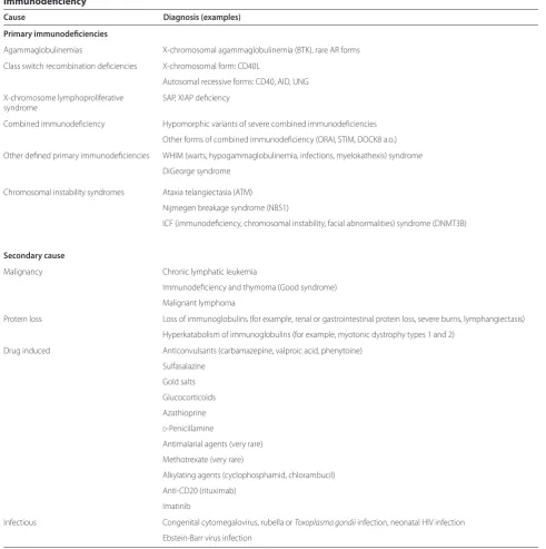
[image:2.612.62.554.104.598.2]show autosomal dominant inheritance. In some larger pedigrees, individuals with selective IgA defi ciency (sIgAD), CVID and intermediate forms can be observed side by side [6,7]. Th is fi nding and cases of progression from sIgAD towards CVID [8] indicate a possible common genetic predisposition. Autosomal recessive CVID is rarely seen in Europe and North America but is Table 1. Primary and secondary causes of hypogammaglobulinemia to be distinguished from common variable
immunodefi ciency
Cause Diagnosis (examples)
Primary immunodefi ciencies
Agammaglobulinemias X-chromosomal agammaglobulinemia (BTK), rare AR forms
Class switch recombination defi ciencies X-chromosomal form: CD40L
Autosomal recessive forms: CD40, AID, UNG
X-chromosome lymphoproliferative syndrome
SAP, XIAP defi ciency
Combined immunodefi ciency Hypomorphic variants of severe combined immunodefi ciencies
Other forms of combined immunodefi ciency (ORAI, STIM, DOCK8 a.o.)
Other defi ned primary immunodefi ciencies WHIM (warts, hypogammaglobulinemia, infections, myelokathexis) syndrome
DiGeorge syndrome
Chromosomal instability syndromes Ataxia telangiectasia (ATM)
Nijmegen breakage syndrome (NBS1)
ICF (immunodefi ciency, chromosomal instability, facial abnormalities) syndrome (DNMT3B)
Secondary cause
Malignancy Chronic lymphatic leukemia
Immunodefi ciency and thymoma (Good syndrome)
Malignant lymphoma
Protein loss Loss of immunoglobulins (for example, renal or gastrointestinal protein loss, severe burns, lymphangiectasis)
Hyperkatabolism of immunoglobulins (for example, myotonic dystrophy types 1 and 2)
Drug induced Anticonvulsants (carbamazepine, valproic acid, phenytoine)
Sulfasalazine
Gold salts
Glucocorticoids
Azathioprine
D-Penicillamine
Antimalarial agents (very rare)
Methotrexate (very rare)
Alkylating agents (cyclophosphamid, chlorambucil)
Anti-CD20 (rituximab)
Imatinib
Infectious Congenital cytomegalovirus, rubella or Toxoplasma gondii infection, neonatal HIV infection
Ebstein-Barr virus infection
more frequent in regions and ethnic groups with higher rates of consanguinity [4,9].
Genetic linkage analysis of large collections of familial CVID/sIgAD patients [10-12] or singular large pedigrees with multiple CVID/sIgAD cases [6] revealed possible genetic loci on chromosome 4q [6], chromosome 6 [10,12] and chromosome 16q [11]. Th ese early genome-wide microsatellite-marker studies found the strongest association with the HLA region [10,12]; they were recently confi rmed by a genome-wide single nucleotide polymorphism (SNP) genotyping array approach in
several hundred CVID patients [13]. Th is study also
revealed several structural chromosomal abnormalities unique to CVID and many novel candidate genes signifi -cantly associated with CVID or its clinical complications [13].
In a minority of patients with CVID, distinct molecular genetic defects have been identifi ed. Th ese genes associated with a CVID phenotype are ICOS (inducible costimulator) [14], TACI (transmembrane activator and calcium-modulating cyclophilin ligand interactor) [15,16], CD19 [17], BAFF-R [18], CD81 [19], CD20 [20], CD21 [21] and LRBA (lipopolysaccharide responsive beige-like
anchor protein) [22]. Th ese defects are very rare,
however, only occurring in single cases or single families and adding up to less than 3% of CVID patients. Th e exceptions are TACI mutations, which are seen in up to 10% of CVID cases but occur also in 1% of the healthy population, and thus must be regarded as disease modifi ers rather than disease-causing gene defects.
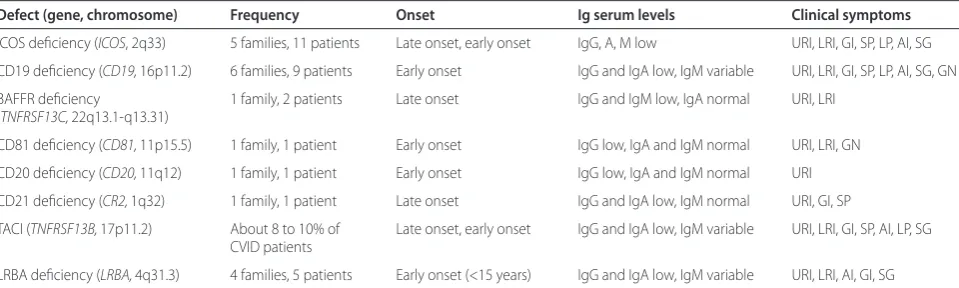
Th e currently known monogenetic defects associated
with CVID are summarized in Table 2. Th ese
mono-genetic defects aff ect only very few patients, but do provide important prototypic disease models by indicat-ing weak points in terminal B-cell diff erentiation.
Immunopathology of common variable immunodefi ciency
Th e immune system of CVID patients has been investi-gated by many studies, describing both phenotypic and functional abnormalities in the adaptive and, more recently, also in the innate immune system. However, the plethora of these defects, their unequal distribution within diff erent CVID cohorts and the lack of a real comprehensive and combined analysis of all of them have so far precluded a defi nitive mapping of all immuno-pathogenic pathways leading to CVID.
Based on the most common defects found in T cells and B cells of the adaptive immune system, several classi-fi cation systems have been introduced [23-26].
Disturbances of T cells
For many years abnormalities of CD4+ and CD8+ T cell numbers or function have been known and described in
subgroups of CVID patients. In a signifi cant proportion of CVID patients a reduction of total CD4+ T-cell counts and the naive CD4+CD45RA+ subset has been observed [23,27,28]. Regulatory CD25+FoxP3+-CD4+ T cells are also diminished [29-32] in a subgroup of CVID patients who present clinically with increased autoimmunity, granulo-mas, splenomegaly and an expansion of CD21low B cells [32].
Th e T-cell compartment of some CVID patients
ex-presses surface marker patterns indicative of chronic activation; in contrast to CD4+ T cells, the CD8+ T cells of these patients may numerically expand, explaining the frequently inverted CD4/CD8 T-cell ratio seen in CVID.
Th ese disturbances of the CD8+ T-cell pool can be
associated with disturbed cytokine secretion [33], lower memory B-cell numbers and severe clinical courses [34], chronic or recurrent cytomegalovirus infections [35] and polyclonal expansions of ‘large granular lymphocytes’ in combination with splenomegaly [36].
Giovanetti and colleagues [23] defi ned clinically rele-vant subgroups of CVID patients based on the reduc tion of naive CD4+ T cells. Th eir group I patients exhibited a severe reduction of naive CD4+ T cells, signs of massive T-cell activation, association with splenomegaly and a more severe course of the disease [23].
Th e French DEFI study group delineated a CVID sub-group with clinically relevant T-cell insuffi ciency and coined the term ‘late onset combined immunodefi ciency’ (LOCID) for these patients [37]. Inclusion criteria were CD4+ T cells below 200/μl or evidence of opportunistic infections, which occurred in 3.5% and 5.4%, respectively, of their studied cohort. LOCID patients often had a consanguine background, and suff ered more often from sarkoid-like granulomas, gastrointestinal complaints, splenomegaly and lymphoma; in general, LOCID patients were more sick and required more intensive therapy [37].
Disturbances of B cells
Th e total number of peripheral B cells is slightly reduced in about 40 to 50% of CVID patients [26]. In some patients elevated numbers of B cells are reported, often associated with polyclonal lymphoid organ infi ltration and autoimmunity [5]. In only about 10% of CVID patients are B cells dramatically reduced or absent [26]. Disease progression tends to be more rapid and severe in these patients [3,38] and the X-linked form of agamma-globulinemia and Good’s syndrome (B-cell aplasia associated with thymoma) have to be excluded (Table 1).
studies [39-41]. Furthermore, class-switched memory B cells are reduced in 80 to 90% of CVID patients [24-26]. Since this fi nding is not specifi c for CVID, it is not suitable as a diagnostic criterion but has been used to classify CVID patients into clinically and
immuno-logically more homogeneous subgroups [24-26]. Th e
[image:4.612.67.547.100.244.2]‘Freiburg’ classifi cation distinguishes three groups of CVID patients [25] based on the percentage of switched memory B cells and the expansion of activated so-called CD21low B cells. Th e ‘Paris’ classifi cation distinguishes three CVID subgroups [24] based on the reduction of total versus switched memory B cells. For both fi cation schemes several studies demonstrated that classi-fi cation of CVID patients based on B-cell phenotypes is useful for identifying clinical subtypes, adapting thera-peutic regimens (vaccination), assessing risks of certain complications and performing pathogenic research [42-46].
In 2008 a European multi-center trial combined both classifi cation systems and proposed the EUROclass classifi cation [26]. B cells were phenotyped for CD19, IgD/IgM, CD27, CD21 and CD38 expression; patients with more than 1% circulating B cells (B+, >90% of all patients) were distinguished from those with less than 1% (B-, <10% of all patients). Th e B+ group was further split into patients with normal or reduced percentages of switched memory B cells (smB+, >2% of total B cells; smB-, <2% of total B cells). Further subgroups were estab-lished depending on the expansion of CD21low B cells or transitional B cells. Th e EUROclass trial confi rmed the clinical association of reduced switched memory B cells
and expanded CD21low B cells with splenomegaly and
granulomatous disease and revealed for the fi rst time an expansion of transitional B cells in patients with lymphadenopathy [26].
Th e disturbed memory B-cell formation points towards an impaired germinal center reaction in secondary
lymphoid organs of most CVID patients. Th is assumption is further supported by decreased rates of somatic hyper-mutations in CD27+ B cells of CVID patients [47,48], a phenomenon that inversely correlates with an increased risk of chronic lung damage [47]. Histopathological studies of secondary lymphoid organs in CVID that would allow a closer look at possible pathomechanisms in situ are still rare. Taubenheim and colleagues [41] showed in three patients an intact development of the centroblast/ centrocyte stage, including the sequential expression of BCL-6 and Blimp-1, but the subsequent development into plasmablasts and plasma cells was disturbed.
B-cell activation is triggered by stimulation of the B-cell receptor, CD40, cytokine receptors and pattern recog-nition receptors such as Toll-like receptors (TLRs). Currently, defects of B-cell receptor activation [44] as well as the TLRs [49-51] have been identifi ed in sub-groups of patients. Th e underlying cause remains un-known for both defects.
Disturbances of antigen presenting cells and innate immunity receptors
Professional antigen presenting cells, such as dendritic cells (DCs), interact with naive T cells in the T-cell areas of secondary lymphoid organs. As part of the germinal center reaction they cooperate with cognate T and B cells to promote their further diff erentiation. Outside of germinal centers plasmacytoid DCs may initiate
immuno globulin class switching and terminal B-cell
diff erentiation independent of T-cell help but via signals through TLRs and the cytokines BAFF (B-cell activating factor) and APRIL (a proliferation inducing ligand). Th ese two pathways are closely linked together parti cu-larly at the level of TLR9 and the BAFF/APRIL receptor TACI [52,53]. When DCs from CVID patients were diff erentiated in cell culture experiments their maturation was impaired, resulting in diminished interleukin-12 Table 2. Monogenetic defects associated with or causing common variable immunodefi ciency
Defect (gene, chromosome) Frequency Onset Ig serum levels Clinical symptoms
ICOS defi ciency (ICOS, 2q33) 5 families, 11 patients Late onset, early onset IgG, A, M low URI, LRI, GI, SP, LP, AI, SG
CD19 defi ciency (CD19, 16p11.2) 6 families, 9 patients Early onset IgG and IgA low, IgM variable URI, LRI, GI, SP, LP, AI, SG, GN
BAFFR defi ciency 1 family, 2 patients Late onset IgG and IgM low, IgA normal URI, LRI (TNFRSF13C, 22q13.1-q13.31)
CD81 defi ciency (CD81, 11p15.5) 1 family, 1 patient Early onset IgG low, IgA and IgM normal URI, LRI, GN
CD20 defi ciency (CD20, 11q12) 1 family, 1 patient Early onset IgG low, IgA and IgM normal URI
CD21 defi ciency (CR2, 1q32) 1 family, 1 patient Late onset IgG and IgA low, IgM normal URI, GI, SP
TACI (TNFRSF13B, 17p11.2) About 8 to 10% of Late onset, early onset IgG and IgA low, IgM variable URI, LRI, GI, SP, AI, LP, SG
CVID patients
LRBA defi ciency (LRBA, 4q31.3) 4 families, 5 patients Early onset (<15 years) IgG and IgA low, IgM variable URI, LRI, AI, GI, SG
production and impaired up-regulation of co-stimulatory molecules. Th is might limit the ability of CVID DCs to contact and successfully interact with T cells [54,55]. In addition, TLR9 expression and response of plasmacytoid DCs and B cells to CpG stimulation is reduced [49]. Further investigations in CVID patients revealed an additional dysfunction of TLR7 and TLR8 signaling [50,51]. Th e recently described relationship between TACI and the TLR9 signaling pathway [52] strengthens the assumption that these disturbances of the TLR system in CVID patients are of pathophysiological rele-vance even though no genetic mutations in the TLR pathway have been established so far.
Clinical presentation of common variable immunodefi ciency
Infections
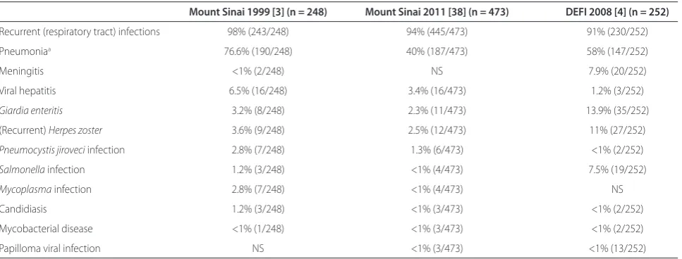
Over 90% of CVID patients suff er from an increased susceptibility to bacterial pathogens aff ecting mucous membranes of the upper and lower airways and, to a lesser extent, of the gastrointestinal tract [3,4,38]. Table 3
summarizes frequencies of specifi c infections and
pathogens encountered in two consecutive studies on the Mount Sinai Hospital CVID cohort in New York [3,38] and the French DEFI cohort study [4]. In the DEFI cohort study, approximately two-thirds of the 252 patients presented with sinusitis or bronchitis and 50% had at least one bout of pneumonia during their life [4]. About one-third of patients had developed bronchiectasis as a result of chronic and recurrent infections. Frequently detected pathogens were Streptococcus pneumoniae,
Haemophilus infl uenzae, Staphylococcus aureus and
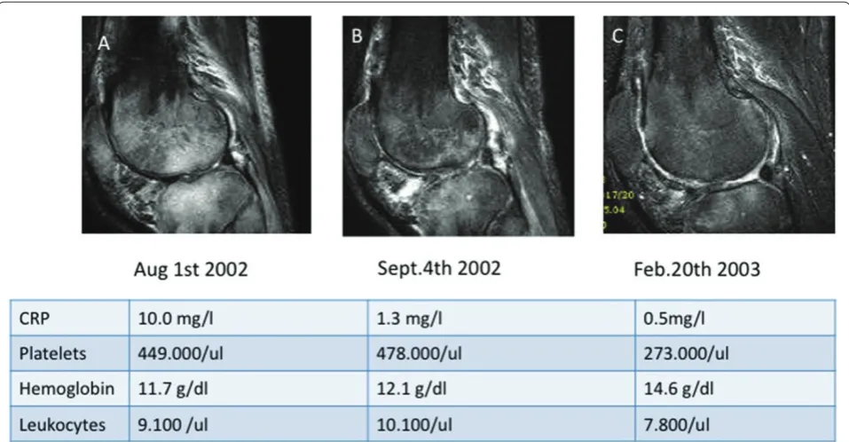
Moraxella catharralis. Recurrent and chronic diarrhea was present in approximately 40% of patients and in about half of them pathogens like Giardia lamblia followed by Salmonella and Campylobacter jejuni were identifi ed. Acute and chronic gastritis caused by Helico-bacter pylori is frequently diagnosed in CVID patients [3]. Up to 10% of CVID patients are described as suff ering from increased rates of Herpes zoster infections. In contrast, typical opportunistic infections are quite unusual and evoke the possibility of an underlying combined immunodefi ciency [37]. A rare but typical complication of hypogammaglobulinemia is oligoarthritis due to Mycoplasma species (Figure 1). Clinically, the condition presents as reactive arthritis with synovial culture techniques being often negative; therefore, the microbiological diagnosis has to include multipathogen PCR in synovial fl uid. In the diff erential diagnosis of hypogammaglobulinemic oligoarthritis, serological investigations are not helpful since most patients do not exhibit an adequate antibody response against the respective pathogens or the test results are infl uenced by intravenous immunoglobulin substitution therapy. In
patients with suspected reactive arthritis, therefore, we recom mend to initially determine IgG and IgA serum concentrations before proceeding to extensive and potentially meaningless antibacterial antibody responses.
Granulomatous lesions
Approximately 10 to 20% of CVID patients develop granulomatous interstitial lung disease. Microbial testing of these lesions often reveals no specifi c pathogen; the reported detection of human herpes virus-8 in a US CVID cohort [56] could not be confi rmed in larger European patient groups (unpublished data), indicating that the underlying cause remains unknown and probably is multifactorial. Patients with granulomatous interstitial lung disease have a signifi cantly poorer prognosis than other CVID patients [38,57]. Th e granulomatous disease to some extent resembles sarcoidosis; in addition to lung and lymph nodes, also the liver, skin, spleen, bone marrow, gastrointestinal tract, brain and kidney (in decreasing frequency) may be aff ected [58].
Gastrointestinal symptoms
Diarrhea is un-bloody if associated with a sprue-like disease and bloody when resulting from chronic infl am-matory bowel disease. Th e sprue-like villous atrophy seen in CVID is often not gluten-sensitive and resembles more autoimmune enteropathy. Th e involvement of the colon in CVID is reminiscent of Crohn’s disease and ulcerative colitis, but can be distinguished histologically [59]. Th e nodular lymphoid hyperplasia that may occur both in the duodenum and ileum may be asymptomatic or associated with unformed stools [3].
Liver disease and abnormal liver function tests are found in 10% of CVID patients [3,60]. Th e most common liver disease in CVID represents nodular regenerative hyperplasia of the liver tissue [60,61] or seronegative, granulomatous hepatitis; autoimmune hepatitis is not a typical entity found in these patients. Usually, liver function in CVID patients is still preserved but portal hypertension may develop [60,61]. Liver disease heralds a poorer prognosis [38]. In any case of a suspected hepatopathy in CVID, seronegative hepatitis B and C as well as cytomegalovirus or Epstein Barr virus hepatitis must be ruled out by searching for hepatitis antigen or viral RNA, respectively.
Autoimmunity
Autoimmunity is present in about 30% of CVID patients [3,26,38,62,63]. Table 4 summarizes frequencies of auto-immune disease encountered in two consecutive studies on the Mount Sinai Hospital CVID cohort in New York [3,38] and the French DEFI cohort study [62].
Particularly common are autoimmune thrombo
to 7%), showing a signifi cant correlation with spleno-megaly [26,64]. Cytopenias can manifest before, simultaneously with or after the diagnosis of immuno-defi ciency. Immunologically, autoimmune cytopenias are associated with low numbers of class-switched memory B cells, low numbers of regulatory T cells, expanded CD21low B cells [25,65], and nodular T-cell infi ltrates of the bone marrow [40]. Autoimmune thyroid disease, vitiligo, pernicious anemia, psoriasis, rheumatoid arthritis and systemic lupus erythematosus are observed in CVID cohorts at decreasing frequency [3,5,26] (Table 4).
Lymphoproliferation and malignancies
Benign lymphoproliferation is found in 40 to 50% of CVID patients, often as splenomegaly, and in approxi-mately 10 to 20% as local or diff use lymphadenopathy [5,26]. Histologically, several subsets can be distin-guished, with follicular hyperplasia and granulomatous infl ammation being the most common ones [66]. In conjunction with lymphoproliferation, CVID patients carry an increased risk of developing lymphoma [3,5]. Most lymphomas are of the B-cell non-Hodgkin lym-phoma type [3]. In addition to lymlym-phomas, stomach cancers represent an important malignant manifestation in CVID [3,5,63,67]. Th e increased risk of cancer in CVID may result from impaired immunity to potentially carcinogenic pathogens (for example, Helicobacter pylori, Epstein-Barr virus) or impaired tumor cell surveillance. In this context it is notable that patients of a CVID subgroup exhibit increased radiosensitivity, known to be a risk factor for increased tumor incidence [68].
Diagnosis and follow-up of common variable immunodefi ciency
Th e diagnosis of CVID can only be made after the exclu sion of a variety of other causes of hypogamma globu linemia
(Table 1). Its rarity and high clinical variability lead to a signifi cant delay in diagnosis between four [69] and nine years [63] after onset of symptoms. Clinically, the leading symptom in most patients is the classical susceptibility to bacterial airway infections as described above. As mentioned before, opportunistic infections are always suggestive of LOCID. Some CVID cases manifest initially with autoimmune cytopenias; thus, CVID needs to be excluded in patients with immune thrombocytopenic
purpura, autoimmune hemo lytic anemia and
auto-immune neutropenia.
An inexpensive, quantitative determination of serum immunoglobulins is the fi rst and most important step in the diagnosis of CVID. Required for the diagnosis of CVID is the diminution of at least two isotypes (IgG and IgA or IgM). IgG is typically below 5 g/L (normal range 7 to 16 g/L) and IgA is markedly reduced or not detectable in most patients. IgM is also below the normal range in up to 80% of patients.
While drug-induced hypogammaglobulinemia (for example, with rituximab; Table 1) may be revealed by the patient’s history, proteinuria is detected by Uristix® and intestinal protein loss may be suspected from the medical history in combination with a decreased serum albumin concentration. Lymphomas tend to be the most diffi cult
diff erential diagnosis in secondary hypogamma
globu-linemia. Th ey require histological examination of lymph node and bone marrow biopsies, notably in CVID patients with ongoing lymphoproliferation. Th e diff er-ential diagnosis of other genetically defi ned immuno-defi ciencies is rare and should be done in a specialized center.
[image:6.612.66.550.99.283.2]Besides quantitative determination of serum immuno-globulins, the basic laboratory tests should include a diff erential blood count, liver and kidney function parameters and C-reactive protein determination. Th e Table 3. Infectious complications in common variable immunodefi ciency
Mount Sinai 1999 [3] (n = 248) Mount Sinai 2011 [38] (n = 473) DEFI 2008 [4] (n = 252)
Recurrent (respiratory tract) infections 98% (243/248) 94% (445/473) 91% (230/252)
Pneumoniaa 76.6% (190/248) 40% (187/473) 58% (147/252)
Meningitis <1% (2/248) NS 7.9% (20/252)
Viral hepatitis 6.5% (16/248) 3.4% (16/473) 1.2% (3/252)
Giardia enteritis 3.2% (8/248) 2.3% (11/473) 13.9% (35/252)
(Recurrent) Herpes zoster 3.6% (9/248) 2.5% (12/473) 11% (27/252)
Pneumocystis jiroveci infection 2.8% (7/248) 1.3% (6/473) <1% (2/252)
Salmonella infection 1.2% (3/248) <1% (4/473) 7.5% (19/252)
Mycoplasma infection 2.8% (7/248) <1% (4/473) NS
Candidiasis 1.2% (3/248) <1% (3/473) <1% (2/252)
Mycobacterial disease <1% (1/248) <1% (3/473) <1% (2/252)
Papilloma viral infection NS <1% (3/473) <1% (13/252)
routine examinations are complemented by the determination of specifi c antibodies against protein antigens (tetanus, diphtheria and hepatitis B virus and
[image:7.612.67.546.89.338.2]hepatitis A virus in vaccinated patients) and antibodies against pneumococcal capsular polysaccharides. Th ese studies are particularly meaningful if the patient has been
Figure 1. Oligoarthritis due to Mycoplasma salivarius as an early manifestation of common variable immunodefi ciency. A male aged 36 years was healthy until he developed recurrent upper respiratory tract infections and a fi rst bout of pneumonia 18 months prior to these images being taken. Five months later he presented with refractory right-sided gonarthritis to an orthopedic surgeon. Despite multiple sterile knee taps, arthroscopy and a Baker cyst resection, joint infl ammation continued and extended to the right shoulder and the right ankle. He was referred to the Division of Rheumatology and Clinical Immunology at Freiburg University Hospital for further diagnosis and treatment of ‘multifocal osteomyelitis and oligoarthritis of unknown origin’. On admission he presented with three tender and swollen joints (right knee, shoulder, ankle), moderately elevated C-reactive protein (CRP) levels (5 to 29 mg/dl) and severe hypogammaglobulinemia: IgG 1.7 g/L, IgA <0.6 g/L, IgM <0.3 g/L. Diagnosis of CVID was established and the patient was started on monthly intravenous immunoglobulin infusions (500 mg/kg) plus various ineff ective antibiotic regimens (initially cefuroxime plus neomycin, then clarithromycin and metronidazol). A diagnostic puncture of the right shoulder eventually revealed Mycoplasma salivarius by multiplex PCR diagnostics. From that point on the patient was put on doxicycline (200 mg/daily orally) and the infl ammatory process rapidly improved. Doxicycline was stopped after 4 weeks, whereas monthly intravenous immunoglobulin was continued. As of today, the patient has been back to work for 7 years and is clinically doing well. Magnetic resonance imaging follow-up (T2, TIRM sequences of the right knee) and laboratory parameters at three time points (A, B, C) nicely show the improvement of the severe arthritis and osteomyelitis of the right knee.
Table 4. Common autoimmune manifestations in common variable immunodefi ciency
Mount Sinai 1999 [3] (n = 248) Mount Sinai 2011 [38] (n = 473) DEFI 2010 [62] (n = 311)
AIHA 4.8% (12/248) 7% (33/473) 5.4% (17/311)
ITP 6% (15/248) 14.2% (67/473) 13.2% (41/311)
Neutropenia <1% (2/248) <1% (<5/473) 3.2% (10/311)
Rheumatoid arthritis 3.6% (9/248) 3.2% (15/473) 2.6% (8/311)
Vitiligo NS <1% (<5/473) 3.9% (12/311)
Sicca syndrome, Sjögren’s syndrome <1% (2/248) <1% (<5/473) 4.2% (13/311)
Autoimmune thyroiditis, diabetes mellitus, <1% (2/248) <1% (<5/473 3.9% (12/311) multiple sclerosis
Alopecia 1.6% (4/248) 1.1% (5/473) NS
Pernicious anemia 1.2% (3/248) <1% (<5/473) NS
Systemic lupus erythematosus <1% (2/248) <1% (<5/473) <1% (1/311)
[image:7.612.65.546.513.678.2]vaccinated for diagnostic purposes prior to the start of immunoglobulin substitution.
Th e next stage of diagnosis is fl ow cytometric analysis of lymphocyte subpopulations, including total T, B and natural killer cells, to distinguish late manifesting X-linked agammaglobulinemia (B cells <0.1%) and com-bined immunodefi ciencies (CD4 cells <200/μl). Th e classi fi cation of CVID patients with the separation of B-cell subpopulations is reserved for specialized immuno-defi ciency centers. A bone marrow biopsy should be performed in patients with low B-cell numbers (<1%) [40] and if lymphoma or myelodysplasia is suspected. In addition, several diagnostic procedures at fi rst visit and during follow-ups are indicated for the control of possible secondary complications (summarized in Table 5).
Therapy, natural course and prognosis
Current therapy of CVID can be categorized as follows: regular and suffi cient substitution with immunoglobulins (IgG trough levels >7.0 g/L); targeted antibiotic treatment of (breakthrough) infections; adequate treatment of
compli cations; and in selected patients with severe
hematological changes (chronic transfusion need, leuko-penia, thrombocytopenia), secondary malignancies and suspected combined immunodefi ciency, allogeneic peripheral stem cell transplantation is being considered in experienced centers [70].
Th e immunoglobulin replacement therapy is the
[image:8.612.63.550.99.499.2]mainstay of therapy; 90% of CVID patients are on either intravenous (IVIg) or subcutaneous (SCIg) treatment [71-74]. Intramuscular administration is no longer Table 5. Initial and follow-up diagnostics in common variable immunodefi ciency
System Type of diagnostic procedure Intervals
Hematopoietic system Blood formula and diff erential blood counts (3-)6 months; more often in case of known
autoimmune cytopenia
Coombs test in any case of newly developing anemia On demand
Bone marrow biopsy in case of suspicion of lymphoma or myelodysplasia On demand at diagnosis or during follow-up
Immune system IgG, IgA, IgM (aiming at trough levels >7g/L)
In intravenous immunoglobulin-substituted patients Monthly trough levels
In subcutaneous immunoglobulin-substituted patients 1-3 months
Immunofi xation in serum and urine, ß2-microglobulin At diagnosis
CD3, CD4, CD8, CD19, CD56 lymphocyte subsets Initially and repeatedly in case of suspected
combined immunodefi ciency
Vaccination responses to tetanus, diphtheria, hepatitis, pneumococcal At diagnosis polysaccharides
Classifi cation (detailed T/B-cell evaluation) At diagnosis
Microbiology (direct culture Purulent sputum: determine colonizing pathogens and resistance pattern On demand of pathogens, PCR, to antibiotics
antigen ELISA)
In case of chronic bronchiectasis, control of colonising pathogens 6-12 months and on demand (Pseudomonas spp., Haemophilius infl uenzae, Streptococcus pneumoniae,
Staphylococcus aureus, Candida spp. and others) with sensitivity to antibiotics
Lungs Spirometry, CO diff usion test, blood gases 12 months
Chest X-ray (12-)24 months
HR-CT in case of proven GILD 24 months and on demand
Bronchoscopy + BAL in case of suspicion of GILD At diagnosis; on demand
Lymphoproliferation Abdomen sonography CT-Abdomen/MRT 12 months
Lymph node biopsy On demand; in case of suspected lymphoma
Gastrointestinal tract Oesophogastroscopy In case of clinical symptoms and every
24 months in case of increased risk for developing intestinal malignancy
Colonoscopy On demand
Central nervous system MRT, liquor analysis in case of neurological symptoms On demand (exclusion of enteroviral infection)
recommended because this route does not ensure eff ective serum levels but is associated with a higher rate
of side eff ects. Th e current standard dosage when
administered intravenously is 400 to 600 mg/kg every 3 to 4 weeks. For subcutaneous administration, this corres-ponds to 100 to 150 mg/kg per week. Th e goal is the control of infections, which is reached at diff erent individual IgG trough levels [63]. As a target value, IgG trough levels of more than 7 g/L are desirable before the next infusion. Patients with existing chronic lung disease (for example, bronchiectasis) or infl ammatory bowel disease often require higher doses of IgG and may not reach the desired trough level. In chronic sinusitis additional careful local therapy (saline lavage, expector-ant and decongestexpector-ant therapy) is mandatory.
First line therapy in autoimmune cytopenias and lympho proliferation are steroids. In case of failure, immunosuppressive drugs, rituximab or splenectomy have been reported as options. Th e infl ammatory and granulomatous lesions of the lungs, liver and intestine respond poorly to the immunoglobulin replacement therapy alone and therefore often require corticosteroids, eventually in combination with immunosuppressants (cyclosporin A, azathioprin and others). Prospective trials on the eff ectiveness of immunosuppressive drugs in CVID are still lacking.
Th e life expectancy of CVID patients has considerably improved over the past 30 years [5,63], from initially 12 years to currently over 50 years [3]. Reduced survival was signifi cantly associated with age at diagnosis, lower baseline IgG, higher IgM and fewer peripheral B cells. Th e risk of death was 11 times higher for patients with non-infectious complications such as lymphoma, chronic hepatitis, structural lung disease and chronic gastrointestinal disease [38].
Th us, the development of better surrogate diagnostic markers for the presence and activity of these secondary complications as well as new therapeutic approaches are a major challenge for the coming years in the care of CVID patients.
Conclusion
CVID represents the most common primary immuno-defi ciency. Besides an increased susceptibility to infec-tions it frequently presents with signs of auto immunity, notably autoimmune cytopenias and rheumatic diseases. Its early diagnosis and treatment are important for a favorable outcome. While in most patients susceptibility
to infections can be suffi ciently covered by
immuno-globulin replacement therapy and antibiotics, other manifes tations, such as autoimmunity, granulomatous disease, interstitial lung disease, chronic diarrhea, lympho proliferation and developing malignancies, need special attention and treatment, which is best off ered in
close collaboration between primary care doctors and specialized immunodefi ciency centers.
Abbreviations
CVID, common variable immunodefi ciency; DC, dendritic cell; LOCID, late onset combined immunodefi ciency; sIgAD, selective IgA defi ciency; TLR, Toll-like receptor.
Competing interests
KW received a grant from Baxter. HHP is member of the scientifi c advisory board of Pfi zer’s Prevenar development program in Germany.
Acknowledgements
The research was funded by grants from the German Research Foundation through the SFB620 projects C1 (KW, HHP) and C7 (US), the 7th European Union framework program grant numer HEALTH-F2-2008-201549 (KW, US), and the Federal Ministry of Education and Research (BMBF 01 EO 0803; KW, HHP, US).
Author details
1Centre of Chronic Immunodefi ciency, University Medical Centre Freiburg, Engesserstr. 4, D-79108 Freiburg i. Breisgau, Germany. 2Department of Rheumatology, University Medical Centre Freiburg, Hugstetterstr. 55, D-79106 Freiburg i. Breisgau, Germany.
Published: 24 September 2012
References
1. Conley ME: Diagnostic guidelines - An International Consensus document. Clin Immunol 1999, 93:189.
2. Bonilla FA, Bernstein IL, Khan DA, Ballas ZK, Chinen J, Frank MM, Kobrynski LJ, Levinson AI, Mazer B, Nelson RP Jr, Orange JS, Routes JM, Shearer WT, Sorensen RU; American Academy of Allergy, Asthma and Immunology; American College of Allergy, Asthma and Immunology; Joint Council of Allergy, Asthma and Immunology: Practice parameter for the diagnosis and management of primary immunodefi ciency. Ann Allergy Asthma Immunol
2005, 94(5 Suppl 1):S1-63.
3. Cunningham-Rundles C, Bodian C: Common variable immunodefi ciency: clinical and immunological features of 248 patients. Clin Immunol 1999,
92:34-48.
4. Oksenhendler E, Gérard L, Fieschi C, Malphettes M, Mouillot G, Jaussaud R, Viallard JF, Gardembas M, Galicier L, Schleinitz N, Suarez F, Soulas-Sprauel P, Hachulla E, Jaccard A, Gardeur A, Théodorou I, Rabian C, Debré P; DEFI Study Group: Infections in 252 patients with common variable
immunodefi ciency. Clin Infect Dis 2008, 46:1547-1554.
5. Chapel H, Lucas M, Lee M, Bjorkander J, Webster D, Grimbacher B, Fieschi C, Thon V, Abedi MR, Hammarstrom L: Common variable immunodefi ciency disorders: division into distinct clinical phenotypes. Blood 2008,
112:277-286.
6. Finck A, Van der Meer JW, Schaff er AA, Pfannstiel J, Fieschi C, Plebani A, Webster AD, Hammarstrom L, Grimbacher B: Linkage of autosomal-dominant common variable immunodefi ciency to chromosome 4q. Eur J Hum Genet 2006, 14:867-875.
7. Vorechovsky I, Zetterquist H, Paganelli R, Koskinen S, Webster AD, Bjorkander J, Smith CI, Hammarstrom L: Family and linkage study of selective IgA defi ciency and common variable immunodefi ciency. Clin Immunol Immunopathol 1995, 77:185-192.
8. Espanol T, Catala M, Hernandez M, Caragol I, Bertran JM: Development of a common variable immunodefi ciency in IgA-defi cient patients. Clin Immunol Immunopathol 1996, 80:333-335.
9. Aghamohammadi A, Farhoudi A, Moin M, Rezaei N, Kouhi A, Pourpak Z, Yaseri N, Movahedi M, Gharagozlou M, Zandieh F, Yazadni F, Arshi S,
10. Kralovicova J, Hammarstrom L, Plebani A, Webster AD, Vorechovsky I: Fine-scale mapping at IGAD1 and genome-wide genetic linkage analysis implicate HLA-DQ/DR as a major susceptibility locus in selective IgA defi ciency and common variable immunodefi ciency. J Immunol 2003,
170:2765-2775.
11. Schaff er AA, Pfannstiel J, Webster AD, Plebani A, Hammarstrom L, Grimbacher B: Analysis of families with common variable immunodefi ciency (CVID) and IgA defi ciency suggests linkage of CVID to chromosome 16q. Hum Genet 2006, 118:725-729.
12. Schroeder HW Jr, Zhu ZB, March RE, Campbell RD, Berney SM, Nedospasov SA, Turetskaya RL, Atkinson TP, Go RC, Cooper MD, Volanakis JE: Susceptibility locus for IgA defi ciency and common variable immunodefi ciency in the HLA-DR3, -B8, -A1 haplotypes. Mol Med 1998, 4:72-86.
13. Orange JS, Glessner JT, Resnick E, Sullivan KE, Lucas M, Ferry B, Kim CE, Hou C, Wang F, Chiavacci R, Kugathasan S, Sleasman JW, Baldassano R, Perez EE, Chapel H, Cunningham-Rundles C, Hakonarson H: Genome-wide association identifi es diverse causes of common variable immunodefi ciency. J Allergy Clin Immunol 2011, 127:1360-1367 e1366. 14. Grimbacher B, Hutloff A, Schlesier M, Glocker E, Warnatz K, Dräger R, Eibel H,
Fischer B, Schäff er AA, Mages HW, Kroczek RA, Peter HH: Homozygous loss of ICOS is associated with adult-onset common variable immunodefi ciency. Nat Immunol 2003, 4:261-268.
15. Castigli E, Wilson SA, Garibyan L, Rachid R, Bonilla F, Schneider L, Geha RS:
TACI is mutant in common variable immunodefi ciency and IgA defi ciency. Nat Genet 2005, 37:829-834.
16. Salzer U, Chapel HM, Webster AD, Pan-Hammarström Q, Schmitt-Graeff A, Schlesier M, Peter HH, Rockstroh JK, Schneider P, Schäff er AA, Hammarström L, Grimbacher B: Mutations in TNFRSF13B encoding TACI are associated with common variable immunodefi ciency in humans. Nat Genet 2005,
37:820-828.
17. van Zelm MC, Reisli I, van der Burg M, Castaño D, van Noesel CJ, van Tol MJ, Woellner C, Grimbacher B, Patiño PJ, van Dongen JJ, Franco JL: An antibody-defi ciency syndrome due to mutations in the CD19 gene. N Engl J Med
2006, 354:1901-1912.
18. Warnatz K, Salzer U, Rizzi M, Fischer B, Gutenberger S, Böhm J, Kienzler AK, Pan-Hammarström Q, Hammarström L, Rakhmanov M, Schlesier M, Grimbacher B, Peter HH, Eibel H: B-cell activating factor receptor defi ciency is associated with an adult-onset antibody defi ciency syndrome in humans. Proc Natl Acad Sci U S A 2009, 106:13945-13950.
19. van Zelm MC, Smet J, Adams B, Mascart F, Schandené L, Janssen F, Ferster A, Kuo CC, Levy S, van Dongen JJ, van der Burg M: CD81 gene defect in humans disrupts CD19 complex formation and leads to antibody defi ciency. J Clin Invest 2010, 120:1265-1274.
20. Kuijpers TW, Bende RJ, Baars PA, Grummels A, Derks IA, Dolman KM, Beaumont T, Tedder TF, van Noesel CJ, Eldering E, van Lier RA: CD20 defi ciency in humans results in impaired T cell-independent antibody responses. J Clin Invest 2010, 120:214-222.
21. Thiel J, Kimmig L, Salzer U, Grudzien M, Lebrecht D, Hagena T, Draeger R, Völxen N, Bergbreiter A, Jennings S, Gutenberger S, Aichem A, Illges H, Hannan JP, Kienzler AK, Rizzi M, Eibel H, Peter HH, Warnatz K, Grimbacher B, Rump JA, Schlesier M: Genetic CD21 defi ciency is associated with hypogammaglobulinemia. J Allergy Clin Immunol 2012, 129:801-810. 22. Lopez-Herrera G, Tampella G, Pan-Hammarström Q, Herholz P, Trujillo-Vargas
CM, Phadwal K, Simon AK, Moutschen M, Etzioni A, Mory A, Srugo I, Melamed D, Hultenby K, Liu C, Baronio M, Vitali M, Philippet P, Dideberg V,
Aghamohammadi A, Rezaei N, Enright V, Du L, Salzer U, Eibel H, Pfeifer D, Veelken H, Stauss H, Lougaris V, Plebani A, Gertz EM, et al.: Deleterious mutations in LRBA are associated with a syndrome of immune defi ciency and autoimmunity. Am J Hum Genet 2012, 90:986-1001.
23. Giovannetti A, Pierdominici M, Mazzetta F, Marziali M, Renzi C, Mileo AM, De Felice M, Mora B, Esposito A, Carello R, Pizzuti A, Paggi MG, Paganelli R, Malorni W, Aiuti F: Unravelling the complexity of T cell abnormalities in common variable immunodefi ciency. J Immunol 2007, 178:3932-3943. 24. Piqueras B, Lavenu-Bombled C, Galicier L, Bergeron-van der Cruyssen F, Mouthon L, Chevret S, Debre P, Schmitt C, Oksenhendler E: Common variable immunodefi ciency patient classifi cation based on impaired B cell memory diff erentiation correlates with clinical aspects. J Clin Immunol
2003, 23:385-400.
25. Warnatz K, Denz A, Drager R, Braun M, Groth C, Wolff -Vorbeck G, Eibel H, Schlesier M, Peter HH: Severe defi ciency of switched memory B cells (CD27(+)IgM(-)IgD(-)) in subgroups of patients with common variable
immunodefi ciency: a new approach to classify a heterogeneous disease. Blood 2002, 99:1544-1551.
26. Wehr C, Kivioja T, Schmitt C, Ferry B, Witte T, Eren E, Vlkova M, Hernandez M, Detkova D, Bos PR, Poerksen G, von Bernuth H, Baumann U, Goldacker S, Gutenberger S, Schlesier M, Bergeron-van der Cruyssen F, Le Garff M, Debré P, Jacobs R, Jones J, Bateman E, Litzman J, van Hagen PM, Plebani A, Schmidt RE, Thon V, Quinti I, Espanol T, Webster AD, et al.: The EUROclass trial: defi ning subgroups in common variable immunodefi ciency. Blood 2008,
111:77-85.
27. De Vera MJ, Al-Harthi L, Gewurz AT: Assessing thymopoiesis in patients with common variable immunodefi ciency as measured by T-cell receptor excision circles. Ann Allergy Asthma Immunol 2004, 93:478-484. 28. Isgro A, Marziali M, Mezzaroma I, Luzi G, Mazzone AM, Guazzi V, Andolfi G,
Cassani B, Aiuti A, Aiuti F: Bone marrow clonogenic capability, cytokine production, and thymic output in patients with common variable immunodefi ciency. J Immunol 2005, 174:5074-5081.
29. Arumugakani G, Wood PM, Carter CR: Frequency of Treg cells is reduced in CVID patients with autoimmunity and splenomegaly and is associated with expanded CD21lo B lymphocytes. J Clin Immunol 2010, 30:292-300. 30. Horn J, Manguiat A, Berglund LJ, Knerr V, Tahami F, Grimbacher B, Fulcher DA:
Decrease in phenotypic regulatory T cells in subsets of patients with common variable immunodefi ciency. Clin Exp Immunol 2009, 156:446-454. 31. Melo KM, Carvalho KI, Bruno FR, Ndhlovu LC, Ballan WM, Nixon DF, Kallas EG, Costa-Carvalho BT: A decreased frequency of regulatory T cells in patients with common variable immunodefi ciency. PLoS One 2009, 4:e6269. 32. Yu GP, Chiang D, Song SJ, Hoyte EG, Huang J, Vanishsarn C, Nadeau KC:
Regulatory T cell dysfunction in subjects with common variable immunodefi ciency complicated by autoimmune disease. Clin Immunol
2009, 131:240-253.
33. Holm AM, Sivertsen EA, Tunheim SH, Haug T, Bjerkeli V, Yndestad A, Aukrust P, Froland SS: Gene expression analysis of peripheral T cells in a subgroup of common variable immunodefi ciency shows predominance of CCR7(-) eff ector-memory T cells. Clin Exp Immunol 2004, 138:278-289.
34. Viallard JF, Blanco P, Andre M, Etienne G, Liferman F, Neau D, Vidal E, Moreau JF, Pellegrin JL: CD8+HLA-DR+ T lymphocytes are increased in common variable immunodefi ciency patients with impaired memory B-cell diff erentiation. Clin Immunol 2006, 119:51-58.
35. Raeiszadeh M, Kopycinski J, Paston SJ, Diss T, Lowdell M, Hardy GA, Hislop AD, Workman S, Dodi A, Emery V, Webster AD: The T cell response to persistent herpes virus infections in common variable immunodefi ciency. Clin Exp Immunol 2006, 146:234-242.
36. Holm AM, Tjonnfj ord G, Yndestad A, Beiske K, Muller F, Aukrust P, Froland SS:
Polyclonal expansion of large granular lymphocytes in common variable immunodefi ciency - association with neutropenia. Clin Exp Immunol 2006,
144:418-424.
37. Malphettes M, Gérard L, Carmagnat M, Mouillot G, Vince N, Boutboul D, Bérezné A, Nove-Josserand R, Lemoing V, Tetu L, Viallard JF, Bonnotte B, Pavic M, Haroche J, Larroche C, Brouet JC, Fermand JP, Rabian C, Fieschi C, Oksenhendler E; DEFI Study Group: Late-onset combined immune defi ciency: a subset of common variable immunodefi ciency with severe T cell defect. Clin Infect Dis 2009, 49:1329-1338.
38. Resnick ES, Moshier EL, Godbold JH, Cunningham-Rundles C: Morbidity and mortality in common variable immune defi ciency over 4 decades. Blood
2011,119:1650-1657.
39. Herbst EW, Armbruster M, Rump JA, Buscher HP, Peter HH: Intestinal B cell defects in common variable immunodefi ciency. Clin Exp Immunol 1994,
95:215-221.
40. Ochtrop ML, Goldacker S, May AM, Rizzi M, Draeger R, Hauschke D, Stehfest C, Warnatz K, Goebel H, Technau-Ihling K, Werner M, Salzer U, Eibel H, Schlesier M, Peter HH: T and B lymphocyte abnormalities in bone marrow biopsies of common variable immunodefi ciency. Blood 2011, 118:309-318. 41. Taubenheim N, von Hornung M, Durandy A, Warnatz K, Corcoran L, Peter HH,
Eibel H: Defi ned blocks in terminal plasma cell diff erentiation of common variable immunodefi ciency patients. J Immunol 2005, 175:5498-5503. 42. Berglund LJ, Wong SW, Fulcher DA: B-cell maturation defects in common
variable immunodefi ciency and association with clinical features. Pathology 2008, 40:288-294.
44. Foerster C, Voelxen N, Rakhmanov M, Keller B, Gutenberger S, Goldacker S, Thiel J, Feske S, Peter HH, Warnatz K: B cell receptor-mediated calcium signaling is impaired in B lymphocytes of type Ia patients with common variable immunodefi ciency. J Immunol 2010, 184:7305-7313.
45. Goldacker S, Draeger R, Warnatz K, Huzly D, Salzer U, Thiel J, Eibel H, Schlesier M, Peter HH: Active vaccination in patients with common variable immunodefi ciency (CVID). Clin Immunol 2007, 124:294-303. 46. Ko J, Radigan L, Cunningham-Rundles C: Immune competence and
switched memory B cells in common variable immunodefi ciency. Clin Immunol 2005, 116:37-41.
47. Andersen P, Permin H, Andersen V, Schejbel L, Garred P, Svejgaard A, Barington T: Defi ciency of somatic hypermutation of the antibody light chain is associated with increased frequency of severe respiratory tract infection in common variable immunodefi ciency. Blood 2005, 105:511-517. 48. Bonhomme D, Hammarstrom L, Webster D, Chapel H, Hermine O, Le Deist F, Lepage E, Romeo PH, Levy Y: Impaired antibody affi nity maturation process characterizes a subset of patients with common variable
immunodefi ciency. J Immunol 2000, 165:4725-4730.
49. Cunningham-Rundles C, Radigan L, Knight AK, Zhang L, Bauer L, Nakazawa A:
TLR9 activation is defective in common variable immune defi ciency. J Immunol 2006, 176:1978-1987.
50. Yu JE, Knight AK, Radigan L, Marron TU, Zhang L, Sanchez-Ramon S, Cunningham-Rundles C: Toll-like receptor 7 and 9 defects in common variable immunodefi ciency. J Allergy Clin Immunol 2009, 124:349-356, 356. e1-3.
51. Yu JE, Zhang L, Radigan L, Sanchez-Ramon S, Cunningham-Rundles C:
TLR-mediated B cell defects and IFN-alpha in common variable immunodefi ciency. J Clin Immunol 2012, 32:50-60.
52. He B, Santamaria R, Xu W, Cols M, Chen K, Puga I, Shan M, Xiong H, Bussel JB, Chiu A, Puel A, Reichenbach J, Marodi L, Döffi nger R, Vasconcelos J, Issekutz A, Krause J, Davies G, Li X, Grimbacher B, Plebani A, Meff re E, Picard C, Cunningham-Rundles C, Casanova JL, Cerutti A: The transmembrane activator TACI triggers immunoglobulin class switching by activating B cells through the adaptor MyD88. Nat Immunol 2010, 11:836-845. 53. Treml LS, Carlesso G, Hoek KL, Stadanlick JE, Kambayashi T, Bram RJ, Cancro
MP, Khan WN: TLR stimulation modifi es BLyS receptor expression in follicular and marginal zone B cells. J Immunol 2007, 178:7531-7539. 54. Bayry J, Lacroix-Desmazes S, Kazatchkine MD, Galicier L, Lepelletier Y, Webster
D, Lévy Y, Eibl MM, Oksenhendler E, Hermine O, Kaveri SV: Common variable immunodefi ciency is associated with defective functions of dendritic cells. Blood 2004, 104:2441-2443.
55. Cunningham-Rundles C, Radigan L: Defi cient IL-12 and dendritic cell function in common variable immune defi ciency. Clin Immunol 2005,
115:147-153.
56. Wheat WH, Cool CD, Morimoto Y, Rai PR, Kirkpatrick CH, Lindenbaum BA, Bates CA, Ellison MC, Serls AE, Brown KK, Routes JM: Possible role of human herpesvirus 8 in the lymphoproliferative disorders in common variable immunodefi ciency. J Exp Med 2005, 202:479-484.
57. Bates CA, Ellison MC, Lynch DA, Cool CD, Brown KK, Routes JM:
Granulomatous-lymphocytic lung disease shortens survival in common variable immunodefi ciency. J Allergy Clin Immunol 2004, 114:415-421. 58. Ardeniz O, Cunningham-Rundles C: Granulomatous disease in common
variable immunodefi ciency. Clin Immunol 2009, 133:198-207.
59. Daniels JA, Lederman HM, Maitra A, Montgomery EA: Gastrointestinal tract pathology in patients with common variable immunodefi ciency (CVID): a clinicopathologic study and review. Am J Surg Pathol 2007, 31:1800-1812. 60. Ward C, Lucas M, Piris J, Collier J, Chapel H: Abnormal liver function in
common variable immunodefi ciency disorders due to nodular regenerative hyperplasia. Clin Exp Immunol 2008, 153:331-337.
61. Malamut G, Ziol M, Suarez F, Beaugrand M, Viallard JF, Lascaux AS, Verkarre V, Bechade D, Poynard T, Hermine O, Cellier C: Nodular regenerative
hyperplasia: the main liver disease in patients with primary hypogammaglobulinemia and hepatic abnormalities. J Hepatol 2008,
48:74-82.
62. Boileau J, Mouillot G, Gerard L, Carmagnat M, Rabian C, Oksenhendler E, Pasquali JL, Korganow AS: Autoimmunity in common variable
immunodefi ciency: correlation with lymphocyte phenotype in the French DEFI study. J Autoimmun 2011, 36:25-32.
63. Quinti I, Soresina A, Spadaro G, Martino S, Donnanno S, Agostini C, Claudio P, Franco D, Maria Pesce A, Borghese F, Guerra A, Rondelli R, Plebani A; Italian Primary Immunodefi ciency Network: Long-term follow-up and outcome of a large cohort of patients with common variable immunodefi ciency. J Clin Immunol 2007, 27:308-316.
64. Sève P, Bourdillon L, Sarrot-Reynauld F, Ruivard M, Jaussaud R, Bouhour D, Bonotte B, Gardembas M, Poindron V, Thiercelin MF, Broussolle C, Oksenhendler E; DEF-I Study Group: Autoimmune hemolytic anemia and common variable immunodefi ciency: a case-control study of 18 patients. Medicine (Baltimore) 2008, 87:177-184.
65. Mouillot G, Carmagnat M, Gérard L, Garnier JL, Fieschi C, Vince N, Karlin L, Viallard JF, Jaussaud R, Boileau J, Donadieu J, Gardembas M, Schleinitz N, Suarez F, Hachulla E, Delavigne K, Morisset M, Jacquot S, Just N, Galicier L, Charron D, Debré P, Oksenhendler E, Rabian C; DEFI Study Group: B-cell and T-cell phenotypes in CVID patients correlate with the clinical phenotype of the disease. J Clin Immunol 2010, 30:746-755.
66. Sander CA, Medeiros LJ, Weiss LM, Yano T, Sneller MC, Jaff e ES:
Lymphoproliferative lesions in patients with common variable immunodefi ciency syndrome. Am J Surg Pathol 1992, 16:1170-1182. 67. Mellemkjaer L, Hammarstrom L, Andersen V, Yuen J, Heilmann C, Barington T,
Bjorkander J, Olsen JH: Cancer risk among patients with IgA defi ciency or common variable immunodefi ciency and their relatives: a combined Danish and Swedish study. Clin Exp Immunol 2002, 130:495-500. 68. Vorechovsky I, Scott D, Haeney MR, Webster DA: Chromosomal
radiosensitivity in common variable immune defi ciency. Mutat Res 1993,
290:255-264.
69. Seymour B, Miles J, Haeney M: Primary antibody defi ciency and diagnostic delay. J Clin Pathol 2005, 58:546-547.
70. Rizzi M, Neumann C, Fielding AK, Marks R, Goldacker S, Thaventhiran J, Tarzi MD, Schlesier M, Salzer U, Eibel H, Warnatz K, Finke J, Grimbacher B, Peter HH:
Outcome of allogeneic stem cell transplantation in adults with common variable immunodefi ciency. J Allergy Clin Immunol 2011, 128:1371-1374.e2. 71. Ballow M: Immunoglobulin therapy: methods of delivery. J Allergy Clin
Immunol 2008, 122:1038-1039.
72. Gardulf A: Immunoglobulin treatment for primary antibody defi ciencies: advantages of the subcutaneous route. BioDrugs 2007, 21:105-116. 73. Gardulf A, Andersen V, Bjorkander J, Ericson D, Froland SS, Gustafson R,
Hammarstrom L, Jacobsen MB, Jonsson E, Moller G, et al.: Subcutaneous immunoglobulin replacement in patients with primary antibody defi ciencies: safety and costs. Lancet 1995, 345:365-369.
74. Orange JS, Hossny EM, Weiler CR, Ballow M, Berger M, Bonilla FA, Buckley R, Chinen J, El-Gamal Y, Mazer BD, Nelson RP Jr, Patel DD, Secord E, Sorensen RU, Wasserman RL, Cunningham-Rundles C; Primary Immunodefi ciency Committee of the American Academy of Allergy, Asthma and Immunology:
Use of intravenous immunoglobulin in human disease: a review of evidence by members of the Primary Immunodefi ciency Committee of the American Academy of Allergy, Asthma and Immunology. J Allergy Clin Immunol 2006, 117(4 Suppl):S525-553.
doi:10.1186/ar4032