Copyright © 2003, American Society for Microbiology. All Rights Reserved.
Unexpected Decrease with Age of
Helicobacter pylori
Seroprevalence
among Swedish Blood Donors
M. So¨rberg,
1O. Nyre´n,
2and M. Granstro¨m
3*
Infectious Diseases Unit, Department of Medicine,1Department of Medical Epidemiology,2and Department of
Clinical Microbiology, MTC,3Karolinska Hospital, Karolinska Institutet, Stockholm, Sweden
Received 17 March 2003/Returned for modification 19 May 2003/Accepted 25 June 2003
Blood donors are often used as proxies for the general population in studies ofHelicobacter pylori
epidemi-ology. Our aim was to test if the age-specific seroprevalence rates among blood donors match with the corresponding rates in a random population sample. This descriptive study was based on sera obtained from 3,502 blood donors representing all Swedish counties and cities. An age-stratified random population sample of 1,030 from Stockholm County served as comparison. Sera were analyzed by an in-house enzyme-linked
immunosorbent assay forH. pylori immunoglobulin G antibodies. In the population sample, we found the
expected increase with age in the seroprevalence ofH. pyloriinfection. This was true also among young blood
donors, while the prevalence-by-age curve showed a deflection downward among blood donors who are>50
years of age. In this age group, the probability of being seropositive was reduced by 73% (95% confidence
interval [CI], 63 to 81%) relative to the population sample. Overall, the adjusted odds ratio forH. pylori
sero-positivity among blood donors was decreased by 43% (95% CI, 28 to 55%). Thus, it appears that blood donors
who areH. pyloriseropositive selectively disappear from the blood donor cohort. We speculate thatH. pylori
-seropositive blood donors may tolerate repeated bleedings less well than do noninfected individuals and/or that the general well-being among those who are infected may be somewhat impaired. Our unexpected observation indicates that blood donors may be less suitable as proxies for the general population in analytic studies of
H. pyloriinfection and that the underlying cause needs further study.
Helicobacter pyloriis accepted as a principal cause of non-autoimmune chronic gastritis (10). Moreover, eradication of
H. pylori prevents recurrence of peptic ulcers (14), and evi-dence for a causal role in gastric cancer is accumulating (11, 17, 30).
H. pyloriinfections are mainly acquired in early childhood (13). In developing countries, the seroprevalence of the infec-tion is higher than in developed countries, where it rises grad-ually with age (12, 23). This latter phenomenon is considered to reflect a birth cohort effect. Hence, the higher infection prevalence in older individuals represents higher childhood infection rates in these birth cohorts rather than acquisition during adult life (1, 9).
In European epidemiological studies, blood donors are of-ten used to represent the general population since all Euro-pean countries have unpaid donors (34, 36). The appropriate-ness of this assumption has not been rigorously tested for
H. pyloriseroprevalence.
We took advantage of a large nationwide sample of blood donors, drawn for the purpose of investigating the seropreva-lence ofBorrelia burgdorferiinfection in different parts of Swe-den (28), to study also the age-specific seroprevalence ofH. pyloriinfection, presuming that the blood donors were repre-sentative for the general population. We then compared the seroprevalence pattern among blood donors with that in a population control series consisting of sera from a representa-tive sample of the adult population of Stockholm County.
MATERIALS AND METHODS
Subjects. (i) Blood donors.Sera were collected from unpaid blood donors from all 25 counties and metropolitan districts of Sweden. Representativity was achieved by including all consecutive blood donors from day 1 until all the predetermined sample size had been attained. All samples were collected during the same period, i.e., from September to December of 1995. The sample size was predetermined at 30:100,000 inhabitants or at least 100 samples from each county or district. A total of 3,502 samples were obtained. Each sample repre-sented 10 to 12 ml of blood or 4 to 5 ml of serum. Each sample was accompanied by information on the county of donation and on the age and gender of the donor. No further information on the donors was available, and the identity of the donors was erased from the records sent to the laboratory.
(ii) General population.A stratified random sample of 1,863 individual was drawn from the computerized and continuously updated population register of Stockholm County. The primary purpose was to measure the effect of a diph-theria vaccination campaign. In total, 1,087 of the selected subjects (58.3%) volunteered for the ensuing blood sampling in 1998 to 1999 (8). Participation rates were 60% among women and 49% among men. Among subjects older than 45 years, the participation rates were 70 and 62% for women and men, respec-tively. Selected subjects were sent two letters of invitation to participate, and a follow-up telephone interview with some of the nonparticipants indicated that the main reasons for nonparticipation (in order of decreasing importance) were as follows: (i) unwillingness to participate, (ii) unavailability, in some cases due to traveling abroad, (iii) disease or impediment that prohibited participation, and (iv) death between selection and blood sample. No further information on the control population was available.
ELISA for immunoglobulin G antibodies.This in-house enzyme-linked immu-nosorbent assay (ELISA) used sonicatedH. pyloriantigen, based on culture of seven clinical isolates and the NCTC 11438 strain (3). The wells of the 96-well microplates were coated at a concentration of 7g/ml. Sera were diluted in 1:1,000 and were tested in duplicate. Serum dilutions were made in phosphate-buffered saline (PBS), first 1:100 in PBS only and then 1:10 in PBS containing 70 mg of sonicatedCampylobacter jejuniantigen/ml (five clinical isolates) to remove cross-reacting antibodies. Alkaline-phosphate conjugated anti-human immuno-globulin G (Euro Diagnostica, Malmo¨, Sweden) was used to detect bound anti-bodies. An optical density at 405 nm of 0.36 was used as a cutoff and had been established in a clinical material of 83H. pyloriculture-positive patients and 45 patients negative by microscopy, culture, and rapid urease test (3). The ELISA
* Corresponding author. Mailing address: Department of Clinical Microbiology, Karolinska Hospital, S-171 76 Stockholm, Sweden. Phone: 46-8-517 73564. Fax: 46-8-308099. E-mail: marta.granstrom@labmed .ki.se.
4038
on May 15, 2020 by guest
http://jcm.asm.org/
was found to have a sensitivity of 100% (83 of 83) and a specificity of 96% (43 of 45), established in the aforementioned 83H. pyloriculture-positive Swedes and 45 seronegative individuals (3).
Statistical methods.First, the seroprevalence rates among blood donors and population controls were compared in 10-year age strata by using Mantel-Haen-szel pooled estimates. Since blood donors and population controls represented only partly overlapping geographical areas (blood donors came from all over Sweden, while population controls came exclusively from Stockholm), we exam-ined the importance of county and health care region in multivariate logistic regression models restricted to blood donors only. In these models, we adjusted for age (in 10-year age categories) and gender. We then fitted multivariate logistic regression models where the effect of blood donor status was estimated while controlling for age, gender, and place of residence (large cities versus other parts of Sweden). In order to avoid confounding by geographical area of resi-dence, a further round of analyses were restricted to blood donors and popula-tion controls from Stockholm. Effect modificapopula-tion by age was evaluated through stratified analyses (17 to 49 and⬎50 years) and by introducing a term for interaction (blood donor status times age category) in the full model. We used the Logistic procedure in the SAS 6.12 statistical package (the SAS Institute, Cary, N.C.).
RESULTS
A total of 4,532 blood samples were analyzed among 3,502 blood donors (mean age ⫾ standard deviation, 50.5⫾ 12.9 years) and 1,030 population control subjects (mean age, 49.9⫾ 16.7 years). Characteristics of the two study groups are given in Table 1. The mean (⫾standard deviation) anti-H. pylori anti-body optical densities among blood donors and population controls were 0.30 (⫾0.40) and 0.36 (⫾0.45), respectively. The corresponding median values were 0.14 and 0.13.
Bar charts of seroprevalence rates by age category revealed two different patterns for blood donors and population con-trols (Fig. 1). While the seroprevalence increased with age in the youngest age groups both among blood donors and in the
population sample (albeit the rates were somewhat higher among the former), the curve for the blood donors deflected after ages 40 to 49. After that age, the seroprevalence fell with age. In the population sample, on the other hand, the sero-prevalence increased monotonically with age throughout the whole age range. This difference was highly significant; the Mantel-Haenszel pooled-odds ratio for being seropositive among blood donors was 0.65 (95% confidence interval, 0.55 to 0.77), relative to the population sample (P⫽0.001).
[image:2.603.305.534.71.256.2]Since blood donors and population controls represented populations that overlapped only in Stockholm, we examined the importance of geographical area of residence in a multi-variate logistic regression model with adjustment for age and
FIG. 1. Prevalence (%) of antibodies toH. pyloriby age among blood donors and in a population sample.
TABLE 1. Characteristics of blood donors and stockholm population sample
Characteristic No. of blood donors(% of total) Stockholm populationNo. of individuals in sample (% of total)
n 3,502 1,030
Gender
Men 2,194 (63) 418 (41)
Women 1,299 (37) 608 (59)
Age (yr)
17–19 32 (1) 0 (0)
20–29 197 (6) 143 (14)
30–39 473 (13) 168 (17)
40–49 905 (26) 190 (19)
50–59 964 (28) 206 (20)
60–69 689 (20) 152 (15)
70–79 231 (7) 121 (12)
ⱖ80 0 (0) 33 (3)
Health care region
South 605 (17) 0 (0)
West 664 (19) 0 (0)
Southeast 352 (10) 0 (0)
Stockholm/Gotland 659 (19) 1,030 (100)
Central Sweden 773 (22) 0 (0)
North 449 (13) 0 (0)
H. pyloristatus
Positive 620 (18) 261 (25)
[image:2.603.42.282.89.362.2]Negative 2,882 (82) 767 (75)
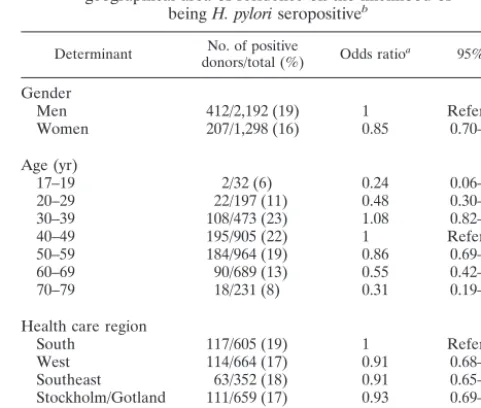
TABLE 2. Multivariate analysis of the effects of gender, age, and geographical area of residence on the likelihood of
beingH. pyloriseropositiveb
Determinant donors/total (%)No. of positive Odds ratioa 95% CI
Gender
Men 412/2,192 (19) 1 Reference
Women 207/1,298 (16) 0.85 0.70–1.02
Age (yr)
17–19 2/32 (6) 0.24 0.06–1.02
20–29 22/197 (11) 0.48 0.30–0.77
30–39 108/473 (23) 1.08 0.82–1.41
40–49 195/905 (22) 1 Reference
50–59 184/964 (19) 0.86 0.69–1.08
60–69 90/689 (13) 0.55 0.42–0.72
70–79 18/231 (8) 0.31 0.19–0.51
Health care region
South 117/605 (19) 1 Reference
West 114/664 (17) 0.91 0.68–1.22
Southeast 63/352 (18) 0.91 0.65–1.28
Stockholm/Gotland 111/659 (17) 0.93 0.69–1.24
Central Sweden 120/773 (16) 0.78 0.59–1.04
North 95/449 (21) 1.22 0.90–1.67
aMutually adjusted for other variables in table.
bThe analysis is restricted to blood donors only. All estimates are mutually adjusted.
on May 15, 2020 by guest
http://jcm.asm.org/
[image:2.603.300.541.496.700.2]gender. This analysis was restricted to blood donors only. Ta-ble 2 shows that the area of residence was of minor importance for the risk of being seropositive. Although the odds ratio for seropositivity tended to be somewhat lower in central Sweden and somewhat higher in northern Sweden, none of the esti-mates attained statistical significance. We also observed an almost statistically significant gender difference in H. pylori
seroprevalence. The odds ratio for being seropositive was 0.85 (95% confidence interval [CI], 0.70 to 1.02) among women relative to men. A similar difference was seen also in the population sample. There, the odds ratio among women was 0.77 (95% CI, 0.57 to 1.04).
Finally, we estimated the odds ratio forH. pylori seroposi-tivity associated with blood donor status, relative to the pop-ulation sample. In these multivariate analyses, performed on the entire material (both blood donors and population con-trols), the estimates were adjusted for gender, age (in 10-year classes), and place of residence (cities of⬎500,000 inhabitants versus the rest of Sweden). Overall, the adjusted odds ratio for
H. pyloriseropositivity among blood donors was decreased by 43% (95% CI, 28 to 55%), relative to population controls (Table 3, first section, left column). In stratified analyses, it was revealed that the effect of blood donor status was modified by age (Table 3, first section, columns 2 and 3). In the age stratum of 17 to 49 years, blood donor status was associated with a 48% (95% CI, 2 to 113%) increased risk ofH. pyloriseropositivity, although this increase was only of borderline significance. Among subjects who wereⱖ50 years of age, the risk of being seropositive for blood donors was reduced by 73% (95% CI, 63 to 81%), relative to the risk in the population sample. This latter interaction was highly statistically significant (P⫽ 0.0001). In order to ensure against confounding by place of residence, we repeated the analyses in the subset of 513 blood donors and 1,030 population controls living in Stockholm. The results were almost identical to those obtained in the entire material (Table 3, second section).
DISCUSSION
The most salient finding in our cross-sectional data was a paradoxical drop inH. pyloriseroprevalence with age among Swedish blood donors over the age of 50 years, who thus markedly differ from the general population with the same age and gender. The seroprevalence among younger blood donors,
on the other hand, was close to, or even somewhat higher than, the age-specific seroprevalence in the general population.
Other studies conducted among blood donors in developed countries have usually not revealed a decline inH. pylori sero-prevalence similar to the one observed by us (5, 7, 15, 20, 26, 33–36). A closer inspection of the previous data, however, unveils lower-than-expected seroprevalence rates above the age of 50 in several studies, (5, 34, 36), but few of them included subjects who were older than 60. In the ages below 60, the deviant pattern was less clear also in our study. An Italian study (31) that included older blood donors found the same seroprevalence pattern as in our study.
The most simplistic explanation for our findings is that blood donations on a regular basis favor disappearance of the infec-tion. This appears biologically implausible, although empirical data are lacking. Removal of antibodies in blood donors could be another possibility but has not been found for other infec-tions. The sample showed no corresponding decrease in the prevalence of antibodies toB. burgdorferi(28).
A biased distribution in socioeconomic background would result in a biasedH. pyloriprevalence but not in a decline with age (12, 23). A successive shift over time in the socioeconomic status of newly recruited blood donors, from low to high, would theoretically lead to lower prevalence rates with increasing age of the donors. However, the prevalence gradient across suc-cessive birth cohorts is likely to be steeper than that across socioeconomic strata. Thus, a shift in the socioeconomic status distribution, if any, would probably not have more than mar-ginal impact on the birth cohort effect, the latter resulting in increasing prevalence rates with age. This argument will also counter another possible variant of selection bias, namely, that only socioeconomically privileged blood donors will remain active after age 50. We therefore tentatively refute this expla-nation, too.
A third alternative explanation would be that subjects who remain active blood donors for the longest time are those with rare and coveted blood groups and that some of these blood groups may be associated with a lower risk ofH. pylori colo-nization. Published data, however, consistently refute any im-portant associations between blood group andH. pylori prev-alence (16, 19, 21, 22, 24, 27). Therefore, this explanation is unlikely.
The fourth, and in our view most probable, explanation is that infected individuals are preferentially removed from the blood donor cohort as they get older. There could be several possible reasons for this selective removal: one would remove them if they are likelier than noninfected individuals to de-velop laboratory test abnormalities that result in active exclu-sion.H. pyloricould potentially affect iron metabolism by caus-ing ulcer-related occult bleedcaus-ing or impaired absorption of nonheme iron or by scavenging heme iron or ferritin (2, 6). Low serum iron and/or ferritin values occur more frequently in infected than in noninfected adults (4, 25, 29). It is conceivable that theH. pylori-induced interference with normal iron me-tabolism will become clinically overt more readily in blood donors than in people without similar blood losses.H. pylori
also seems to be a causative agent in the development of adult vitamin B12deficiency (18). However, this reason does not hold
up, since few blood donors develop persistent anemia and few are excluded for that reason (J. Lundahl, Department of
Clin-TABLE 3. Adjusted odds ratios for beingH. pyloriseropositive, overall and in analyses stratified by age
Study group
Data for: Donors
of all ages aged 17–49 yrDonors agedDonorsⱖ50 yr
ORa 95% CI OR 95% CI OR 95% CI
Entire country
Population control 1 Reference 1 Reference 1 Reference
Blood donors 0.57 0.45–0.72 1.48 1.02–2.13 0.27 0.19–0.37
Stockholmers
Population control 1 Reference 1 Reference 1 Reference
Blood donors 0.57 0.44–0.76 1.47 0.98–2.20 0.26 0.18–0.39
aOdds ratio adjusted for gender, age (in 10-year classes), and place of resi-dence (big cities versus rest of Sweden).
on May 15, 2020 by guest
http://jcm.asm.org/
[image:3.603.43.283.88.204.2]ical Immunology and Transfusion Medicine, Karolinska Hos-pital, personal communication).
The most common reason for exclusion among older Swed-ish blood donors is that they themselves decide to stop giving blood. The reason why is not fully understood, but we hypoth-esize that H. pylori-infected subjects may feel less well after donations than do noninfected blood donors and that the re-covery time may be prolonged. Moreover, ifH. pyloriinfection is causally or noncausally linked to other severe diseases such as coronary heart disease, then these diseases will also contrib-ute to a selective disappearance of infected individuals from the blood donor category.
Our study confirmed the previously reported gender differ-ence (32) with a tendency towards higherH. pyloriprevalence in men than in women, both among blood donors and in the population sample. Although not quite reaching statistical sig-nificance, women had an approximately 20% lower probability of being infected than did men. A recent meta-analysis (32) showed that only in two studies out of six was the H. pylori
seroprevalence higher in women than in men and that the summary odds ratio for seropositivity among men, relative to women, was 1.2 (95% CI, 1.02 to 1.4). Interestingly, the gender difference has not been seen in children, an observation also confirmed in our recent study of 10- to 12-year-old Swedish schoolchildren (13). The reasons behind the gender difference among adults is unlikely to be due to a preferential eradication treatment in men since screening and eradication, at the time of the drawing of study samples, were little practiced in Swe-den. The gender difference may account in part for the in-creased incidence of H. pylori-related diseases (peptic ulcer and stomach cancer) among men in later decades of life (32). Strengths of our study include representativity of our sample vis-a`-vis Swedish blood donors. As an indication of this repre-sentativity, the sample showed the expected geographical vari-ation as well as age- and sex-dependent variability inB. burg-dorferi seroprevalence (28). Another strength is our truly population-based comparison sample. The low participation rate may raise concerns about possible selection bias. The control population did, however, show exactly the expected cohort effect described in numerous studies from developed countries (12, 23). Also, in the age groups where our important findings were made (⬎45 years), the participation rate (66%) was acceptable.
In conclusion, our data, supported by findings in other stud-ies, are consistent with a falling prevalence ofH. pylori infec-tion with age among elderly blood donors. The reasons for this paradoxical decrease remain conjectural, but the most reason-able explanation is thatH. pylori-positive subjects are prefer-entially removed from the blood donor cohort. Since active exclusions due to persistent anemia are uncommon, our results imply that elderly individuals infected withH. pylorimay tol-erate repeated bleedings less well than do noninfected individ-uals and/or that the general well-being among those who are infected may be somewhat impaired. This observation ought to prompt more studies of the role of H. pyloriin the general health status of elderly people. And our findings emphasize the dangers of using blood donor data in lieu of epidemiological sound, population-based observations.
ACKNOWLEDGMENTS
This work was supported by the Swedish Society of Medicine and by the Richard and Ruth Juhlins Foundation.
REFERENCES
1. Banatvala, N., K. Mayo, F. Megraud, R. Jennings, J. J. Deeks, and R. A. Feldman.1993. The cohort effect andHelicobacter pylori.J. Infect. Dis.168: 219–221.
2. Barabino, A.2002.Helicobacter pylori-related iron deficiency anemia: a re-view. Helicobacter7:71–75.
3. Befrits, R., M. Granstro¨m, M. Rylander, and C. Rubio.1993.Helicobacter pyloriin 205 consecutive endoscopy patients. Scand. J. Infect. Dis.25:185– 191.
4. Berg, G., G. Bode, M. Blettner, H. Boeing, and H. Brenner.2001. Helico-bacter pyloriinfection and serum ferritin: a population-based study among 1806 adults in Germany. Am. J. Gastroenterol.96:1014–1018.
5. Bergenzaun, P., K. G. Kristinsson, B. Thjodleifsson, E. Sigvaldadottir, S. Molstad, M. Held, and T. Wadstro¨m.1996. Seroprevalence ofHelicobacter pyloriin south Sweden and Iceland. Scand J. Gastroenterol.31:1157–1161. 6. Bini, E. J.2001.Helicobacter pyloriand iron deficiency anemia: guilty as
charged? Am. J. Med.111:495–497.
7. Breuer, T., T. Sudhop, J. Hoch, T. Sauerbruch, and P. Malfertheiner.1996. Prevalence of and risk factors forHelicobacter pyloriinfection in the western part of Germany. Eur. J. Gastroenterol. Hepatol.8:47–52.
8. Christenson, B., U. Hellstro¨m, S. P. Sylvan, L. Henriksson, and M. Gran-stro¨m.2000. Impact of a vaccination campaign on adult immunity to diph-theria. Vaccine19:1133–1140.
9. Cullen, D. J., B. J. Collins, K. J. Christiansen, J. Epis, J. R. Warren, I. Surveyor, and K. J. Cullen.1993. When isHelicobacter pylori infection acquired? Gut34:1681–1682.
10. Dixon, M. F., and G. M. Sobala.1992. Gastritis and duodenitis: histopatho-logical spectrum. Eur. J. Gastroenterol. Hepatol.4(Suppl. 2):17–23. 11. Ekstro¨m, A. M., M. Held, L. E. Hansson, L. Engstrand, and O. Nyre´n.2001.
Helicobacter pyloriin gastric cancer established by CagA immunoblot as a marker of past infection. Gastroenterology121:784–791.
12. The EUROGAST Study Group.1993. Epidemiology of, and risk factors for,
Helicobacter pyloriinfection among 3194 asymptomatic subjects in 17 popu-lations. Gut34:1672–1676.
13. Granstro¨m, M., Y. Tindberg, and M. Blennow.1997. Seroepidemiology of
Helicobacter pyloriinfection in a cohort of children monitored from 6 months to 11 years of age. J. Clin. Microbiol.35:468–470.
14. Hentschel, E., G. Brandsta¨tter, B. Dragosics, A. M. Hirschl, H. Nemec, K. Schutze, M. Taufer, and H. Wurzer.1993. Effect of ranitidine and amoxicillin plus metronidazole on the eradication ofHelicobacter pyloriand recurrence of duodenal ulcer. N. Engl. J. Med.328:308–312.
15. Holtmann, G., N. J. Talley, H. Mitchell, and S. Hazell.1998. Antibody response to specificH. pyloriantigens in functional dyspepsia, duodenal ulcer disease, and health. Am. J. Gastroenterol.93:1222–1227.
16. Hook-Nikanne, J., P. Sistonen, and T. U. Kosunen.1990. Effect of ABO blood group and secretor status on the frequency ofHelicobacter pylori
antibodies. Scand. J. Gastroenterol.25:815–818.
17. IARC Working Group on the Evaluation of Carcinogenic Risks to Humans. 1994. Schistosomes, liver flukes andHelicobacter pylori.IARC Monogr. Eval. Carcinog. Risks Hum.61:1–241.
18. Kaptan, K., C. Beyan, A. U. Ural, T. Cetin, F. Avcu, M. Gulen, R. Finci, and A. Yalcin.2000.Helicobacter pylori—is it a novel causative agent in Vitamin B12 deficiency? Arch. Intern. Med.160:1349–1353.
19. Kopanski, Z., E. Golec, B. Witkowska, E. Slowakiewicz, A. Migas-Nirska, and A. Cienciala.1996. The relationship between the frequency of the appearance of IgG againstHelicobacter pyloriand the main blood groups among patients with ulcer sickness and stomach cancer. Eur. J. Med. Res. 1:280–282.
20. Kosunen, T. U., J. Hook, H. I. Rautelin, and G. Myllyla.1989. Age-depen-dent increase ofCampylobacter pyloriantibodies in blood donors. Scand. J. Gastroenterol.24:110–114.
21. Martin de Argila, C., D. Boixeda, S. Valdezate, N. Mir, R. Barcena, J. P. Gisbert, A. Garcia Plaza, and R. Canton.1998. ABO blood groups, rhesus factor andHelicobacter pylori.Rev. Esp. Enferm. Dig.90:263–268. 22. McKeown, I., P. Orr, S. Macdonald, A. Kabani, R. Brown, G. Coghlan, M.
Dawood, J. Embil, M. Sargent, G. Smart, and C. N. Bernstein.1999. Heli-cobacter pyloriin the Canadian arctic: seroprevalence and detection in com-munity water samples. Am. J. Gastroenterol.94:1823–1829.
23. Megraud, F., M. P. Brassens-Rabbe, F. Denis, A. Belbouri, and D. Q. Hoa. 1989. Seroepidemiology ofCampylobacter pyloriinfection in various popu-lations. J. Clin. Microbiol.27:1870–1873.
24. Mentis, A., C. C. Blackwell, D. M. Weir, C. Spiliadis, A. Dailianas, and N. Skandalis.1991. ABO blood group, secretor status and detection of Helico-bacter pyloriamong patients with gastric or duodenal ulcers. Epidemiol. Infect.106:221–229.
25. Milman, N., S. Rosenstock, L. Andersen, T. Jorgensen, and O. Bonnevie. 1998. Serum ferritin, hemoglobin, andHelicobacter pyloriinfection: a
on May 15, 2020 by guest
http://jcm.asm.org/
epidemiologic survey comprising 2794 Danish adults. Gastroenterology115: 268–274.
26. Nandurkar, S., N. J. Talley, H. Xia, H. Mitchell, S. Hazel, and M. Jones. 1998. Dyspepsia in the community is linked to smoking and aspirin use but not toHelicobacter pyloriinfection. Arch. Intern. Med.158:1427–1433. 27. Oberhuber, G., A. Kranz, C. Dejaco, B. Dragosics, I. Mosberger, W. Mayr,
and T. Radaszkiewicz.1997. Blood groups Lewis(b) and ABH expression in gastric mucosa: lack of inter-relation withHelicobacter pyloricolonisation and occurrence of gastric MALT lymphoma. Gut41:37–42.
28. O’Connell, S., M. Granstro¨m, J. S. Gray, and G. Stanek.1998. Epidemiology of European Lyme borreliosis. Zentbl. Bakteriol.287:229–240.
29. Parkinson, A. J., B. D. Gold, L. Bulkow, R. B. Wainwright, B. Swaminathan, B. Khanna, K. M. Petersen, and M. A. Fitzgerald.2000. High prevalence of
Helicobacter pyloriin the Alaska native population and association with low serum ferritin levels in young adults. Clin. Diagn. Lab. Immunol.7:885–888. 30. Parsonnet, J., G. D. Friedman, D. P. Vandersteen, Y. Chang, J. H. Vogelman, N. Orentreic, and R. K. Sibley.1991.Helicobacter pyloriinfection and the risk of gastric carcinoma. N. Engl. J. Med.325:1127–1131.
31. Pellicano, R., M. G. Mazzarello, S. Morelloni, M. Allegri, V. Arena, M.
Ferrari, M. Rizzetto, and A. Ponzetto.1999. Acute myocardial infarction and
Helicobacter pyloriseropositivity. Int. J. Clin. Lab. Res.29:141–144. 32. Replogle, M. L., S. L. Glaser, R. A. Hiatt, and J. Parsonnet.1995. Biologic
sex as a risk factor forHelicobacter pyloriinfection in healthy young adults. Am. J. Epidemiol.142:856–863.
33. Russo, A., M. Eboli, P. Pizzetti, G. Di Felice, F. Ravagnani, P. Spinelli, A. M. Hotz, P. Notti, G. Maconi, S. Franceschi, D. Ferrari, and L. Bertario.1999. Determinants ofHelicobacter pyloriseroprevalence among Italian blood do-nors. Eur. J. Gastroenterol. Hepatol.11:867–873.
34. Vaira, D., M. Miglioli, P. Mule, J. Holton, M. Menegatti, M. Vergura, G. Biasco, R. Conte, R. P. Logan, and L. Barbara.1994. Prevalence of peptic ulcer inHelicobacter pyloripositive blood donors. Gut35:309–312. 35. Wagtmans, M. J., A. M. Witte, D. R. Taylor, I. Biemond, R. A. Veenendaal,
H. W. Verspaget, C. B. Lamers, and R. A. van Hogezand.1997. Low sero-prevalence ofHelicobacter pyloriantibodies in historical sera of patients with Crohn’s disease. Scand. J. Gastroenterol.32:712–718.
36. Wilhoite, S. L., D. A. Ferguson, Jr., D. R. Soike, J. H. Kalbfleisch, and E. Thomas.1993. Increased prevalence ofHelicobacter pyloriantibodies among nurses. Arch. Intern. Med.153:708–712.