Copyright © 2004, American Society for Microbiology. All Rights Reserved.
Evaluation of DNA Extraction and PCR Methods for Detection of
Enterocytozoon bienuesi
in Stool Specimens
Ittisak Subrungruang,
1Mathirut Mungthin,
2* Porntip Chavalitshewinkoon-Petmitr,
1Ram Rangsin,
3Tawee Naaglor,
2and Saovanee Leelayoova
2Department of Protozoology, Faculty of Tropical Medicine, Mahidol University,1and Department of Parasitology2and
Department of Military and Community Medicine, Phramongkutklao College of Medicine,3Ratchathewi,
Bangkok 10400, Thailand
Received 27 February 2004/Returned for modification 11 April 2004/Accepted 4 May 2004
An evaluation of the sensitivities of three DNA extraction methods, i.e., FTA filter paper, a QIAamp stool mini kit, and a conventional phenol-chloroform method, by using specimens with known concentrations of
Enterocytozoon bieneusispores was performed. FTA filter paper and the QIAamp stool mini kit were the most
sensitive methods, which could detectE. bieneusiin specimens with a concentration of 800 spores/ml. We also
compared five previously described PCR methods that use five different primer pairs for the detection ofE.
bieneusiand showed that MSP3-MSP4B and EBIEF1-EBIER1 were the most sensitive primers. Although both sets of primers showed the same sensitivity, using the MSP3-MSP4B primers can directly provide genotypic information by sequencing. A blinded diagnostic test to compare PCR and light microscopy methods for the
detection ofE. bieneusiin stool specimens was also conducted. The use of FTA filter paper for DNA extraction
together with the PCR method using the primer pair MSP3-MSP4B showed 100% sensitivity and 100%
specificity for the detection of E. bieneusi in stool specimens, while the light microscopy method gave a
sensitivity of 86.7% and a specificity of 100%.
Enterocytozoon bieneusi is an emerging pathogen causing
diarrhea in patients with human immunodeficiency virus infec-tion and other immunosuppressive condiinfec-tions (9, 20, 33). Self-limited diarrhea as well as chronic diarrhea in immunocompe-tent patients has also been reported (25, 32). The prevalence of
E. bieneusiin human immunodeficiency virus-infected patients
with diarrhea was 2 to 50%, depending on the study population and methods of diagnosis (2, 8, 16, 20). Several staining meth-ods such as Gram-chromotrope (17), modified trichrome (31), and chemofluorescence stains such as Calcofluor White M2R (29) have been developed for the detection of E. bieneusi. However, the accuracy of microscopic diagnosis of this organ-ism depends on the experience of the microscopist. Moreover, staining methods cannot differentiate down to the species level. Thus, electron microscopy is still necessary for confirmation of the diagnosis and species identification (6). Molecular tech-niques that rely on PCR-based methods to amplify different regions of the small subunit rRNA (SSU rRNA) gene for the identification ofE. bieneusihave been successfully developed (4, 7, 26, 30, 35). More recently, a real-time PCR method was used to quantifyE. bieneusiDNA in stool specimens for mon-itoring treatment in immunocompromised patients (15). A multicenter study has shown that PCR can detectE. bieneusiin concentrations as low as 102spores/g of stool, while a detection
limit of 104spores/g of stool was apparent for light microscopy
(22). Thus, epidemiological studies based on only light micros-copy may give prevalence data which do not reflect the true prevalence ofE. bieneusi.
PCR amplification using stool specimens for the detection of
E. bieneusicould be insensitive because of PCR inhibitors and
the difficulty of spore disruption. To raise the sensitivity of PCR to diagnoseE. bieneusi infection, an efficient DNA ex-traction method is needed. Commercial DNA exex-traction kits such as the QIAamp stool mini kit (QIAGEN, Hilden, Ger-many), Instagene Matrix (Bio-Rad, Hercules, Calif.), and RapidPrep Micro Genomic DNA isolation kit (Pharmacia Bio-tech Inc., Piscataway, N.J.) have shown their usefulness for DNA extraction from stool specimens. Recently, the extrac-tion-free FTA filter method (Whatman Bioscience, Cam-bridge, United Kingdom) has been demonstrated to have high sensitivity for DNA detection by PCR (19). These DNA ex-traction methods, however, have never been compared. We aimed to evaluate the sensitivities of three DNA extraction methods, i.e., a QIAamp stool mini kit, FTA filter paper, and a conventional phenol-chloroform method.
In recent years, researchers have developed PCR methods for the detection ofE. bieneusiin stool specimens. These meth-ods have also never been compared. We chose to evaluate five previously described single-step PCR methods (4, 7, 14, 26, 30) with species-specific primer sets for the detection ofE. bieneusi
in stool specimens. Moreover, the sensitivities and specificities of the most sensitive PCR method using the most sensitive DNA extraction method were compared with results obtained from light microscopy by using electron microscopy as the gold standard.
MATERIALS AND METHODS
Stool samples.Stool specimens were collected from 290 children who lived in an orphanage situated in Bangkok, Thailand, during a routine stool examination that was performed every 6 months by the Department of Parasitology, Phramongkutklao College of Medicine, Bangkok, Thailand. Stool specimens were stained with Gram-chromotrope as previously described (17) and examined
* Corresponding author. Mailing address: Department of Parasitol-ogy, Phramongkutklao College of Medicine, 315 Ratchawithi Rd., Rat-chathewi, Bangkok 10400, Thailand. Phone and fax: 662 354 7761. E-mail: [email protected].
3490
on May 15, 2020 by guest
http://jcm.asm.org/
under a 100⫻objective by light microscopy forE. bieneusi. Confirmation of the presence ofE. bieneusiwas performed by electron microscopy or PCR. From these samples, a single positive specimen was used to evaluate the sensitivities of three DNA extraction and five species-specific PCR methods. All positive spec-imens confirmed by electron microscopy were used for the evaluation of sensi-tivities and specificities of light microscopy and the PCR method. These speci-mens were stored at 4°C for less than 3 months. Stool specispeci-mens with noE. bieneusi spores were from healthy persons who lived in a rural community outside Bangkok. These samples were ruled out forE. bieneusiby negative Calcofluor staining.
Evaluation of DNA extraction methods.Three DNA extraction methods, i.e., FTA filter paper, a QIAamp stool mini kit, and a conventional phenol-chloro-form method were compared. For extraction with the FTA filter paper, 6-mm disks were punched out from FTA filter paper (Whatman Bioscience) by using a modified hole punch and placed in a 1.5-ml microcentrifuge tube. Fifteen mi-croliters of each diluted sample was spotted onto the FTA disks and dried on a heating block at 56°C. One quarter of each disk was used for one test since it could fit in a 0.2-ml PCR tube. The FTA disk was washed twice with 200l of FTA purification buffer (Life Technologies, Gaithersburg, Md.) for 15 min and then washed twice with 200l of TE buffer (10 mM Tris-HCl [pH 8.0], 0.1 mM EDTA [pH 8.0]) for 5 min and again dried on a heating block at 56°C. The washed FTA disks were used as the DNA template in PCR amplification. It is critical that the TE buffer used to wash the FTA disks have a pH of 8.0; otherwise, there may be interference with the DNA amplification. Moreover, FTA disks have to be completely dried before the PCR is performed. For the QIAamp stool mini kit (QIAGEN), 200l of stool specimen was used for DNA extraction, following the manufacturer’s instructions. The extracted DNA of each sample was kept frozen at⫺20°C until used. The phenol-chloroform ex-traction method was performed as described by Katzwinkel-Wladarsch et al. (7). Two hundred microliters of diluted stool was added with 33.3l of 1 M KOH and 9.3l of 1 M dithiothreitol and mixed thoroughly in a microcentrifuge tube. After incubation at 65°C for 15 min, the samples were neutralized with 4.3l of 25% HCl, buffered with 80l of 2 M Tris-HCl (pH 8.3), and the suspension was mixed again. DNA was extracted by shaking with 250l of phenol-chloroform-isoamyl alcohol (25:24:1) saturated with 10 mM Tris (pH 8.0) and 1 mM EDTA. To precipitate nucleic acids from the aqueous phase, 0.1 volume of 3 M sodium acetate, pH 5.0, and 2 volumes of cold absolute ethanol were used. DNA was collected immediately by centrifugation at 10,000⫻gfor 20 min; then ethanol was removed, and the pellet was collected after washing with 0.5 ml of cold 70% ethanol. To remove the residual ethanol, 1 ml of acetone was added, and the pellet was then dried at 37 to 50°C with the lid open. The DNA pellet was resuspended in 200l of TE buffer and kept at⫺20°C until used.
To determine the sensitivities of these three DNA extraction methods, a stool specimen with a known concentration of microsporidial spores was used. A spore count ofE. bieneusiwas performed by using a positive stool sample dissolved in 200l of phosphate-buffered saline, pH 7.5. This specimen was stained by Gram-chromotrope and counted by light microscopy. Dilutions of homogenized stools were made to give different spore concentrations at 100,000, 20,000, 4,000, 800, and 160 spores/ml. For the QIAamp stool mini kit and phenol-chloroform methods, 200l of diluted stool sample was used for each test. A total of 10l of each extracted DNA specimen was used in the PCR amplification to obtain the equivalent of 1,000, 200, 40, 8, and 1.6 spores per PCR mixture. The sensi-tivity of the FTA filter was assessed by using the same samples as used for the QIAamp stool mini kit and conventional phenol-chloroform method. Since the amount of specimen placed on the FTA disk was limited to 15l and one-fourth of the FTA disk was used for each test, the number of spores was equivalent to 375, 75, 15, 3, and 0.6 spores per PCR mixture. The most sensitive extraction method is defined as the method that can extract DNA from the specimen with the lowest spore concentration and give a positive band ofE. bieneusiby PCR amplification with the primer pair MSP3-MSP4B with PCR conditions as
de-scribed by Katzwinkel-Wladarsch et al. (7). The evaluation of each method was performed three times.
Evaluation of PCR amplification methods.Five single-step PCR methods for the detection ofE. bieneusiwere compared for their sensitivities. Stool specimens with five different concentrations ofE. bieneusispores, i.e., 100,000, 20,000, 8,000, 400, and 160 spores/ml, were extracted for DNA by FTA filter paper. The most sensitive PCR method is defined as the method that can amplify the specimen containing the lowest spore concentration. These five sets of primer pairs included MSP3-MSP4B (7), EBIEF1-EBIER1 (4), Primer set 2 (26), Eb.gc-Eb.gt (30), and V1-Mic3 (14). Genomic DNA and the primer pairs were used with PCR conditions as previously described (4, 7, 14, 26, 30). PCR amplification was performed by using a Perkin Elmer 480 thermal cycler. A 10-l PCR product from each reaction mixture was run on a 2% agarose gel (FMC Bioproducts, Rockland, Maine) with 1% Tris-borate-EDTA buffer. Gels were stained with ethidium bromide and visualized under UV light and documented on high-density printing paper by using a UV-save gel documentation system I (UVItech, Cambridge, United Kingdom). The evaluation of the sensitivities of each PCR method was performed three times.
Comparison of PCR and light microscopy for the detection ofE. bieneusiin stool specimens. The most sensitive DNA extraction and PCR method was chosen for the comparison of the light microscopy and PCR methods for the detection ofE. bieneusiin stool samples. The blinded evaluation of both tech-niques was performed by using 30 positive and 30 negative stool samples. Stool specimens were examined microscopically by an experienced microscopist by using Gram-chromotrope staining. The sources of all positive and negative stool samples were described above. Sensitivities and specificities were calculated by using two-by-two tables and Epi Info version 6.01 software. A chi-square test was used to determine the significance of the difference between two proportions for sensitivities and specificities of the two diagnostic methods.
RESULTS
Sensitivities of DNA extraction methods.To evaluate the
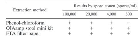
[image:2.603.300.541.90.154.2]sensitivities of these three extraction methods, specimens with known spore concentrations were used for the experiment. As shown in Table 1, FTA filter paper and the QIAamp stool mini kit could detect E. bieneusiat concentrations as low as 800 spores/ml. The sensitivity of the phenol-chloroform extraction method was equivalent to 4,000 spores/ml. The comparison shows that FTA filter paper and the QIAamp stool mini kit are more sensitive compared to the conventional phenol-chloro-form method for DNA extraction ofE. bieneusiin stool spec-imens.
TABLE 1. Comparison of the sensitivities of three DNA extraction methods for detection ofE. bieneusiin stool specimens
Extraction method Results by spore concn (spores/ml)
100,000 20,000 4,000 800 160
Phenol-chloroform ⫹ ⫹ ⫹ ⫺ ⫺
QIAamp stool mini kit ⫹ ⫹ ⫹ ⫹ ⫺
FTA filter paper ⫹ ⫹ ⫹ ⫹ ⫺
TABLE 2. Comparison of the sensitivities of five PCR methods for the detection ofE. bieneusiin stool specimens
Primer pair Expected amplicon(bp) Reference Results by spore concn (spores/ml)
100,000 20,000 4,000 800 160
MSP3-MSP4B 508 7 ⫹ ⫹ ⫹ ⫹ ⫺
EBIEF1-EBIER1 607 4 ⫹ ⫹ ⫹ ⫹ ⫺
Primer set 2 1,294 24 ⫺ ⫺ ⫺ ⫺ ⫺
Eb.gc-Eb.gt 210 29 ⫹ ⫹ ⫺ ⫺ ⫺
V1-Mic3 446 14 ⫹ ⫹ ⫺ ⫺ ⫺
on May 15, 2020 by guest
http://jcm.asm.org/
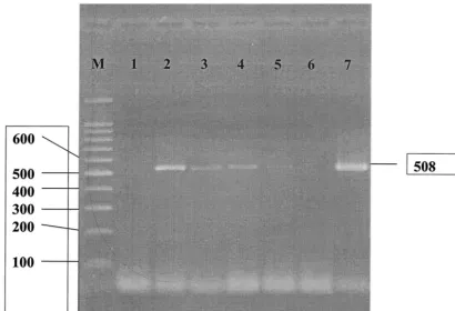
[image:2.603.43.543.645.725.2]Sensitivities of PCR methods.A comparison of the sensitiv-ities of five PCR methods is shown in Table 2. Our results demonstrated that the two protocols that use the MSP3-MSP4B and EBIEF1-EBIER1 primer pairs were the most sensitive methods for detectingE. bieneusiat concentrations as low as 800 spores/ml. Figure 1 shows the sensitivity testing of the PCR method using the primer pair MSP3-MSP4B to am-plifyE. bieneusiin stool specimens. The protocols which used the Eb.gc-Eb.gt and V1-Mic3 primers could detectE. bieneusi
in specimens with 20,000 spores/ml. Following the technique of Schuitema et al. (26) and using Primer set 2, we could not amplify the DNA ofE. bieneusiat a concentration of 100,000 spores/ml. Positive results were found only in specimens with spore concentrations higher than 100,000 spores/ml.
Sensitivities and specificities of PCR and light microscopy
for the detection of E. bieneusi. Based on the above results,
FTA filter paper was chosen for DNA extraction together with the PCR method using the primer pair MSP3-MSP4B to test their sensitivities and specificities for the detection ofE.
bie-neusiin stool specimens. Although the PCR method using the
EBIEF1-EBIER1 primer pair gave the same sensitivity as the primer pair MSP3-MSP4B, we chose the MSP3-MSP4B primer pair because of its usefulness in terms of genotypic character-ization. Table 3 shows the sensitivities and specificities of the PCR method and light microscopy with Gram-chromotrope staining. The sensitivities of PCR and light microscopy with Gram-chromotrope staining were 100% (95% confidence in-terval [CI], 90.5 to 100) and 86.7% (95% CI, 70.9 to 95.6), respectively. The specificities of both PCR and light
micros-copy with Gram-chromotrope staining were 100% (95% CI, 90.5 to 100). The difference between the sensitivities of both methods was 13.3% (95% CI, 4.4 to 29.1). There were no significant differences between the results from PCR and light microscopy with Gram-chromotrope staining in terms of sen-sitivity (P⫽0.9) and specificity (P⫽0.36).
DISCUSSION
The detection of protozoan parasites in stool specimens by PCR usually requires a highly sensitive DNA extraction method because of the presence of inhibitors in stools. To choose a sensitive method for the PCR detection ofE. bieneusi
[image:3.603.87.497.70.349.2]DNA in stool specimens, we evaluated three DNA extraction methods: a QIAamp stool mini kit, FTA filter paper, and a conventional phenol-chloroform method. Our study showed FIG. 1. PCR analysis using the MSP3-MSP4B primer pair specific to the SSU rRNA gene ofE.bieneusi. Lane M, molecular markers of 100-bp DNA ladder; lane 1, negative control; lanes 2 to 6, different concentrations of spores (100,000, 20,000, 4,000, 800, and 160 spores/ml, respectively); lane 7, positive control.
TABLE 3. Comparison of PCR and light microscopy for the detection ofE. bieneusi
Method and result E. bieneusispecimens
No. positive (%) No. negative (%)
PCR
Positive 30 (100) 0 (0)
Negative 0 (0) 30 (100)
Light microscopy
Positive 26 (86.7) 0 (0)
Negative 4 (13.3) 30 (100)
on May 15, 2020 by guest
http://jcm.asm.org/
[image:3.603.301.542.635.725.2]that the detection limit varied between 800 to 4,000 spores/ml, depending on the extraction method. Although the phenol-chloroform method has been widely used as a standard method for DNA extraction, our data showed that this less sensitive method could detect spores at a concentration of 4,000 spores/ ml. Since DNA was extracted directly from stool samples, poor DNA extraction efficiency or incomplete removal of inhibitors could have occurred. The extraction protocol involved several steps; thus, limited numbers of specimens could be tested each time. To raise the sensitivity of DNA extraction, spore concen-tration or purification and/or DNA purification afterward may be required (12, 18, 27, 28).
Our experiments showed that both the QIAamp stool mini kit and FTA filter paper could detect E. bieneusi in stool specimens with the same concentration of 800 spores/ml. The QIAamp stool mini kit procedure involves digestion of pro-teins, elimination of inhibitors, binding DNA to silica gel mem-brane, and elution of DNA by spin column. In this study, stool homogenate was incubated at 70°C for 5 min with lysis buffer for efficient spore lysis before the extraction. Since spores ofE.
bieneusi are resistant to disruption and lysis, increasing the
incubation temperature of the stool homogenates in the lysis buffer up to 95°C may be helpful to improve sensitivity. The QIAamp stool mini kit involves several steps and takes approx-imately 1 h to be completed, thus limiting the numbers of specimens that can be handled at one time.
FTA filter paper is an extraction-free, filter-based template which is impregnated with denaturants, chelating agents, and a free-radical trap. Lysis of organisms occurs upon contact with the FTA filter paper, and then DNA is trapped on the matrix. Inhibitors or other debris are effectively removed by washing reagent. Since the application of samples onto the FTA filters is very simple, a large number of samples for field epidemio-logical studies can be prepared simultaneously by untrained personnel with less technical equipment. The whole procedure, including drying and washing to get the DNA template ready for PCR amplification, takes less than 3 h after spotting the specimens. The disks can be stored at room temperature so that they are easy to handle and transport for further analysis. FTA filter paper and the QIAamp stool mini kit are commer-cially available with approximately the same price on a sample basis. Modification of the FTA filters by using an individual hole punch to get an FTA disk with a 6-mm diameter for tested samples can reduce the cost. Thus, for long-term use, FTA filters are a low-cost method with high sensitivity, which makes them a good choice for use with large numbers of samples. In addition, the amount of sample required for FTA filter paper is less than that for the QIAamp stool mini kit.
A number ofE. bieneusi-specific PCR protocols, which are usually based on the amplification of the intergenic transcribed spacer region of the SSU rRNA gene, have been developed. However, there has been no evaluation of these PCR methods. We evaluated single-step PCR rather than nested PCR meth-ods for the detection ofE. bieneusiin stool specimens because the single-step PCR method is convenient and less time con-suming. Based on the above data, we used the FTA filter method for DNA extraction to evaluate five PCR protocols, i.e., PCR with the primer pairs MSP3-MSP4B, EBIEF1-EBIER1, Primer set 2, Eb.gc-Eb.gt, and V1-Mic3. Our study showed that the MSP3-MSP4B and EB1EF1-EB1ER primer
pairs were the most sensitive primers to detectE. bieneusiin stool specimens. Both MSP3-MSP4B and EB1EF1-EB1ER have been shown to beE. bieneusispecific and do not cross-amplify with other human microsporidia (4, 7). These two PCR protocols have been used for the identification ofE. bieneusi
both in humans and animals (1, 12, 13, 21). The MSP3-MSP4B primer set has shown satisfactory results for species-specific identification (5, 23, 24). For the purpose of genotypic char-acterization, comparisons of the intergenic transcribed spacer sequences amplified by MSP3-MSP4B can differentiate their genotype.
The protocol of the Eb.gc-Eb.gt primer set was also tested and was shown to have less sensitivity in the present study. In contrast to the protocols using the MSP3-MSP4B and EB1EF1-EB1ER primer pairs, the Eb.gc-Eb.gt primer pair requires PCR-restriction fragment length polymorphism for genotypic characterization of E. bieneusi(11). However, the PCR-restriction fragment length polymorphism technique has the disadvantage that any mutations or deletions between the restriction enzyme recognition sites are not accessible. The usefulness of genotypic differentiation might be limited.
The use of forward primer V1 (35) and reverse primer Mic3 was developed for E. bieneusi identification (14). The PCR protocol with the V1-Mic3 primer pair and in situ hybridiza-tion procedures was a sensitive diagnostic tool and enabled the differentiation ofE. bieneusifrom other microsporidia (3). Our study demonstrated that the PCR protocol using the V1-Mic3 primer pair was not as sensitive as the protocols with the MSP3-MSP4B and EB1EF1-EB1ER primer pairs. Primer set 2 was designed by Schuitema et al. (26) and was shown to be useful for the detection ofE. bieneusiin stool specimens (10, 14). However, we found that Primer set 2 was the least sensi-tive protocol in this study.
Rinder et al. (22) conducted a blinded, externally controlled multicenter evaluation of light microscopy and PCR for the detection of E. bieneusi in stool specimens. The sensitivities reported from six different laboratories were between 71 to 100%. The differences in the sensitivities might be dependent on DNA extraction methods and PCR protocols. Recently, a real-time PCR method for the detection of Encephalitozoon
intestinalis from stool specimens has been developed. It has
been shown that real-time PCR is more sensitive than light microscopy (34). In the present study, we evaluated the sensi-tivities and specificities of PCR and light microscopy for the detection ofE. bieneusiin stool specimens. We used the most sensitive techniques to detectE. bieneusi, i.e., FTA filter paper for DNA extraction and the MSP3-MSP4B primer pair for PCR amplification. Although light microscopy seemed to be less sensitive than the PCR method, there is no statistically significant difference between these two methods. The nonsig-nificant difference might be due to the specimens used in this study, which contained high concentrations of spores and were examined by a highly experienced microscopist. Although light microscopy is an inexpensive method, it cannot differentiate species of microsporidia. Light microscopy, thus, is suitable for a routine screening test. The PCR method using the MSP3-MSP4B primer pair together with FTA filter paper for DNA extraction gave 100% sensitivity and 100% specificity for the detection ofE. bieneusiin stool specimens. Hence, this method is a powerful diagnostic method for evaluating clinical
on May 15, 2020 by guest
http://jcm.asm.org/
mens and also a meaningful tool for epidemiological study of this infection. Moreover, genotype can be characterized by sequence analysis of the PCR product. Understanding the ge-netic variations ofE. bieneusistrains among the population will be useful for exploring the source, transmission, and pathogen-esis of this organism.
In conclusion, we demonstrate that both FTA filter paper and the QIAamp stool mini kit were sensitive methods for the DNA extraction ofE. bieneusiin stool specimens. We chose FTA filter paper for further investigation since it is easy to use, is a rapid test, does not require experienced persons to handle specimens, and requires smaller amounts of stool specimens. Comparison of five PCR protocols showed that the PCR pro-tocols using the primer pairs MSP3-MSP4B and EB1EF1-EB1ER are the most sensitive methods for the detection ofE.
bieneusi in the present study. Using these sensitive DNA
ex-traction and PCR methods together gave high sensitivity and specificity for the detection ofE. bieneusi.
ACKNOWLEDGMENTS
This work was supported by Thailand-Tropical Diseases Research Programme (T-2), ID 02-2-ARI-24-007.
REFERENCES
1. Breitenmoser, A. C., A. Mathis, E. Bu¨rgi, R. Weber, and P. Deplazes.1999. High prevalence ofEnterocytozoon bieneusiin swine with four genotypes that differ from those identified in humans. Parasitology118:447–453. 2. Canning, E. U., and W. S. Hollister.1990.Enterocytozoon bieneusi
(Micros-pora): prevalence and pathogenicity in AIDS patients. Trans. R. Soc. Trop. Med. Hyg.84:181–186.
3. Carville, A., K. Manfield, G. Widmer, A. Lackner, D. Kotler, P. Wiest, T. Gumbo, S. Sarbah, and S. Tzipori.1997. Development and application of genetic probes for detection ofEnterocytozoon bieneusiin formalin-fixed stools and in intestinal biopsy specimens from infected patients. Clin. Diagn. Lab. Immunol.4:405–408.
4. da Silva, A. J., D. A. Schwartz, G. S. Visvesvara, H. de Moura, S. B. Sle-menda, and N. J. Pieniazek.1996. Sensitive PCR diagnosis of infections by Enterocytozoon bieneusi(microsporidia) using primers based on the region coding for small-subunit rRNA. J. Clin. Microbiol.34:986–987.
5. Dengjel, B., M. Zahler. W. Hermanns, K. Heinritzi, T. Spillmann, A. Thom-schke, T. Loscher, R. Gothe, and H. Rinder.2001. Zoonotic potential of Enterocytozoon bieneusi.J. Clin. Microbiol.39:4495–4499.
6. Franzen, C., and A. Mu¨ller.1999. Molecular techniques for detection, spe-cies differentiation, and phylogenetic analysis of microsporidia. Clin. Micro-biol. Rev.12:243–285.
7. Katzwinkel-Wladarsch, S., M. Lieb, W. Helse, T. Lo¨scher, and H. Rinder. 1996. Direct amplification and species determination of microsporidian DNA from stool specimen. Trop. Med. Int. Health1:373–378.
8. Kotler, D. P., and J. M. Orenstein.1994. Prevalence of enteric pathogens in HIV-infected individuals referred for gastrointestinal evaluation. Am. J. Gastroenterol.89:1998–2002.
9. Kotler, D. P., and J. M. Orenstein.1998. Clinical syndromes associated with microsporidiosis. Adv. Parasitol.40:321–349.
10. Ligoury, O., F. David, C. Sarfati, A. R. J. Schuitema, R. A. Hartskeerl, F. Derouin, J. Modaï, and J. M. Molina.1997. Diagnosis of infections caused byEnterocytozoon bieneusiandEncephalitozoon intestinalisusing polymerase chain reaction in stool specimens. AIDS11:723–726.
11. Ligoury, O., F. David, C. Sarfati, F. Derouin, and J. M. Molina.1998. Determination of types ofEnterocytozoon bieneusistrains isolated from pa-tients with intestinal microsporidiosis. J. Clin. Microbiol.36:1882–1885. 12. Lores, B., C. del Aguila, and C. Arias.2002.Enterocytozoon bieneusi
(mi-crosporidia) in faecal samples from domestic animal from Galicia, Spain. Mem. Inst. Oswaldo Cruz97:941–945.
13. Lores, B., I. Lopez-Miragaya, C. Arias, S. Fenoy, J. Torres, and C. del Aguila.2002. Intestinal microsporidiosis due toEnterocytozoon bieneusiin elderly human immunodeficiency virus-negative patients from Vigo, Spain. Clin. Infect. Dis.34:918–921.
14. Manfield, K. G., A. Carville, D. Shvetz, J. Mackey, S. Tzipori, and A. A. Lackner.1997. Identification of anEnterocytozoon bieneusi-like
microspo-ridian parasite in simian-immunodeficiency-virus-inoculated macaques with hepatobiliary disease. Am. J. Pathol.150:1395–1405.
15. Menotti, J., B. Cassinat, R. Porcher, C. Sarfati, F. Derouin, and J. M. Molina.2003. Development of a real-time polymerase-chain-reaction assay for quantitative detection ofEnterocytozoon bieneusiDNA in stool speci-mens from immunocompromised patients with intestinal microsporidiosis. J. Infect. Dis.187:1469–1474.
16. Molina, J. M., C. Sarfati, B. Beauvais, M. Lemann, A. Lesourd, F. Ferchal, I. Casin, P. Lagrange, R. Modigliani, F. Derouni, and J. Modai.1993. Intestinal microsporidiosis in human immunodeficiency virus-infected pa-tients with chronic unexplained diarrhea: prevalence and clinical and bio-logic features. J. Infect. Dis.167:217–221.
17. Moura, H., J. L. da Silva, F. C. Sodre, P. Brasil, D. Walmo, S. Wahlquist, S. Wallace, G. P. Croppo, and G. S. Visvesvara.1996. Gram-chromotrope: a new technique that enhances detection of microsporidia spores in clinical specimens. J. Eukaryot. Microbiol.43:94S–95S.
18. Muller, A., K. Stellermann, P. Hartmann, M. Schrappe, G. Fatkenheuer, B. Salzberger, V. Diehl, and C. Franzen.1999. A powerful DNA extraction method and PCR for detection of microsporidia in clinical stool specimens. Clin. Diag. Lab. Immunol.6:243–246.
19. Orlandi, P. A., and K. A. Lampel.2000. Extraction-free, filter-based template preparation for rapid and sensitive PCR detection of pathogenic parasitic protozoa. J. Clin. Microbiol.38:2271–2277.
20. Rabenek, L., F. Gyorkey, R. M. Genta, P. Gyorkey, L. W. Foote, and J. M. Risser.1993. The role of Microsporidia in the pathogenesis of HIV-related chronic diarrhea. Ann. Intern. Med.119:895–899.
21. Reetz, J., H. Rinder, A. Thomschke, H. Manke, M. Schwebs, and A. Bru-derek.2002. First detection of the microsporidiumEnterocytozoon bieneusiin non-mammalian hosts (chickens). Int. J. Parasitol.32:785–787.
22. Rinder, H., K. Janitschke, H. Aspo¨ck, A. J. Da Silva, P. Deplazes, D. P. Fedorko, C. Franzen, U. Futh, F. Hu¨nger, A. Lehmacher, C. G. Meyer, J. M. Molina, J. Sandfort, R. Weber, T. Lo¨scher, and the Diagnostic Multicenter Study Group on Microsporidia.1998. Blinded, externally controlled multi-center evaluation of light microscopy and PCR for detection of microspo-ridia in stool specimens. J. Clin. Microbiol.36:1814–1818.
23. Rinder, H., A. Thomschke, B. Dengjel, R. Gothe, T. Luscher, and M. Zahler. 2000. Close genotypic relationship betweenEnterocytozoon bieneusi from humans and pigs and first detection in cattle. J. Parasitol.86:185–188. 24. Sadler, F., N. Peake, R. Borrow, P. L. Rowl, E. G. L. Wilkin, and A. Curry.
2002. Genotyping ofEnterocytozoon bieneusiin AIDS patients from the north west of England. J. Infect.44:39–42.
25. Sandfort, J., A. Hannemann, H. Gelderblom, K. Stark, R. L. Owen, and B. Ruf.1994.Enterocytozoon bieneusiinfection in an immunocompetent patient who had acute diarrhea and who was not infected with the human immu-nodeficiency virus. Clin. Infect. Dis.19:514–516.
26. Schuitema, A. R. J., R. A. Hartskeerl, T. van Gool, R. Laxminarayan, and W. J. Terpstra.1993. Application of the polymerase chain reaction for the diagnosis of microsporidiosis. AIDS7(Suppl. 3):S62–S63.
27. Sulaiman, I. M., R. Fayer, A. A. Lal, J. M. Trout, F. W. Schaefer III, and L. Xiao.2003. Molecular characterization of microsporidia indicated that wild mammals harbor host-adaptedEnterocytozoonspp. as well as human-patho-genicEnterocytozoon bieneusi.Appl. Environ. Microbiol.69:4495–4501. 28. van Gool, T., E. U. Canning, and J. Dankert.1994. An improved practical
and sensitive technique for the detection of microsporidian spores in stool samples. Trans. R. Soc. Trop. Med. Hyg.88:189–190.
29. Va´vra, J., R. Dahbiova´, W. S. Hollister, and E. U. Canning.1993. Staining of microsporidian spores by optical brighteners with remarks on the use of brighteners for the diagnosis of AIDS associated human microsporidioses. Folia Parasitol.40:267–272.
30. Velasquez, J. N., S. Carnevale, E. A. Guarnera, J. H. Labbe, A. Chertcoff, M. G. Cabrera, and M. I. Rodriguez.1996. Detection of the microsporidian parasiteEnterocytozoon bieneusiin specimens from patients with AIDS by PCR. J. Clin. Microbiol.34:3230–3232.
31. Weber, R., R. T. Bryan, R. L. Owen, C. M. Wilcox, L. Gorelkin, and G. S. Visvesvara.1992. Improved light-microscopical detection of microsporidia spores in stool and duodenal aspirates. N. Engl. J. Med.326:161–166. 32. Weber, R., and R. T. Bryan.1994. Microsporidial infections in
immunode-ficient and immunocompetent patients. Clin. Infect. Dis.19:517–521. 33. Weber, R., R. T. Bryan, D. A. Schwartz, and R. L. Owen.1994. Human
microsporidial infection. Clin. Microbiol. Rev.7:426–461.
34. Wolk, D. M., S. K. Schneider, N. L. Wengenack, L. M. Sloan, and J. E. Rosenblatt.2002. Real-time PCR method for detection ofEncephalitozoon intestinalisfrom stool specimens. J. Clin. Microbiol.40:3922–3928. 35. Zhu, X., M. Wittner, H. B. Tanowitz, D. Kolter, A. Cali, and L. M. Weiss.
1993. Small subunit rRNA sequence ofEnterocytozoon bieneusiand its po-tential diagnostic role with use of the polymerase chain reaction. J. Clin. Microbiol.168:1570–1575.