R E S E A R C H A R T I C L E
Open Access
Synergistic study of a Danshen (Salvia
Miltiorrhizae Radix et Rhizoma) and Sanqi
(Notoginseng Radix et Rhizoma)
combination on cell survival in EA.hy926
cells
Xian Zhou
1*, Valentina Razmovski-Naumovski
1,2, Antony Kam
1,3, Dennis Chang
1, Chun Guang Li
1,
Kelvin Chan
1,4*and Alan Bensoussan
1Abstract
Background:This study investigated the protective effects of theDanshen(DS) andSanqi(SQ) herb pair on cell survival in the human cardiovascular endothelial (EA.hy926) cell line exposed to injury.
Methods:Nine combination ratios of Danshen-Sanqi extracts (DS-SQ) were screened for their protective effects in the EA.hy926 cell line against two different cellular impairments induced by DL-homocysteine (Hcy)–adenosine (Ado)–tumour necrosis factors (TNF) and oxidative stress (H2O2), respectively. The type of interaction (synergistic, antagonistic, additive) between DS and SQ was analysed using a combination index (CI) model. The effects of key bioactive compounds from DS and SQ were tested using the same models. The compound from each herb that demonstrated the most potent activity in cell viability was combined to evaluate their synergistic/antagonistic interaction using CI.
Results:DS-SQ ratios of 6:4 (50–300μg/mL) produced synergistic effects (CI < 1) in restoring cell viability, reducing lactate dehydrogenase (LDH) leakage and caspase-3 expressions against Hcy-Ado-TNF. Additionally, DS-SQ 6:4 (50–150μg/mL) was found to synergistically protect endothelial cells from impaired cellular injury induced by oxidative damage (H2O2) by restoring reduced cell viability and inhibiting excessive expression of reactive oxygen species (ROS). In particular, the combination of salvianolic acid A (SA) and ginsenoside Rb1 (Rb1) at 4:6 (1–150μM) showed synergistic effects in preventing cytotoxic effects caused by Hcy-Ado-TNF (CI < 1). This simplified combination also demonstrated synergistic effects on H2O2-induced oxidative damage on EA.hy926 cells.
Conclusions: This study provides scientific evidence to support the traditional use of the DS-SQ combination on protecting endothelial cells through their synergistic interactions.
Keywords: Danshen-Sanqi, Synergy, Cell injury, Oxidative stress, Homocysteine, Tumour necrosis factor, Combination index
* Correspondence:p.zhou@westernsydney.edu.au;k.k.chan@ljmu.ac.uk
1NICM Health Research Institute, Western Sydney University, Sydney 2751,
Australia
Full list of author information is available at the end of the article
Background
It is well known that complex pathological conditions require combinational therapies that can act on multiple biological targets to efficiently manage and treat the underlying mechanistic pathways. In modern medical re-search, synergy can be understood as augmented bioactiv-ity of compounds on the same target/receptor, and/or multi-target behaviour, and/or enhanced bioavailability. This produces an effect which is greater than the sum of the effect from the individual agents [1]. Although modern medicine has recently developed multiple active drugs based on this synergy concept, traditional Chinese medi-cine (TCM) has incorporated synergy through herbal prescriptions for centuries. It is believed that multiple ingredients in a herbal formula could enhance the thera-peutic outcome, reduce toxicity and systematically manage the complexities of the condition [1].
Endothelial dysfunction is an early marker of vascular dysfunction prior to the development of vascular struc-tural changes and clinical symptoms. This contributes to the progression of atherosclerotic plaques and leads to various types of vascular diseases [2–4]. There are many risk factors that are related to endothelial dysfunction. For example, it has been repeatedly demonstrated that an elevated level of homocysteine (Hcy) in blood is an inde-pendent risk factor for atherosclerotic vascular disease af-fecting the coronary, cerebral and peripheral arteries [5–8]. Coupled with adenosine (Ado), S-adenosylhomocysteine accumulates and leads to cellular DNA hypomethylation [9,
10], which disrupts cell survival and results in cellular in-jury [10]. Previous literature has reported that tumour ne-crosis factor (TNF) not only has a direct impact on endothelial dysfunction (by down-regulating endothelial ni-tric oxide synthase (eNOS) expression), but is also associ-ated with endothelial cell apoptosis by modulating the interactions of cell apoptosis suppressors and inducers [11,
12]. Several in vitro studies reported that Hcy and TNF with Ado significantly impaired endothelial cell survival and induced cell apoptosis [3,13].
Reactive oxygen species (ROS) is another important biomarker for detecting endothelial cell death in endo-thelial dysfunction. It is known to induce endoendo-thelial cell death by modulating a series of intracellular signal-ing pathways [14, 15]. ROS directly reacts with eNOS and forms peroxynitrite, which triggers endothelium dysfunction [16–18]. In in vitro studies, H2O2-induced
endothelial apoptosis has been extensively used to induce cellular injury caused by oxidative stress [19]. Given the complexity of the pathological pathways of endothelial dysfunction, a combinational therapy that can multi-target those pathways may be considered as a better option than using a single agent only.
The herb-pair of Danshen-Sanqi (DS-SQ) has been widely used in Chinese herbal medicines in Asian countries
for the prevention and treatment of vascular diseases, including angina pectoris, stroke and myocardial infarc-tion [20]. A study by Zeng et al (2006) revealed that the combination of DS-SQ at 5:3 and 1:1 showed potent protective effects on human umbilical vein endothelial cells (HUVECs) against hypoxia [21]. There are numer-ous in vivo and in vitro studies that have demonstrated the protective effects of DS and SQ as a single extract on cell injury/apoptosis induced by various stimulants. Moreover, these studies have elucidated the multi-target ac-tivities attributed to its chemical compounds. For example, the aqueous extract of DS prevented oxysterol-induced endothelial cell apoptosis in Sprague-Dawley rats [22] and reduced the infarct volume size in cerebral ischaemia reper-fusion (CIR) rats [23]. DS extract exhibited anti-apoptotic activity using platelet-derived growth factor (PDGF)-BB (20 ng/mL) and TNF (10 ng/mL) stimulated-HUVECs via mitogen-activated protein kinase (MAPK) and NF-κB sig-nalling pathways [24]. Additionally, studies showed that the anti-apoptotic effects were attributed to phenolic acids in-cluding salvianolic acid A (SA) [25–28] and salvianolic acid B (SB) [29–32], and tanshinones such as tanshinone IIA (TIIA) and cryptotanshinone (CT) [33–35]. Although the effects of SQ and its chemical compounds have yet been in-vestigated on endothelial cells, several studies have shown the protective effects of notoginseng saponins on bone marrow stromal cells (BMSCs) induced by oxidative stress [36]. Among all major bioactives in SQ, ginsenoside Rd was the most extensively investigated compound for its anti-apoptotic activity in various cell lines [37, 38]. Al-though there have been several studies on the protective actions of DS and SQ as individual herbs on various cell lines including endothelial cells, their combinational effects remain to be investigated.
Therefore, this study reports the combination effects of DS-SQ on protecting endothelial cells against endo-thelial injuries induced by Hcy-Ado-TNF and oxidative stress (H2O2). Combination index (CI), a popular
math-ematical model, is used in this study to investigate the interaction (synergism, addition or antagonism) of the DS-SQ combination [23]. In addition, the underlying mechanistic behaviour of the synergistic interactions were further investigated by the evaluating the associ-ated activities of purified major bioactives of these herbs.
Methods
Cell line and culture conditions
incubator at 37 °C. Human TNF recombinant protein, the cell culture reagents including Dulbecco’s Modified Eagle’s Medium (DMEM)/Ham’s F12 containing 15 mM HEPES and L-glutamine and foetal bovine serum were purchased from Life Technologies (Australia). Penstrep (penicillin and streptomycin) was purchased from Gibco™(Australia). All chemicals and reagents, including Hcy and Ado, were from Sigma-Aldrich (Australia) unless otherwise stated.
Preparation of herbal samples and their bioactive compounds
Crude DS and SQ herbal materials were sourced from PuraPharm International Ltd., Hong Kong. They were labeled with a reference number and kept in the herbal laboratory of NICM Health Research Institute, Australia. The raw herbal materials were authenticated by Professor Si-bao Chen from the Department of Applied Biology and Chemical Technology, the Hong Kong Polytechnic University, Hong Kong, China, according to the Hong Kong Materia Medica Standards. The aqueous extracts were prepared as follows: 1 g of ground DS/SQ crude herbal powder (30-meshsize) was weighed and soaked with 30 mL water for 0.5 h, followed by refluxing with boiling water for another 1 h. The solution was then cen-trifuged at 3000 rpm for 5 min and the supernatant was separated and evaporated to dryness using a freeze dryer. For the preparation of the combined aqueous extracts of DS-SQ, the crude herbal powder of DS and SQ was com-bined in nine different ratios (1:9, 2:8, 3:7,..., 8:2, 9:1,w/w) and was extracted in the same manner as for the single ex-tract. The chemical fingerprint of DS and SQ are shown in Additional file1. The content of chemical standards in both single and combined extracts of DS and SQ is shown in Additional file2.
Chemical standards for DS including sodium danshensu (DSS), salvianolic acid B (SB), salvianolic acid A (SA), tan-shinone TIIA (TIIA), dihydrotantan-shinone I (DT), tanshi-none I (TI) and cryptotanshitanshi-none (CT); and those for SQ including ginsenoside Rg1 (Rg1), ginsenoside Rg2 (Rg2), ginsenoside Rd (Rd), ginsenoside Rb1 (Rb1) and notogin-senoside R1 (NR1) were purchased from Chengdu Biopur-ify Phytochemicals Ltd. (Chengdu, China; purity > 98%) and were verified via liquid chromatography-mass spec-trometry. The most potent bioactives from each extract were further combined in nine different ratios (1:9, 2:8, 3:7,..., 8:2, 9:1,v/v) and were tested using the same models as for the extracts. The standard stock solutions of these reference compounds were prepared in methanol and stored at 4∘C until further use.
Hcy-ado-TNF induced cell injury
EA.hy926 cells were seeded on a 96-well cell culture plate for 24–48 h until confluent. DS, SQ, DS-SQ ex-tracts (10–150μg/mL), and their bioactive compounds
(0.1–150μM) [25–28] were treated on EA.hy926 cells for 4 h, followed by Hcy (0.5 mM), Ado (0.5 mM) and TNF (0.5 ng/mL) for another 20 h before further analysis [3,13].
Hcy-ado-TNF induced cell injury determined by MTT assay
After the treatment period, MTT solution (final concen-tration of 0.5 mg/mL in PBS) was added to the cells and incubated for 4 h at 37 °C. Dimethyl sulfoxide (DMSO) was then added to dissolve the insoluble formazan crys-tal. The absorbance was measured at 540 nm using a mi-croplate reader (BMG Labtech Fluostar Optima, Mount Eliza, Victoria, Australia) [3, 13]. The density of forma-zan formed in the control (medium with vehicle) cells was taken as 100% of cell viability.
Hcy-ado-TNF induced cell injury determined by LDH leakage assay
After the treatment period, the supernatants from the cells were subjected to LDH measurement using a com-mercial kit according to the manufacturer’s instructions [39]. Briefly, the cells in the 96-well cell culture plate with supernatant after the treatment period were centri-fuged at 250 x g for 4 min. Then, 50μL of the super-natant was transferred from each tested well to a fresh 96-well flat-bottom plate. Prepared CytoTox 96 substrate mix (50μL) obtained from the kit was added to each well of the new 96-well plate containing samples trans-ferred from the original cytotoxicity assay plate. The plate was then covered with aluminum foil to avoid light and incubated for 30 min at room temperature. Finally, 50μL of stop solution (from the kit) was added to each well and the absorbance was monitored at 490 nm.
Hcy-ado-TNF induced cell injury determined by caspase-3 activity colorimetric assay
was incubated at 37 °C overnight, and the absorbance was read on a microplate reader (BMG Labtech Fluos-tar Optima, Mount Eliza, Victoria, Australia) at a wave-length of 410 nm.
Oxidative stress (H2O2) induced cell injury
To investigate the protective effects of DS, SQ, DS-SQ extracts and their bioactive compounds (0.1–150μM) on endothelial cell toxicity caused by hydrogen perox-ide (H2O2) [25–28], EA.hy926 cells were pre-treated
with increasing concentrations of the herbal extracts (10–300μg/mL) for 30 min, followed by H2O2(0.5 mM)
incubation for 20 h.
H2O2induced cell injury determined by intracellular ROS
production
The assay was conducted according to the instruction of the cellular ROS detection assay kit manufacturer (Abcam, Australia) [19]. EA.hy926 cells were seeded on a 96-well cell culture plate at a concentration of 2.5 × 10⁵ cells/mL and allowed to reach confluence overnight. The medium was then removed, and the cells were washed once with 1X assay buffer (from the kit). The cells were than stained with 2′,7′ – dichloro-fluorescin diacetate (DCFDA) (20μM) at 100μL per well. The plate was incubated with a staining solution for 45 min at 37 °C in the dark. After the incubation, the DCFDA staining was discarded and the plate’s initial absorbance (A0) was immediately read under a
mi-croplate reader (BMG Labtech Fluostar Optima, Mount Eliza, Victoria, Australia). The cells were then washed again with PBS, and tested herbal extracts of DS, SQ, DS-SQ with increasing concentrations (10–150μg/mL) were added to the cells and treated for 1 h. The absorb-ance at the endpoint (A1) was measured in the presence
of the treatments under the same microplate reader. The wavelength was set up with excitation at 455 nm and emission 535 nm at fluorescence mode. The final absorbance was calculated as A1 normalised with its
corresponding A0(A1/A0).
Synergism determination
CI models are practical methods for evaluating the inter-actions of multiple agents on the same target. Computa-tional software, CompuSyn (Biosoft, US), was used to analyse the interaction between DS and SQ on each of the anti-apoptosis bioassays.
For the analysis, the concentration-response curves of DS, SQ and DS-SQ on the same bioassay were gener-ated. The data of each concentration and response were entered into the CompuSyn software 2.0. The specific measurement for the CI value of a combination of two constituents was based on Chou-Talalay method, and
the equation for the calculation of CI value is shown below [1,41]:
CI¼ d1 D1þ
d2
D2
In this equation, d1 and d2 refer to the concentrations of constituent 1 (DS) and 2 (SQ) used alone to reach certain effect level, whereas D1 and D2 refers to the dose of constituent 1 (DS) and 2 (SQ) in the combin-ation 12 (DS-SQ) to reach the same effect level, respectively.
Through the software process, the CI-Fa (fraction af-fected level) curve and the relevant statistic regarding the synergistic/antagonistic interactions were then gen-erated automatically [1]. In this model, the CI value demonstrates the interaction level, where CI < 1, CI = 1 and CI > 1 suggest synergistic, additive and antagonistic interaction, respectively. Here, the CI-Fa curve demon-strates the relationship between the CI value and the various effective level (Fa). For example, CI = 1 at ED50
suggests that an additive effect is found (CI = 1) when the effect reaches 50% (ED50). Thus, the
synergistic/an-tagonistic interaction at effective ranges can be inter-preted through this CI-Fa curve.
Statistical analysis
All statistic comparisons were performed using GraphPad Version 5.02 (US). The significance was analysed by one-way ANOVA test. Data was expressed as a mean ± SEM,n> 3.P< 0.05 was considered statistically significant.
Results
Synergistic effects of DS, SQ and DS-SQ and their bioactive compounds on Hcy-ado-TNF impaired cell survival in EA.hy926 cells
Synergistic effects of DS, SQ and DS-SQ on impaired cell viability
The cytotoxic effects of TNF associated with Hcy and Ado were demonstrated in a concentration-dependent manner in EA.hy926 cells. A steadily decrease in cell via-bility was observed when Hcy and Ado were co-incubated with TNF (0.1–10 ng/mL) in EA.hy926 cells. Significant cell viability was reduced by over 40% in comparison to blank control when the concentration of TNF was greater than 0.5 ng/mL (P< 0.01) (Additional file3).
level was compared, DS-SQ extracts at 6:4 ratios (10–150μg/mL) showed the highest cell viability and this was constantly higher compared to DS or SQ alone. Figure1d demonstrated the synergistic cytoprotective effects in DS-SQ 6:4 (10–150μg/mL) via the MTT assay (CI < 1).
Synergistic effects of DS, SQ and DS-SQ 6:4 on LDH leakage suppression
EA.hy926 cells incubated with Hcy-Ado-TNF for 4 h caused a significant increase in LDH leakage compared to the control group (P< 0.01), indicating significant cell damage. As Fig.2a demonstrates, all single and combin-ational extracts (6:4) attenuated the increased LDH leak-age in a concentration-dependent manner from 10 to 300μg/mL. However, only DS-SQ 6:4 showed a statisti-cally significant inhibitory effect on LDH at the concen-trations of 150 and 300μg/mL which were greater than DS and SQ alone at the same concentration levels (P< 0.05 and P< 0.001, respectively) (Fig. 2a).
Synergistic effects were observed when the suppressive effect of DS-SQ 6:4 was greater than 38% (Fa > 0.38), as shown in Fig.2c. The synergistic effect was stronger until the LDH inhibitory level reached 94%, and then it tended to be additive when the effect was predicted to be higher than 95%. The doses of DS and SQ in the combination were generally much lower than that of DS and SQ used alone to reach the same effect. For example, at 50% sup-pressive effect level, 69.32 and 46.22μg/mL was needed for DS and SQ in the combination, respectively, which was much lower than that of DS (287.18μg/mL) and SQ (35,407.2μg/mL, predicted by CompuSyn) used alone to reach the same effect.
Synergistic effects of DS, SQ and DS-SQ 6:4 on caspase-3 total protein suppression
[image:5.595.59.542.87.432.2]indicating significant cell apoptosis. As shown in Fig. 3a, the single extract of DS and SQ from 50 to 300μg/mL did not cause significant attenuation of the increased caspase-3 total protein levels, compared to the Hcy-Ado-TNF group (P> 0.05). However, a significant inhibitory effect was observed with DS-SQ 6:4 at 300μg/mL (P< 0.001). A syn-ergistic effect of DS-SQ 6:4 was generally observed in inhibiting caspase-3 total protein production (Fig.3c). The synergistic effect already became apparent at 10% effect level (Fa = 0.1) and continued to increase at higher effect levels.
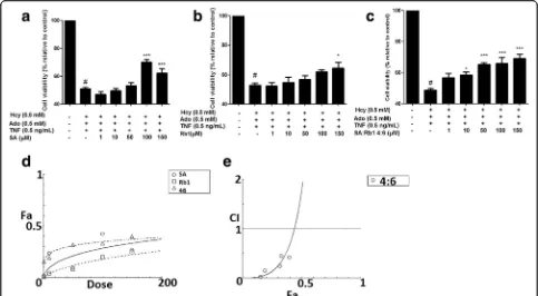
Synergistic effects of bioactive compounds on impaired cell viability
Fourteen bioactive compounds from DS and SQ were tested for their cytoprotective effects against the cellular
damage induced by Hcy-Ado-TNF, using MTT assay. Among all compounds from DS (Additional file4), only SA showed improvement in cell viability in a dose-dependent manner. As shown in Fig. 4, SA significantly restored the cell viability at the concentration greater than 100μM (P< 0.05). SB, the most abundant bioactive compound in DS, did not show any significant protective effects (P> 0.05). Among all the tested compounds in SQ (Additional file 5), only ginsenoside Rb1 showed signifi-cant improvement at 150μM (P < 0.05). Therefore, SA and Rb1 from DS and SQ, respectively, were found to be the most active compounds in protecting EA.hy926 cells from Hcy-Ado-TNF. Thus, SA and Rb1 were further stud-ied by combining the specifstud-ied ratios (1:9, 2:8…8:2, 9:1) and tested using MTT assays. The results suggested that SA-Rb1 4:6 demonstrated significant effects when the Fig. 2Inhibitory effects of DS, SQ and DS-SQ 6:4 on elevated levels of LDH leakage in EA.hy926 cells under cellular damage induced by Hcy-Ado-TNF.a. Inhibitory activity (% relative to control) of DS, SQ and DS-SQ 6:4 (10–300μg/mL) on increased LDH activity (relative to control). Data shown are mean ± SEM, n > 3.b. Concentration-effect curves of LDH inhibitory activity of DS, SQ and DS-SQ 6:4 generated by CompuSyn software.c. Log (CI) values were plotted as a function of suppressing LDH leakage (Fa) by‘CompuSyn’software. Dotted line is the reference line, where Log (CI) value equals to 0; solid line represents CI values at different Fa. Fa values correspond to % LDH inhibitory effect relative to control
[image:6.595.56.539.89.227.2] [image:6.595.60.539.515.641.2]tested concentration was greater than 10μM (P< 0.05, Fig. 4). Synergistic effects were generally observed for SA-Rb1 4:6 ratio at all tested concentrations (1–150μM) for restoring impaired cell viability on MTT assay (CI < 1, Fig. 4e). The synergistic interaction tended to be weaker when the concentration of SA-Rb1 was higher than 150μM. The interaction was predicted to be antagonistic, when the concentration was higher than 465.51μM.
Synergistic effects of SA-Rb1 on LDH leakage suppression
As shown in Fig.5a, SA, Rb1 and SA-Rb1 (4:6) significantly reduced the LDH leakage induced by Hcy-Ado-TNF at all tested concentrations (P < 0.05), except for Rb1 at 50μM (P> 0.05). A strong synergistic effect (CI < 1 with Fa from 0.1 to 0.97) was generally shown at the tested concentra-tion range (50–150μM) for SA-Rb1 4:6 (Fig.5c). The doses of SA and Rb1 in the combination were generally much lower than that of SA and Rb1 used alone to reach the same effect. For example, to reach 50% suppres-sive level, 15.82 and 23.73μM were needed for SA and Rb1 in the combination, respectively, and the doses were much lower than that of SA (78.1074μM) and Rb1 (20,697μM, predicted by CompuSyn) used alone to reach the same effect.
Synergistic effects of DS, SQ, DS-SQ 6:4 and their bioactive compounds on H2O2impaired cell survival in EA.hy926 cells
Synergistic effects of DS, SQ and DS-SQ on impaired cell viability
H2O2 (0.1–10 mM) concentration-dependently reduced
cell viability. As 0.5 mM H2O2caused a significant cellular
injury (P< 0.001), this concentration was selected for the H2O2 induced cell injury model. The cell viability with
pre-treatment of DS, SQ and DS-SQ 6:4, followed by H2O2
(0.5 mM), was shown in Fig.6. There were no significant protective effects from DS on restoring impaired cell viabil-ity against H2O2 (P> 0.05). SQ (50–150μg/mL) showed
concentration-dependent effects and significantly restored the impaired cell viability induced by H2O2 (P< 0.05). It
was found that DS-SQ 6:4 (150μg/mL) significantly re-stored cell viability against H2O2(0.5 mM) (P< 0.01).
Syn-ergistic effect was generally observed for DS-SQ 6:4 in restoring impaired cell viability after the cells were exposed to H2O2, with CI values of 0.072 at ED50(Fig.6d).
Synergistic effects of DS, SQ and DS-SQ on ROS generation
As shown in Fig. 7, EA.hy926 cells incubated with H2O2
[image:7.595.56.539.88.354.2]Fig. 6Protective effect of DS, SQ and DS-SQ 6:4 on EA.hy926 cells against H2O2-induced cell injury by restoring reduced cell viability. Cell viability was
assayed by the MTT method (n= 4) in EA.hy926 cells for 20 h. Cell viability was expressed as a percentage compared to H2O2stimulation only. Data
shown are means ± SEM,n> 3. Cell viability (% relative to control) versus increased concentrations of (a) DS, (b) SQ and (c) DS-SQ 6:4 (10–300μg/mL) against H2O2induced cell damage. #P< 0.05 vs control. *P< 0.05 vs H2O2treatment only. **P< 0.01 vs H2O2treatment only. ***P< 0.001 vs H2O2
treatment only.d. CI values were plotted as a function of restored cell viability (Fa) by‘CompuSyn’software. Fa values correspond to restored cell viability % relative to control. Dotted line is the reference line, where CI value equals 1; solid line represents CI values at different Fa
Fig. 5Effects of SA, Rb1 and SA-Rb1 4:6 in inhibiting LDH leakage on EA.hy 926 cells against cellular damage induced by Hcy-Ado-TNF.a
[image:8.595.61.539.88.228.2] [image:8.595.54.542.315.665.2]with DS (10–150μg/mL) concentration-dependently at-tenuated the ROS generation by H2O2, with significance
at 50–150μg/mL. SQ extract showed no significant ef-fect in inhibiting ROS from 10 to 150μg/mL. DS-SQ 6:4 showed significant activity in decreasing the ROS gener-ation at 50, 100 and 150 μg/mL compared to the H2O2
group (P< 0.001). Gallic acid was used as a positive con-trol in this assay, as its prominent anti-oxidant activity showed significant ROS inhibitory effect at 10μg/mL. A one-way ANOVA test suggested that the inhibitory ef-fect of DS and DS-SQ 6:4 at 150μg/mL were compar-able with the effect of gallic acid (10μg/mL) (P> 0.05). A synergistic effect was also observed for DS-SQ 6:4 at lower effects in inhibiting ROS production induced by H2O2 (Figs. 7b), when the Fa level (ROS inhibitory
ef-fect) was lower than 0.2 (20% inhibition), and the con-centration range for DS-SQ 6:4 was calculated to be smaller than 80.51μg/mL. Whereas, the antagonistic ef-fect was observed when the concentration of DS-SQ 6:4 was above 80.511μg/mL. The CI values were calculated to be 1.39 and 2.31 at ED50and ED90, respectively.
Synergistic effect of SA-Rb1 4:6 on impaired cell viability
The cell viability with pre-incubation of SA, Rb1, and SA-Rb1 (4:6) with different concentrations (1–300μM) followed by H2O2 (0.5 mM) cell injury was shown in
Fig.8. Both SA (300μM) and Rb1 (100–300μM) showed significant effects for restoring the impaired cell viability of H2O2-treated EA.hy926 cells (P< 0.05). Additionally, a
prominent effect was observed for SA-Rb1 4:6 (1– 150μM), with significant improvement of cell viabil-ity when the concentration was greater than 10μM (P< 0.001). Similarly, a synergistic effect for SA-Rb1 4:6 was observed against H2O2-induced cell damage
with concentration less than 35.14μM. At the above concentrations, it became antagonistic as the pro-tective effect of SA alone was stronger than the combination (Fig. 8c).
Synergistic effect of SA-Rb1 4:6 on ROS production
As shown in Fig. 9, both SA and SA-Rb1 significantly inhibited ROS production from 50 to 150μM (P< 0.001), whereas Rb1 did not show any significant inhibitory effect for ROS production (P> 0.05). Additionally, gallic acid (10μg/mL) was used as a negative control in this assay, which inhibited ROS significantly. The non-parametric test suggested that the inhibitory effect of SA and SA-Rb1 4:6 from 50 to 150μM was comparable to that of gallic acid (P > 0.05). For the inhibitory effect of SA-Rb1 4:6 on ROS production, a synergistic effect was only observed below 50μM. It was predicated to be antagonistic at con-centrations greater than 100μM (Fig.9c).
Discussions
This study shows for the first time the cytoprotective ac-tivity of SQ extract and compounds of both DS and SQ in endothelial cells. Further to previous studies, we ap-plied CI model and systematically analysed the possible Fig. 7Protective effects of DS, SQ and DS-SQ 6:4 against H2O2-induced endothelial cell injury by attenuating excessively expressed ROS
production.aInhibitory effect of DS, SQ and DS-SQ 6:4 on ROS production induced by H2O2in EA.hy926 cells. The ROS expression level was
estimated by cellular ROS detection assay kit manufacturer (Abcam, Australia). TBPH (from kit) was used as negative control and gallic acid (10μg/ mL) was used as a positive control in the assay. Results were shown as absorbance by a microplate reader, with the means ± SEM,n> 3. ***P< 0.001 compared with H2O2group.bLog (CI) values were plotted as a function of suppression fractional of ROS level (Fa) by‘CompuSyn’software.
[image:9.595.57.540.86.297.2]Fig. 9Protective effect of SA, Rb1 and SA-Rb1 4:6 on EA.hy926 cells against H2O2-induced cell injury by inhibiting excessive intracellular ROS
production.aInhibitory effect of SA, Rb1 and SA-Rb1 4:6 on ROS production induced by H2O2in EA.hy926 cells. The ROS expression level was
estimated by cellular ROS detection assay kit manufacturer (Abcam, Australia). TBPH (from kit) was used as positive control in the assay. Results were shown as absorbance by a microplate reader, with the means ± SEM, n > 3.bConcentration-effect curves of ROS inhibitory effects of SA, Rb1 and SA-Rb1 4:6 generated by CompuSyn software.cLog (CI) values were plotted as a function of suppression fractional of ROS (Fa) by
‘CompuSyn’software. Fa values correspond to % ROS inhibitory effect relative to control. Dotted line is the reference line, where Log (CI) value equals 0; solid line represents CI values at different Fa
Fig. 8Protective effect of SA, Rb1 and SA-Rb1 4:6 on EA.hy926 cells against H2O2-induced cell injury was determined by MTT assay.aCell
viability (% relative to control) verses increased concentrations of SA, Rb1 and SA-Rb1 4:6 (1–150μM) against H2O2induced cell damage.b
Concentration-effect curves of restored cell viability against H2O2treatment with increasing concentrations of SA, Rb1 and SA-Rb1 4:6 (1–150μM)
were generated by CompuSyn software. Data shown are mean ± SEM,n> 3.cCI values were plotted as a function of restored cell viability (Fa) by
[image:10.595.54.540.88.220.2] [image:10.595.54.539.294.652.2]synergistic effects for the DS-SQ combinations with nine ratios (1:9 to 9:1,w/w) in protecting endothelial cell in-jury against both Hcy-Ado-TNF and H2O2. Our results
revealed that the DS-SQ paired herb at all tested ratios restored impaired cell injury against Hcy-Ado-TNF. The combinational effect was more prominent than the sin-gle herbs at the same concentration level. This suggested the superior effects of using the combination for en-hanced bioactivity on the same target.
Among all the tested combinations, DS-SQ 6:4 was found to be the optimal ratio for its potent protective effects compared to that of other ratios, not only for re-storing cell viability, but also reducing the cell apoptosis (LDH, caspase-3). Furthermore, this particular combin-ation consistently restored the cell injury against oxida-tive stress by directly suppressing ROS level. For the first time, this finding suggests that synergistic interactions exist with the DS and SQ combination in protecting endothelial cells against both Hcy-Ado-TNF- and oxida-tive stress-mediated cell injury.
Moreover, the observed synergistic effect is concentration-dependent as revealed by CI. These results suggest that the interactions in the combination are dependent on the com-position of the ingredients. In TCM clinical practice, the ratio (dosage) of herbal ingredients is tailored to in-dividual patient in order to reach a desirable clinical outcome based on syndrome differentiation treatment strategy. Our findings provide the preliminary evidence for this theory. The optimal ratio of DS and SQ against cytotoxicity on EA.hy926 cells tended to be equal in this study. In traditional practice, a higher ratio (dos-age) of DS in the combination is usually used for condi-tions at the early stage of disease, when pathological changes in the organs are not obvious (possibly point-ing to the early inflammatory stage), and the proportion of SQ should be increased at the later stage of the dis-ease, when pathological changes have been diagnosed (later stage for vascular dysfunction) [42]. Our observa-tions support this theory and clinical use, however fur-ther in vivo and human studies are needed to confirm this finding. There are several possible mechanisms for the pharmacodynamics synergy in combinational ther-apy, including increased extraction yield of bioactive compounds, chemical reactions among compounds, multi-target behaviour or enhanced bioavailability [43]. While contradictory, current chemical analysis studies on the DS-SQ combination indicate that the chemical composition of DS-SQ mixed preparation may not sim-ply be the sum of chemical constituents from the single herb, and the new compounds are likely formed during the mixture preparation which contribute to the en-hanced bioactivity of the combination. Our ultra-performance liquid chromatography chemical analysis for the combination revealed that the chemical
fingerprint was altered in the combined extract com-paring to that of individual extract, and the amount of bioactive compounds (DSS, SB, NR1, Rg1 and Rb1) was not directly proportional to the ratio of their herbal in-gredients in the combination (Additional file2).
To investigate the mechanistic actions of the observed synergistic actions for the mixed extracts by multi-target behaviour, the effects of fourteen key bioactive com-pounds of DS and SQ were further investigated using the same assays. SA and Rb1 were found to be effective in restoring impaired cell injury against Hcy-Ado-TNF and oxidative stress. Other tested compounds showed no significant effects in the current experimental condi-tion, indicating that these compounds may be inactive on the cellular mechanisms studied. However, this also does not exclude the possibility that these compounds may become active in vivo due to metabolism or trans-formation [44]. Additionally, the present findings do not exclude the possibility of the involvement of the other active components in the actions of the DS-SQ extract, as more than 100 single compounds have currently been isolated from DS and SQ. A systems biology approach can be applied to predict the key constituents for the po-tential targets, and the experimental data can be con-firmed by bioassay-guided fractionation. Previously, the multi-target behaviour from DS-SQ was demonstrated in several studies using systems biology. For example, Li et al. (2012) established a systems biology model for DS-SQ on cardiovascular diseases targets and found that the candidate compounds from DS tackled 39 out of the total 41 validated CVD targets, whereas those from SQ interacted with 36 potential targets, 34 of which over-lapped with DS’s targets [45].
In addition, synergistic effects were generally observed for SA-Rb1 combination on anti-apoptosis. This demon-strated that the combination of two prominent com-pounds may contribute to the effects of the mixed extract and engender a stronger effect. The two com-pounds may act as an agonist for each other on the same receptor, thus the effect is enhanced. This simplified compounds combination which maintained the effect and exhibited synergy maybe a potential pharmaceutical agent to be used on endothelial dysfunction. However, the effect and toxicity need to be further confirmed in in vivo and human studies.
Conclusions
DS-SQ 6:4 showed prominent and synergistic effects in restoring reduced cell viability and attenuating excessively expressed LDH and caspase −3 against Hcy-Ado-TNF. In addition, this combination exerted a synergistically-enhanced cytoprotective effect from oxidative damage (caused by H2O2) by restoring the
SA and Rb1 were found to be the most potent component in each extract for restoring reduced cell viability. Further-more, SA-Rb1 4:6 combination showed synergistically -en-hanced effects in preventing cytotoxic effects caused by Hcy-Ado-TNF as analysed by CI. This simplified combin-ation demonstrated prominent effects on H2O2-induced
oxidative damage. Our findings provided a theoretical basis for the purported synergistic efficiency of DS and SQ as a herbal drug pair on vascular function through its protection on endothelial cells against major stressors: Hcy, TNF and oxidative stress.
Additional files
Additional file 1:Chemical fingerprint of raw herb extract of DS and SQ analysed by UPLC-PDA. (PDF 110 kb)
Additional file 2:Contents (mg/g, mean ± SD,n= 3) of DSS, SB, NR1, Rg1 and Rb1 in DS-SQ combinations extract by UPC-PDA. (PDF 43 kb)
Additional file 3:Cytotoxic effects of Hcy-Ado-TNF on EA.hy926 cells as determined by MTT dye reduction assay, following treatments with Hcy-Ado-TNF in EA.hy926 endothelial cells for 20 h. (DOCX 27 kb)
Additional file 4:Effects of bioactive compounds from DS on restoring cell viability against Hcy-Ado-TNF in EA.hy926 cells determined by MTT assay. (DOCX 284 kb)
Additional file 5:Effects of bioactive compounds from SQ on restoring cell viability against Hcy-Ado-TNF in EA.hy926 cells determined by MTT assay. (DOCX 282 kb)
Abbreviations
Ado:Adenosine; BMSCs: Bone marrow stromal cells; CI: Combination index;
CIR: Cerebral ischaemia reperfusion; CT: Cryptotanshinone; CT: Cryptotanshinone;DCFDA: 2′,7′ –dichlorofluorescin diacetate; DMEM: Dulbecco’s Modified Eagle’s Medium; DMSO: Dimethyl sulfoxide; DSS: Sodium danshensu; DS-SQ: Danshen-Sanqi extracts;
DT: Dihydrotanshinone I; EA.hy926: Human cardiovascular endothelial; eNOS: Endothelial nitric oxide synthase; Hcy: Homocysteine; HUVECs: Human umbilical vein endothelial cells; LDH: Lactate dehydrogenase;
MAPK: Mitogen-activated protein kinase; NR1: Notoginsenoside R1; PDGF: Platelet-derived growth factor;Penstrep: Penicillin and streptomycin; Rb1: Ginsenoside Rb1; Rb1: Ginsenoside Rb1; Rd.: Ginsenoside Rd.;
Rg1: Ginsenoside Rg1;Rg2: Ginsenoside Rg2; ROS: Reactive oxygen species; ROS: Reactive oxygen species; SA: Salvianolic acid A; SB: Salvianolic acid B; TCM: Traditional Chinese Medicine; TI: Tanshinone I; TIIA: Tanshinone IIA; TNF: Tumour necrosis factor
Acknowledgments
We are thankful to Dr. Sai Seto for trouble -shooting.
Funding
The work was supported by the Western Sydney University Research Grant Scheme (P00021202), the International Postgraduate Research Scholarships, and the Joint Chair in Traditional Chinese Medicines Program (JCTCM), which was jointly funded by the Office of Science Research in NSW, The University of Sydney, and Western Sydney University, Australia.
Availability of data and materials
The datasets used and/or analysed during the current study are presented within the manuscript and Additional files1,2,3,4and5). Detailed datasets are available from the corresponding author on reasonable request.
Authors' contributions
KC, VRN and AB conceived and designed the experiments; XZ and AK performed the experiments; XZ and AK analysed the data; CGL and DC contributed materials/analysis tools; XZ and VRN wrote the paper. All authors
contributed to the final manuscript. All authors read and approved the final manuscript.
Ethics approval and consent to participate
Not applicable.
Consent for publication
Not applicable.
Competing interests
The authors declare that they have no competing interests.
Publisher’s Note
Springer Nature remains neutral with regard to jurisdictional claims in published maps and institutional affiliations.
Author details
1NICM Health Research Institute, Western Sydney University, Sydney 2751,
Australia.2South Western Sydney Clinical School, UNSW Medicine, University of New South Wales Sydney, Sydney 2170, Australia.3School of Biological
Sciences, Nanyang Technological University, Singapore 637551, Singapore.
4School of Pharmacy and Biomolecular Sciences, Liverpool John Moores
University, L3 3AF, Liverpool, UK.
Received: 18 April 2018 Accepted: 13 February 2019
References
1. Zhou X, Seto SW, Chang D, Kiat H, Razmovski-Naumovski V, Chan K, Bensoussan A. Synergistic effects of Chinese herbal medicine: a comprehensive review of methodology and current research. Front Pharmacol. 2016;7.https://doi.org/10.3389/fphar.2016.00201. 2. Bonetti P, Lerman L, Lerman A. Endothelial dysfunction: a marker of
atherosclerotic risk. Arterioscler Thromb Vasc Biol. 2003;23:168–75.https:// doi.org/10.1161/01.ATV.0000051384.43104.FC.
3. Kam A, Li KM, Razmovski-Naumovski V, Nammi S, Chan K, Li GQ. Combination of TNF-α, homocysteine and adenosine exacerbated cytotoxicity in human cardiovascular and cerebrovascular endothelial cells. Cell Physiol Biochem. 2012;30(3):805–14.https://doi.org/10.1159/000341459. 4. De Caterina R. Endothelial dysfunctions: common denominators in vascular disease. Curr Opin Clin Nutr Metab Care 2000; 3(6):453–467.https://doi.org/ 10.1097/00075197-200011000-00007.
5. Boushey CJ, Beresford SA, Omenn GS, Motulsky AG. A quantitative assessment of plasma homocysteine as a risk factor for vascular disease: probable benefits of increasing folic acid intakes. Jama. 1995;274(13):1049– 57.https://doi.org/10.1001/jama.1995.03530130055028.
6. Hankey GJ, Eikelboom JW. Homocysteine and vascular disease. Lancet. 1999; 354(9176):407–13.https://doi.org/10.1016/S0140-6736(98)11058-9. 7. Fryer RH, Wilson BD, Gubler DB, Fitzgerald LA, Rodgers GM. Homocysteine, a
risk factor for premature vascular disease and thrombosis, induces tissue factor activity in endothelial cells. Arterioscler Thromb Vasc Biol. 1993;13(9): 1327–33.https://doi.org/10.1161/01.ATV.13.9.1327.
8. Graham IM, Daly LE, Refsum HM, Robinson K, Brattström LE, Ueland PM, Palma-Reis RJ, Boers GH, Sheahan RG, Israelsson B. Plasma homocysteine as a risk factor for vascular disease: the European concerted action project. Jama. 1997;277(22):1775–81.https://doi.org/10.1001/jama.1997. 03540460039030.
9. James SJ, Melnyk S, Pogribna M, Pogribny IP, Caudill MA. Elevation in S-adenosylhomocysteine and DNA hypomethylation: potential epigenetic mechanism for homocysteine-related pathology. J Nutr. 2002;132(8):2361S– 6S.https://doi.org/10.1093/jn/132.8.2361S.
10. Gopisetty G, Ramachandran K, Singal R. DNA methylation and apoptosis. Mol Immunol. 2006;43(11):1729–40.https://doi.org/10.1016/j. molimm.2005.11.010.
11. Zhang HR, Park YJ, Wu JX, Chen XP, Lee SW, Yang JY, Dellsperger KC, Zhang CH. Role of TNF-αin vascular dysfunction. Clin Sci. 2009;116(3):219–30.
https://doi.org/10.1042/CS20080196.
13. Kam A, Li KM, Razmovski-Naumovski V, Nammi S, Chan K, Li GQ. Gallic acid protects against endothelial injury by restoring the depletion of DNA methyltransferase 1 and inhibiting proteasome activities. Int J Cardiol. 2014; 171(2):231–42.https://doi.org/10.1016/j.ijcard.2013.12.020.
14. Thannickal VJ, Fanburg BL. Reactive oxygen species in cell signaling. Am J Phys Lung Cell Mol Phys. 2000;279(6):L1005–28.https://doi.org/10.1152/ ajplung.2000.279.6.L1005.
15. Kojda G, Harrison D. Interactions between NO and reactive oxygen species: pathophysiological importance in atherosclerosis, hypertension, diabetes and heart failure Cardiovasc Res 1999; 43(3):652–671.https://doi.org/10. 1016/S0008-6363(99)00169-8.
16. Förstermann U. Nitric oxide and oxidative stress in vascular disease. Pflugers Arch. 2010;459(6):923–39.https://doi.org/10.1007/s00424-010-0808-2. 17. Simon HU, Haj Yehia A, Levi Schaffer F. Role of reactive oxygen species
(ROS) in apoptosis induction. Apoptosis. 2000;5(5):415–8.
18. Garcia-Martinez R, Noiret L, Sen S, Mookerjee R, Jalan R. Reactive oxygen species in the vasculature: molecular and cellular mechanisms. Liver Int. 2015;35:335–43.https://doi.org/10.1161/01.HYP.0000100443.09293.4F.
19. Coyle CH, Martinez LJ, Coleman MC, Spitz DR, Weintraub NL, Kader KN. Mechanisms of H 2 O 2-induced oxidative stress in endothelial cells. Free Radic Biol Med. 2006;40(12):2206–13.https://doi.org/10.1016/j. freeradbiomed.2006.02.017.
20. Zhou X, Razmovski-Naumovski V, Kam A, Chang D, Li C, Bensoussan A, Chan K. Synergistic effects of Danshen (Salvia Miltiorrhizae Radix et Rhizoma) and Sanqi (Notoginseng Radix et Rhizoma) combination in angiogenesis behavior in EAhy 926 cells. Medicines. 2017;4(4):85.https://doi.org/10.3390/ medicines4040085.
21. Zeng GF, Liu JX, Li P, Fu SP, Xiao HB, Liang XM. Protective effects of different ratios of Danshen to Sanqi on hypoxia and reoxygenation -induced HUVECs injury Fine Chem ICALS. 2006; 23(2):126–129.
22. Nakazawa T, Xui N, Hesong Z, Kinoshita M, Chiba T, Kaneko E, Yui K, Shimokado K. Danshen inhibits oxysterol-induced endothelial cell apoptosis in vivo. J Atheroscler Thromb. 2005;12(3):132–7.https://doi.org/10.5551/jat.12.132. 23. Liang XY, Li HN, Yang XY, Zhou WY, Niu JG, Chen BD. Effect of Danshen
aqueous extract on serum hs-CRP, IL-8, IL-10, TNF-αlevels, and IL-10 mRNA, TNF-αmRNA expression levels, cerebral TGF-β1 positive expression level and its neuroprotective mechanisms in CIR rats. Mol Biol Rep. 2013;40(4): 3419–27.https://doi.org/10.1007/s11033-012-2419-9.
24. Cho YH, Ku CR, Hong ZY, Heo JH, Kim EH, Choi DH, Kim DK, Kim AJ, Lee CS, Jung MK. Therapeutic effects of water soluble danshen extracts on atherosclerosis. Evid Based Complement Alternat Med. 2013;2013.
https://doi.org/10.1155/2013/623639.
25. Guo J, Zhang YJ, Zeng LH, Liu JY, Liang J, Guo GZ. Salvianic acid a protects L-02 cells againstγ-irradiation-induced apoptosis via the scavenging of reactive oxygen species. Environ Toxicol Pharmacol. 2013;35(1):117–30.
https://doi.org/10.1016/j.etap.2012.11.010.
26. Zhang HA, Gao M, Zhang L, Zhao Y, Shi LL, Chen BN, Wang YH, Wang SB, Du GH. Salvianolic acid a protects human SH-SY5Y neuroblastoma cells against H 2 O 2-induced injury by increasing stress tolerance ability. Biochem Biophys Res Commun. 2012;421(3):479–83.https://doi.org/10.1016/ j.bbrc.2012.04.021.
27. Wang H, Gao XM, Zhang BL. Tanshinone: an inhibitor of proliferation of vascular smooth muscle cells. J Ethnopharmacol. 2005;99(1):93–8.
https://doi.org/10.1016/j.jep.2005.01.057.
28. Wang XJ, Xu JX. Salvianic acid a protects human neuroblastoma SH-SY5Y cells against MPP+-induced cytotoxicity. Neurosci Res. 2005;51(2):129–38.
https://doi.org/10.1016/j.neures.2004.10.001.
29. Wang M, Sun GB, Sun X, Wang HW, Meng XB, Qin M, Sun J, Luo Y, Sun XB. Cardioprotective effect of salvianolic acid B against arsenic trioxide-induced injury in cardiac H9c2 cells via the PI3K/Akt signal pathway. Toxicol Lett. 2013;216(2):100–7.https://doi.org/10.1016/j.toxlet.2012.11.023. 30. Sun LQ, Zhao J, Zhang TT, Qu L, Wang X, Xue B, Li XJ, Mu YM, Lu JM.
Protective effects of Salvianolic acid B on Schwann cells apoptosis induced by high glucose. Neurochem Res. 2012;37(5):996–1010.https://doi.org/10. 1007/s11064-011-0695-8.
31. Tian LL, Wang XJ, Sun YN, Li CR, Xing YL, Zhao HB, Duan M, Zhou Z, Wang SQ. Salvianolic acid B, an antioxidant from Salvia miltiorrhiza, prevents 6-hydroxydopamine induced apoptosis in SH-SY5Y cells. Int J Biochem Cell Biol. 2008;40(3):409–22.https://doi.org/10.1016/j.bbrc.2006.07.110.
32. Sun LQ, Xue B, Li XJ, Wang X, Qu L, Zhang TT, Zhao J, Wang BA, Zou XM, Mu YM. Inhibitory effects of salvianolic acid B on apoptosis of Schwann cells
and its mechanism induced by intermittent high glucose. Life Sci. 2012; 90(3):99–108.https://doi.org/10.1016/j.lfs.2011.10.001.
33. Jia LQ, Yang GL, Ren L, Chen WN, Feng JY, Cao Y, Zhang L, Li XT, Lei P. Tanshinone IIA reduces apoptosis induced by hydrogen peroxide in the human endothelium-derived EA. hy926 cells. J Ethnopharmacol. 2012;143(1): 100–8.https://doi.org/10.1016/j.jep.2012.06.007.
34. Fu JJ, Huang HQ, Liu JJ, Pi RB, Chen JW, Liu PQ. Tanshinone IIA protects cardiac myocytes against oxidative stress-triggered damage and apoptosis. Eur J Pharmacol. 2007;568(1):213–21.https://doi.org/10.1016/j.ejphar.2007.04.031. 35. Wang W, Zheng LL, Wang F, Hu ZL, Wu WN, Gu J, Chen JG. Tanshinone IIA
attenuates neuronal damage and the impairment of long-term potentiation induced by hydrogen peroxide. J Ethnopharmacol. 2011;134(1):147–55.
https://doi.org/10.1016/j.jep.2010.11.069.
36. Qiang H, Zhang C, Shi ZB, Yang HQ, Wang KZ. Protective effects and mechanism of Panax Notoginseng saponins on oxidative stress-induced damage and apoptosis of rabbit bone marrow stromal cells. Chin J Integr Med. 2010;16(6):525–30.https://doi.org/10.1007/s11655-010-0566-1. 37. Li SY, Wang XG, Ma MM, Liu Y, Du YH, Lv XF, Zhou JG, Tang YB, Guan YY.
Ginsenoside-Rd potentiates apoptosis induced by hydrogen peroxide in basilar artery smooth muscle cells through the mitochondrial pathway. Apoptosis. 2012;17(2):113–20.https://doi.org/10.1007/s10495-011-0671-4. 38. Ye RD, Kong XW, Yang QZ, Zhang YX, Han JL, Zhao G. Ginsenoside Rd
attenuates redox imbalance and improves stroke outcome after focal cerebral ischemia in aged mice. Neuropharmacology. 2011;61(4):815–24.
https://doi.org/10.1016/j.neuropharm.2011.05.029.
39. Promega. Cell viability.https://www.promega.com.au/-/media/files/ resources/protocols/technical-bulletins/0/cytotox-96-nonradioactive-cytotoxicity-assay-protocol.pdf?la=en. Accessed 1 Feb 2016.
40. R&D A. Caspase-3 Colorimetric Assay. 2016.https://resources.rndsystems. com/pdfs/datasheets/bf3100.pdf. Accessed 01 Februray 2016.
41. Chou TC. Drug combination studies and their synergy quantification using the Chou-Talalay method. Cancer Res. 2010;70(2):440–6.https://doi.org/10. 1158/0008-5472.CAN-09-1947.
42. Xu Q. Chinese medicine for summa. Beijing: Chinese Medicine Press; 1996. 43. Wagner H. Synergy research: approaching a new generation of
phytopharmaceuticals. Fitoterapia. 2011;82(1):34–7.https://doi.org/10.1016/j. fitote.2010.11.016.
44. Huttunen KM, Raunio H, Rautio J. Prodrugs—from serendipity to rational design. Pharmacol Rev. 2011;63(3):750–71.https://doi.org/10.1124/pr.110.003459. 45. Li XX, Xu X, Wang JN, Yu H, Wang X, Yang HJ, Xu HY, Tang SH, Li Y, Yang L. A