R E S E A R C H A R T I C L E
Open Access
Pharmacological potential of
Bidens pilosa
L. and determination of bioactive
compounds using UHPLC-QqQ
LIT
-MS/MS
and GC/MS
Garima Singh
1, Ajit Kumar Passsari
1, Pratibha Singh
2, Vincent Vineeth Leo
1, Sarathbabu Subbarayan
1,
Brijesh Kumar
2, Bhim Pratap Singh
1, Hauzel lalhlenmawia
3and Nachimuthu Senthil Kumar
1*Abstract
Background:Research of natural products from traditionally used medicinal plants to fight against the human ailments is fetching attention of researchers worldwide.Bidens pilosaLinn. var. Radiata (Asteraceae) is well known for its folkloric medicinal use against various diseases from many decades. Mizoram, North East India, has high plant diversity and the use of this plant as herbal medicine is deep rooted in the local tribes. The present study was executed to understand the pharmacological potential ofB. pilosaleaves extract.
Methods:The antimicrobial potential was determined using agar well diffusion and broth microdilution method against bacterial and yeast pathogens. Cytotoxicity was evaluated using MTT and apoptotic DNA fragmentation assays. Further, the antioxidant ability of the extract was analysed using DPPH and ABTS free radical scavenging assay. Mosquitocidal activity was evaluated against third in-star larvae ofC. quinquefasciatususing dose response and time response larvicidal bioassay. Additionally, the major phenolic and volatile compounds were determined using UHPLC-QqQLIT-MS/MS and GC/MS respectively.
Results:We found that the extract showed highest antimicrobial activity againstE. coli(MIC 80μg/mL and IC50 110.04μg/mL) and showed significant cytotoxicity against human epidermoid carcinoma (KB-3-1) cells with IC50 values of 99.56μg/mL among the tested cancer cell lines.
The IC50values for scavenging DPPH and ABTS was 80.45μg/mL and 171.6μg/mL respectively. The extract also showed the high phenolics (72μg GAE/mg extract) and flavonoids (123.3μg Quercetin /mg extract). Lastly, five bioactive and six volatile compounds were detected using UHPLC-QqQLIT-MS/MS and GC-MS respectively which may be responsible for the plant’s bioactivities. An anticancerous compound, Paclitaxel was detected and quantified for the first time fromB. pilosaleaves extract, which further showed the anticancerous potential of the tested extract.
Conclusion:On the basis of the present investigation, we propose that the leaf extract ofB. pilosamight be a good candidate for the search of efficient environment friendly natural bioactive agent and pharmaceutically important compounds.
Keywords:Bidens pilosaL., Antioxidant, Antimicrobial, Cytotoxicity, GS-MS analysis, UPLC-ESI-MS/MS
* Correspondence:nskmzu@gmail.com
1Department of Biotechnology, Mizoram University, Aizawl, Mizoram 796004,
India
Full list of author information is available at the end of the article
Background
Bidens pilosa Linn. var. Radiata (Spanish needles or beggar ticks) from the family Asteraceae is an annual weed widely distributed throughout the tropical and sub-tropical regions of the world [1]. In some part of the world the plant is eaten as food whereas in other coun-tries B. pilosa is used in traditional medicines [2]. In Mizoram, it is called as vawkpuithal and is reported to treat various diseases and infections, commonly rheuma-tism, diarrhoea, ear, eyes and tooth ache problems [3]. Plant has a long ethno-medicinal history for treating mal-aria, skin infections, stomach and liver disorders. This plant is very well documented as a source of natural anti-microbials [4, 5], anti-inflammatory [6, 7], hepatoprotec-tive [8], and cytotoxic against various cancer cells [9, 10]. Phytochemical screening studies of B. pilosa showed the presence of phenylpropanoids, polyacetylenes, polyphe-nols, triterpenes, saponins and alkaloids [11]. The pharmaceutical property of the plant seems to be associ-ated with the bioactive phytochemical compounds, espe-cially sesquiterpene lactones and polyacetylenes, which inhibit the growth of pathogenic microorganisms and the flavonoids, which are considered as effective anti-inflammatory agents [6, 11, 12]. Phytochemicals and es-sential oil of B. pilosa reported to possess exploitable amount phenolic compounds with free readical scaven-ging potential [11].
Osmotic stress and autoxidation are the natural phenomenon of human physiology resulted in the over-production of reactive oxygen species that plays an im-portant pathophysiological role in the development of several human diseases including cancer [13]. Natural antioxidants are stable molecules capable to donate an electron to neutralize these free radicals, but sometimes overwhelmed by excessive stress. Intake of antioxidants counteracts the oxidative damage in the human body, protects DNA, and improves biological antioxidant mechanism by trapping the free radicals [14].
On the other hand, development of drug resistance is becoming serious issue to fight against the diseases [15]. For instance, few bacteria have developed resistance against available antimicrobial agents which has resulted in significant public health problems [16, 17]. Herbal medicine has emerges as a health aid during the last 56 decades and showed the bio prospecting for new plant derived drugs [18, 19]. Previous studies has proved the efficacy of several isolated compounds from B. pilosa and suggested the plant as a potential anticancer medi-cinal plant [10, 20]. The specific polyphenols and flavo-noids present in B. pilosa were not fully elucidated, although caffeoylquinic acid, luteolin, quercetin and others have been reported so far [21, 22].
Keeping these findings in mind, the present work was designed to assess the in vitro antioxidant, antimicrobial,
antitumor and mosquitocidal activities of the B. pilosa leave extract. Furthermore, the phenolic, anticancerous and volatile compounds were detected and quantified using UHPLC-QqQLIT-MS/MS and GC MS respectively,
which further proves the potentiality of the selected plant to be used in health care system.
Methods
Plant collection and extract preparation
Fresh leaves ofB. pilosawere collected from the Botanical Garden, Mizoram University, Mizoram, India during September 2015 based on traditional uses and identified by Dr. Kalidas Upadhyay, Department of Forestry, Mizoram University. Moreover, the collected plant is also identified by the amplification of internal transcribed spaces (ITS) rRNA gene and the sequence has been submitted in NCBI genebank with the accession number MF440588. A vou-cher specimen was prepared and kept at the collection of Department of Biotechnology, Mizoram University (MZU/ BT/26). The healthy leaves were shade dried at room temperature (30 °C ± 2 °C) for 3 days and grounded to make powder by using a blender. Fifty grams of powder was extracted thrice in 750 ml of methanol for 48 h with occasional stirring. The extract was prepared using rotary evaporator (Buchi, India) at 40 °C under reduced pressure and the obtained crude extract was stored at 4 °C.
Reagents
2,2- Azinobis-3-ethylbenzothiazoline-6-sulphonic acid disodium salt (ABTS), 2,2-diphenyl-1-picrylhydrazyl (DPPH), Dimethyl Sulphoxide, Sodium acetate trihydrate ACS, Ferric chloride hexahydrate A.R., Ferrous sulphate heptahydrate A.R., Folin ciocalteu’s reagent L.R., Gallic acid monohydrate, L-Ascorbic acid A.R., Acetic acid gla-cial A.R., Sodium carbonate ACS, Potassium persulphate A.R., were purchased from Hi-media, Mumbai, India. 6-hydroxy-2,5,7,8-tetramethylchromane-2-carboxylic acid (trolox), Aluminium chloride AR, and Quercetin ≥95% (HPLC) solid were purchased from Sigma-Aldrich, USA. Acetonitrile, methanol (LC-MS grade) and formic acid (ana-lytical grade) were purchased from Fluka, Sigma-Aldrich (St. Louis, MO, USA). Ultra pure water was obtained from a Direct-Q 8 UV water purification system (EMD Millipore Corporation, Billerica, MA, USA). All other reagents includ-ing solvents were of analytical grade and were procured from Hi-Media, Mumbai, India.
Phytochemical analysis
Total phenolic content (TPC) determination
reagent (1:10v/v in water) and 100μl of 15% Na2CO3to
make the 200 μl volume in a 96 well microplate. The mixture was incubated for 1 h in dark and absorbance was recorded using a UV/Vis microplate spectropho-tometer (Multiscan™GO, Thermo Scientific, MA, USA) at 725 nm. The result was expressed as gallic acid equivalent (GAE) per gram of extract based on the standard curve of gallic acid.
Determination of total flavonoids
Total flavonoids content of the plant extract was deter-mined by using modified aluminium colorimetric method [24]. 150 μl of methanol extract is mixed with 150 μl of 2% ethanolic AlCl3 and allowed to incubated in dark for 1 h and the absorbance was recorded at 420 nm. The total flavonoids content was expressed as μg quercetin equivalent (QE) per mg of plant extract compared with the standard curve of quercetin.
Determination of antioxidant potential
By using DPPH (2,2-Diphenyl-1-picrylhydrazyl) assay
Free radical scavenging capability of methanolic leaves extract of B. pilosa was determined by DPPH assay as described by Brand-Williams et al. [25]. Briefly, plant extract (100 μl) was added at different concentration (10–100 μg/ml) in a 200 μl of freshly prepared DPPH methanolic solution (0.1 mM). Reaction mixture was in-cubated for 30 min in dark and the absorbance was re-corded at 517 nm. Ascorbic acid was used as standard and methanol with DPPH used as blank. Triplicate mea-surements were taken and the ability to scavenge the DPPH radical was noted by using the given formula: % decolouration = [1-(OD Sample/OD Control] X 100. The concentration that reduced the DPPH colour by 50% was determined as IC50.
By ABTS+.Radical Cation discoloration assay
The ABTS free radical scavenging activity was performed by using the method described by Re et al. [26]. ABTS+ Inhibition percentage was measured as described earlier (27). The IC50value was analyzed from the graph plotted
as the inhibition percentage against the concentration.
Antimicrobial assays
Sample preparation for antimicrobial assay
10 mg sample of crude methanolic extract of B. pilosa leaves was resuspended in dimethyl sulfoxide (DMSO). The final concentration was made to 10 mg/ml, which was Diluted to obtain different concentrations (1.0, 5.0, 7.5 and 10.0 mg/mL) to evaluate the antimicrobial po-tential against all selected test organisms.
Test strains
Antimicrobial activity of methanolic leaves extract of B. pilosa was checked by the agar well diffusion and broth micro dilution methods. Pathogens used for the study were gram positive bacteria Staphylococcus aureus (MTCC-96); Bacillus subtilis (MTCC-2097) and Micro-coccus luteus (MTCC-2070); gram negative bacteria Escherichia coli (MTCC-739); Pseudomonas aeruginosa (MTCC-2453) and a yeast pathogen Candida albicans (MTCC-3017), obtained from microbial type culture col-lection (MTCC), Chandigarh, India.
Antimicrobial assay by using agar well diffusion method
Agar well diffusion assay was used for initial antimicro-bial screening [27]. Briefly, the optical densities of the tested organisms were adjusted to match a 0.5 McFar-land standard with 108 colony forming unit (cfu) /ml and spreaded on agar plates. A 50μL of extract at differ-ent concdiffer-entrations was added into the 6 mm wells pre-pared using the sterilized cork borer. DMSO was served as the negative control and readymade impregnated disc of antibiotic tetracycline (20μg/disc) as positive control. A clear halo zone around the filled wells showed the antibacterial potential [28]. The experiments were per-formed in triplicates.
Antimicrobial assay by using broth micro dilution method
Minimum Inhibitory Concentration (MIC) of was evalu-ated using broth micro dilution method on 96-well mi-crotiter plate against all selected test organisms [29]. The bacterial culture suspension was prepared to make the final concentration of 1.0 × 10⁴CFU/mL (OD = 0.403). Plant extract of different concentrations (1– 10 mg/ml) was added in 96-well microtiter plate with bacterial culture suspension. Different concentrations of plant extract were kept as blank, bacterial culture in DMSO was used as negative control, and standard anti-biotics i.e. ampicillin was used as positive control. The 96 well plates were incubated for 36 h at 37 °C and the OD was taken as 630 nm. Results were documented as IC50 values which indicate 50% reduction of bacterial
growth. The IC50 values were calculated by using
cali-bration curve drawn by using linear regression.
Cytotoxicity potential of plant extract
Cell lines and cell culture
Three cancer cell lines [Cervical cancer cell (HeLa), Human hepato carcinoma (HepG2) and epidermoid car-cinoma (KB-3-1)] were selected and screened against the obtained extract as described earlier [30].
MTT assay
cell lines were grown with cell density of 10 × 10−4cells/ well in 100 μl of medium on 96-well plates and incu-bated for 24 h at 37 °C in 5% CO2 incubator chamber.
5% methanolic plant extracts (1–200μg/mL) were added to the plates. Cells incubated with 5% methanol were used as blank while untreated cells represented positive control. Experiment was performed in triplicate. After the incubation of 72 h, the culture medium was replaced with 20μl of MTT in each well and again incubated for 4 h. of incubation. DMSO was added to each well and absorbance was recorded at 570 nm. The percentage of cell viability was calculated as previously described [30].
DNA fragmentation assay
The selected three cell lines were plated at a density of 1x10⁶cells/well, in a 96 well plate. Cells were treated with the methanolic extract (100μg/ml) and allowed for 48 h incubation. The DNA fragmentation was carried out as per Sarathbabu et al. [32].
Determination of phenolic compounds by using UHPLC-QqQLIT-MS/MS
Preparation of standard solution
Standard bioactive compounds were prepared in metha-nol with a final concentration of 1 mg/mL in acetonitrile as mentioned earlier by Singh et al. [30]. Briefly, a mixed standard stock solution (1 mg/mL) of five reference compounds was prepared in methanol. The working standard solutions were prepared by appropriate dilution of the mixed standard solution with acetonitrile to a series of concentration ranges from 0.1–1000 ng/mL. The standard stock and working solutions were stored at −20 °C until use and vortexed for 30 s prior to injection.
UHPLC-QqQLIT-MS/MS conditions
The UHPLC-QqQLIT-MS/MS analysis was performed by following the protocol of Pandey et al. [33] with minor modifications. The optimized compound dependent Multiple Reaction Monitoring (MRM) parameters of each analyte are presented in Table 1.
Determination of volatile compounds by using gas chromatography mass spectroscopy (GC/MS)
Bioactive volatile compounds present in theB. pilosa meth-anolic leaves extract was analysed and identified using GC/ MS as described by Sen et al. [34] and Rufatto et al. [35] with some minor modifications. Analysis was performed on Perkin Elmer Turbo mass with single quadrapole fitted with PE-5MS column (thickness 0.25μm, length 30 m, internal diameter 25 mm, composed of 100% Dimethyl polysilox-ane), operating in electron ionization (EI) mode in 220 °C at 70 eV. Helium (99.999%) was used as carrier gas at a constant flow of 1 ml/min and 1 μl of the sample was injected at 250 °C (split at the ratio of 1:30; ion-source temperature 280 °C). The oven temperature was started at 75 °C held for 5 min and ramped at 10 °C per min up to 280 °C, ending with a 10 min. Mass spectrometer was run in the electron ionization (EI) mode in 220 °C at 70 eV with a scan range of 10 to 620 m/z. The peaks were analysed and identified the mass by comparing the mass stored in the National Institute of Standards and Technology (NIST, USA) library.
Mosquitocidal potential
Mosquito culture and maintenance
C. quinquefasciatuslarvae were collected from Mizoram University campus during the month of March–April, 2016. The larvae were grown and maintained as per Lalrotluanga et al. [36].
Larvicidal bioassay
The larvicidal bioassay was carried out according to WHO standard protocols [37] with slight minor modifi-cations. Five different concentrations (concentrations of 50, 100, 200, 400 and 500 ppm) of methanolic plant ex-tract were prepared with sterilized distilled water. For experimental treatment, 1.0 ml of different concentra-tions of plant extracts individually dissolved in 249 ml of water with around 25 third instar larvae of C. quinque-fasciatus. No foods were supplied during the treatment. 1 ml of 5% methanol mixed with 249 ml of dH2O was
[image:4.595.57.538.638.723.2]used as control. Mortality and dead larvae was docu-mented after 24 h of post-exposure period. The experi-ments were performed in triplicates at 27 ± 2 °C with
Table 1Multiple reaction monitoring (MRM) compound dependent parameters for reference analytes
Peak No. tR(min) Analytes Q1 (Da) Q3 (Da) DPa(V) EPb(V) CEc(eV) CXPd(V) Polarity
1 0.83 Catechin 289.0 203.0 −110 −10 −29 −8 Negative
2 1.50 Kaempferol 285.0 239.0 −95 −5 −39 −15 Negative
3 2.82 Ferulic acid 193.0 134.0 −58 −5 −23 −9 Negative
4 3.15 Gallic acid 169.0 125.0 −59 −8 −21 −10 Negative
5 4.16 Paclitaxel 852.3 525.1 −57 −9 −17 −16 Negative
75–85% relative humidity. Larval susceptibility (LC50) in
ppm and LT50 were calculated by probit analysis as per
Lallawmawma et al. [38].
Statistical analysis
The data obtained as the mean of three replicates was analyzed using Microsoft Excel XP 2007. One way ANOVA was used to determine the significant differ-ences (P≤0.05) by using SPSS software version 16.0 (IBM SPSS, USA).
Results
Total phenolics and flavonoids contents
Total phenolic content (TPC) of B. pilosaleaves extract was detected by Folin-Ciocalteu method and result was expressed as mg/GAE equivalent. The extract showed a significant amount of phenolic content of 72μg of GAE per mg of DW. Total flavonoids content was expressed as milligram of quercetin equivalent and was found to be 123.3μg Quercetin per mg of DW (Fig 1).
Antioxidant potential
DPPH and ABTS based antioxidant potential of the studied plant extract was estimated by using the IC50
values, which is the concentration of the plant extract required for 50% scavenging of DPPH and ABTS radicals in a specific time. The IC50values with respect to DPPH
and ABTS scavenging assay were found as 80.45 μg/ml and 171.6μg/ml respectively, which is a significant anti-oxidant amount in leaves of B. pilosa (Fig 2). Smaller IC50value means higher antioxidant of the plant extract.
Antimicrobial assay
Antimicrobial assay using agar well diffusion method
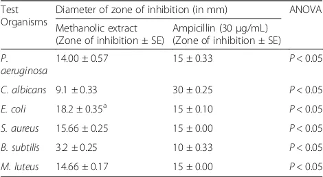
The results representing the antimicrobial potential of crude methanolic leaves extract of B. pilosa is docu-mented in Table 2. The extract showed significant anti-bacterial activity ranges from 9.1–18.2 mm. B. pilosa showed maximum antibacterial activity against E. coli with 18.2 mm (10 mg/mL) inhibition zone as compared to standard Ampicillin (30 μg/mL). The extract showed significant antimicrobial inhibition of S. aureus, M. luteusandP. aeruginosawith 15.66, 14.66 and 14 mm at the concentration of 10 mg/mL and was less active against C. albicanswith 9.1 mm inhibition at the same concentration. However, the extract showed moderate activity at higher concentrations.
Antimicrobial assay using broth micro dilution method
The minimum inhibitory concentrations (MICs) against selected pathogens are represented in Table 3. The ex-tract of B. pilosa showed significant activity against se-lected bacterial pathogens with MIC ranging from 80 to 870 μg/ml. The extract showed maximum activity againstE. coli(80μg/mL) followed byS. aureus(110μg/ mL) andP. aeruginosa(220μg/mL) [Table 3]. The MIC of extract showed significant effect against pathogenic bacterial strains that means the plant extract has a po-tential to develop antimicrobial agent.
Cytotoxicity assay
MTT [3-(4, 5-dimethylythiazol-2-yl)-2, 5-diphenyl-2H-tetrazolium hydrobromide] assay was employed to evalu-ate the cytotoxicity activity against three cancer cell lines: human epithelial carcinoma (HeLa), human hepato carcinoma (HepG2) and human epidermoid carcinoma
[image:5.595.59.540.518.703.2](KB-3-1). The IC50 value was determined as compared
to that of untreated cells and percentage viability curve was plotted against the extract concentration. Micro-scopic and colorimetric measurements were done after 24 h of treatment with the tested extract. The extract showed significant inhibitory effect against tumour cell growth with varying efficiency. Among the screened cell
lines, plant extract showed highest activity against KB-3-1cell lines with IC50values of 99.56 μg/mL (Fig. 3). The
IC50 values for the inhibition of HepG2 and HeLa cells
were found to be 210.8μg/mL and 179.3μg/mL respect-ively. Figure 3 explained that decrease in cell viability which indicated apoptosis induced by methanolic extract of B. pilosa. The results indicated that the leaves of B. pilosamight contain some anticancerous compounds.
DNA fragmentation assay
DNA fragmentation assay was carried out to understand the possible mechanism of cell death on selected cancer cell lines by the methanolic extract of B. pilosa. All the cells were grown and were treated by the IC50
concen-tration of the extract for 72 h. Further, DNA was ex-tracted from the treated cells using 2.0% agarose gel electrophoresis. A typical ladder like pattern was ob-served which shows the internucleosomal fragmentation. The findings suggested that the methanolic leaf extract of B. pilosa is a potent inducer of apoptosis in HeLa, HepG2, and KB-3 cells (Fig. 4).
[image:6.595.61.540.88.444.2]Fig. 2Antioxidant potential of leaves extract ofB. pilosa.aABTS assay (b) DPPH assay
Table 2Antimicrobial activity of methanolic extract ofBidens
pilosaleaves using agar well diffusion method
Test Organisms
Diameter of zone of inhibition (in mm) ANOVA
Methanolic extract (Zone of inhibition ± SE)
Ampicillin (30μg/mL) (Zone of inhibition ± SE)
P. aeruginosa
14.00 ± 0.57 15 ± 0.33 P< 0.05
C. albicans 9.1 ± 0.33 30 ± 0.25 P< 0.05
E. coli 18.2 ± 0.35a 15 ± 0.10 P< 0.05
S. aureus 15.66 ± 0.25 15 ± 0.00 P< 0.05
B. subtilis 3.2 ± 0.25 10 ± 0.33 P< 0.05
M. luteus 14.66 ± 0.17 15 ± 0.00 P< 0.05
a
[image:6.595.56.291.594.723.2]Detection and quantification of phenolic compounds by UHPLC-QqQLIT-MS/MS
Analytical method validation
Determination and quantitative analysis of bioactive compounds was performed using UHPLC-MRM method as described earlier Chandra et al. [39].
Linearity, limits of detection (LOD) and quantification (LOQ) Calibration curves of standard compounds were established using different concentrations of reference analytes. LOD and LOQ were determined using diluted standard com-pounds when the signal-to-noise rations of reference ana-lytes were about 3 and 10, respectively. The obtained results are listed in Table 4. The calculations for calibrations curves and correlation coefficients (r2) were from 0.9996 to 1.0000 within test ranges. LOD and LOQ of reference analytes was 0.01 to 0.20 ng/ml and 0.03 to 0.61 ng/ml respectively.
Precision, stability and recovery
Relative standard deviation (RSD) was used to meas-ure precision and intra-day and inter-day variations
were evaluated by using six replicates and repeating the experiments for 3 days. The intra-day and inter-day precision was found to be less than 1.21%. Stabil-ity was also measured by replicating the injections at 0, 2, 4, 8, 12 and 24 h. The percentage of RSD of five standard analytes was found to be 2.55. The method developed for evaluation of bioactive compounds from B. pilosa leaves extract has good accuracy, with recovery ranges from 94.87% to 105.19% for all ana-lytes (Table 4).
Quantitative analysis
In this study, the UHPLC-QqQLIT-MS/MS method was
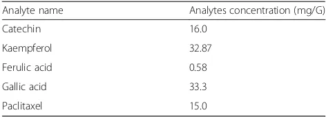
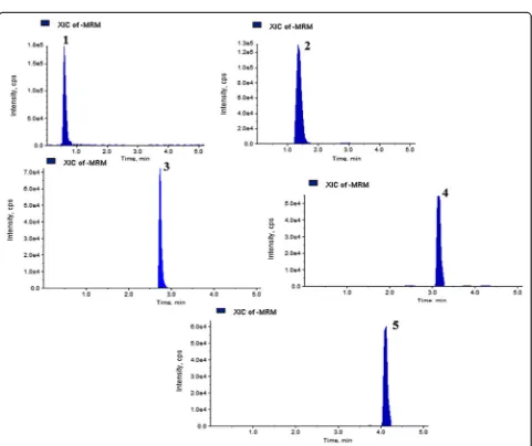
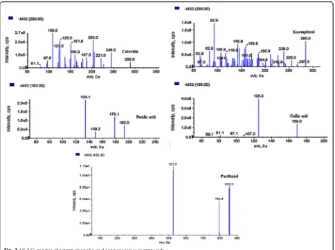
[image:7.595.57.541.99.209.2]applied to five quantitative reference compounds. Quantitative results are listed in Table 5. Gallic acid (33.3 mg/g) was present at the highest amounts, while ferulic acid (0.58 mg/g) was lowest in B. pilosa. The findings of the study prove the existence of variations among the tested reference analytes in B. pilosa. The MRM, extracted ion chromatogram and MS/MS spectra of five mixed standards are shown in Figs. 5, 6 and 7. Table 3Minimum Inhibitory Concentration (MIC) of methanolic extract ofBidens pilosaleaves
Test Organisms MIC values (inμg/mL) ANOVA
Methanolic extract (MIC ± SE) IC50value Ampicillin (MIC ± SE)
P. Aeruginosa 220 ± 0.17 250.52 110 ± 0.05 P< 0.05
C. Albicans 870 ± 0.25 640.04 210 ± 0.30 P< 0.05
E. Coli 80 ± 0.05a 110.67a 60 ± 0.05 P< 0.05
S. Aureus 110 ± 0.17 150.71 82 ± 0.25 P< 0.05
B. Subtilis 380 ± 0.27 520.83 230 ± 0.15 P< 0.05
M. Luteus 250 ± 0.15 290.11 320 ± 0.05 P< 0.05
a
Values indicate significant activity against the pathogen
[image:7.595.57.542.516.714.2]Analysis of volatile compounds by gas chromatography-mass spectroscopy (GC-MS)
GC-MS analysis of compounds was performed in methano-lic leaf extract ofB. pilosa, shown in Table 6. The identifica-tion of volatile compounds is based on the peak area, retention time, percentage of area, molecular weight and molecular formula. Several compounds were detected in the methanolic leaves extract of B. pilosa including
1,3,6,10-Dodecatetraene, 3,7,11-trimethyl-(Z,E); 1H-3A, 7-Methanozulene, Octahydro-1,4,9,9-tetramethyl; 9H– Fluor-ene, 9-Diazo; 1-Octadecyne; N-Hexadecanoic acid and 3,7,11,15-Tetramethyl-2-Hexadecen-1-ol. The spectrum profile of GC-MS analysis showing six components individ-ual fragmentation pattern with retention time 14.05, 16.18, 17.80, 18.90, 20.16 and 21.62 is demonstrated in Fig. 8. The highest peak area (%) of 57.82 was found in 3,7,11,15-Tetra-methyl-2-Hexadecen-1-ol with retention-time 21.62 and the lowest peak area (%) of 3.96 was detected in 1,3,6,10-Dodecatetraene, 3,7,11-trimethyl-(Z,E) with retention-time 14.05 (Table 6).
Mosquitocidal bioassay
Mortality
Mortality rate (MR) of the third instar larva ofC. quinque-fasciatustreated with methanolic extracts ofB. pilosais il-lustrated in Table 7. The MR of B. pilosa was highest at 1000 ppm concentration at different time intervals (P< 0.05) at 24 and 48 h of exposure (Table 8). We have found that the highest larvicidal activity (100%) was detected in metha-nolic extract of B. pilosaafter 12 h. At higher concentra-tions, the larvae moved for some time and then died.
Dose-response (LC50) and time-response (LT50) larvicidal
bioassay
Table 8 described the lethal concentration (LC50) values
of the larvicidal assay after 24 and 48 h ofB. pilosa. The
[image:8.595.58.290.84.371.2]Fig. 4The methanolic extract ofB. pilosainduced DNA fragmentation. L1–100 bp DNA Ladder; L2- Untreated KB-3 cells DNA; L3- DNA KB-3 cells treated with extract; L4- Untreated HeLa cells DNA; L5- DNA HeLa cells treated with extract; L6- Untreated HepG2 cells DNA; L7- DNA HepG-2 cells treated with extract
Table 4Method validation parameters for five reference analytes
Parameters Analytes
Catechin Kaempferol Ferulic acid Gallic acid Paclitaxel
Regression equation 6.01×+ 0.33 5.53×+ 0.31 40.02×+ 0.05 41.44×+ 0.02 306× + 1.92
Correlation coefficient (r2) 0.9998 0.9999 0.9995 1.0000 0.9996
Linearity range (ng/mL) 1–250 1–250 0.5–100 0.1–100 5–500
LOD (ng/mL) 0.14 0.20 0.03 0.01 0.02
LOQ (ng/mL) 0.43 0.61 0.09 0.03 0.07
Precision RSD % (Intra-day,n= 6) 0.34 1.02 0.62 0.25 0.61
Precision RSD % (Inter-day, n = 6) 1.01 1.22 1.11 0.94 1.21
Stability RSD % (n= 5) 1.83 2.55 1.88 1.92 1.80
Recovery (n= 3) Mean 105.19 97.50 94.87 95.50 98.86
RSD % 0.76 1.02 1.13 1.82 0.96
Table 5Content (mg/g) of five bioactive compounds detected
inB. pilosa
Analyte name Analytes concentration (mg/G)
Catechin 16.0
Kaempferol 32.87
Ferulic acid 0.58
Gallic acid 33.3
[image:8.595.303.539.109.194.2] [image:8.595.57.540.566.733.2]Fig. 5MRM extracted chromatogram of standards bioactive compounds mixture obtained by UPLC-ESI–MS/MS in negative mode
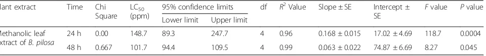
[image:9.595.59.539.87.278.2] [image:9.595.58.539.311.715.2]highest larvicidal activity was found in methanolic ex-tract of B. pilosa (LC50= 148.7) after 24 h and (LC50=
101.7) after 48 h. Chi-square value was highly significant at P< 0.045 to 0.0004 levels in B. pilosa plant extract. The result of one way ANOVA of methanolic extract of B. pilosaat different concentrations (50–1000 ppm) and at different time intervals (24 and 48 h) also exhibited significant difference in larval mortality (P< 0.0004). Higher slope value (0.168 ± 0.015 at 24 h; 0.063 ± 0.022 at 48 h) and lower and upper limits at 95% confident level of LC50 (89.3–247.7 ppm at 24 h; 94.4–109.5 ppm
at 48 h) were observed for methanolic extract of B.
pilosa. The regression analysis showed a positive correl-ation among the mortality rate (Y) and the concentra-tion of exposure (X) having a regression coefficient (R2) of 0.96 and 0.99 respectively. Time response larvicidal bioassay was carried out in methanolic extract of B. pilosa at different concentrations (50–1000 ppm) for 48 h againstC. quinquefaciatus. Methanolic extract ofB. pilosahas taken minimum lethal time (LT50= 6 h) to kill
50% ofC. quinquefasciatusat 500 ppm (Table 9). Stasti-cal analysis showed a positive correlation between the LT50 values and mortality rate was found. Significant
Chi-square value (at P< 0.008 to 0.0001 level), higher
[image:10.595.57.541.86.447.2]Fig. 7MS/MS spectra of target phenolic and anticancerous compounds
Table 6Volatile compounds identified in the methanolic leaf extract ofBidens pilosaby GC-MS
Sl. No. Name of the Compound RT Peak Area Area (%) Height Molecular Weight Nature of compound
1 1,3,6,10-Dodecatetraene, 3,7,11-trimethyl-(Z,E) 14.053 683,540.1 3.96 18,457,168 204.3511
2 1H-3A, 7-Methanozulene, Octahydro-1,4,9,9-tetramethyl 16.184 1,116,642.1 6.46 30,036,296 206.3669
3 9H–Fluorene, 9-Diazo 17.8 3,135,375.3 18.14 82,788,704 192.22 Alkene
4 1-Octadecyne 18.9 1,195,249.8 6.92 33,715,172 252.486 Alkene
5 N-Hexadecanoic acid 20.16 1,158,552.5 6.70 27,304,302 256.4241 Fatty acid
[image:10.595.56.539.632.730.2]slope value (2.212 ± 0.101 at 300 ppm) and lower and upper limits at 95% confident level of LT50 (4.28 at
500 ppm & 29.98 at 50 ppm) were also observed in methanolic extract ofB. pilosa.
Discussion
Phenolics are one of the vital groups of secondary me-tabolites present in plants. Rose and Kasum, [40] sug-gested that the phenolic compounds helps in the maintenance of human health by protecting against vari-ous diseases. Moreover, flavonoids are a group of pheno-lics which have broad spectrum antioxidant properties.
In the present study, the TPC was estimated to be 72μg of GAE/mg of DW significantly high than the reported by Lee et al. [41] from B. pilosa (38.1 mg of GAE/g of DW). The total flavonoids content was found to be 123.3 μg Quercetin/mg of DW. The findings were in support of Lee et al. [38] who demonstrated the TFC as 235.06 mg Quercetin/g of DW. The higher amount of phenolic and flavonoids production showed better anti-oxidant capacity of the tested extract [42]. Cortés-Rojas et al. [43] suggested that the leaves and flowers of B. pilosahave highest TPC and TFC contents as compared to other parts. The main role of flavonoids in the plants
[image:11.595.59.538.86.524.2]is to protect plants from sun radiation and scavenge free radicals. Hence, it is quite expected that the plant parts exposed to sunlight are high in the TFC [44].
Free radicals are well known to play quiet effective role in pathological symptoms [44]. Antioxidant helps us from various diseases by protecting against free radicals either by scavenging the reactive oxygen species or pro-tecting the cells by antioxidant defence mechanisms [45].B. pilosamethanolic extract was tested for free rad-ical scavenging ability using DPPH and ABTS method. In our study, we found that DPPH IC50 value of
80.45μg/ml in methanolic extract ofB. pilosa. Adedapo et al. [46] reported that DPPH IC50value of 94.2 mg/mL
which was higher than our reported value. Deba et al. [1] reported that antioxidant activity of essential oils fromB. pilosaand showed that leaves and flowers essen-tial oil having DDPH IC50 value of 47 and 50μg/ml
re-spectively which further proved that leaves has the highest antioxidant potential as compared to other parts of the selected plants. ABTS, A more appropriate decolorization technique assay in which the radicals are generated directly in a stable form prior to reaction with putative antioxidants [26]. In our study, ABTS IC50value
of 171.6 μg/ml which is higher than the previously re-ported by Adedapo et al. [47] who showed IC50value of
ABTS as 0.75 mg/mL.
The antimicrobial activity showed that B. pilosa have significant antimicrobial potential against four human bac-terial pathogen (S. aureus, P. aeruginosa, M. luteusandE. coli) and yeastC. albicans which are the most common cause of different food borne diseases. In this study, meth-anolic extract of B. pilosaexhibited significant inhibitory effect against gram-negative bacteria (18.1 mm diameter zone of inhibition) than the gram positive bacteria (14.6 mm diameter zone of inhibition) which is compared to standard known antibiotics ampicillin (50μg/disc). The highest zone of inhibition was found against E. coli (18.2 mm). The findings of zone of inhibition was slightly higher than a study reported by Falowo et al. [47] who stated that methanolic extract ofB. pilosashowed zone of inhibition againstE. coli(16.0 mm).
[image:12.595.58.541.112.311.2]We found that B. pilosaleaves extract exhibited signifi-cant antibacterial activity againstS. aureus(15.6 mm). This result was similarly reported by Ashafa and Afolayan, [48] who demonstrated that methanolic extract ofB. pilosahave suppressed the growth of Gram positive bacteriaS. aereus (5.0 mm). According to some previous researchers, metha-nolic extract ofB. pilosawas inactive againstP. aeruginosa andS. aureus[46, 47]. As these bacteria are having resistant capacity against the extracts could be characterized to their cell wall which has been mentioned to inhibit the penetra-tion of the plant extract [49, 50].
Table 7Time dependent mortality check of larvicidal activity of crude methanolic extract ofB. pilosatill 48 h at different concentrations
Plant extract Concentration in PPM
% Mortality ± SE (Time in h)
1 3 6 12 18 24 30 36 42 48
Methanolic leaf extract ofB. pilosa
50 0 0 0 4.1 ± 0.25 11.6 ± 0.12 21.5 ±
0.25
37.2 ± 0.17 46.3 ± 0.10
54.7 ± 0.20 67.1 ± 0.20
100 0 0 6.3 ± 0.10 11.6 ±
0.25
22.4 ± 0.10 38.2 ± 0.10
47.6 ± 0.17 59.2 ± 0.10
67.4 ± 0.25 83.4 ± 0.27
200 0 0 14.7 ±
0.17
29.4 ± 0.12
42.3 ± 0.27 54.6 ± 0.25
67.0 ± 0.10 76.2 ± 0.05
85.4 ± 0.15 97.1 ± 0.10
300 0 11.2 ±
0.10
19.8 ± 0.05
27.8 ± 0.15
39.4 ± 0.25 59.4 ± 0.15
74.4 ± 0.17 86.5 ± 0.25
100.0 ± 0.00
–
400 16.2 ±
0.17 27.8 ± 0.25 41.6 ± 0.05 58.3 ± 0.15
74.8 ± 0.10 89.7 ± 0.15
100.0 ±
0.00 – – –
500 24.2 ±
0.10 47.1 ± 0.05 69.8 ± 0.15 90.8 ± 0.17 100.0 ± 0.00 – – – – –
1000 24.1 ±
0.27 42.8 ± 0.10 67.6 ± 0.25 100 ±
0.00 – – – – – –
Control 0 0 0 0 0 0 0 0 0 0
Table 8Log probit and regression analysis of third larval instars ofC. quinquefasciatusin different concentrations of methanolic extract ofB. pilosafor 24 h and 48 h
Plant extract Time Chi
Square LC50 (ppm)
95% confidence limits df R2Value Slope ± SE Intercept ± SE
Fvalue Pvalue
Lower limit Upper limit
Methanolic leaf extract ofB. pilosa
24 h 0.00 148.7 89.3 247.7 4 0.96 0.168 ± 0.015 17.02 ± 4.69 118.7 0.0004
[image:12.595.61.541.676.732.2]The minimum inhibitory concentration (MICs) of methanolic extracts ofB. pilosaagainst selected bacterial pathogens is represented in Table 3. The methanolic extracts of B. pilosa inhibited bacterial and yeast patho-gen with MIC ranging from 80 to 380μg/mL. B. pilosa showed highest activity against E. coli (80 μg/mL) followed by S. aureus (110 μg/mL) and P. aeruginosa (220 μg/mL). Previous reports also showed that the methanolic leaves extract was more active which indi-cates that the methanolic leaves extract has the potential antimicrobials [48]. TheB. pilosaextract showed signifi-cant inhibitory activity against bacterial and yeast patho-gen which suggest as an exploitable source for the discovery of antimicrobial agents [30, 51].
Previous reports have stated that isolated new com-pounds from B. pilosahave anticancer activities against various types of cancer. According to Kviecinski and Felipe, [20], different crude extract like chloroform, ethyl acetate and methanol fractions ofB. pilosapossess anti-tumor activity which has assessed using brine shrimp, hemolytic, MTT, and neutral red uptake (NRU) assays. In present study, the methanolic extract of B. pilosa inhibited the growth of three cancer cell lines KB-3-1, HepG2 and HeLa with IC50 values of 99.56 μg/mL,
210.8 μg/mL and 179.3 μg/mL respectively. Percentage of inhibition was found significantly high than the previ-ous studies reported by Sundararajan et al. [9] and Wu et al. [52] who stated that the methanol extract of B. pilosa showed anticancer activity against HeLa, HepG2 and KB cells with IC50values of 965.2μg/mL, 119.55μg/
mL and 586.2μg/mL respectively. Steenkamp and Gouws, [53] reported that several members of Astera-ceae family such as B. pilosa showed cytotoxic activity on some tumor cell lines. Furthermore, Kumari et al. [10] reported that the isolated compound phenyl-1, 3, 5-heptatriene from B. pilosa has antiproliferating effect against human oral, liver, colon, and breast cancer cell lines with IC50 values of 8.0 ± 0.01, 0.49 ± 0.45, 0.7 ±
0.01and 10 ± 0.01 μg/mL respectively. Further, DNA fragmentation was observed in HeLa, HepG2, and KB-3 cells treated withB. pilosaextract, thereby indicating the
onset of apoptotic cell death. Thus, the results obtained in this study suggest that the methanolic extract of B. pilosa might have an apoptosis-inducing property, iso-lated from the leaves ofB. pilosacan act as potential an-ticancer agents in cancer chemotherapy.
A few phenolic compounds like gallic acid, Kaempferol, Catechin, Paclitaxel and Ferulic acid was detected for the first time from methanolic extract of B. pilosa plant. Kaempferol, phenolic compound was also reported first time from B. pilosa which is used for the treatment of various types of cancers [54, 55]. Ferulic acid was detected in less quantity (0.58 mg/G). This compound was similarly reported by Muchuweti et al. [56] who has detected from 50% aqueous methanol of B. pilosa using HPLC system. Paclitaxel, brand name taxol is a chemotherapy medica-tion which was reported first time from B. pilosa. This compound was isolated first time from the bark of the Pa-cific yew,Taxus brevifoliaand its given name“taxol”[57].
GC-MS analysis of the methanolic extract of B. pilosa showed the presence of six volatile compounds i.e. 1,3,6,10-Dodecatetraene, 3,7,11-trimethyl-(Z,E); 1H-3A, 7-Methanozulene, Octahydro-1,4,9,9-tetramethyl; 9H– Fluorene, 9-Diazo; 1-Octadecyne; N-Hexadecanoic acid and 3,7,11,15-Tetramethyl-2-Hexadecen-1-ol. These com-pounds are responsible for numerous pharmacological ac-tions like antimicrobial activities useful in a treatment of variety of diseases and anticancer activities against various cancers [58, 59]. Recently, Kale, [59] reported that two volatile compound namely N-Hexadecanoic acid and 3,7,11,15-Tetramethyl-2-Hexadecen-1-ol from ethanolic leaf extract of Adiantum capillus-veneris L which has similarly reported in our study from methanolic extract of B. pilosaplant. To best our knowledge, this is first time re-ported six compound 1,3,6,10-Dodecatetraene, 3,7,11-tri-methyl-(Z,E); 1H-3A, 7-Methanozulene, Octahydro-1,4,9,9-tetramethyl; 9H–Fluorene, 9-Diazo; 1-Octadecyne; N-Hexadecanoic acid and 3,7,11,15-Tetramethyl-2-Hexa-decen-1-ol from methanolic extract ofB. pilosa.
[image:13.595.58.539.110.221.2]Larvae are mainly killing by using different synthetic che-micals like – organochlorine (DDT), organophosphates (malathion, temephos and fenthion), synthetic pyrethroids Table 9Log probit and regression analysis of time dependent larvicidal efficacy of methanolic extract ofB. pilosaat different concentrations against third instar larvae ofC. quinquefasciatus
Plant name Concentration Chi Square
LT50 (h)
95% confidence limits df R2
Value Slope ± SE Intercept ± SE Fvalue Pvalue
Lower limit Upper limit
Methanolic extract ofBidens pilosa
50 2.8 28.32 26.74 29.98 8 0.96 1.488 ± 0.097 −8.47 ± 2.64 232.9 0.0001
100 0.8 25.36 23.09 27.86 8 0.99 1.788 ± 0.057 −5.734 ± 1.566 959.1 0.0001
200 0.8 19.17 16.74 21.95 8 0.98 2.087 ± 0.083 0.76 ± 2.25 628.8 0.0001
300 0.8 18.85 15.67 22.67 8 0.98 2.212 ± 0.101 3.196 ± 2.76 471.3 0.0001
400 5.4 10.17 8.34 12.4 4 0.86 1.815 ± 0.258 30.91 ± 7.0 49.45 0.0001
(deltamethrin), insect growth regulators (diflubenzuron and methoprene) etc. A high amount of DDT and Malathion re-sistance was used inC. quinquefasciatuslast several years in Northeast India. The use of DDT is stopped in several places of India due to development of resistance in vector populations. Though, this chemicals are still used for control of Kala-azar vector and malaria vectors of different parts of North-eastern India especially Mizoram [60]. Since insecti-cide resistance threatens to contribute towards the reintro-duction of vector borne diseases in many parts of the world, efforts have been focused on finding an alternative form of mosquito control. Therefore, several compounds of plant have been reported as insecticides-larvicides which are very essential to improve their formulations with enhanced activ-ity. So, this improved product may be useful to control insecticides and mosquito. Previous researchers reported that different plant families – Asteraceae, Solanaceae, Euphorbiaceae, Leguminoceae, Cladophoraceae, Labiatae, Meliaceae, Solanaceae, Umbelliferae, Compositae, Myrtaceae, Lauraceae, Lamiaceae, Apiaceae, Cupressaceae, Poaceae, Zingiberaceae, Piperaceae, Aristolochiaceae, Caesalpinaceae, Chenopodiaceae, Oocystaceae, Fabaceae and Rutaceae showed larvicidal and insecticidal activity against different species of mosquitoes [61–64]. The crude methanolic extract ofB. pilosashowed larvicidal effect against third instar larva ofC. quinquefasciatus. The methanolic extract of B. pilosa exhibited 100% mortality rate after 12 h of incubation at the concentration of 1000 ppm. Similarly, Macêdo et al. [65] checked that ethanolic extract ofBidens pilosashowed larvi-cidal effect against fourth instar larva ofAedes fluviatiliswho stated that 12.2% of mortality at 100 mg/L concentration. To best our knowledge, this is first time reported that methano-lic extract of B. pilosa exhibited larvicidal activity against third instar larva ofC. quinquefasciatus.
Conclusions
The overall findings of our study provide evidence for the bioactive potential of methanolic leaves extract of B. pilosaand the ecological significance of human well be-ing. The results obtained bring up supporting data for future investigation of the studied plant which could lead to their use in cancer, oxidative stress and anti-microbial therapy.
Abbreviations
ABTS+:2,2- Azinobis-3-ethylbenzothiazoline-6-sulphonic acid disodium salt; AlCl3: Aluminium chloride; ANOVA: Analysis of variance; CFU: Colony forming unit; DMSO: Dimethyl sulfoxide; DPPH: 2,2-diphenyl-1-picrylhydrazyl; GC-MS: Gas chromatography-mass spectrometry; HeLa: Human epithelial carcinoma; HepG2: Human hepato carcinoma; IC50: The Inhibitory concentration required for 50% scavenging of DPPH and ABTS radicals in a specific time; KB-3-1: Human epidermoid carcinoma; LC50: Lethal concentration required to kill 50% of mosquito larvae; LT50: Lethal time till half the mosquito larvae dies; MIC: Minimum inhibitory concentrations; MRM: Multiple Reaction Monitoring; MTCC: The Microbial Type Culture Collection and Gene Bank; MTT: 3-(4, 5-dimethylythiazol-2-yl)-2, 5-diphenyl-2H- tetrazolium hydrobromide; UHPLC-QqQLIT-MS/MS: Ultra high
performance liquid chromatography-hybrid linear ion trap triple quadrupole mass spectrometry
Acknowledgments
Authors are thankful to the Department of Biotechnology, New Delhi for establishment of DBT-State Biotech Hub in Mizoram University which has been utilized for the present study.
Funding
GS wishes to thank University Grants Commission (UGC), New Delhi for the fellowship provided through Rajiv Gandhi National Fellowship for to pursue Ph.D. Degree (F1–17.1/2015–16/RGNF-2015-17-SC-UTT-9023).
Availability of data and materials
All data generated or analysed during this study are included in this article.
Authors’contributions
GS carried out the full experiments, and drafted the manuscript; BPS and NSK helped her to conceptualize and supervise the experiment; AKP and VVL helped GS in Data analysis and statistical analysis; SS helped to evaluate cytotoxicity assays; PS, BK and HL helped to carry out UHPLC-QqQLIT-MS/MS and GC/MS analysis. All authors have read and approved the final manuscript.
Ethics approval and consent to participate Not applicable.
Consent for publication Not applicable.
Competing interests
The authors declare that they have no competing interests.
Publisher’s Note
Springer Nature remains neutral with regard to jurisdictional claims in published maps and institutional affiliations.
Author details
1Department of Biotechnology, Mizoram University, Aizawl, Mizoram 796004,
India.2SAIF, CSIR-Central Drug Research Institute (CSIR-CDRI), Lucknow 226012, India.3Department of Pharmacy, Regional Institute of Paramedical and Nursing Sciences, Aizawl, Mizoram 796017, India.
Received: 2 August 2017 Accepted: 10 November 2017
References
1. Deba F, Xuan TD, Yasuda M, Tawata S. Chemical composition and antioxidant, antibacterial and antifungal activities of the essential oils from Bidens pilosaLinn. Var. Radiata. Food Control. 2008;19:346–52.
2. Pozharitskaya ON, Shikov AN, Makarova MN, Kosman VM, Faustova NM, Tesakova SV, Makarov VG, Galambosi B. Anti-inflammatory activity of a HPLC-fingerprinted aqueous infusion of aerial part of Bidens Tripartita L. Phytomedicine. 2010;17:463–8.
3. Sharma HK, Chhangte L, Dolui AK. Traditional medicinal plants in Mizoram, India. Fitoterapia. 2001;72:146–61.
4. Silva JJ, Cerdeira CD, Chavasco JM, Cintra AB, Silva CB, Mendonça AN, et al. In vitroscreening antibacterial activity ofBidens pilosa linnéandAnnona crassifloramart against oxacillin resistantStaphylococcus aureus(orsa) from the aerial environment at the dental clinic. Rev Inst Med Trop Sao Paulo. 2014;56:333–40.
5. Khan MR, Kihara M, Omoloso AD. Anti-microbial activity ofBidens pilosa, Bischofia javanica,Elmerillia papuanaandSigesbekia orientalis. Fitoterapia. 2001;72:662–5.
6. Pereira LC, Ibrahim T, Lucchetti L, Da Silva ADR, De Moraes VKG. Immunosuppressive and anti-inflammatory effects of methanolic extract and the polyacetylene isolated fromBidens pilosaL. Immunopharmacology. 1999;43:31–7.
8. Yuan LP, Chen FH, Ling L, Dou PF, Bo H, Zhong MM, et al. Protective effects of total flavonoids ofBidens pilosaL. (TFB) on animal liver injury and liver fibrosis. J Ethnopharmacol. 2008;116:539–46.
9. Sundararajan P, Dey A, Smith A, Doss AG, Rajappan M, Natarajan S. Studies of anticancer and antipyretic activity ofBidens pilosawhole plant. Afri Health Sci. 2006;6:27–30.
10. Kumari P, Misra K, Sisodia BS, Faridi U, Srivastava S, Luqman S, et al. A promising anticancer and anti-malarial component from the leaves of Bidens pilosa. Planta Med. 2009;75:59–61.
11. Silva FL, Fischer DCH, Tavares JF, Silva MS, De-Athayde-Filho PF, Barbosa-Filho JM. Compilation of secondary metabolites fromBidens pilosaL. Molecules. 2011;16:1070–102.
12. Bartolome F, Wu HC, Burchell VS, Preza E, Wray S, Mahoney CJ. Pathogenic VCP mutations induce mitochondrial uncoupling and reduced ATP levels. Neuron. 2013;78:57–64.
13. Pacome OA, Bernard DN, Sekou D, Joseph DA, David NJ, Mongomaké K, et al. Phytochemical and antioxidant activity of roselle (hibiscus Sabdariffa L.) petal extracts. Res J Pharm Biol Chem Sci. 2014;5:1453–65.
14. Lobo V, Patil A, Phatak A, Chandra N. Free radicals, antioxidants and functional foods: impact on human health. Pharmacogn Rev. 2010;4(8):118–26. 10.4103/0973-7847.70902.
15. Prabuseenivasan S, Jayakumar M, Ignacimuthu S.In vitroantibacterial activity of some plant essential oils. BMC Complement Alter Med. 2006;6:39. 16. Hersch-Martinez P, Leanos-Miranda BE, Solorzano-Santos F. Antibacterial
effects of commercial essential oils over locally prevalent pathogenic strains in Mexico. Fitoterapia. 2005;76:453–7.
17. De Esparza RR, Bye R, Meckes M, Torres LJ, Estrada J. Antibacterial activity of Piqueria trinervia, a Mexican medicinal plant used to treat diarrhea. Pharm Biol. 2007;45:446–52.
18. Chang JS, Chiang LC, Chen CC, Liu LT, Wang KC, Lin CC. Anti-leukemic activity ofBidens pilosaL. Var. Minor (Blume) Sherff andHouttuynia cordata Thunb. Am J Chin Med. 2002;29:303–12.
19. Ojekale AB, Lawal OA, Lasisi AK, Adeleke TI. Phytochemistry and spermatogenic potentials of extract ofCissus populnea(Guill and per) stem bark. TSW Holistic Health Med. 2006;1:176–82.
20. Kviecinski MR, Felipe KB, Schoenfelder T. Study of the antitumor potential of Bidens pilosa(Asteraceae) used in Brazilian folk medicine. J Ethnopharmacol. 2008;117:69–75.
21. Chiang YM, Chuang DY, Wang SY, Kuo YH, Tsai PH, Shyur LF. Metabolite profiing and chemopreventive bioactivity of plant extracts fromBidens pilosa. J Ethnopharmacol. 2004;95:409–19.
22. Liang X, Xu Q. Separation and identification of phenolic compounds in Bidens pilosaL. by ultra high performance liquid chromatography with quadrupole time-of-flight mass spectrometry. J Sep Sci. 2016;29:1853–62. 23. Liu M, Li WQ, Weber C, Lee CY, Brown J, Liu RH. Antioxidant and
antiproliferative activities of raspberries. J Agric Food Chem. 2002;50:2926–30. 24. Chang C, Yang M, Wen H, Chern J. Estimation of total flavonoid content in
propolis by two complementary colorimetric methods. J Food Drug Analysis. 2002;10:178–82.
25. Brand-Williams W, Cuvelier ME, Berset C. Use of a free radical method to evaluate antioxidant activity. Food Sci Technol. 1995;28:25–30. 26. Re R, Pellegrini N, Proteggente A, Pannala A, Yang M, Rice-Evans C.
Antioxidant activity applying an improved ABTS radical cation decolorization assay. Free Radic Biol Med. 1999;26:231–1237.
27. Perez C, Paul M, Bazerque P. An antibiotic assay by the agar well diffusion method. Acta Bio Med Exp. 1990;15:113–5.
28. Osato MS. Antimicrobial susceptibility testing for helicobacter pylori: sensitivity test results and their clinical relevance. Curr Pharm Design. 2000;6:1545–55. 29. King T, Dykes G, Kristianti R. Comparative evaluation of methods commonly
used to determine antimicrobial susceptibility to plant extracts and Phenolic compounds. J AOAC Int. 2008;91:1423–9.
30. Singh G, Passsari AK, Leo VV, Mishra VK, Subbarayan S, Sigh BP, et al. Detection of phenolic compounds, antioxidant, antimicrobial and cytotoxic activities ofselected traditional medicinal plants from Northeast India. Front Plant Sci. 2016;7:407.
31. Mosmann T. Rapid colorimetric assay for cellular growth and survival: application to proliferation and cytotoxicity assays. J Immunol Methods. 1983;65:55–63. 32. Subbarayan S, Marimuthu SK, Nachimuthu SK, Zhang W, Subramanian S.
Characterization and cytotoxic activity of apoptosis-inducing pierisin-5 protein from white cabbage butterfly. Int J Biol Macromolec. 2016;87:16–27. 10.1016/j.ijbiomac.2016.01.072.
33. Pandey R, Mahar R, Hasanain M, Shukla SK, Sarkar J, Rameshkumar KB, Kumar B. Rapid screening and quantitative determination of bioactive compounds from fruit extracts of Myristica species and their in vitro antiproliferative activity. Food Chem. 2016;211:483–93. 10.1016/j.foodchem.2016.05.065.
34. Sen A, Batra A. Chemical composition of methanol extract of the leaves of Melia AzedarachL. Asian J Pharm Clin Res. 2012;5:42–5.
35. Rufattoa LC, Finimundy TC, Roesch-Ely M, Mouraa S. Mikania Laevigata: chemical characterization and selective cytotoxic activity ofextracts on tumor cell lines. Phytomed. 2013;20:883–9.
36. Lalrotluanga LN, Senthil NK, Gurusubramanian G. Insecticidal and repellent activity ofHiptage benghalensisL. Kruz (Malpighiaceae) against mosquito vectors. Parasitol Res. 2012;111:1007–17.
37. WHO - World Health Organization. Instructions for determining the susceptibility or resistance of mosquito larvae to insecticides. Geneva: WHO; 1981. p. 6.
38. Lallawmawma H, Kumar GS, Sarathbabu S, Ghatak S, Sivaramakrishnan S, Gurusubramanian G, et al. Synthesis of silver and gold nanoparticles using Jasminum nervosumleaf extract and its larvicidal activity against filarial and arboviral vectorCulex quinquefasciatussay (Diptera: Culicidae). Environ Sci Pollut Res. 2015;22:17753–68.
39. Chandra P, Pandey R, Kumar B, Srivastva M, Pandey P, Sarkar J, Singh BP. Quantification of multianalyte by UPLC–QqQLIT–MS/MS and in-vitro anti-proliferative screening in Cassia species. Ind Crop Prod. 2015;76:1133–41. 10. 1016/j.indcrop.2015.08.030.
40. Rose JA, Kasum CM. Dietary flavonoids: bioavalibility, metabolic effects and safety. Ann Rev Nutr. 2002;22:19–34.
41. Lee WC, Peng CC, Chang CH, Huang SH, Chyau CC. Extraction of antioxidant components fromBidens pilosaflowers and their uptake by human intestinal Caco-2 cells. Molecules. 2013;18:1582–601.
42. Ghasemzadeh A, Jaafar HZE, Rahmat A. Effects of solvent type on phenolics and flavonoids content and antioxidant activities in two varieties of young ginger (Zingiber officinaleroscoe) extracts. J Med Plants Res. 2011;5:1147–54. 43. Cortes-Roja DF, De Souza CRF, Pereira OW. Clove (Syzygium aromaticum): a
precious spice. Asian Pac J Trop Biomed. 2014;4:90–6.
44. Dorman HJD, Peltoketo A, Hiltunen R, Tikkanen MJ. Characterization of the antioxidant properties of de-odourised aqueous extracts from selected Lamiaceae herbs. Food Chem. 2003;83:255–62.
45. Umamaheswari M, Chatterjee TK.In vitroantioxidant activities of the fractions ofCoccinia grandisL. leaf extracts. Afr J Trad Camp Alter Med. 2008;5:61–73.
46. Adedapo A, Jimoh F, Afolayan A. Comparison of the nutritive value and biological activities of the acetone, methanol and water extracts of the leaves ofBidens pilosaandChenopodium album. Acta Pol Pharm ñ Drug Res. 2011;68:83–92.
47. Falowo AB, Muchenje V, Hugo CJ, Charimba G.In vitroantimicrobial activities ofBidens pilosaandMoringa oleiferaleaf extracts and their effects on ground beef quality during cold storage. Cyta J Food. 2016;14:541–6. 48. Ashafa AOT, Afolayan AJ. Screening the root extracts fromBiden pilosa
L. Var.radiata(Asteraceae) for antimicrobial potentials. J Med Plants Res. 2009;3:568–72.
49. Hayek SA, Ibrahim SA. Antimicrobial activity of Xoconostle pears (Opuntiamatudae) againstEscherichia coliO157: H7 in laboratory medium. Int J Microbiol. 2012;2012:1–6.
50. Nisa H, Kamili AN, Bandh SA, Amin S, Lone BA, Parray PA. Phytochemical screening, antimicrobial and antioxidant efficacy of different extracts of Rumex dentatusL.–a locally used medicinal herb of Kashmir Himalaya. Asian Pac J Trop Dis. 2013;3:434–40.
51. Borrás-Linares I, Fernández-Arroyo S, Arráez-Roman D, Palmeros-Suárez PA, Val-Díaz RD, Andrade-Gonzáles I. Characterization of phenolic compounds, anthocyanidin, antioxidant and antimicrobial activity of 25 varieties of Mexican Roselle (Hibiscus sabdariffa). Indus Crop Prod. 2015;69:385–94. 52. Wu J, Wan Z, Yi J, Wu Y, Peng W, Wu W. Investigation of the extracts from
Bidens pilosaLinn. Var. radiate Sch. Bip. For antioxidant activities and cytotoxicity against human tumor cells. J Nat Med. 2013;67:17–26. 53. Steenkamp V, Gouws MC. Cytotoxicity of six south African medicinal plant
extracts used in the treatment of cancer. South Afri J Bot. 2006;72:630–3. 54. Calderon-Montaño JM, Burgos-Moron E, Perez-Guerrero C, Lopez-Lazaro M. A
review on the dietary flavonoid kaempferol. Mini Rev Med Chem. 2011;11:298–344. 55. Kim SH, Choi KC. Anti-cancer effect and underlying mechanism(s) of
56. Muchuweti M, Mupure C, Ndhlala A, Murenje T, Benhura MAN. Screening of antioxidant and radical scavenging activity ofVigna ungiculata, Bidens pilosa and Cleome gynandra. Ame J Food Technol. 2007;2:161–8.
57. Goodman J, Walsh V. The Story of Taxol: Nature and Politics in the Pursuit of an Anti-Cancer Drug Cambridge University Press, Cambridge, UK. 2001. ISBN 978-0-521-56123-5.
58. Selvamangai C, Bhaskar A. GC-MS analysis of Phytocomponents in the Methanolic extract ofEupatorium triplinerve. Int J Drug Dev Res. 2012;4:148–53. 59. Kale MV. GC-MS analysis of phytocomponents on whole plant extracts
Adiantumcapillus-venerisL.-a potential folklore medicinal plant. Res J Life Sci Bioinfor Pharmaceu Chem Sci. 2015;2:117.
60. Sarkar M, Bhattacharyya IK, Borkotoki A, Goswami D, Rabha B, Baruah I. Insecticide resistance and detoxifying enzyme activity in the principal bancroftian filariasis vector,Culex quinquefasciatusin Northeastern India. Med Vet Entomol. 2009a;23:122–31.
61. Shrestha PM, Dhillion SS. Medicinal plant diversity and use in the highlands of Dolakha district, Nepal. J Ethnopharmacol. 2003;86:81–96.
62. Teodora DB, Ashlyn KDB. Ethnomedical knowledge of plants and healthcare practices among the Kalanguya tribe in Tinoc, Ifugao, Luzon, Philippines. Indian J Trad Know. 2011;10:227–38.
63. Ghosh A, Nandita C, Chandra G. Plant extracts as potential mosquito larvicides. Indian J Med Res. 2012;135:581–98.
64. Dua VK, Kumar A, Pandey AC, Kumar S. Insecticidal and genotoxic activity of Psoralea corylifoliaLinn. (Fabaceae) againstCulex quinquefasciatussay, 1823. ParasiteVector. 2013;6:30.
65. Macedo ME, Consoli RAGB, Grandi TSM, dos-Anjos AMG, de Oliveira AB, Mendes NM, et al. Screening of Asteraceae (Compositae) plant extracts for Larvicidal activity againstAedes fluviatilis(Diptera: Culicidae). Mem Inst Oswaldo Cruz Rio de Janeiro. 1997;92:565–70.
• We accept pre-submission inquiries
• Our selector tool helps you to find the most relevant journal • We provide round the clock customer support
• Convenient online submission • Thorough peer review
• Inclusion in PubMed and all major indexing services • Maximum visibility for your research
Submit your manuscript at www.biomedcentral.com/submit