Changes in Dynamic Pedobarography after Extensive
Plantarmedial Release for Paralytic Pes Cavovarus
Yong Uk Kwon,
1Hyun Woo Kim,
2Jin Ho Hwang,
2Hoon Park,
3Hui Wan Park,
4and Kun Bo Park
5 1Department of Orthopaedic Surgery, Busan Paik Hospital, Inje University College of Medicine, Busan;2Department of Orthopaedic Surgery, Severance Children’s Hospital, Yonsei University College of Medicine, Seoul;
3Department of Orthopaedic Surgery, Gangnam Severance Hospital, Yonsei University College of Medicine, Seoul;
4Department of Orthopaedic Surgery, International St. Mary’s Hospital, Incheon;
5Department of Orthopedic Surgery, Haeundae Paik Hospital, Inje University College of Medicine, Busan, Korea.
Received: May 14, 2013 Revised: August 10, 2013 Accepted: September 27, 2013 Corresponding author: Dr. Kun Bo Park, Department of Orthopaedic Surgery, Haeundae Paik Hospital,
Inje University College of Medicine, 875 Haeun-daero, Haeundae-gu, Busan 612-896, Korea.
Tel: 82-51-797-0611, Fax: 82-51-797-0032 E-mail: kunbopark@gmail.com
∙ The authors have no financial conflicts of interest.
© Copyright:
Yonsei University College of Medicine 2014
This is an Open Access article distributed under the terms of the Creative Commons Attribution Non-Commercial License (http://creativecommons.org/ licenses/by-nc/3.0) which permits unrestricted non-commercial use, distribution, and reproduction in any medium, provided the original work is properly cited.
Purpose: Plantarmedial release and first ray extension osteotomy are often com-bined to treat paralytic cavovarus foot deformity. The purpose of this study is to evaluate the effect of additional first ray extension osteotomy in terms of dynamic pedobarography. Materials and Methods: We reviewed findings of pre- and post-operative plain radiography and dynamic pedobarography for 25 patients in whom the flexibility of the hindfoot was confirmed by the Coleman block test. The re-sults of treatment by extensive plantar medial release with first ray osteotomy (group I) were compared with the results of treatment by extensive plantar medial release alone (group II). Results: Plain radiographs obtained pre- and postopera-tively showed no statistically significant improvement in each group. Only in group I, peak forces at the 1st metatarsal head, 2nd metatarsal head and medial calcaneus were increased after operation. Conclusion: In paralytic hindfoot flexi-ble cavovarus, extensive plantarmedial release with first ray osteotomy improve foot pressure distribution more than extensive plantarmedial release alone.
Key Words: Paralysis, pes cavus, osteotomy
INTRODUCTION
Foot deformity is one of the most frequent orthopedic problems in neuromuscular disease and is caused by imbalances between the intrinsic and extrinsic muscles. Cavovarus foot deformity, which increases the height of the medial longitudinal arch and induces heel varus, may develop in patients with cerebral palsy, myelo-meningocele, poliomyelitis, Charcot-Marie-Tooth disease, and other neuromuscu-lar diseases.1-3
included in this study. This included 15 boys and 10 girls with a mean age of 9 years and 11 months (range, 4 years and 3 months to 15 years and 6 months) at the time of sur-gery. The average follow-up period was 25 months (range, 16-58 months). Cavovarus foot deformity was caused by myelomeningocele in 26 cases, Charcot-Marie-Tooth dis-ease in 7 cases, and spinal tumor resection in 4 cases. All patients were able to walk independently before and after surgery, and weight-bearing radiological measurements and dynamic pedobarography were performed.
Operative techniques
Extensive plantarmedial release began with a medial inci-sion, followed by release of the abductor hallucis from the first metatarsal bone and exposure of the plantarmedial area. After confirming the medial and lateral plantar nerves and blood vessels, Z-lengthening was performed on the flexor hallucis longus and the flexor digitorum longus, if re-quired. Contractured ligaments of the subtalar joint, talona-vicular joint, plantar fascia, and muscles originating from the tarsal bone were also released. As a first ray osteotomy, a first metatarsal extension wedge osteotomy was per-formed in 21 feet, and a medial cuneiform extension wedge osteotomy was performed in 1 foot.
For correcting equinus, the Achilles tendon was length-ened in 27 feet. In 5 feet in which equinus deformity was not sufficiently corrected by lengthening of the Achilles tendon, posterolateral release was performed by additional release of calcaneofibular ligament and capsulotomy of tib-iotalar and subtalar joint. In 4 feet, although the tibia exter-nal rotation was below 40 degrees, distal tibial rotatioexter-nal surgical technique appropriate for the cause of disease, age
at the time of surgery, and especially the flexibility of the foot.5,7,8 Cavovarus creates a tripod effect, in which plantar
flexion of the first ray induces forefoot pronation and hind-foot varus develops.5,9 When the flexibility of the hindfoot
varus is confirmed by the Coleman block test,9 selective
soft tissue release and midfoot or forefoot osteotomies are generally recommended.6,9-11
However, the combination of soft tissue release and mid-foot or foremid-foot osteotomies was decided subjectively by the operator. Although radiographic correction or clinical improvement of cavovarus with flexible hindfoot after sur-gery has been reported, these outcomes cannot directly be compared because the investigators have used different techniques for treatment. Furthermore, many studies had only evaluated static changes in terms of radiological ob-servations.
We reviewed medical records for patients who under-went extensive plantar medial release with elective first ray osteotomy for paralytic cavovarus foot deformity with flex-ible hindfoot varus. Using plain radiography and dynamic pedobarography, we evaluated the effect of the addition of the first ray extension osteotomy in paralytic cavovarus foot deformity with flexible hindfoot varus.
MATERIALS AND METHODS
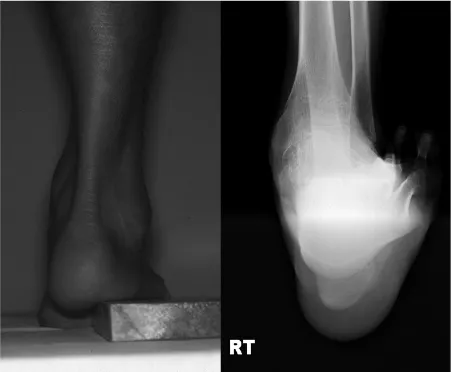
This study was approved by the Institutional Review Board of our hospital. Pediatric patients below 16 years old with neuromuscular disease and cavovarus foot deformity who were able to walk without assistive devices and underwent extensive plantar medial release from March 2005 to April 2009 were recruited. The exclusion criteria were: 1) use of any kind of tendon transfer surgery around ankle, such as tibialis anterior or posterior split transfer, 2) presence of oth-er skeletal deformity, such as hip dislocation or subluxation, knee contracture, excessive femoral anteversion above 50 degrees, excessive external tibial torsion above 40 degrees, excessive internal tibial torsion above 15 degrees and ex-cessive varus or valgus ankle deformity. Flexibility of the hindfoot was confirmed in these patients using the Cole-man block test (Fig. 1), and hindfoot varus was determined to be <5 mm using the radiological test suggested by Pau-los, et al.5
Twenty five patients (37 affected feet) who were available
[image:2.595.313.540.521.707.2]es (PF) of each area were obtained during the stance phase. To evaluate the effects of first ray osteotomy on the out-come, we compared results for 22 feet treated with a first metatarsal extension wedge osteotomy and medial cuneiform extension wedge osteotomy (group I) with results for 15 feet not treated with osteotomy (group II). At the time of surgery, the mean age of the patients was 10 years and 8 months (range, 4 years and 3 months to 15 years and 6 months) in group I and 8 years and 5 months (range, 4 years and 3 months to 15 years and 2 months) in group II (p=0.765). In group I, posterolateral release was performed in 2 feet and Achilles tendon lengthening in 18 feet. In group II, postero-lateral release was performed in 3 feet and Achilles tendon lengthening in 9 feet.
Statistical analysis
The plantar force measurements and radiographic findings of patients were compared with corresponding data for 16 normal individuals older than 4 years. To minimize measure-ment errors, 2 fellowship-trained pediatric orthopedic sur-geons obtained all radiographic and pedobarographic mea-surements. All parameters were measured twice by each author, and these measurements were then averaged. Statis-tical analyses were performed using the SAS software pack-age (version 9.1, SAS Institute, Cary, NC, USA).
To ensure validity of the results and to identify significant group differences, a mixed model was used to enable repeat-osteotomy was performed to make tibia rotation similar to
the contralateral side. Tendon transfer was not performed in any case.
Outcome measurement
The first metatarsal-talus angle and the talocalcaneal angle were measured on pre- and postoperative weight-bearing anteroposterior radiographs. The talocalcaneal angle, calca-neal pitch, first metatarsal-talus angle (Meary angle), and first metatarsal-calcaneus angle (Hibb’s angle) were mea-sured on lateral radiographs.
F-scan (Tekscan High Resolution Pressure Assessment System, South Boston, MA, USA) was used to measure dynamic foot pressure. Pressure was recorded at 50 Hz us-ing a pressure-sensitive insole consistus-ing of a 0.15-mm-thick sensor with an embedded grid-work of 960 pressure-sens-ing cells distributed evenly at 0.5-cm intervals. Before use, a disposable insole was trimmed to fit into each patient’s shoes. Patients walked approximately 20 meters to acquaint themselves with the system. Foot pressure in 9 areas, name-ly, the hallux, 5 metatarsal heads, midfoot, medial calcane-us, and lateral calcanecalcane-us, was recorded for 5 steps in the middle of the test walk, and the mean value was calculated. The pressure-reading data were saved and then processed using custom-made software (FSCAN version 4.19F). The pressure-time data for each individual area were graphed using a normalized pressure and time scale. The peak
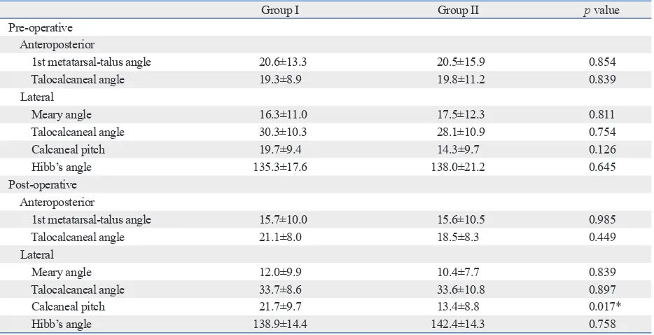
forc-Table 1. Comparisons of Pre- and Post-Operative Radiographic Parameters (Degree) between Group I and II
Group I Group II p value
Pre-operative Anteroposterior
1st metatarsal-talus angle 20.6±13.3 20.5±15.9 0.854
Talocalcaneal angle 19.3±8.9 19.8±11.2 0.839
Lateral
Meary angle 16.3±11.0 17.5±12.3 0.811
Talocalcaneal angle 30.3±10.3 28.1±10.9 0.754
Calcaneal pitch 19.7±9.4 14.3±9.7 0.126
Hibb’s angle 135.3±17.6 138.0±21.2 0.645
Post-operative Anteroposterior
1st metatarsal-talus angle 15.7±10.0 15.6±10.5 0.985
Talocalcaneal angle 21.1±8.0 18.5±8.3 0.449
Lateral
Meary angle 12.0±9.9 10.4±7.7 0.839
Talocalcaneal angle 33.7±8.6 33.6±10.8 0.897
Calcaneal pitch 21.7±9.7 13.4±8.8 0.017*
Hibb’s angle 138.9±14.4 142.4±14.3 0.758
[image:3.595.59.526.471.710.2]ed measurements from individual patients and was adjusted for intra-person correlations. In addition, to adjust for the ef-fects of age, gender, leg length, and time to assessment, we performed multivariate analysis. The values measured on plain radiographs and dynamic pedobarographs were com-pared between groups. The level of significance was set at
p<0.05.
RESULTS
Radiological measurements
[image:4.595.313.540.67.287.2]Values measured on the anteroposterior and lateral views of weight-bearing radiographs did not differ significantly be-tween groups, except for postoperative calcaneal pitch (Ta-ble 1). Although anteroposterior 1st metatarsal-talus angle, Meary angle and Hibb’s angle were slightly improved after operation, there was no statistically significant improve-ment in the radiologic indices in both groups (p=0.192, 0.520, 0.205, 0.269, 0.529, 0.476 in group I and 0.369, 0.745,
Table 2. Comparisons of Preoperative Peak Force (N) between Groups I and II and Normal Controls
Group I Group II Normal Group I vs. p value
Group II Group I vs. Normal Group II vs. Normal
Hallux 8.4±11.6 10.0±12.0 65.8±39.7 0.768 0.001* 0.001*
1st metatarsal head 24.3±17.9 18.6±17.9 57.3±22.8 0.311 0.001* 0.001* 2nd metatarsal head 30.2±20.2 21.8±23.0 93.6±36.8 0.261 0.001* 0.001* 3-4th metatarsal head 34.7±21.6 22.3±21.2 80.3±28.3 0.094 0.001* 0.001*
5th metatarsal head 29.0±21.3 18.8±22.7 36.1±14.4 0.117 0.122 0.002*
Lateral midfoot 23.9±19.6 23.4±16.9 23.8±13.4 0.850 0.618 0.612
Lateral calcaneus 27.6±13.7 18.2±9.7 78.5±19.5 0.045* 0.001* 0.001*
Medial calcaneus 29.6±14.7 17.5±9.4 84.2±18.7 0.016* 0.001* 0.001*
Values are mean±standard deviation. *p<0.05.
Table 3. Comparison of Postoperative Peak Force (N) between Groups I and II and Normal Controls
Group I Group II Normal Group I vs. p value
Group II Group I vs. Normal Group II vs. Normal
Hallux 10.9±10.4 4.1±5.4 65.8±39.7 0.132 0.001* 0.001*
1st metatarsal head 47.1±34.9 17.3±17.0 57.3±22.8 0.012* 0.079 0.001*
2nd metatarsal head 52.6±36.6 26.3±28.8 93.6±36.8 0.048* 0.001* 0.001* 3-4th metatarsal head 41.0±15.7 25.6±22.3 80.3±28.3 0.044* 0.001* 0.001*
5th metatarsal head 22.6±11.7 21.6±20.7 36.1±14.4 0.414 0.003* 0.009*
Lateral midfoot 16.4±12.6 19.0±14.6 23.8±13.4 0.854 0.029* 0.454
Lateral calcaneus 32.5±12.2 24.0±16.2 78.5±19.5 0.120 0.001* 0.001*
Medial calcaneus 41.8±21.6 27.7±17.8 84.2±18.7 0.087 0.001* 0.001*
[image:4.595.75.540.381.517.2]Values are mean±standard deviation. *p<0.05.
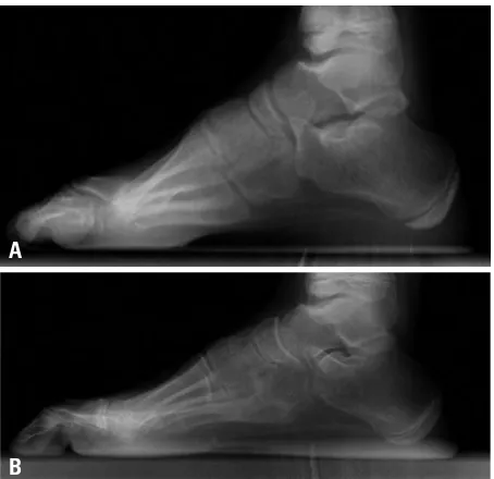
Fig. 2. Ten year old boy with myelomeningocele has a cavovarus deformity. He underwent extensive plantarmedial release with Achilles tendon lengthening. Preoperative (A) and postoperative (B) standing lateral foot radiography was compared. After operation, Hibb’s angle was increased from 124 degree to 126 degree and Meary angle was decreased from 21 degree to 15 degree.
A
[image:4.595.72.539.573.710.2]ble hindfoot varus, and that extensive plantar medial release or calcaneal osteotomy be used for patients aged >12 years with stiff hindfoot varus.
Some conflicting results from previous studies indicate a primary dependence on the clinical judgment and experi-ence of the surgeon to decide the course of treatment.3,5,6,12
For example, the Coleman block test,9 is judged by clinical
criteria. Furthermore, studies that used combined surgical methods cannot evaluate outcomes for a single procedure. In this study, therefore, we selected patients with paralytic cavovarus and confirmed the flexibility in the hindfoot us-ing the Coleman block test.9 Furthermore, we further
se-lected patients with hindfoot varus <5 mm, measured by the radiological reference suggested by Paulos, et al.,5 and
extensive plantarmedial release alone was the only soft tis-sue surgery to correct cavovarus in this study.
If extensive plantar medial release alone did not provide satisfactory correction, first ray osteotomy was added. First ray osteotomy is performed to improve the cavus and varus of the hindfoot by correcting the forefoot pronation result-ing from the tripod effect.6 When soft tissue release does
not adequately correct the cavovarus, an extension osteoto-my on the first metatarsal bone or the medial cuneiform bone may improve the hindfoot varus.3 Nevertheless, the
use of osteotomy is based on the age of the patient or the surgeon’s discretion during surgery, rather than on objective criteria. Azmaipairashvili, et al.13 reported that the degree of
forefoot pronation, as measured using lateral radiographic views obtained while performing the Coleman block test, may indicate the need for metatarsal osteotomy. In our study, although we did not assess lateral radiographic views dur-ing the Coleman block test, groups I and II did not differ significantly in the other radiological assessments, either pre- or postoperatively. These measurements of static de-0.090, 0.210, 0.799, 0.537 in group II, respectively) (Fig. 2).
Comparison of peak forces
Preoperatively, the PF at the medial and lateral calcaneus were higher in group I than in group II. PF at the hallux, first through fourth metatarsal heads, and medial and lateral cal-caneus were lower in both groups than in normal controls, but did not differ significantly from controls in the midfoot (Table 2).
Postoperatively, PF at the first through fourth metatarsal heads were higher in group I than in group II. PF at each area, except for the first metatarsal head in group I and the midfoot in group II, were lower than those in normal con-trols (Table 3).
Only in group I, PF at the 1st metatarsal head (p=0.015), 2nd metatarsal head (p=0.024) and medial calcaneus (p=0.041) were increased after operation (p=0.483, 0.362, 0.200, 0.148, 0.293 in other areas, respectively). There were no significant changes in group II after operation (p= 0.079, 0.833, 0.614, 0.686, 0.753, 0.457, 0.232, 0.079, respectively).
DISCUSSION
Pes cavovarus is a multidimensional deformity involving several joints in the foot and may require a combination of surgical treatments depending on the level of deformity. The choice of surgical technique generally depends on the level of flexibility in the hindfoot varus.9 Reports suggest that
simple plantar release provides satisfactory results for feet with hindfoot flexibility, whereas feet with relative stiffness respond better to extensive plantarmedial release.5
McClus-key, et al.2 recommended that soft tissue release and tendon
transfer be performed for patients aged <8 years with
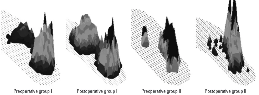
flexi-Fig. 3. After operation, the peak forces in the forefoot area were increased in group I, but still decreased in group II.
[image:5.595.84.498.565.718.2]quired only extensive plantarmedial release. In terms of dy-namic pedobarography, patients who received a first ray os-teotomy showed improvement in peak pressure distribution than patients in group II.
Our study considered only paralytic cavovarus foot de-formity, apart from the underlying disease. Because of the different manifestation according to the different neuromus-cular disease entity, research about the single disease entity would give more information about the effect of the sur-gery. We did not consider about the imbalance of the extrin-sic tendon, because we excluded patients who had undergone tendon transfer. However, the imbalance of the extrinsic ten-don can affect the foot pressure distribution. Results of our study can apply to patients with minimal imbalance. Al-though we found differences in dynamic foot function that may indicate the need for first ray osteotomy, a larger pro-spective study is required to confirm and extend these indi-cations. Nevertheless, when selecting a surgical procedure for patients with paralytic cavovarus, findings of dynamic pedobarography should be considered in addition to radio-graphic findings.
ACKNOWLEDGEMENTS
This work was supported by the 2013 Inje University re-search grant.
REFERENCES
1. Holmes JR, Hansen ST Jr. Foot and ankle manifestations of Char-cot-Marie-Tooth disease. Foot Ankle 1993;14:476-86.
2. McCluskey WP, Lovell WW, Cummings RJ. The cavovarus foot deformity. Etiology and management. Clin Orthop Relat Res 1989:27-37.
3. Schwend RM, Drennan JC. Cavus foot deformity in children. J Am Acad Orthop Surg 2003;11:201-11.
4. Mubarak SJ, Van Valin SE. Osteotomies of the foot for cavus de-formities in children. J Pediatr Orthop 2009;29:294-9.
5. Paulos L, Coleman SS, Samuelson KM. Pes cavovarus. Review of a surgical approach using selective soft-tissue procedures. J Bone Joint Surg Am 1980;62:942-53.
6. Samilson RL, Dillin W. Cavus, cavovarus, and calcaneocavus. An update. Clin Orthop Relat Res 1983:125-32.
7. Sherman FC, Westin GW. Plantar release in the correction of de-formities of the foot in childhood. J Bone Joint Surg Am 1981;63: 1382-9.
8. Jahss MH. Evaluation of the cavus foot for orthopedic treatment. Clin Orthop Relat Res 1983:52-63.
9. Coleman SS, Chesnut WJ. A simple test for hindfoot flexibility in
formity alone cannot be used to evaluate the outcome of the addition of first ray osteotomy or suggest indications of the additional first ray osteotomy.
Previous studies have been based on radiological mea-surements of the Meary angle, reflecting the level of cav-ovarus correction, or on measurements of the talocalcaneal angle, reflecting the correction of hindfoot varus. However, the cavovarus foot deformity increases pressure on the lat-eral midfoot, which promotes ulceration of the foot and in-stability while walking. This condition requires dynamic evaluation. The measurement of dynamic foot pressure can be used to evaluate the pressure on the foot during walking at a single point in time. This measurement can also be used to evaluate the imbalances at each area of the foot as force changes throughout the gait cycle. Static radiological as-sessment cannot provide the same type or quantity of infor-mation.11,14-16
In both groups, peak forefoot and calcaneal forces were lower than those in normal controls, and only the peak mid-foot force was similar. Metaxiotis, et al.16 used
measure-ments of dynamic plantar pressure to classify adult cavovar-us into 3 types. In type 1, the pressure at the first metatarsal head and the midfoot are high; in type 2, the pressure at each metatarsal head is high; and in type 3, pressure is high at the fifth metatarsal head and the lateral midfoot area. Al-though parameters of groups I and II in our study, did not fit well into the Metaxiotis classification, they did resemble a type 3, because forces at the metatarsal heads were de-creased and midfoot were inde-creased.
In dynamic foot pressure, patients in group I had increased PF at calcaneus than group II. However, those are lower than normal control. After operation, only PF at the 1st metatarsal head, 2nd metatarsal head and medial calcaneus in group I were increased, but only PF at the 1st metatarsal head was comparable to the normal control. In group II, there were no significant changes after operation and postoperatively PF at 1-4th metatarsal heads were lower than group I. In our study, we cannot suggest the indication of the addition of first ray extension osteotomy because there was a little difference between groups. However, the addition of first ray extension osteotomy increased forefoot peak pressure compared with extensive plantarmedial release alone (Fig. 3).
re-14. Chang CH, Miller F, Schuyler J. Dynamic pedobarograph in eval-uation of varus and valgus foot deformities. J Pediatr Orthop 2002;22:813-8.
15. Chan G, Sampath J, Miller F, Riddle EC, Nagai MK, Kumar SJ. The role of the dynamic pedobarograph in assessing treatment of cavovarus feet in children with Charcot-Marie-Tooth disease. J Pediatr Orthop 2007;27:510-6.
16. Metaxiotis D, Accles W, Pappas A, Doederlein L. Dynamic pedo-barography (DPB) in operative management of cavovarus foot deformity. Foot Ankle Int 2000;21:935-47.
the cavovarus foot. Clin Orthop Relat Res 1977:60-2.
10. Drennan JC. Current concepts in myelomeningocele. Instr Course Lect 1999;48:543-50.
11. Herring JA. Tachdjian’s pediatric orthopaedics. 4th ed. Philadel-phia: WB Saunders Co; 2008. p.1139-86.
12. Bradley GW, Coleman SS. Treatment of the calcaneocavus foot deformity. J Bone Joint Surg Am 1981;63:1159-66.