R E S E A R C H A R T I C L E
Open Access
The traditional Tibetan medicine
Yukyung
Karne
exhibits a potent anti-metastatic
activity by inhibiting the epithelial to
mesenchymal transition and cell migration
Tenzin Choedon
1,2, Ganeshan Mathan
2and Vijay Kumar
1*Abstract
Background:In Traditional Tibetan medicine,Yukyung Karnehas been used for the treatment of ovarian cancer. ThoughYukyung Karnehas been reported to be clinically effective, the molecular mechanism of its anti-metstatic action remains elusive.
Methods:The cytotoxic property ofYukyung Karnewas evaluated by crystal violet staining while its ability to induce ceramide production was analyzed by sphingomyelinase assay. The anti-metastatic property was investigated using adhesion, invasion, migration and colony formation assays. The effect ofYukyung Karneon the expression of extracellular matrix components, and epithelial and mesenchymal markers were evaluated by confocal microscopy and western blotting.
Results:Yukyung Karneexhibited a strong anti-metastatic property by significantly reducing the invasion, migration and colony formation ability of ovarian cancer cells. Besides it inhibited the levels of biomarkers involved in epithelial to mesenchymal transition such as down-regulation of vimentin and N-cadherin and up-regulation of epithelial E-cadherin.Yukyung Karnealso induced the neutral sphingomyelinase II (nSMNaseII) enzyme activity that is known to hydrolyze sphingomyelins into pro-apoptotic intracellular molecule ceramide.
Conclusions:The study provides some compelling evidences supporting the anti-metastatic potential ofYukyung
Karnewhich strongly suggests its possible usage as a promising alternative medicine. Thus,Yukyung Karnemay be used as an anticancer and anti-metastatic agent along with other conventional anticancer therapeutics to increase their efficacy.
Keywords:Yukyung Karne, Traditional Tibetan medicine, Metastasis, Cell migration, Epithelial mesenchymal transition, Extracellular matrix
Background
Ovarian cancer is one of the leading gynecological ma-lignancies worldwide in which tumor metastasis is asso-ciated with poor survival of the ovarian cancer patients [1, 2]. Tumor invasion and metastasis are recognized as complex and multi-step cellular processes which involve cell detachment, invasion, migration, intravasation, cir-culation, implantation, angiogenesis and proliferation
[3]. Degradation of the extracellular matrix (ECM) by cancer cells through proteases such as matrix metallo-proteinase’s (MMPs) and serine proteases aids to the separation of intercellular matrix and promotes tumor invasion. Incidentally, MMPs such as MMP2 (Gelatinase A) and MMP9 (Gelatinase B) are secreted by ovarian cancer cells which in turn correlates with increased occur-rence of tumor invasion and metastasis. MMPs promote the epithelial to mesenchymal transition (EMT) by cleav-ing cell adhesion molecule E-cadherin [4]. Therefore, interventional strategies against these steps are considered important to prevent metastasis [5, 6]. Further, loss or * Correspondence:vijay@icgeb.res.in
1
Virology Group, International Centre for Genetic Engineering and Biotechnology, Aruna Asaf Ali Marg, New Delhi 110067, India Full list of author information is available at the end of the article
reduction of trans membrane glycoprotein E-cadherin is another important manifestation in the EMT program of tumor cells. It is now well established that loss of E-cadherin and up-regulation of mesenchymal markers re-sult in subsequent activation of signal transduction events such as Wnt/β-catenin pathway that aid to the progres-sion of metastasis [7]. Not surprisingly, the levels of Wnt effectorβ-catenin are tightly regulated in order to control the expression of its target genes such asc-mycthat exerts a profound effect on cell proliferation, differentiation and migration [8].
Chemotherapy, surgery and radiotherapy are widely used in the management of cancer but these are often associated with adverse effects. To remove these short-comings in cancer treatment, much attention has shifted towards the use of complementary and alternative medi-cines (CAM). Not surprisingly, CAM is now fast emerging as an effective measure to aid conventional therapies [9]. Natural products have been widely used for centuries and recent preclinical studies have shown their potential appli-cations in pharmacology and cancer therapy. Yukyung Karne has been used for many decades in traditional Tibetan medicine or Sowa Rigpa for treatment of cancer [10]. Recently, we have reported the anticancer potential of Yukyung Karne using ovarian cancer cells in culture [11]. In the present study, we have investigated the anti-metastatic properties ofYukyung Karneon ovarian cancer cells by evaluating its inhibitory effects on cell migration, cell invasion, EMT and ECM.
Methods
Cell lines and culture conditions
Ovarian cancer cell line SKOV6 was a kind gift of Dr. Anil Suri (National Institute of Immunology, New Delhi). All cultures were grown in Dulbecco’s modified Eagle’s medium containing 10 % heat inactivated fetal bovine serum, penicillin 100μg/ml and streptomycin (100μg/ml). Cells were incubated at 37 °C in a humidified chamber under 5 % CO2atmosphere.
Traditional Tibetan medicine
Yukyung Karne was purchased from the Tibetan Medical and Astrological Institute, Dharamshala, India. According to the Tibetan Pharmacopeia [10]. Yukyung Karne is a mixture of various plant components including: Roots of Saussurea lappa (C.B. Clarke) (family: Asteraceae), Zingi-ber officinalis (Roscoe) (family: Zingiberaceae), Aconitum ferox(Wall.ex Ser) (family:), Corydalis hendersoni(Fedde) (family: Fumariaceae), and Acorus calamus (L) (family: Araceae); Fruits ofEmblica officinalis(L) (family: Euphorbia-ceae),Piper longum(L) (family: Piperaceae), andTerminalia chebula(Rety) (family: Combretaceae); Leaves of Adhatoda vasica (NEES) (family: Acanthaceae); Seeds of Elletra cardomomum (L) (family: Zingiberaceae), Coriandrum
sativum (L) (family: Apiaceae), Embelia ribes (Burm, F) (Family: Myrsinaceae), and Delphinium brunonianum (Royale) (family: Ranunculaceae); resin of Commiphora mukul(Hook) (family: Burseraceae); Whole plant of Mecon-opsis horridula(Hook) (Papaveraceae), andDracocephyllum tanguticum (Maxim) (family: Lamiaceae); and Tsothel (detoxified mercury) as mineral ingredient. All the herbs were identified by Dr. Tsering Norbu (Menrampa) and the voucher numbers of plant specimens are available at the herbarium department of Tibetan Medical and Astrological Institute for reference.
Sphingomyelinase activity
Neutral sphingomyelinase II (nSMNaseII) enzyme activity was determined in cell lysate using Amplex Red Sphingo-myelinase Assay Kit (Molecular probes, Invitrogen) as per manufacturer’s instructions. The reaction was carried out for 30 min and fluorescence data was measured in a fluor-escence microplate reader using excitation at 530 nm and emission 590 nm. The fluorescence values were normal-ized with total protein to obtain activity/μg of protein.
Gelatin zymography
Gelatin zymography was performed as described by Al Dhaheriet al., [12]. Briefly, cells were incubated in serum free DMEM for 24 h in the presence of Paclitaxel (P), Yukyung Karne (YK) or P + YK (PY). The conditioned media was collected and protein was precipitated (1:4) by acetone. Samples were resuspended in 2x Laemmli buffer without reducing agent. After boiling, the samples were re-solved in a 10 % poly acrylamide gel containing 0.1 % gel-atin. After electrophoresis, gels were washed with 2.5 % Triton X100 at RT to remove SDS and then incubated overnight at 37 °C with substrate incubation buffer (50 mM Tris–HCl pH 7.5, 200 mM NaCl, 10 mM CaCl2). Bands corresponding to MMP2/9 activity were visualized by negative staining using Coomassie brilliant blue. Images were captured using Fluorchem M (Protein Simple, USA).
Crystal violet staining of cells
SKOV6 cells were treated with Yukyung Karne. After 24 h, media was discarded and adhered cells were washed with 1xPBS and fixed in methanol. Crystal violet (1 %) solution was added to the cells for 20 min followed by a wash, and the stained cells were resuspended in methanol and absorbance was read at 600 nm.
Cell adhesion assay
were fixed in ice cold methanol: acetone (1:1) for 10 min at−20 °C. Cells were stained with Hoechst dye for 15 min at RT. After rinsing with 1x PBS, images were captured using confocal microscopy (Nikon A1R). Adherent cells were counted in five random fields, and the mean number of cells/field ± SD was calculated.
Colony formation assay
The colony formation assay was performed as described by Shuklaet al., [13]. Briefly, treated cells were incubated at 37 °C for 14 days and observed regularly for colony formation and the bright field images were captured with a Nikon ECLIPSE TE 2000-S inverted microscope and the number of foci were counted in random fields.
In-vitroinvasion assay
The cell migration of SKOV6 cells was studied using Trans-well plate with 8 μm pore size membrane insert. Complete medium was added to the membrane and lower chamber 1 h prior to seeding of cells at 37 °C, 5 % CO2 humidified incubator. Cells were seeded to the membrane and treatment was simultaneously given for 4 h. After incubation, inserts were carefully removed and cells were scraped off using cotton swabs from the upper surface of the membrane. Cells on lower side of the membrane were fixed in 10 % methanol followed by staining with crystal violet (1 %) for 20 min. The mem-branes were air dried and the number of infiltrated cells on the lower side of filter was counted by microscopy.
Wound healing assay
Briefly, SKOV6 cells were grown as monolayer at 37 °C, 5 % CO2 in a humidified incubator. A scratch wound was made using 200μl yellow pipette across the conflu-ent monolayer. Cells were cultured with fresh DMEM supplemented with 10 % fetal bovine serum in the pres-ence or abspres-ence of Yukyung Karne (100 μg) for 24 h. The extent of wound closure was captured using Nikon ECLIPSE TE 2000-S inverted microscope.
RNA extraction and quantitative real time PCR (RT-qPCR) Total RNA was isolated from cells using Trizol reagent (Invitrogen) as per the manufacturer’s protocol. Reverse transcriptase-PCR (RT-PCR) was performed with MMuLV reverse transcriptase (Fermentas) according to the manu-facturer’s protocol. qRT-PCR was done in an ABI Step One plus system (Applied Bio system) using specific primers for N- Vimentin Forward:5’GAGACAGGTGC
AGTCCCTCA, Vimentin Reverse: 5’GAAGGTGACG
AGCCATTTCCT.βActin served as an internal control.
Western blotting
Cell lysates were prepared in 1x cell lysis buffer (Promega, USA). Protein concentration was determined by Bradford’s
reagent (Biorad). Equal amounts of protein samples were resolved in a 10-15 % SDS-PAGE followed by western blotting (WB). Primary antibodies-E-cadherin, N-cadherin were procured from Cell Signaling Technology, pFAK (Tyr397), Caveolin1 (N-20), Integrin5α, Beta catenin (E5), GSK-3β (H-76) and GAPDH (FL-335) were from Santa Cruz Biotechnology (USA). The protein bands were visualized using enhanced chemiluminescence (ECL) from Santa Cruz Biotechnology (California, USA).
Statistical analysis
Data are expressed as mean ± S.D. Statistical significance was calculated using Student’s t test. p values of <0.05 were considered significant.
Results
Yukyung Karneelevates the nSMNase II activity
Working on the pharmacological basis of anticancer prop-erties ofYukyung Karne, we have earlier reported that it is able to induce cell cycle arrest and apoptosis as well as re-store the levels of the key tumor suppressor factor p53 in
0 20 40 60 80 100 120 140
Control Pac PY YK
B
A
cti
v
ity
per
µg
protein
Cell
v
iabilit
y
(%)
0 200 400 600 800 1000 1200 1400 1600 1800
Control Pac PY YK
A
[image:3.595.306.539.351.648.2]ovarian cancer cell line SKOV6. Since manipulation of sphingolipid level for ceramide production has been shown to improve the efficacy of anticancer therapy [14, 15], we investigated the effect ofYukyung Karneon the activity of nSMNaseII enzyme in SKOV6 cells. Interestingly, we ob-served the elevated levels of nSMaseII in SKOV6 cells treated withYK(100μg) (p<0.001) confirming its positive role in ceramide production (Fig. 1a). We also observed that Yukyung Karne treatment significantly inhibited the viability of the cells (p<0.001) as evident from the crystal violet staining of cells (Fig. 1b) further strengthening its pro-apoptotic property.
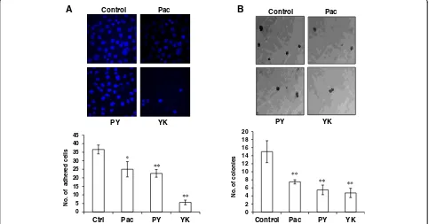
Yukyung Karneinhibits collagen -mediated cell adhesion Since the migratory and metastatic potential of cancer cells is fore timed by their interaction with endothelial cells and ECM proteins like collagen [16], we assessed the effect ofYukyung Karnetreatment on cell to matrix interaction in vitro by collagen binding ability using ovarian cancer cells. According to our data, a quantum reduction in adhesion of cancer cells to collagen (84 %) was observed for Yukyung Karne treated SKOV6 cells when compared to untreated cells. Interestingly, a sig-nificant decrease in cell adhesion was also observed for positive control paclitaxel (p<0.05) and combination of paclitaxel withYK(p<0.001) suggesting its strong ability
of Yukyung Karne to suppress the adhesion of SKOV6 cells to collagen (Fig. 2a).
Yukyung Karneinterferes with the transformation of SKOV6 cells
Widely accepted hallmarks of cancer include the resistance of cancer cells to contact inhibition and the enhanced ability to form independent colonies. We next examined the inhibitory potential of Yukyung Karne on the colony forming ability of SKOV6 cells. While the untreated SKOV6 cells produced 15 ± 3 colonies/field, it was sup-pressed to 5 ± 1 (p<0.001) on exposure toYK, Pac 7.5 ± 1 (p<0.001) and PY 5.5 ± 1 (p<0.001) (Fig 2b). This result confirmed the negative effect of Yukyung Karne on the transformation potential of ovarian cancer cells.
Yukyung Karneinhibits migration and invasion of ovarian cancer cells
Metastasis of ovarian cancer cells from the primary site to the neighboring secondary tissues mainly peritoneum and omentum is the main event in transition of benign to ma-lignant cancer. To elucidate the role ofYukyung Karnein inhibiting the migratory potential of SKOV6 cells, we per-formed trans-well migration and wound healing assays. A close examination of the invasive potential of Yukyung Karne using transwell insert (Fig. 3a) revealed a significant reduction in the number of infiltrated cells in a dose
No
. o
f ad
h
ered
cells
0 5 10 15 20 25 30 35 40 45
Ctrl Pac PY YK
PY YK
Control Pac A
0 2 4 6 8 10 12 14 16 18 20
Control Pac PY YK
No. of colonies
Control Pac
PY YK
B
[image:4.595.57.540.431.684.2]0 10 20 30 40 50 60
No. of infiltrated cells ** ** ** **
*
Control YK
6h
24h
B
Control Pac PY
YK
50 100 200
A
Control Pac PY 50 100 200
YK
[image:5.595.58.542.88.684.2]dependent manner (p<0.001). Further, in wound healing assay (Fig. 3b), Yukyung Karne severely impaired the migratory behavior of SKOV6 cells when compared to un-treated cells by 20 % and 80 % at 6 h and 24 h respectively. Taken together these results confirm the inhibitory effect ofYukyung Karneon the migration and invasion potential of SKOV6 cells.
Yukyung Karneacts as a MMPs inhibitor
Degradation of the ECM by MMPs aids in tumor inva-sion and thus is a crucial step in the initiation of metas-tasis [17]. In line with its strong anticancer properties, we next investigated the inhibitory effect of Yukyung Karneon both MMP2 and MMP9 activities using gelatin zymography. As shown in Fig. 4a, there was a marked inhibition (p<0.05) in the gelatinolytic band correspond-ing to MMP9 and MMP2 (proenzyme forms) in the con-ditioned media of the cells treated withYukyung Karne when compared to control untreated cells.
Effect ofYukyung Karneon EMT
In order to metastasize, tumor cell undergo an EMT that is marked by increase in mesenchymal markers such as vimentin, vitronectin, N-cadherin and decrease in epithelial markers like E–Cadherin [18]. Since Yukyung Karne caused down regulation of MMPs, we hypothesized that it may have a strong bearing on the expression of EMT markers. Western blot analysis of the Yukyung Karne treated cells showed low levels of N-cadherin protein (p<0.05) (Fig. 4b) and a significant inhibition (p<0.01) in the levels of vimentin mRNA in theYukyung Karne treated cells (Fig. 4c). In contrast, as shown in Fig. 4b, the levels of E-cadherin were found to be significantly elevated following treatment with PY (p<0.01) andYK (p <0.01). Thus, the reciprocal expression of epithelial and mesenchymal markers induced by Yukyung Karne, suggest its potential as an anti-metastatic agent.
Effect ofYukyung Karneon ECM
To further support the anti-metastatic property of Yukyung Karne, we also investigated its role in the modulation of Wnt/βcatenin pathway. Binding of Wnt protein to the receptors triggers activation of β catenin and its inappropriate activation was often found deregu-lated in many cancers. Under normal circumstances onco-protein β-catenin gets phosphorylated by GSK3β and
subsequently ubiquitinylated and targeted for degradation. Western blot analysis of the Yukyung Karne-treated SKOV6 cells exhibited a significant reduction in the Wnt/β catenin effector molecule β-Catenin (p <0.05), GSK3β(p<0.05), and also its downstream target c-Myc (p <0.05), (Fig. 5c) [19, 20]. Consistent with a marked inhibition of ECM components and its cytoskeletal dynam-ics, the levels of integrinα5 which is crucial for cell to cell and ECM interaction for adhesion was down-regulated fol-lowingYukyung Karnetreatment (p<0.001) (Fig. 5c).
To further understand the mechanism of action of Yukyung Karne,we investigated its effect on the levels of Caveolin 1, whose down regulation is associated with ac-tivation of MAP kinase and anchorage independent growth [21]. Surprisingly, the Yukyung Karne- treated cells exhibited no appreciable change in the levels of Caveolin-1 (Fig. 5a, c) while there was a marginal (40 %) inhibition in the levels of of pFAK which is crucial for transmitting signals during tumorogenesis (Fig. 5b, c). Taken together, our present sets of results strongly sug-gest the inherent potential of Yukyung Karne. Yukyung Karne exerts an inhibitory effect on the migration of ovarian cancer cells by regulating the expression of EMT markers and ECM components.
Discussion
CAMs are fast emerging as an acceptable choice for treating various chronic diseases including ovarian can-cer where extensive metastasis to the neighboring organs like fallopian tubes, uterus, bladder and peritoneal cavity is a major challenge. In traditional Tibetan medicine, Yukyung Karnehas been used as an effective alternative treatment for cancer metastasis specifically for ovarian cancer. Recently, we have shown thatYukyung Karne ex-hibits several therapeutic effects on ovarian cancer cells such as induction of growth arrest and apoptosis [11]. In addition to our earlier observation, we observed a marked increase in the levels of a critical enzyme nSMNaseII which is involved in generation of ceramide from sphingo-myelins in response to apoptotic inducers such as chemo-therapeutic agents leading to growth inhibition and apoptosis [14]. Thus, increase in the nSMNaseII activity appears to be one of theYukyung Karne’s cytotoxic mech-anisms. However, the pharmacological basis of its anti-metastatic property remains elusive. In the present study, we investigated the anti-metastatic potential of Yukyung
(See figure on previous page.)
Karne using ovarian cancer cells. Adhesion of cells to ECM is a crucial event in modulation of cellular processes including the regulation of anchorage-dependent pro-cesses. Interestingly, a significant number of cancer cells failed to adhere to the collagen-coated culture plates in
the presence of Yukyung Karne(Fig. 2). Further, cell mi-gration is an important mechanism in the metastasis of high-grade ovarian cancer [22]. We observed a negative correlation between the number of migratory cells and the dose ofYukyung Karne. Further, the colony forming ability
0 0.2 0.4 0.6 0.8 1 1.2 1.4
Control Pac
PY
YK
B
Control Pac PY YK
MMP9
MMP2
A
Relativ
e v
imentin
mRNA
lev
el
C
*
Control Pac PY YK
Zymography
WB
1.0
1.2
2.3
2.5
1.0
0.5
0.6
0.5
1.0
0.7
0.3 0.2
1.0
1.1 0.6 0.3
[image:7.595.61.538.89.595.2]of SKOV6 cells were also seen reduced significantly upon treatment withYukyung Karne.
Here EMT plays a major role in aiding the tumor cells to migrate out from the primary site, enter into the cir-culation and adhere to endothelial cells of target organs. Cadherin- mediated adhesion plays an important role in maintaining cell-cell contacts and reducing tumor me-tastasis [23, 24]. Accordingly, the E-cadherin negative tumor is a predictor of poor overall survival [25]. In the present study, we observed a striking correlation in the E-cadherin level and decrease in mesenchymal markers such as Vimentin and N-cadherin supporting epithelial characteristics following treatment with Yukyung Karne and correlates with decrease in tumorogenicity [26].
Degradation of ECM components is a prerequisite step for initiation of metastasis [27] and intraperitoneal ovar-ian cancer metastasis is mediated by adhesion via integ-rins to peritoneal mesothelial cells. On binding to ECM, integrin 5αregulates diverse functions in tumor cells in-cluding migration, invasion, proliferation and survival through activation of various pathways such as MAPK. In our study, we observed a significant decrease in integ-rin 5α expression in the Yukyung Karne treated cells. Hence, use of known integrin inhibitors could also be a novel therapeutic strategy to combat cancer. The key mediator of integrin signal is focal adhesion kinase (FAK) which functions in cell motility and proliferation [28, 29]. Increased tumor apoptosis has been reported earlier following pharmacological inhibition of FAK in a xenograft cancer model [30]. In the Yukyung
Karne-treated cancer cells, we observed a significant inhibition in integrin 5α and pFAK gene expression corroborating the tumor growth inhibitory property ofYukyung Karne (Fig. 5b, c). E-cadherin is frequently down regulated in epithelial tumors and a number of studies have shown that disruption of E-cadherin leads to transcriptional ac-tivation of oncoprotein ß-catenin [23]. The accumula-tion of ß-catenin stimulated transcripaccumula-tion factors that enhanced cell proliferation and poor prognosis. Our study confirmed decreased levels of ß-catenin, pGSK3-ß and downstream target c-Myc (Fig. 5c) and increased levels of E-cadherin (Fig. 4b) following Yukyung Karne treatment.
Besides the well-defined role of major ECM compo-nents, we also investigated the status of scaffolding pro-tein Caveolin in this study. Caveolin 1 is ubiquitously expressed and encodes a major component of membrane caveolae. It functions as tumor suppressor [31] and con-tributes to the organization and stability of adherent junctions through its association with E-cadherin at junction level. Caveolin 1 is consistently seen lost or down regulated in malignant ovarian cancers [23]. Inter-estingly, the level of caveolin1 levels were found up-regulated in the Yukyung Karne treated SKOV6 cells suggestingYukyung Karneas a strong candidate in indu-cing expression of tumor suppressor caveolin 1 (Fig. 5c). Consistent with anti-metastatic property of Yukyung Karne, further our gelatin zymography studies also indi-cated a marked reduction in the activity of key enzymes MMP2/9 (Fig. 4a) that are involved in the cleavage of
B
A
Control YK
Mitochondria Caveolin 1
DAPI
Merge
C
Control Pac PY YK WB
1.0 0.7 0.6 0.4
1.0 0.8 0.8 0.7
1.0 0.7 0.41 0.43
1.0 0.9 1.0 1.15
1.0 0.47 0.3 0.3 1.0 2.1 1.0 0.6
Control YK
pFAK
Merge DAPI
Fig. 5Effect ofYukyung Karneon extracellular matrix proteins. SKOV6 cells were cultured on cover slips and treated with Paclitaxel (Pac, 10 nM),
Yukyung Karne(YK, 100μg) or PY [Pac (10 nM) + YK (100μg)] for 24 h and processed for immunofluorescence analysis using anti caveolin 1
[image:8.595.58.539.89.315.2]ECM components during tumor invasion and metastasis and are abundantly expressed in various malignant can-cers [32, 33].
Thus,Yukyung Karneseems to exert its anti-metastatic potential by regulating EMT with increase in E- cadherin expression and loss of critical intracellular oncoprotein ß-catenin that has been shown implicated in induction of EMT in various cancers [7]. However, further in vivo studies are needed to ascertain its effectiveness in the treatment of ovarian cancer. Nevertheless,Yukyung Karne appears to be a good candidate medicine for treating ovar-ian cancer patients as it exhibits better efficacy than pacli-taxel. Further, its use in combination therapy along with conventional chemotherapeutic agent such as paclitaxel should improve the overall efficacy of treatment.
Conclusions
Our findings provide a multitude of evidences supporting a strong and potent anti-metastatic property associated TTMYukyung Karne. Yukyung Karneeffectively counters a range of anti-metastatic properties of cancer cells in-cluding adhesion, invasion migration, colony formation, ECM components, loss of mesenchymal marker and gain of epithelial marker. Thus,Yukyung Karneappears to be an ideal CAM that could be used for developing new therapeutic strategies in the management of ovarian can-cer metastasis.
Competing interests
The authors declare that they have no competing interests.
Authors’contributions
TC and GM carried out experiments and drafted the manuscript. VK designed the study, arranged funds and finalized the manuscript. All authors have read and approved the final manuscript.
Acknowledgements
This work was supported by the J.C. Bose fellowship to VK (Grant No. SR/S2/ JCB-80)/2012) from the Department of Science of Technology, Government of India, New Delhi. The authors thank Dr. Anil Suri (Cancer Microarray, Genes and Proteins Laboratory, National Institute of Immunology, New Delhi) for kindly providing SKOV6 cells and Dr. Dawa Dolma (Tibetan Medical and Astrological institute) for kindly sharing her clinical experience withYukyung
Karne.
Author details
1Virology Group, International Centre for Genetic Engineering and
Biotechnology, Aruna Asaf Ali Marg, New Delhi 110067, India.2Department of Biomedical Science, Bharathidasan University, Tiruchirappalli 620024, India.
Received: 20 January 2015 Accepted: 4 June 2015
References
1. Ocaña OH, Córcoles R, Fabra A, Moreno-Bueno G, Acloque H, Vega S, et al. Metastatic colonization requires the repression of the epithelial-mesenchymal transition inducer Prrx1. Cancer Cell. 2012;22:709–24. 2. Ween MP, Oehler MK, Ricciardelli C. Role of Versican, Hyaluronan and CD44
in Ovarian Cancer Metastasis. Inl J Mol Sci. 2011;12:1009–29. 3. Blumenthal RD, Hansen HJ, Goldenberg DM. Inhibition of adhesion,
invasion, and metastasis by antibodies targeting CEACAM6 (NCA-90) and CEACAM5 (Carcino-embryonic Antigen). Cancer Res. 2005;65:8809–17.
4. Zhao H, Yang Z, Wang X, Zhang X, Wang M, Wang Y, et al. Triptolide inhibits ovarian cancer cell invasion by repression of matrix metalloproteinase 7 and 9 and upregulation of E-cadherin. Exp Mol Med. 2012;44:633–41.
5. Faça VM, Ventura AP, Fitzgibbon MP, Pereira-Faça SR, Pitteri SJ, Green AE, et al. Proteomic analysis of ovarian cancer cells reveals dynamic processes of protein secretion and shedding of extra-cellular domains. PLoS One. 2008;3:e2425.
6. Zhu H, Liu XW, Cai TY, Cao J, Tu CX, Lu W, et al. Celastrol acts as a potent antimetastatic agent targeting beta1 integrin and inhibiting cell-extracellular matrix adhesion, in part via the p38 mitogen-activated protein kinase pathway. J Pharmacol Exp Ther. 2010;334:489–99.
7. Onder TT, Gupta PB, Mani SA, Yang J, Lander ES, Weinberg RA. Loss of E-cadherin promotes metastasis via multiple downstream transcriptional pathways. Cancer Res. 2008;68:3645–54.
8. Wu G, Huang H, Garcia Abreu J, He X. Inhibition of GSK3 phosphorylation of beta-catenin via phosphorylated PPPSPXS motifs of Wnt coreceptor LRP6. PLoS One. 2009;4:e4926.
9. Cragg GM, Newman DJ. Plants as a source of anti-cancer agents. J Ethnopharmacol. 2005;100:72–9.
10. Dawa.Bod kyi Gso Ba Rigpa Las Sman Rdzas Sbyor lag len Gsan Sgo byed Pai
Lde Mig. Dharamshala, HP, India: RigDrag publications; 2003.
11. Choedon T, Dolma D, Mathan G, Kumar V. Molecular insights into the anti-cancer properties of traditional Tibetan medicine Yukyung Karne. BMC Complement Altern Med. 2014;14:380.
12. Al Dhaheri Y, Attoub S, Arafat K, Abuqamar S, Viallet J, Saleh A, et al. Anti-metastatic and anti-tumor growth effects of Origanum majorana on highly metastatic human breast cancer cells: inhibition of NFκB signaling and reduction of nitric oxide production. PLoS One. 2013;8, e68808. 13. Shukla SK, Kumar V. Hepatitis B virus X protein and c-Myc cooperate in the
upregulation of ribosome biogenesis and in cellular transformation. FEBS J. 2012;279:3859–71.
14. Park B, Lee YM, Kim JS, Her Y, Kang JH, Oh SH, et al. Neutral sphingomyelinase 2 modulates cytotoxic effects of protopanaxadiol on different human cancer cells. BMC Complement Altern Med. 2013;13:194. 15. Kolesnick R. The therapeutic potential of modulating the ceramide/
sphingomyelin pathway. J Clin Invest. 2002;110:3–8.
16. Wever OD, Hendrix A, Boeck DA, Westbroek W, Braems G, Emami S, et al. 2010. Modeling and quantification of cancer cell invasion through collagen type I matrices. Int J Devl Biol. 2010;54:887–96.
17. Yang J, Zhang Z, Lin J, Lu J, Liu BF, Zeng S, et al. Detection of MMP activity in living cells by a genetically encoded surface-displayed FRET sensor. Biomembranes. 2007;1773:400–7.
18. Kalluri R, Weinberg RA. The basics of epithelial-mesenchymal transition. J Clin Invest. 2009;119:1420–8.
19. Metcalfe C, Bienz M. Inhibition of GSK3 by Wnt signalling–two contrasting models. J Cell Sci. 2011;124:3537–44.
20. He TC, Sparks AB, Rago C, Hermeking H, Zawel L, da Costa LT, et al. Identification of c-MYC as a target of the APC pathway. Science. 1998;281:1509–12.
21. Wiechen K, Sers C, Agoulnik A, Arlt K, Dietel M, Schlag PM, et al. Down-regulation of caveolin-1, a candidate tumor suppressor gene, in sarcomas. Am J Pathol. 2001;158:833–9.
22. Chen X, Brewer MA, Zou C, Campagnola PJ. Adhesion and migration of ovarian cancer cells on crosslinked laminin fibers nanofabricated by multiphoton excited photochemistry. Integr Biol. 2009;1:469–76. 23. Miotti S, Tomassetti A, Facetti I, Sanna E, Berno V, Canevari S. Simultaneous
expression of caveolin-1 and E-cadherin in ovarian carcinoma cells stabilizes adheren junctions through inhibition of Src-related kinases. Am J Pathol. 2005;167:1411–27.
24. Burkhalter RJ, Symowicz J, Hudson LG, Gottardi CJ, Stack MS. Integrin regulation of beta-catenin signaling in ovarian carcinoma. J Biol Chem. 2011;286:23467–75.
25. Fujioka T, Takebayashi Y, Kihana T, Kusanagi Y, Hamada K, Ochi H, et al. Expression of E-cadherin and beta-catenin in primary and peritoneal metastatic ovarian carcinoma. Oncol Rep. 2001;8:249–55.
26. Strauss R, Zong-Yi L, Liu Y, Beyer I, Persson J, Sova P, et al. Analysis of Epithelial and Mesenchymal Markers in Ovarian Cancer Reveals Phenotypic Heterogeneity and Plasticity. PLoS ONE. 2011;6, e16186.
28. Voulgari A, Pintzas A. Epithelial-mesenchymal transition in cancer metastasis: mechanisms, markers and strategies to overcome drug resistance in the clinic. Biomembranes. 2009;1796:75–90.
29. Eke I, Deuse Y, Hehlgans S, Gurtner K, Krause M, Baumann M, et al.
β1Integrin/FAK/cortactin signaling is essential for human head and neck cancer resistance to radiotherapy. J Clin Invest. 2012;122:1529–40. 30. Tancioni I, Uryu S, Sulzmaier FJ, Shah NR, Lawson C, Miller NL, et al. FAK
Inhibition Disrupts aβ5 Integrin Signaling Axis Controlling Anchorage-Independent Ovarian Carcinoma Growth. Mol Cancer Ther. 2014;13:2050–61. 31. Hehlgans S, Cordes N. Caveolin-1: an essential modulator of cancer cell
radio- and chemoresistance. Am J Cancer Res. 2011;1:521–30. 32. Tan ML, Choong PF, Dass CR. 2010. Direct anti-metastatic efficacy by the
DNA enzyme Dz13 and down regulated MMP-2, MMP-9 and MT1-MMP in tumours. BMC Cancer Cell Int. 2010;10:9.
33. Liabakk NB, Talbot I, Smith RA, Wilkinson K, Balkwill F. Matrix
metalloproteinase 2 (MMP2) and matrix metalloproteinase 9 (MMP9) type IV collagenases in colorectal cancer. Cancer Res. 1996;56:190–6.
Submit your next manuscript to BioMed Central and take full advantage of:
• Convenient online submission
• Thorough peer review
• No space constraints or color figure charges
• Immediate publication on acceptance
• Inclusion in PubMed, CAS, Scopus and Google Scholar
• Research which is freely available for redistribution


![Fig. 5 Effect of Yukyung Karne on extracellular matrix proteins. SKOV6 cells were cultured on cover slips and treated with Paclitaxel (Pac, 10 nM),Yukyung Karne (YK, 100 μg) or PY [Pac (10 nM) + YK (100 μg)] for 24 h and processed for immunofluorescence an](https://thumb-us.123doks.com/thumbv2/123dok_us/6097.1000339/8.595.58.539.89.315/yukyung-extracellular-proteins-cultured-paclitaxel-yukyung-processed-immunofluorescence.webp)
![Poly[aqua[μ N′ (carboxymethyl)ethylenediamine N,N,N′ triacetato]neodymium(III)]](data:image/gif;base64,R0lGODlhAQABAIAAAP///wAAACH5BAEAAAAALAAAAAABAAEAAAICRAEAOw==)