D
E
V
E
LO
P
M
E
N
T
INTRODUCTION
Müllerian inhibiting substance (MIS; also known as anti-Müllerian hormone, AMH) is essential for normal male sexual differentiation. In mammalian males, the fetal testes produce and secrete both MIS, which causes Müllerian (paramesonephric) ducts to regress, and testosterone, which promotes the differentiation of Wolffian (mesonephric) ducts. Müllerian ducts, in the absence of MIS, continue to develop and differentiate as the oviduct, uterus, cervix and upper part of the vagina, whereas Wolffian ducts, which give rise to the male internal reproductive tract structures, epididymides, vas deferens and seminal vesicles, degenerate without testosterone stimulation. Defects in either the gene for MIS or its receptor can result in a form of male pseudohermaphroditism characterized by retained Müllerian ducts (Behringer et al., 1994; Mishina et al., 1996; Belville et al., 1999; Hoshiya et al., 2003).
The molecular mechanisms leading to Müllerian duct regression have yet to be clarified. MIS functions, like other members of the transforming growth factor (TGF) superfamily, by binding to its specific type II receptor (MISRII), which presumably must recruit and phosphorylate a type I receptor to initiate a downstream signaling cascade (for a review, see Teixeira et al., 2001; Josso and di Clemente, 2003). When the Müllerian duct is first developing, the coelomic epithelial cells are thought to invaginate and migrate in a cranial-to-caudal manner to form the Müllerian duct (Gruenwald, 1941). The Müllerian duct is subsequently eliminated in a cranial-to-caudal fashion as a result of MIS action (Picon, 1969; Tsuji et al., 1992), which is attributed to the cranial-to-caudal expression of
MISRII (Allard et al., 2000). Expression of MISRII was found in the mesenchyme but not in the Müllerian duct epithelial cells at the time of regression in the male (Baarends et al., 1994; di Clemente et al., 1994; Teixeira et al., 1996), thus MIS is believed to function via a paracrine mechanism to cause apoptosis in the Müllerian duct (Tsuji et al., 1992; Catlin et al., 1997; Roberts et al., 1999; Allard et al., 2000). By contrast, MISRII transcripts are present in a polarized pattern in the coelomic epithelium of female urogential ridges during the corresponding period (Parr and McMahon, 1998; Clarke et al., 2001). The cause of this sexually dimorphic pattern of MISRII expression has heretofore been uncharacterized.
Whereas the type II receptor is unique for MIS signaling, several type I receptors may mediate MIS signaling in different tissue contexts. Dominant-negative (Clarke et al., 2001) and antisense (Visser et al., 2001) Alk2can reverse the function of MIS in p19 embryonic carcinoma cells and in the rat urogenital ridge in organ culture, respectively. ALK6 can have MIS ligand-dependent interaction with MISRII in Chinese hamster ovary (CHO) cells (Gouédard et al., 2000); however, Müllerian ducts regress normally in male Alk6 (Bmpr1b) knockout mice (Clarke et al., 2001). Conditional inactivation of Alk3(Bmpr1a) prevents Müllerian duct regression in male mice (Jamin et al., 2002), creating a phenotype identical to that seen by inactivating the MIS ligand or its type II receptor, and thus providing strong evidence that ALK3 is an MIS type I receptor in the mouse. When transgenic mice carrying the conditional mutation of Alk3 were bred with transgenic mice overexpressing human MIS, the female progeny had no uterus (Jamin et al., 2003), suggesting possible redundancy among different MIS type I receptors in the presence of high levels of MIS. ALK2, ALK3 and ALK6 also mediate the signaling of bone morphogenetic proteins (BMPs). These type I receptors phosphorylate receptor-regulated SMADs (R-SMADs) 1, 5 and 8 at the C-terminal SSXS motifs to transduce BMP signals. The
Müllerian inhibiting substance regulates its receptor/SMAD
signaling and causes mesenchymal transition of the coelomic
epithelial cells early in Müllerian duct regression
Yong Zhan, Akihiro Fujino, David T. MacLaughlin, Thomas F. Manganaro, Paul P. Szotek, Nelson A. Arango, Jose Teixeira and Patricia K. Donahoe*
Examination of Müllerian inhibiting substance (MIS) signaling in the rat in vivo and in vitro revealed novel developmental stage-and tissue-specific events that contributed to a window of MIS responsiveness in Müllerian duct regression. The MIS type II receptor (MISRII)-expressing cells are initially present in the coelomic epithelium of both male and female urogenital ridges, and then migrate into the mesenchyme surrounding the male Müllerian duct under the influence of MIS. Expression of the genes encoding MIS type I receptors, Alk2and Alk3, is also spatiotemporally controlled; Alk2expression appears earlier and increases
predominantly in the coelomic epithelium, whereas Alk3expression appears later and is restricted to the mesenchyme, suggesting sequential roles in Müllerian duct regression. MIS induces expression of Alk2, Alk3and Smad8, but downregulates Smad5in the urogenital ridge. Alk2-specific small interfering RNA (siRNA) blocks both the transition of MISRII expression from the coelomic epithelium to the mesenchyme and Müllerian duct regression in organ culture. Müllerian duct regression can also be inhibited or accelerated by siRNA targeting Smad8and Smad5, respectively. Thus, the early action of MIS is to initiate an epithelial-to-mesenchymal transition of MISRII-expressing cells and to specify the components of the receptor/SMAD signaling pathway by differentially regulating their expression.
KEY WORDS: MIS, MIS type I/II receptor, SMAD, Epithelial-to-mesenchymal transition, RNA interference, Organ culture, Rat
Development 133, 2359-2369 (2006) doi:10.1242/dev.02383
Pediatric Surgical Research Laboratories, Department of Surgery, Massachusetts General Hospital and Harvard Medical School, Boston, MA 02114, USA. *Author for correspondence (e-mail: donahoe.patricia@mgh.harvard.edu)
D
E
V
E
LO
P
M
E
N
T
phosphorylated SMADs translocate into the nucleus complexed with SMAD4 and transcriptionally regulate specific sets of targeted genes (for reviews, see Massagué, 2000; Attisano and Wrana, 2002). MIS has been shown to activate SMAD1 (Gouédard et al., 2000; Clarke et al., 2001; Visser et al., 2001) and SMAD5 (Visser et al., 2001) in vitro, implying that R-SMADs 1, 5 and 8 may mediate Müllerian duct regression (Kobayashi and Behringer, 2003).
The present study was undertaken to define when and where the MIS type I receptors are employed and to determine which SMADs transduce MIS signals in the urogenital ridge during Müllerian duct regression. We adapted RNA interference (RNAi) (Calegari et al., 2002; Sakai et al., 2003; Soutschek et al., 2004) to test functional activity of the components of the MIS signaling pathway in a urogenital ridge organ culture assay, which recapitulates the morphological events occurring in vivo during Müllerian duct regression (Donahoe et al., 1977). We show that ALK2-mediated MIS signaling induces migration of MISRII-expressing cells from the coelomic epithelium into the Müllerian duct mesenchyme, and thus is responsible for the sexual dimorphism of MISRII expression. MIS also orchestrates the spatiotemporal expression of its type I receptors and R-SMADs, which is necessary for Müllerian duct regression.
MATERIALS AND METHODS
Animals, organ culture and recombinant human MIS
Urogenital ridges were dissected from the embryos of timed pregnant rats (Harlan) and studied at developmental stages from E14 to E15 to determine gene expression patterns and morphological changes in vivo. Male or female urogenital ridges from timed pregnant rats at E14.5 were also dissected and then cultured, either immediately or after special treatment, on MilliCell-CM membranes (Millipore) over MilliCell-CMRL1066 medium (Life Technologies) supplemented with 10% female (to avoid an effect of bovine MIS in male serum) fetal bovine serum, penicillin/streptomycin and 10 nM testosterone. Cultures were carried out with or without recombinant human MIS at a final
concentration of 6 g/ml (42.5 nM).
To obtain bioactive recombinant MIS, the human MIS cDNA was stably transfected into CHO cells. MIS was purified from the serum containing media by immunoaffinity chromatography as described previously in detail (Ragin et al., 1992), using a monoclonal antibody developed in this laboratory (Hudson et al., 1990).
In situ hybridization
Immediately after dissection at various times of gestation or after organ culture, urogenital ridges were fixed overnight at 4°C in 4% paraformaldehyde. Tissues were dehydrated, rehydrated, treated with proteinase K, pre-hybridized and then hybridized with sense or antisense
riboprobes (1 ng/l) overnight at 65-70°C. After hybridization, samples
were placed in 1% blocking solution (Roche) for 1.5 hours at room temperature, then incubated with anti-digoxigenin-AP antibody (Roche) at 1:1000 overnight at 4°C. BM-Purple AP substrate (Roche) was used to detect probe hybridization colorimetrically. Samples were subsequently
cryosectioned at 10 m.
RNA probes
Riboprobes were synthesized with digoxigenin-labeled nucleotide mix
(Roche). A full-length coding sequence of Wnt7awas subcloned from an
IMAGE consortium clone (GenBank Accession Number BC049093) into
pYX-ASC vector using EcoRI and NotI restriction sites. The Wnt7aplasmid
was digested with EcoRI and transcribed with T3 RNA polymerase to make
antisense probes. A full-length coding sequence of Alk3was subcloned from
an IMAGE consortium clone (GenBank Accession Number BI735174) into
the pCMV-sport6 vector. To make antisense probes, the Alk3plasmid was
digested with SalI and transcribed with T7 RNA polymerase. The antisense
probes for Smad1 and Smad5 were made from linearized IMAGE
consortium clones (GenBank Accession Numbers BI695704 and BI695413)
and produced with T7 RNA polymerase. The probes for Smad8, Misr2and
Alk2, all cloned in the laboratory, were made as described previously (Clarke
et al., 2001).
Histology, immunofluorescent staining and immunohistochemistry
For histology, urogenital ridges were fixed in Bouin’s fixative, dehydrated
and embedded in paraffin. Sections were cut at 8 m and stained with
Hematoxylin and Eosin (HE). For immunofluorescent staining, urogenital
ridges were fixed in 4% paraformaldehyde, embedded and cut at 7-10 m.
For vimentin staining, sections were blocked using 5% normal donkey serum, then incubated with anti-vimentin antibody at a dilution of 1:100 (Santa Cruz Biotechnology) and FITC-conjugated secondary antibody. For laminin staining, sections were blocked using 3% BSA, and then incubated
with anti-laminin 1 antibody (1:50, Santa Cruz Biotechnology) and Alexa
fluor 568 secondary antibody (Invitrogen). For immunohistochemistry, urogenital ridges were fixed overnight at 4°C in 4% paraformaldehyde,
embedded in paraffin wax and sectioned at 6 m. Deparaffinized and
hydrated sections were microwaved in 0.01 M sodium citrate to unmask antigens by heating at 80-85°C for 10 minutes. Sections were blocked with 5% normal goat serum; incubated with rabbit anti-phosphoSMAD1/5/8 antibody (Cell Signaling) diluted at 1:100, with biotin-labeled goat anti-rabbit antibody (Vector) and with ABC reagent (Vector); developed with DAB reagent; and counterstained with 1% Methyl Green.
Whole-mount immunofluorescent microscopy
Urogenital ridges were fixed in 4% paraformaldehyde overnight at 4°C, followed by washes with PBS, and permeabilized in 0.2% Triton X-100 in PBS for 15 minutes at room temperature. Samples were quenched in 0.1% sodium borohydride for 10 minutes at room temperature, blocked (1% BSA/5% normal goat serum in PBS) for 3 hours at room temperature, and incubated with rabbit anti-phosphoSMAD1/5/8 antibody (1:100, Cell Signaling) in 1% BSA/PBST overnight at 4°C and FITC-conjugated goat anti-rabbit IgG for 1 hour at room temperature.
siRNAs and RNAi in organ culture
After testing multiple small interfering RNAs (siRNAs), the optimal targeting siRNA for each gene was selected as indicated in Table 1. The siRNAs were chemically synthesized, purified and duplexed by
Qiagen-Xeragon, and resuspended to 20 M following the manufacture’s protocol.
[image:2.612.53.563.659.741.2]siRNA concentrations between 50 and 400 nM were tested for optimal silencing efficiency with less toxicity, and 200 nM was selected for further studies. Urogenital ridges were transfected with siRNA duplex in serum-free culture medium by using Oligofectamine reagent (Invitrogen). siRNAs and Oligofectamine were diluted in separate tubes, combined and incubated for 20 minutes at room temperature. The siRNA:Oligofectamine mixture was added to the medium and incubated with immersed urogenital ridges for 10-12 hours. The urogenital ridges were subsequently placed on MilliCell-CM membranes (Millipore) to continue culture at the air media interface over complete medium.
Table 1. The sequences for siRNA targeting
Position GenBank Name Sequence (5⬘-3⬘) (in coding sequence) Accession Number
Control AATTCTCCGAACGTGTCACGT – Random sequence
Alk2 AACGTCGGAGATAGCACTCTA 519~539 NM_024486
Alk3 AACCGTGACTTGGAACAGGAT 567~587 NM_030849
Smad1 AACCGGAACTCCACTATTGAA 361~381 AF067727
Smad5 AATGCCACGTTTCCTGATTCC 508~528 AB010955
D
E
V
E
LO
P
M
E
N
T
CM-DiI labeling and tracking of cell migration
Urogenital ridges at E14.5 were incubated with Cell Tracker CM-DiI
(chloromethylbenzamido, Molecular Probes) at 1 M in CMRL 1066
medium for 10 minutes at 37°C. Then, the tissues were extensively washed, directly fixed or cultured on MilliCell-CM membranes with or without MIS followed by fixation in 4% paraformaldehyde overnight at 4°C. Tissues were
subsequently cryosectioned at 8 m and localization of DiI labeled cells was
examined by fluorescent microscopy.
RESULTS
Dynamic expression of MIS receptors and SMADs early in Müllerian duct regression
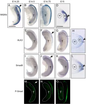
In the rat embryo, the window of MIS responsiveness is from E14-E15. MIS has to be present during this period in order to achieve complete regression of the Müllerian ducts (Picon, 1969; Josso et al., 1976; Donahoe et al., 1977; Tsuji et al., 1992). Regression events, i.e. disruption of the basement membrane of the Müllerian duct and apoptosis of the Müllerian duct epithelial cells, occur after E15 in male rat urogenital ridges (Price et al., 1977; Trelstad et al., 1982; Allard et al., 2000). These facts prompted us to examine the expression profiles of the MIS receptors and SMADs in male rat urogenital ridges at E14-E15 in order to understand how they participate and cooperate in MIS signaling. At early E14 (E14.25), MISRII mRNA was expressed strongly in the cranial urogenital ridge (Fig. 1A), and then the expression was seen to extend craniocaudally along the urogenital ridge (Fig. 1B,C). Unexpectedly, MISRII expression was found predominantly in the coelomic epithelium, but not in the mesenchyme between the Müllerian and
Wolffian ducts in male urogenital ridges (Fig. 1D). At this time, the pattern of MISRII expression was seen as different from that detected at E15.5 when MISRII expression appeared in a circumferential pattern around the male Müllerian duct epithelium (Clarke et al., 2001).
Previous studies have shown that MIS can activate or phosphorylate R-SMADs1, 5 and 8 in cell culture (Gouédard et al., 2000; Clarke et al., 2001; Visser et al., 2001). In this study, we examined the expression of phosphorylated SMAD1/ SMAD5/SMAD8 (P-SMAD) in the urogenital ridge. Whole-mount immunofluorescence analysis showed no obvious P-SMAD expression in the urogenital ridge at early E14 (data not shown). After E14.5, expression of P-SMAD could be detected in male urogenital ridges. It appeared at low level at E14.5 and increased craniocaudally thereafter (Fig. 1M-O). Presence of P-SMAD in the male urogenital ridge after E14.5 implies that MIS is eliciting a functional response and MIS signaling may contribute to subsequent molecular events in the male urogenital ridge.
[image:3.612.51.338.390.740.2]Sexually dimorphic pattern of Alk2and Smad8expression has previously been found in rat urogenital ridges at E15.5 (Clarke et al., 2001), and we examined their expression in male rat urogenital ridges at earlier stages. Little Alk2expression was detected in the urogenital ridge before E14.5 (Fig. 1E). Thereafter, increased expression of Alk2was seen in the anterior male urogenital ridge and extended craniocaudally (Fig. 1F,G). More Smad8transcripts were also detected after E14.5 in male rat urogenital ridges (Fig. 1J,K). Cryosections showed that Alk2and Smad8 mRNA was
Fig. 1. MIS signaling and dynamic expression of
D
E
V
E
LO
P
M
E
N
T
mainly localized in the coelomic epithelium (Fig. 1H,L). At this developmental stage, Alk3 expression was not detected in the coelomic epithelium but in the mesenchyme (data not shown).
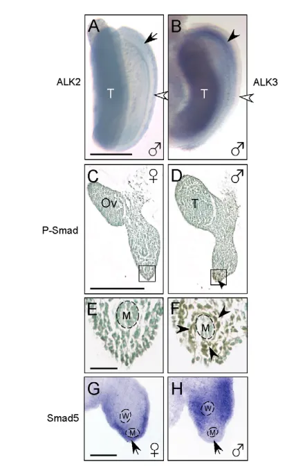
At E15.5, Alk2expression was increased in the fetal gonad but markedly reduced in the coelomic epithelium of the male urogenital ridge, and it began to disappear craniocaudally (Fig. 2A, arrow). Meanwhile, more Alk3was detected in the mesenchyme, and its expression was much higher at E15.5 (Fig. 2B) than at E14.5 in the Müllerian duct mesenchyme in male urogenital ridges (data not shown). Concomitantly, prominent expression of P-SMAD was detected in the mesenchymal cells surrounding the Müllerian ducts of male urogenital ridges (Fig. 2D,F), but absent in the same area in the female (Fig. 2C,E), suggesting that functional MIS signaling continues in the peri-Müllerian duct mesenchyme. Smad5also has
a sexually dimorphic expression pattern at E15.5; its transcripts were expressed in the coelomic epithelium of female urogenital ridges (Fig. 2G), whereas male urogenital ridges expressed much less Smad5in the coelomic epithelium adjacent to the Müllerian duct (Fig. 2H, arrow). Smad1expression was weak and indistinguishable between male and female urogenital ridges from E14.5 to E15.5 (data not shown) (Clarke et al., 2001).
MIS signaling induces a shift of MISRII expression from the coelomic epithelium to the mesenchyme To confirm that the expression of P-SMAD in male urogenital ridges at E14.5-E15.5 was the result of MIS action, we treated E14.5 female rat urogenital ridges in organ culture (Donahoe et al., 1977) with MIS at concentrations known to cause Müllerian duct regression (Ragin et al., 1992; Lorenzo et al., 2002). MIS treatment induced P-SMAD expression in two hours in female urogenital ridges (Fig. 3B). P-SMAD expression was first noted to increase along the outer region of the urogenital ridge lateral to the Müllerian duct (Fig. 3B,C, arrows), and later was also visualized medial to the Müllerian duct following treatment with MIS for 30 hours (Fig. 3D, arrowhead). This pattern is similar to that normally seen in male urogenital ridges (Fig. 3F), but not in untreated female urogenital ridges (Fig. 3E). These data suggest that the dynamic change of P-SMAD in male urogenital ridges at the corresponding developmental stage resulted from MIS activity.
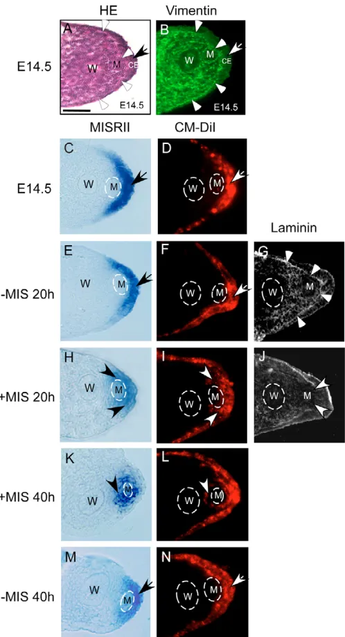
We next investigated whether MIS directs Misr2expression from the coelomic epithelium into the mesenchyme of the Müllerian duct. Female urogenital ridges were treated with MIS in organ culture, and the pattern of Misr2expression was compared with that observed in untreated female counterparts. At E14.5, the coelomic epithelium adjacent to the Müllerian duct appeared thicker than that in other regions in both female (Fig. 4A, arrow) and male urogenital ridges (data not shown). The coelomic epithelium was separated from subjacent mesenchyme by a prominent basement membrane (Fig. 4A,G) (Ikawa et al., 1984), and was noted to have less vimentin expression (Fig. 4B, arrow). Before treatment commenced, Misr2 transcripts were localized to the coelomic epithelium lateral to the Müllerian duct (Fig. 4C). After treatment with MIS for 20 hours, Misr2mRNA was observed in the mesenchyme adjacent to the Müllerian duct with reduced expression in the coelomic epithelium (Fig. 4H), in contrast to the untreated conterpart (Fig. 4E), in which Misr2expression is indistinguishable from that at E14.5 (Fig. 4C). Prolonged treatment with MIS for 40 hours caused expression of Misr2to diminish markedly in the coelomic epithelium and increase in the mesenchyme surrounding the Müllerian duct, notably, between the Müllerian and Wolffian ducts (Fig. 4K, arrowhead). In the untreated female urogenital ridges (without MIS for 40 hours), expression of Misr2 remained lateral to the Müllerian duct, predominantly in the coelomic epithelium (Fig. 4M). When MIS was removed from organ culture before Misr2expression appeared around the Müllerian duct, the change of Misr2expression did not proceed (data not shown). These data indicate that constitutive MIS signaling early in Müllerian duct regression contributes to the distinct male pattern of Misr2expression.
[image:4.612.51.257.237.573.2]MIS induces migration of Misr2-expressing cells To determine whether a mechanism of epithelial-to-mesenchymal transition underlies the switch of Misr2expression, we labeled the coelomic epithelium of female urogenital ridges at E14.5 with CM-DiI, which incorporates into cell membranes, with photostable fluorescence and no apparent adverse effects (Austin, 1995; Karl and Capel, 1998), and tracked the migration of fluorescent-labeled cells Fig. 2. Expression patterns of the MIS type I receptors (Alk2and
Alk3), Smad5and P-SMAD in E15.5 rat urogenital ridges. (A,B) Whole-mount in situ hybridization shows decreased Alk2 expression in the cranial coelomic epithelium (A, arrow) and increased Alk3expression in the mesenchyme (B, black arrowhead) of male urogenital ridges. White arrowhead indicates the coelomic epithelium. (C-F) Sexually dimorphic expression of P-SMAD detected by
D
E
V
E
LO
P
M
E
N
T
in the presence of MIS. After a short incubation with CM-DiI, fluorescence could be detected in the coelomic epithelium (Fig. 4D, arrow). The fluorescence-labeled coelomic epithelium adjacent to the Müllerian duct comprises two to three layers of cells, thicker than that in other regions. Deeper uptake of CM-DiI beyond the coelomic epithelial cells appeared to be prohibited by the basement membrane. In female urogenital ridges cultured at E14.5 for 20 hours, the basement membrane was continuous (Fig. 4G, arrowheads) and CM-DiI remained in the coelomic epithelium (Fig. 4F). However, in the urogenital ridges treated with MIS for 20 hours, CM-DiI fluorescence appeared beneath the disrupted basement membrane, which was shown with loss of laminin staining (Fig. 4J, arrowheads), and was detected in the area adjacent to the Müllerian duct (Fig. 4I, arrowheads). The extension of CM-DiI was colocalized with Misr2 expression (Fig. 4H). Longer treatment resulted in localization of fluorescence around the Müllerian duct (Fig. 4L, arrowhead). At this time, Misr2expression was also found in the mesenchyme around the Müllerian duct (Fig. 4K). The Misr2 -expressing mesenchymal cells were stained for vimentin (data not shown). In untreated urogenital ridges, CM-DiI labeled cells remained in the thick surface epithelium (Fig. 4N). Our data suggest that one of the earliest actions of MIS is to cause epithelial-to-mesenchymal transition and drive the Misr2-expressing cells to migrate from the coelomic epithelium into the mesenchyme surrounding the Müllerian duct.
Exogenous MIS differentially regulates R-SMADs 1, 5 and 8 expression
To investigate whether the sexually dimorphic expression of Smad8 and Smad5 is dependent upon MIS, we treated E14.5 female urogenital ridges in organ culture with MIS and examined their expression. MIS treatment of female urogenital ridges resulted in increased expression of Smad8(Fig. 5B,E). Smad8expression was also induced by MIS added to female urogenital ridges after removal of the gonad (data not shown), indicating that this effect is not a result of other gonadal factors. In situ hybridization with probes targeting different regions in the Smad8transcript (data not shown) confirmed that the regulated Smad8was full length, not an isoform encoding C-terminus deleted Smad8 (Nishita et al., 1999). Treatment of E14.5 female urogenital ridges with MIS also resulted in decreased Smad5expression in the coelomic epithelium adjacent to the Müllerian duct (Fig. 5H,K), similar to that seen in male urogenital ridges at the same developmental stage in vivo and in vitro (Fig. 2H, Fig. 5I,L). MIS had no noticeable effect on Smad1 expression in urogenital ridges (data not shown).
MIS spatiotemporally regulates Alk2and Alk3 expression
To investigate whether MIS regulates Alk2 and Alk3expression during Müllerian duct regression, we treated E14.5 female urogenital ridges in organ culture with MIS and examined expression over time. Treatment of E14.5 female urogenital ridges with MIS for 12 hours induced Alk2expression (Fig. 5N,P) when compared with untreated ridges (Fig. 5M,O). Moreover, increased Alk2expression was detected in the coelomic epithelium as early as 4-6 hours after treatment, and decreased after treatment for 24 hours (data not shown). Alk3expression was increased only after culture for more than 24 hours with MIS. It was upregulated predominantly in the mesenchyme surrounding the Müllerian duct (Fig. 5R,T) when compared with untreated ridges (Fig. 5Q,S). Upregulation of both Alk2and Alk3both followed a cranial-to-caudal pattern.
Alk2mediates the change of MISRII expression and is required for Müllerian duct regression The functional importance of the MIS type I receptors and R-SMADs1, 5 and 8 in Müllerian duct regression was investigated by RNAi in organ culture of male rat urogenital ridges. Multiple siRNAs designed to target Alk2, Alk3, Smad1, Smad5and Smad8 were first studied in cultured MIS-responsive and MISRII-expressing R2C rat Leydig cells (data not shown) (Teixeira et al., 1999). The siRNAs that showed significant silencing of mRNA expression for each gene in cell culture were selected for subsequent use in organ culture (Table 1). Transfection of fluorescein-labeled siRNA into urogenital ridges could be visualized in the urogenital ridge, where it was seen to penetrate the coelomic epithelium, but not beyond (data not shown).
Male urogenital ridges were treated with control- or Alk2-siRNA, and expression of Misr2and P-SMAD was examined. P-SMAD expression was markedly decreased in Alk2-siRNA treated male urogenital ridges (compare Fig. 6B with Fig. 6A, arrows). In the urogenital ridges treated with control-siRNA, Misr2mRNA was detected in the mesenchyme around the Müllerian duct (Fig. 6C, arrowhead); however, in those treated with Alk2-siRNA, Misr2 expression was not evident in the area between the Müllerian and Wolffian ducts (Fig. 6D, arrowhead).
[image:5.612.52.282.57.328.2]The selective expression of Wnt7a, which drives the expression of MISRII, in the Müllerian duct epithelium of urogenital ridges (Parr and McMahon, 1998) makes it a particularly useful marker Fig. 3. MIS activates R-SMADs 1, 5 and 8 in urogenital ridges in
D
E
V
E
LO
P
M
E
N
T
with which to study the Müllerian duct (data not shown), as it faithfully reflects Mullerian duct formation and regression. Detection of Wnt7aexpression, which was able to locate remaining Müllerian duct epithelium in urogenital ridges, allowed us to monitor the effects of RNAi on Müllerian duct regression in organ culture and to examine the contribution of Alk2as an MIS type I receptor in Müllerian duct regression.
Male urogenital ridges were treated with siRNAs and then cultured for additional 2 days. In situ hybridization showed that the Müllerian duct epithelium expressing Wnt7awas retained in the urogenital ridges treated with Alk2-siRNA (Fig. 6F,H, arrows), but not in control-siRNA treated urogenital ridges (Fig. 6E,G). Multiple siRNAs targeting different regions of Alk2had similar effects (data not shown). These results suggest that Alk2mediates essential MIS signaling in the transition of Misr2-expressing cells from the coelomic epithelium to the peri-Müllerian duct mesenchyme.
SMAD8 but not SMAD5 mediates MIS signaling in Müllerian duct regression
The role of SMAD1, SMAD5 or SMAD8 in MIS signaling and Müllerian duct regression was also investigated by RNAi. When male ridges were treated with control-siRNA for 12 hours, the entire Müllerian duct was still evident after culture for additional 36 hours (Fig. 7A). However, in Smad5-siRNA-treated urogenital ridges, regression was accelerated, as discontinuous Wnt7a expression was seen in the cranial area after culture for the same period (Fig. 7B, arrowheads). Moreover, when RNAi effect was examined in urogenital ridges after prolonged culture for additional 12 hours, Wnt7aexpression still remained in the posterior region of control-siRNA urogenital ridges (Fig. 7C, arrow), but not in Smad5 -siRNA-treated ridges (Fig. 7D), indicating that SMAD5 deficiency led to enhanced Müllerian duct regression. By contrast, treatment with Smad8-siRNA delayed Müllerian duct regression in male urogenital ridges, as Wnt7aexpression was detected in the Smad8 -siRNA (Fig. 7F, arrow) but not control--siRNA treated urogenital ridges (Fig. 7E). Moreover, the effect of Smad8-siRNA on Müllerian duct regression was consistent with its specific gene silencing in cell culture, demonstrated by both RT-PCR and western (data not shown). Smad1-siRNA had no effect alone, and RNAi with both Smad1-siRNA and Smad8-siRNA simultaneously did not show a further inhibitory effect on Müllerian duct regression than that caused by Smad8-siRNA alone (data not shown).
DISCUSSION
MIS-induced epithelial-to-mesenchymal transition underlies the change of MISRII expression
[image:6.612.50.299.59.519.2]During male sexual development, Müllerian ducts first form and then are eliminated as a consequence of MIS signaling. In the rat, MIS expression is first detected at E13 in fetal testes (Hirobe et al., 1992). However, a functional signaling pathway is not initiated until MISRII appears in the urogenital ridge. Our present work shows that functional MIS signaling, as documented by activation of R-SMADs1, 5 and 8, is not observed in the male urogenital ridge immediately until after the expression of MISRII. Interestingly, expression of MISRII and phosphorylated SMAD1/5/8 are localized in the coelomic epithelium at this stage. The downstream signaling events (e.g. upregulation of Alk2and Smad8, and downregulation of Smad5) also appear initially in the coelomic epithelium. This explains why RNAi with the lipid-based transfection technique, which could only penetrate the surface coelomic epithelium, was effective in knocking down the components of MIS signaling in the urogenital ridge.
D
E
V
E
LO
P
M
E
N
T
The epithelial cells of the Müllerian duct originate from the coelomic epithelium (Gruenwald, 1941), where expression of Misr2 is also first detected (Fig. 1A-D). MIS signaling leads to the appearance of Misr2 expression in the peri-Müllerian duct mesenchyme. By tracking the migration of DiI-labeled cells, we found that MIS induces the Misr2-expressing cells that originally reside in the coelomic epithelium to migrate into the mesenchymal compartment around the Müllerian duct, following disruption of basement membrane. MIS causes epithelial cells to become migratory, and thereby, initiates an epithelial-to-mesenchymal transition (for a review, see Thiery, 2002), driving Misr2 expression into the peri-Müllerian duct mesenchyme. Although we cannot completely rule out that non-Misr2-expressing cells migrate in response to an indirect effect of MIS and then begin to express Misr2 in the mesenchyme, our data strongly suggest that MIS directs Misr2-expressing cells from the coelomic epithelium into mesenchyme. The timeframe of the migration is in agreement with the period required for apoptosis to be observed in the Müllerian duct epithelium (Price et al., 1977; Roberts et al., 1999; Allard et al., 2000), implying that the Misr2-expressing cells as MIS effectors may have to arrive in the peri-Müllerian duct mesechyme and/or become mesenchymal cells to exert significant paracrine effects on Müllerian ducts. This early epithelio-mesenchymal transformation is reminiscent of the subsequent transition of the epithelial duct cells to mesenchyme later during the regression phase (Trelstad et al., 1982; Austin, 1995; Allard et al., 2000), and illustrates that this cellular process is a key mechanism in Müllerian duct regression.
Before the Müllerian ducts develop, the Wolffian ducts occupy the lateral area of urogenital ridges beneath the coelomic epithelium where the Müllerian ducts are later destined to emerge
[image:7.612.49.410.60.381.2](Gruenwald, 1941; Trelstad et al., 1982). The Müllerian duct forms between the Wolffian duct and the coelomic epithelium, and the Müllerian duct is initially separated from the coelomic epithelium only by a shared basement membrane and no intervening mesenchyme (Trelstad et al., 1982; Ikawa et al., 1984). The coelomic epithelium adjacent to the Müllerian duct expresses Misr2and appears thicker than the epithelium covering other regions of the urogenital ridge (Fig. 4A-G). After peri-Müllerian duct mesenchyme forms under the influence of MIS, the coelomic epithelium adjacent to the male Müllerian duct becomes thinner and indistinguishable from that in lateral regions (Trelstad et al., 1982). MIS induces the Misr2-expressing epithelial cells to lose polarity and manifest a migratory phenotype, and thus facilitates the formation and patterning of the peri-Müllerian duct mesenchyme (Fig. 8). WNT signaling is associated with the epithelial and mesenchymal patterning of the female reproductive tract (Miller and Sassoon, 1998). -Catenin, which transduces canonical WNT signaling, has been linked to the regulation of epithelial cell migration and epithelial-to-mesenchymal transition (Müller et al., 2002; Lu et al., 2003). Misr2-directed -catenin knockout mice show defects in Müllerian mesenchymal development (Arango et al., 2005). MIS is able to activate the NF-B pathway (Segev et al., 2001; Segev et al., 2002), which is also a stimulatory signal leading to epithelial-to-mesenchymal transition (Sosic et al., 2003; Huber et al., 2004). Translocation of -catenin to the nucleus has also been correlated with MIS signaling (Allard et al., 2000). Therefore, MIS and WNT signaling pathways may function cooperatively in mediating epithelial-to-mesenchymal transition early in Müllerian duct regression.
Fig. 5. MIS upregulates SMAD8, downregulates SMAD5 and induces
D
E
V
E
LO
P
M
E
N
T
Alk2and Alk3may act as sequential MIS type I receptors in Müllerian duct regression
Specificity and versatility in the signaling responses of TGF- family members are defined particularly by the type I receptors that a ligand can activate. For example, TGF-activates ALK5 in Mink lung cells (Bassing et al., 1994), while ALK1 acts as a TGF-type I receptor in vascular smooth muscle differentiation (Oh et al., 2000). Alk3and ALK6 can serves as sequential type I receptors in BMP signaling, and control the production and fate of dorsal precursor cells from neural stem cells (Panchision et al., 2001). Alk2, as a functionally essential MIS type I receptor in the rat urogenital ridge (Visser et al., 2001), mediates the change of MISRII expression, and thus the migration and transition of the coelomic epithelial cells (Fig. 6). Alk2 has also been shown to regulate
epithelial-to-mesenchymal transition during cardiac valve formation (Desgrosellier et al., 2005). Interestingly, constitutively active Alk3 can stimulate E-cadherin expression and antagonize the process of epithelial-to-mesenchymal transition (Zeisberg et al., 2003).
[image:8.612.52.248.54.382.2]Analysis of Alk2expression in the male urogenital ridge of the rat has previously shown that it is present in the Müllerian mesenchyme at E15, but not at E16 (He et al., 1993) when Müllerian duct regression is not yet complete. Alk3is ubiquitously expressed in embryonic organs including the urogenital system by the time that MIS and MISRII are expressed (Dewulf et al., 1995). However, we noted that ALK3 expression favors the mesenchyme of the urogenital ridge instead of the coelomic epithelium where functional MIS signaling initially occurs (Fig. 2B). Alk3expression is increased after E15.5, coincident with the appearance of Misr2 in the peri-Müllerian duct mesenchyme in male rat urogenital ridges (Fig. 2B), and this could also be recapitulated in female urogenital ridges upon MIS treatment (Fig. 5Q-T). Moreover, regulation of Alk3expression appears to be a downstream event, as diminution of Alk2-mediated signaling with Alk2-siRNA also inhibits the upregulation of Alk3in male urogenital ridges (data not shown). The spatiotemporal patterns of Alk2and Alk3expression imply that they may act sequentially as type I receptors for MIS signaling (Fig. 8); Alk2functions early in MIS signaling and mediates the migration and transition of coelomic epithelial cells, Fig. 6. Alk2is essential for MIS signaling, transition of Misr2
[image:8.612.323.504.58.345.2]expression and Müllerian duct regression in the rat.E14.5 male urogenital ridges were treated with control-siRNA (A,C,E) or Alk2-siRNA (B,D,F) for 10 hours. (A,B) Cultured for additional 10 hours followed by whole-mount immunofluorescence analysis of activated R-SMAD1, 5, 8 (P-SMAD). (C,D) Cultured for an additional 20 hours followed by in situ hybridization to detect Misr2. (E-H) Cultured for an additional 48 hours followed by in situ hybridization to detect Wnt7aexpression. White arrows indicate the cranial regions with high (A) or low (B) P-SMAD expression for comparison. The presence (C) or absence (D) of Misr2 expression can be noted in the regions between the Müllerian and Wolffian ducts (arrowheads). Black arrows indicate the persistence of Wnt7aexpression in the remaining Müllerian duct epithelium (F,H). The position of transverse sections (G,H) is marked by broken lines on E and F, respectively. Cranial is oriented towards the top and Müllerian duct to the right of individual images. M, Müllerian duct; T, testis; W, Wolffian duct. Scale bar: 500 m for all the whole-mount samples; 50 m for all the sections.
D
E
V
E
LO
P
M
E
N
T
whereas ALK3 may participate in later MIS signaling in Müllerian duct regression, which has been well documented in the mouse (Jamin et al., 2002). The indispensable role of ALK2 in Müllerian duct regression was also documented in mouse urogenital ridges by performing RNAi at E12.5 (data not shown). Moreover, we also observed increased expression of Alk3in the mouse at E14.5 (developmentally equivalent to ~E15.5 in the rat) in the Müllerian duct mesenchyme (data not shown), which appears later than Alk2 (Visser et al., 2001). Although incubation with siRNAs for Alk3was able to knockdown Alk3 expression in cultured cells (data not shown), we could not achieve a commensurate decrease in ALK3 expression in the peri-Müllerian mesenchyme owing to limited penetration of siRNAs (data not shown). This precluded further pursuit of the ALK3 function in rat Müllerian duct regression using our current RNAi techniques. However, given that the timing of increased Alk3expression seen in Müllerian duct mesenchyme coincides with regression of Müllerian duct epithelium, which occurs predominantly after E15.5 in male rat urogenital ridges, it would be reasonable to speculate that in the rat (like the mouse) Misr2-expressing mesenchymal cells favor Alk3 as the type I receptor in late stage Müllerian duct regression.
Specificity of SMADs in MIS signaling during Müllerian duct regression
Regression of the Müllerian duct requires the simultaneous action of the MIS ligand, the type II receptor and the type I receptor(s). R-SMADs 1, 5 and 8 are the intracellular effectors of Alk2/Alk3
signaling, and their functional redundancy has been suggested in BMP signaling. It is also noteworthy that they can function specifically in particular tissue and developmental contexts. SMAD5 mediates BMP2 signaling in developing cerebellum (Rios et al., 2004) and knockout of SMAD5 reveals its importance in regulating endothelial-mesenchymal interactions during embryonic angiogenesis (Yang et al., 1999), whereas SMAD1 signaling controls the growth of extra-embryonic structures at postimplantation stages (Tremblay et al., 2001). MIS-induced upregulation of Smad8and downregulation of Smad5correlate with Müllerian duct regression, suggesting that SMAD5 and SMAD8 can transduce specific signals in MIS pathways. In addition, targeting Smad5expression with siRNA promoted Müllerian duct regression. SMAD5 has been shown to mediate BMP7 signaling and to cause reversal of TGF-1-induced epithelial-to-mesenchymal transition (Zeisberg et al., 2003). Downregulation of SMAD5 by MIS seems to favor the transition of the coelomic epithelial cells to mesenchymal cells.
Increased SMAD8, similar to upregulated ALK2, can act to sustain and amplify the signaling cascade. Disruption of the feed-forward circuit by RNAi-mediated gene silencing of either SMAD8 or ALK2 affected the subsequent downstream signaling events, resulting in retained Müllerian ducts. However, our investigation did not reveal a clear role for SMAD8 in MIS-induced earlier epithelial-to-mesenchymal transition (data not shown). It is possible that this process is independent of SMAD signaling. Prolonged induction of Smad8at E15~E16 over Alk2expression was seen in the male peri-Müllerian mesenchyme (data not shown), suggesting that SMAD8 may play a role in later ALK3-mediated molecular events during Müllerian duct regression.
In conclusion, we identified the coelomic epithelium as the first target for MIS and found that MIS exerts a profound influence on the expression of its own signaling components early in Müllerian duct regression. These events elicit epithelial-to-mesenchymal transition and amplify the MIS signaling for subsequent regression of the Müllerian duct. Knowledge of the downstream MIS signaling events in the urogenital ridge will be important to the study of MIS at other target sites such as the coelomic epithelium of the ovary where oncogenic changes lead to ovarian cancer in mouse models (Orsulic et al., 2002; Connolly et al., 2003; Dinulescu et al., 2005) and presumably in humans.
We thank Drs Allan Goldstein, Liz Perkins and Drucilla Roberts for suggestions and critical reading of the manuscript; Drs Trent Clarke, Makiko Hoshiya and Yasunori Hoshiya for sharing techniques and reagents, and members of the Donahoe laboratory for helpful discussions. This work was supported by a grant form the NIH (NICHD-HD-32112 to P.K.D. and D.T.M., and J.T.), a fellowship from the Ovarian Cancer Research Training Program of the Department of Defense (Y.Z.), and a National Research Service Award (NRSA) fellowship (to N.A.A.). Recombinant human MIS used in this study was provided under the auspices of NCI-CA-17393 (D.T.M. and P.K.D.).
References
Allard, S., Adin, P., Gouédard, L., di Clemente, N., Josso, N., Orgebin-Crist, M. C., Picard, J. Y. and Xavier, F.(2000). Molecular mechanisms of hormone-mediated Müllerian duct regression: involvement of -catenin. Development 127, 3349-3360.
Arango, N. A., Szotek, P. P., Manganaro, T. F., Oliva, E., Donahoe, P. K. and Teixeira, J.(2005). Conditional deletion of -catenin in the mesenchyme of the developing mouse uterus results in a switch to adipogenesis in the myometrium.
Dev. Biol. 288, 276-283.
Attisano, L. and Wrana, J. L.(2002). Signal transduction by the TGF- superfamily. Science296, 1646-1647.
Austin, H. B.(1995). DiI analysis of cell migration during Müllerian duct regression. Dev. Biol. 169, 29-36.
[image:9.612.54.300.60.236.2]Baarends, W. M., van Helmond, M. J., Post, M., van der Schoot, P. J., Fig. 8. A schematic model of MIS actions at the early stage of
D
E
V
E
LO
P
M
E
N
T
Hoogerbrugge, J. W., de Winter, J. P., Uilenbroek, J. T., Karels, B., Wilming, L. G., Meijers, J. H. et al.(1994). A novel member of the transmembrane serine/threonine kinase receptor family is specifically expressed in the gonads and in mesenchymal cells adjacent to the Müllerian duct.
Development120, 189-197.
Bassing, C. H., Yingling, J. M., Howe, D. J., Wang, T., He, W. W., Gustafson, M. L., Shah, P., Donahoe, P. K. and Wang, X. F.(1994). A transforming growth factor type I receptor that signals to activate gene expression. Science 263, 87-89.
Behringer, R. R., Finegold, M. J. and Cate, R. L.(1994). Müllerian-inhibiting substance function during mammalian sexual development. Cell79, 415-425. Belville, C., Josso, N. and Picard, J. Y.(1999). Persistence of Müllerian derivatives
in males. Am. J. Med. Genet. 89, 218-223.
Calegari, F., Haubensak, W., Yang, D., Huttner, W. B. and Buchholz, F.(2002). Tissue-specific RNA interference in postimplantation mouse embryos with endoribonuclease-prepared short interfering RNA. Proc. Natl. Acad. Sci. USA 99, 14236-14240.
Clarke, T. R., Hoshiya, Y., Yi, S. E., Liu, X., Lyons, K. M. and Donahoe, P. K. (2001). Müllerian inhibiting substance signaling uses a bone morphogenetic protein (BMP)-like pathway mediated by ALK2 and induces Smad6 expression.
Mol. Endocrinol. 15, 946-959.
Catlin, E. A., Tonnu, V. C., Ebb, R. G., Pacheco, B. A., Manganaro, T. F., Ezzell, R. M., Donahoe, P. K. and Teixeira, J. (1997). Müllerian inhibiting substance inhibits branching morphogenesis and induces apoptosis in fetal rat lung.
Endocrinology 138, 790-796.
Connolly, D. C., Bao, R., Nikitin, A. Y., Stephens, K. C., Poole, T. W., Hua, X., Harris, S. S., Vanderhyden, B. C. and Hamilton, T. C.(2003). Female mice chimeric for expression of the simian virus 40 TAg under control of the MISIIR promoter develop epithelial ovarian cancer. Cancer Res. 63, 1389-1397. Desgrosellier, J. S., Mundell, N. A., McDonnell, M. A., Moses, H. L. and
Barnett, J. V.(2005). Activin receptor-like kinase 2 and Smad6 regulate epithelial-mesenchymal transformation during cardiac valve formation. Dev. Biol. 280, 201-210.
Dewulf, N., Verschueren, K., Lonnoy, O., Moren, A., Grimsby, S., Vande Spiegle, K., Miyazono, K., Huylebroeck, D. and Ten Dijke, P.(1995). Distinct spatial and temporal expression patterns of two type I receptors for bone morphogenetic proteins during mouse embryogenesis. Endocrinology 136, 2652-2663.
di Clemente, N., Wilson, C., Faure, E., Boussin, L., Carmillo, P., Tizard, R., Picard, J. Y., Vigier, B., Josso, N. and Cate, R.(1994). Cloning, expression, and alternative splicing of the receptor for anti-Müllerian hormone. Mol. Endocrinol. 8, 1006-1020.
Dinulescu, D. M., Ince, T. A., Quade, B. J., Shafer, S. A., Crowley, D. and Jacks, T.(2005). Role of K-ras and Pten in the development of mouse models of endometriosis and endometrioid ovarian cancer. Nat. Med. 11, 63-70. Donahoe, P. K., Ito, Y. and Hendren, W. H., 3rd(1977). A graded organ culture
assay for the detection of Müllerian inhibiting substance. J. Surg. Res. 23, 141-148.
Gouédard, L., Chen, Y. G., Thevenet, L., Racine, C., Borie, S., Lamarre, I., Josso, N., Massagué, J. and di Clemente, N.(2000). Engagement of bone morphogenetic protein type IB receptor and Smad1 signaling by anti-Müllerian hormone and its type II receptor. J. Biol. Chem. 275, 27973-27978.
Gruenwald, P.(1941). The relation of the growing Müllerian duct to the Wolffian duct and its importance for the genesis of malformations. Anat. Rec. 81, 1-19. He, W. W., Gustafson, M. L., Hirobe, S. and Donahoe, P. K.(1993).
Developmental expression of four novel serine/threonine kinase receptors homologous to the activin/transforming growth factor-type II receptor family.
Dev. Dyn. 196, 133-142.
Hirobe, S., He, W. W., Lee, M. M. and Donahoe, P. K.(1992). Müllerian inhibiting substance messenger ribonucleic acid expression in granulosa and Sertoli cells coincides with their mitotic activity. Endocrinology 131, 854-862. Hoshiya, M., Christian, B. P., Cromie, W. J., Kim, H., Zhan, Y., MacLaughlin,
D. T. and Donahoe, P. K.(2003). Persistent Müllerian duct syndrome caused by both a 27-bp deletion and a novel splice mutation in the MIS type II receptor gene. Birth Defects Res. Part A Clin. Mol. Teratol. 67, 868-874.
Huber, M. A., Azoitei, N., Baumann, B., Grunert, S., Sommer, A.,
Pehamberger, H., Kraut, N., Beug, H. and Wirth, T.(2004). NF-B is essential for epithelial-mesenchymal transition and metastasis in a model of breast cancer progression. J. Clin. Invest. 114, 569-581.
Hudson, P. L., Dougas, I., Donahoe, P. K., Cate, R. L., Epstein, J., Pepinsky, R. B. and MacLaughlin, D. T.(1990). An immunoassay to detect human Müllerian inhibiting substance in males and females during normal development. J. Clin. Endocrinol. Metab. 70, 16-22.
Ikawa, H., Trelstad, R. L., Hutson, J. M., Manganaro, T. F. and Donahoe, P. K. (1984). Changing patterns of fibronectin, laminin, type IV collagen, and a basement membrane proteoglycan during rat Müllerian duct regression. Dev. Biol. 102, 260-263.
Jamin, S. P., Arango, N. A., Mishina, Y., Hanks, M. C. and Behringer, R. R. (2002). Requirement of Bmpr1a for Müllerian duct regression during male sexual development. Nat. Genet. 32, 408-410.
Jamin, S. P., Arango, N. A., Mishina, Y., Hanks, M. C. and Behringer, R. R. (2003). Genetic studies of the AMH/MIS signaling pathway for Müllerian duct regression. Mol. Cell. Endocrinol. 211, 15-19.
Josso, N. and di Clemente, N.(2003). Transduction pathway of anti-Müllerian hormone, a sex-specific member of the TGF-family. Trends Endocrinol. Metab. 14, 91-97.
Josso, N., Picard, J. Y. and Trah, D.(1976). The antimüllerian hormone. Recent Prog. Horm. Res. 33, 117-167.
Karl, J. and Capel, B.(1998). Sertoli cells of the mouse testis originate from the coelomic epithelium. Dev. Biol. 203, 323-333.
Kobayashi, A. and Behringer, R. R.(2003). Developmental genetics of the female reproductive tract in mammals. Nat. Rev. Genet. 4, 969-980. Lorenzo, H. K., Teixeira, J., Pahlavan, N., Laurich, V. M., Donahoe, P. K. and
MacLaughlin, D. T.(2002). New approaches for high-yield purification of Müllerian inhibiting substance improve its bioactivity. J. Chromatogr. B Analyt. Technol. Biomed. Life Sci. 766, 89-98.
Lu, Z., Ghosh, S., Wang, Z. and Hunter, T.(2003). Downregulation of caveolin-1 function by EGF leads to the loss of E-cadherin, increased transcriptional activity of -catenin, and enhanced tumor cell invasion.
Cancer Cell4, 499-515.
Massagué, J.(2000). How cells read TGF-signals. Nat. Rev. Mol. Cell Biol. 1, 169-178.
Miller, C. and Sassoon, D. A.(1998). Wnt-7a maintains appropriate uterine patterning during the development of the mouse female reproductive tract.
Development125, 3201-3211.
Mishina, Y., Rey, R., Finegold, M. J., Matzuk, M. M., Josso, N., Cate, R. L. and Behringer, R. R.(1996). Genetic analysis of the Müllerian-inhibiting substance signal transduction pathway in mammalian sexual differentiation. Genes Dev. 10, 2577-2587.
Müller, T., Bain, G., Wang, X. and Papkoff, J.(2002). Regulation of epithelial cell migration and tumor formation by -catenin signaling. Exp. Cell Res. 280, 119-133.
Nishita, M., Ueno, N. and Shibuya, H.(1999). Smad8B, a Smad8 splice variant lacking the SSXS site that inhibits Smad8-mediated signalling. Genes Cells 4, 583-591.
Oh, S. P., Seki, T., Goss, K. A., Imamura, T., Yi, Y., Donahoe, P. K., Li, L., Miyazono, K., ten Dijke, P., Kim, S. et al.(2000). Activin receptor-like kinase 1 modulates transforming growth factor-1 signaling in the regulation of angiogenesis. Proc. Natl. Acad. Sci. USA 97, 2626-2631.
Orsulic, S., Li, Y., Soslow, R. A., Vitale-Cross, L. A., Gutkind, J. S. and Varmus, H. E.(2002). Induction of ovarian cancer by defined multiple genetic changes in a mouse model system. Cancer Cell1, 53-62.
Panchision, D. M., Pickel, J. M., Studer, L., Lee, S. H., Turner, P. A., Hazel, T. G. and McKay, R. D.(2001). Sequential actions of BMP receptors control neural precursor cell production and fate. Genes Dev. 15, 2094-2110.
Parr, B. A. and McMahon, A. P.(1998). Sexually dimorphic development of the mammalian reproductive tract requires Wnt-7a. Nature395, 707-710. Picon, R.(1969). Action of the fetal testis on the development in vitro of the
Müllerian ducts in the rat. Arch. Anat. Microsc. Morphol. Exp. 58, 1-19. Price, J. M., Donahoe, P. K., Ito, Y. and Hendren, W. H., 3rd(1977).
Programmed cell death in the Müllerian duct induced by Müllerian inhibiting substance. Am. J. Anat. 149, 353-375.
Ragin, R. C., Donahoe, P. K., Kenneally, M. K., Ahmad, M. F. and MacLaughlin, D. T.(1992). Human Müllerian inhibiting substance: enhanced purification imparts biochemical stability and restores antiproliferative effects.
Protein Expr. Purif. 3, 236-245.
Rios, I., Alvarez-Rodriguez, R., Marti, E. and Pons, S.(2004). Bmp2 antagonizes sonic hedgehog-mediated proliferation of cerebellar granule neurones through Smad5 signalling. Development131, 3159-3168. Roberts, L. M., Hirokawa, Y., Nachtigal, M. W. and Ingraham, H. A.(1999).
Paracrine-mediated apoptosis in reproductive tract development. Dev. Biol. 208, 110-122.
Sakai, T., Larsen, M. and Yamada, K. M.(2003). Fibronectin requirement in branching morphogenesis. Nature423, 876-881.
Segev, D. L., Hoshiya, Y., Stephen, A. E., Hoshiya, M., Tran, T. T.,
MacLaughlin, D. T., Donahoe, P. K. and Maheswaran, S.(2001). Müllerian inhibiting substance regulates NF-B signaling and growth of mammary epithelial cells in vivo. J. Biol. Chem. 276, 26799-26806.
Segev, D. L., Hoshiya, Y., Hoshiya, M., Tran, T. T., Carey, J. L., Stephen, A. E., MacLaughlin, D. T., Donahoe, P. K. and Maheswaran, S.(2002). Müllerian-inhibiting substance regulates NF-B signaling in the prostate in vitro and in vivo.
Proc. Natl. Acad. Sci. USA 99, 239-244.
Sosic, D., Richardson, J. A., Yu, K., Ornitz, D. M. and Olson, E. N.(2003). Twist regulates cytokine gene expression through a negative feedback loop that represses NF-B activity. Cell112, 169-180.
Soutschek, J., Akinc, A., Bramlage, B., Charisse, K., Constien, R., Donoghue, M., Elbashir, S., Geick, A., Hadwiger, P., Harborth, J. et al.(2004). Therapeutic silencing of an endogenous gene by systemic administration of modified siRNAs. Nature432, 173-178.
D
E
V
E
LO
P
M
E
N
T
Hudson, P. L., Wing, J., Maclaughlin, D. T. and Donahoe, P. K.(1996). Developmental expression of a candidate Müllerian inhibiting substance type II receptor. Endocrinology137, 160-165.
Teixeira, J., Kehas, D. J., Antun, R. and Donahoe, P. K.(1999). Transcriptional regulation of the rat Müllerian inhibiting substance type II receptor in rodent Leydig cells. Proc. Natl. Acad. Sci. USA 96, 13831-13838.
Teixeira, J., Maheswaran, S. and Donahoe, P. K.(2001). Müllerian inhibiting substance: an instructive developmental hormone with diagnostic and possible therapeutic applications. Endocr. Rev. 22, 657-674.
Thiery, J. P.(2002). Epithelial-mesenchymal transitions in tumour progression. Nat. Rev. Cancer2, 442-454.
Trelstad, R. L., Hayashi, A., Hayashi, K. and Donahoe, P. K.(1982). The epithelial-mesenchymal interface of the male rat Müllerian duct: loss of basement membrane integrity and ductal regression. Dev. Biol. 92, 27-40. Tremblay, K. D., Dunn, N. R. and Robertson, E. J.(2001). Mouse embryos
lacking Smad1 signals display defects in extra-embryonic tissues and germ cell formation. Development128, 3609-3621.
Tsuji, M., Shima, H., Yonemura, C. Y., Brody, J., Donahoe, P. K. and Cunha, G. R.(1992). Effect of human recombinant Müllerian inhibiting substance on isolated epithelial and mesenchymal cells during Müllerian duct regression in the rat. Endocrinology 131, 1481-1488.
Visser, J. A., Olaso, R., Verhoef-Post, M., Kramer, P., Themmen, A. P. and Ingraham, H. A.(2001). The serine/threonine transmembrane receptor ALK2 mediates Müllerian inhibiting substance signaling. Mol. Endocrinol. 15, 936-945. Yang, X., Castilla, L. H., Xu, X., Li, C., Gotay, J., Weinstein, M., Liu, P. P. and
Deng, C. X.(1999). Angiogenesis defects and mesenchymal apoptosis in mice lacking Smad5. Development 126, 1571-1580.