Development 139, 2751-2762 (2012) doi:10.1242/dev.082974 © 2012. Published by The Company of Biologists Ltd
INTRODUCTION
Animal development is achieved through the coordination of cell growth, cell division and cell death. During the early embryogenesis of Drosophila melanogaster, cells divide rapidly, which is facilitated by maternal deposition of components of the cell cycle machinery (Foe et al., 1993). After the beginning of zygotic transcription and three rounds of mitosis (14th-16th), most cells enter their final interphase, while cells in the CNS continue to divide (Edgar and O’Farrell, 1990). At late embryogenesis, the larval tissues initiate endoreplication cycles and become polyploid (Edgar and Orr-Weaver, 2001). Endoreplication continues throughout the larval stages, supporting cell growth that results in a dramatic increase of larva body size. By contrast, cells of the imaginal discs (appendage primordia destined to form the adult structures at metamorphosis) continue to proliferate throughout larval development (Bryant and Schmidt, 1990).
Although transcriptional regulation is known to play a vital role in controlling patterning and proliferation during Drosophila development, the contribution of post-translational mechanisms, such as SUMOylation, are less well understood. SUMO (Small Ubiquitin-related Modifier) belongs to a family of highly-conserved Ubiquitin-like proteins that can be conjugated to other proteins to alter their properties. SUMO modification is found in all eukaryotic organisms (Meulmeester and Melchior, 2008), and the biochemical pathway that regulates SUMO conjugation to target proteins is evolutionarily conserved. SUMO proteins are genetically encoded as precursor molecules that are proteolytically cleaved to expose the glycine-glycine motif used for conjugation. SUMO processing is carried out by specific proteases that can also
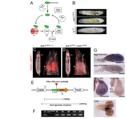
remove SUMO from substrates (Mukhopadhyay and Dasso, 2007). SUMO conjugation to substrates (SUMOylation) starts with the activation of processed SUMO, performed by an E1 SUMO-activating complex comprising the Aos1/Uba2 heterodimeric pair (Johnson et al., 1997). SUMO is activated by ATP hydrolysis-dependent formation of a thioester bond between the catalytic cysteine of the Uba2 subunit and the C-terminal glycine of SUMO (Bossis and Melchior, 2006). Activated SUMO is then transferred to the catalytic cysteine of the E2 SUMO-conjugating enzyme Ubc9. Finally, SUMO is transferred to the -amino group of a lysine side chain of the target protein with the assistance of E3 SUMO protein ligases (Fig. 1A), and an isopeptide bond is formed between the lysine of the substrate and the glycine of SUMO (Bossis and Melchior, 2006). Aos1/Uba2 and Ubc9 are the only E1 and E2 enzymes that facilitate SUMO conjugation in each organism. By contrast, there are several proteins that have been found to have E3 SUMO ligase activity so far, including members of PIAS family (Johnson and Gupta, 2001), the nuclear pore protein RanBP2 (Kirsh et al., 2002; Pichler et al., 2002; Tatham et al., 2005) and the Pc2 protein of the polycomb group (Kagey et al., 2003; Kagey et al., 2005).
SUMOylation is essential in most organisms and is thought to take place in all tissues and at all developmental stages (Geiss-Friedlander and Melchior, 2007). Previous investigations have found SUMOylation to be required for numerous cellular processes, including transcription, cell cycle progression, DNA repair, subcellular localization and signal transduction (Johnson, 2004; Hay, 2005; Dasso, 2008). Although the role of SUMOylation in development, growth and differentiation at the organismal level is not thoroughly studied, there is evidence that ablation of the SUMOylation pathway leads to dramatic developmental defects and early lethality. Ubc9 (SUMO E2) depletion in yeast causes cell cycle arrest at G2/M (Seufert et al., 1995). Likewise, Aos1 or Uba2 depletion in yeast cells leads to the formation of microcolonies comprising enlarged cells (Johnson et al., 1997). In mice, a Ubc9 null allele causes early embryonic lethality and apoptosis of the inner cell mass (ICM) with reported mitotic chromosome 1Stowers Institute for Medical Research, 1000 East 50th Street, Kansas City, MO
64110, USA. 2The Open University, Milton Keynes, MK7 6AA, UK. 3Department of Anatomy and Cell Biology, University of Kansas School of Medicine, 3901 Rainbow Boulevard, Kansas City, KS 66160, USA.
*Author for correspondence (mg2@stowers.org)
Accepted 14 May 2012
SUMMARY
SUMOylation is a highly conserved post-translational modification shown to modulate target protein activity in a wide variety of cellular processes. Although the requirement for SUMO modification of specific substrates has received significant attention in vivo and in vitro, the developmental requirements for SUMOylation at the cell and tissue level remain poorly understood. Here, we show that in Drosophila melanogaster, both heterodimeric components of the SUMO E1-activating enzyme are zygotically required for mitotic progression but are dispensable for cell viability, homeostasis and DNA synthesis in non-dividing cells. Explaining the lack of more pleiotropic effects following a global block of SUMO conjugation, we further demonstrate that low levels of global substrate SUMOylation are detected in mutants lacking either or both E1 subunits. These results not only suggest that minimal SUMOylation persists in the absence of Aos1/Uba2, but also show that the process of cell division is selectively sensitive to reductions in global SUMOylation. Supporting this view, knockdown of SUMO or its E1 and E2 enzymes robustly disrupts proliferating cells in the developing eye, without any detectable effects on the development or differentiation of neighboring post-mitotic cells.
KEY WORDS: Aos1, Drosophila, SUMOylation, Uba2, Imaginal disc
A differential requirement for SUMOylation in proliferating
and non-proliferating cells during Drosophila development
Kiriaki Kanakousaki1,2and Matthew C. Gibson1,3,*
D
E
V
E
LO
P
M
E
N
condensation and segregation defects (Nacerddine et al., 2005). Similarly, lack of Ubc9 function causes embryonic lethality in zebrafish (Nowak and Hammerschmidt, 2006). In Drosophila germline clones that remove maternal smt3(DrosophilaSUMO), 70% of embryos fail to hatch and those that survive die during the first larval instar (Nie et al., 2009). Zygotic mutant animals for the same smt3mutation die in the early second instar (Nie et al., 2009). Additionally, P-element induced lesswrightalleles (lwr, Drosophila E2) cause late embryonic or first instar larval lethality owing to the inability of maternal Bicoid to enter the nuclei during embryogenesis (Epps and Tanda, 1998). Apart from these more general defects, SUMO is also reported to have specific roles in individual developmental processes. RNAi knockdown of smt3 (SUMO) in the prothoracic gland results in a prolonged larval life and pupariation is impeded due to inefficient production of Ecdysone (Talamillo et al., 2008). In addition, transcription factors that are involved in wing disc patterning, such as Vestigial (vg), Spalt (sal) and Spalt-related (salr), are regulated by SUMOylation (Takanaka and Courey, 2005; Sanchez et al., 2010).
In the present study, we examine the role of the E1 SUMO-activating enzyme subunits Aos1 and Uba2 during Drosophila development. Surprisingly, we report that aos1and uba2mutants exhibit specific defects in imaginal disc development and lethality at the larval/pupal transition, without any obvious defects in larval cell growth or survival. We show that Aos1 and Uba2 control global SUMOylation, but also present evidence that low levels of global SUMOylation can persist in the absence of Aos1/Uba2 activity. Finally, based on zygotic null phenotypes and RNAi knockdowns, we show that SUMO E1 activity is crucial for cell division, but may be only minimally required for DNA replication, cell survival and homeostasis in non-dividing cells.
MATERIALS AND METHODS
Fly strains
The UAS-RNAitransgenic stocks for aos1and uba2were obtained from the National Institute of Genetics (NIG-FLY, Kyoto). The UAS-RNAiline for ubc9(lwr) was obtained from the Vienna DrosophilaRNAi Center (VDRC), and the UAS-RNAi for smt3 was provided by Rosa Barrio (Talamillo et al., 2008). The Bloomington DrosophilaStock Center was the source for the PBac-element insertion line in aos1(aos1c06048), the
P-element insertion P{EP}G4672 and the following deficiencies:
Df(3R)ED5554, Df(3R)ED5591and Df(3L)Exel6112. The FRT82B, Ubi-GFP(S65T)nls, RpS3Plac92flies that were used for the clone generation in
a Minute/+background and the fly stocks for UAS-p35, GMR-Gal4, eyeless-Gal4 and Histone 2Av-mRFP1 were also obtained from Bloomington DrosophilaStock Center. All crosses for complementation tests and RNAi expression were cultured at room temperature (25oC).
Molecular cloning
To generate Aos1 probes for in situ hybridization, the cDNA clone
LD33652was digested with SacI-PstI and cloned into SacI/PstI-digested
pBluescript(SK+). pUAST-Aos1was generated by amplification ofaos1
cDNA from LD33652 clone with primers 5GCGGGGCGTAAT -TGGTACGTCG 3(F) and 5XhoI-GGAATTCTTTCTTGTTGCG 3(R), and subsequent cloning into XhoI-digested pUAST. The pCaSpeR4-Aos1
genomic rescue construct was generated by genomic sequence amplification with primers 5XbaI-GAATGGGTCCGGACTACC 3(F) and 5XbaI-CTCCATCACGCGCAGATC 3(R), and cloning into Xba I-digested pCaSpeR4. To generate pUAST AttB-Uba2, we cloned BglII-Xho I-digested LD22577into BglII/XhoI-digested pUAST AttB (Bischof et al., 2007). To generate a pCaSpeR4-AttB-Uba2rescue construct, we amplified the Uba2genomic region with primers 5PstI-TCACATACCTATCCG 3 (F) and 5XbaI-AAGTGGAGCCAGGCGT 3(R), and cloned the PCR product into a PstI/XbaI-digested pCaSpeR4-AttB(Markstein et al., 2008).
Generation of aos1and uba2mutants
aos1-null alleles were generated by the ‘ends-out’ homologous recombination method (Gong and Golic, 2003). Approximately half of
aos1(591 bp, starting 90 bp before the ATG and including the whole first and most of the second exon) was deleted and replaced with mini white. The DNA used for recombination was generated by amplification of the upstream flanking sequence of the deleted area with the primers 5 SpeI-GAAACTGGTAATAGGTGCG 3 (F) and 5AvrIIACGCCTTT -TGAAAATG 3 (R), followed by cloning into a SpeI/AvrII-digested CMC105 vector (a gift from Gary Struhl, Columbia University, NY, USA). In the second step, the downstream flanking sequence was amplified with the primers 5NheI-TGACACCATTTGCCG 3 (F) and 5 NheI-GCGAAATAAGTCGGC 3(R), and then cloned into a NheI-digested CMC105 containing the upstream flanking sequence. The resulting plasmid was used to generate transgenic flies and the gene disruption procedure was completed by Genetic Services (Cambridge, MA, USA).
Bothuba2alleles were generated by imprecise excision of a P-element (P{EP}G4672) located in the uba25UTR. Excision lines were screened for lethality in homozygous animals and lethal lines were tested for complementation with Df(3L)Exel6112. The lethal lines uba2N2and uba2C3
failed to complement the deficiency and were also rescued by the presence of one copy of an uba2genomic rescue construct (described above). We molecularly characterized the lesions in both alleles by sequencing, and found that the P-element was incompletely excised, resulting in an internal deletion that did not disrupt the uba2-coding sequence. Nevertheless, this sequence rearrangement within the P-element was sufficient to cause a defect in uba2transcription or translation, as no protein was observed by western blot analysis of mutant protein extracts.
Genomic rescue of aos1and uba2mutants
For the genomic rescue of aos1 mutant animals, we performed the following cross for each of the three alleles and scored for survival of eclosed non-Sb adults: pCaSpeR4-Aos1/CyO; aos1c06048/TM6C pCaSpeR4-Aos1/CyO; aos1c06048/TM6C.
For the genomic rescue of uba2/Df(3L)Exel6112, we performed the following crosses: uba2N2/TM6BpCaSpeR AttB-Uba2/pCaSpeR 4-AttB-Uba2; Df(3L)Exel6112/TM6Cand uba2C3/TM6CpCaSpeR 4-AttB-Uba2/pCaSpeR 4-AttB-Uba2; Df(3L)Exel6112/TM6C.
Adult progeny from these crosses were scored for the presence or absence of the TM6Cor TM6B balancer chromosomes and therefore for being heterozygous or homozygous for the uba2lesion. The genomic rescue of the uba2mutants was also repeated without the deficiency, following the crossing scheme described above for aos1. All crosses were maintained at 25°C.
Mosaic analysis and Drosophilagenetics
aos1c06048and uba2N2mutant cell clones were generated with the MARCM
system (Lee and Luo, 2001). Mitotic clones were induced by heat shock (1 hour at 37oC) ~48 hours after egg laying (AEL). Larvae were dissected at
late third instar, 2.5-3.0 days after heat shock. For p35-expressing
aos1c06048mutant clones, larvae were dissected at different time points
(2.5-3.5 days after heat shock) so that the mutant cells could be analyzed at different stages of extrusion. aos1c06048mutant clones in the Minute/+
background were generated using the conventional hsFLP-FRT system (Golic, 1991). Clones were induced 4 days after egg lay and larvae were dissected 2.5 days after heat shock. aos1c06048and uba2N2clones marked
by loss of GFP were induced with the hsFLP-FRT system (Golic, 1991) ~48 hours AEL; larvae were dissected 2.5 days after heat shock. Cultures were maintained at room temperature for these experiments.
Genetic crosses used in this study were:
hs-flp, UAS-srcEGFP; actGal4, UAS-GFP; FRT82B, Gal80 (a gift from Ting Xie, Stowers Institute, MO, USA)FRT82B
hs-flp, UAS-srcEGFP; actGal4, UAS-GFP; FRT82B, Gal80 FRT82B, aos1c06048/ TM6B
hs-flp, UAS-srcEGFP; actGal4, UAS-GFP; FRT82B, Gal80 UAS-aos1; FRT82B, aos1c06048/TM6C
hs-flp, UAS-srcEGFP; actGal4, UAS-GFP; FRT82B, Gal80 UASp35;
FRT82B
D
E
V
E
LO
P
M
E
N
hs-flp, UAS-srcEGFP; actGal4, UAS-GFP; FRT82B,Gal80 UASp35; FRT82B, aos1c06048/TM6C
hs-flp, tubGal4, UAS-nlsGFP; FRT80B, tubGal80 FRT80B hs-flp, tubGal4, UAS-nlsGFP; FRT80B, tubGal80 FRT80B, uba2N2/TM6C
hs-flp, tubGal4, UAS-nlsGFP; FRT80B, tubGal80 UAS-uba2; FRT80B, uba2N2
hs-flp; FRT82B FRT82B, ubiGFP, RpS3Plac92/TM6B
hs-flp; FRT82B, aos1c06048/TM6B FRT82B, ubiGFP, RpS3Plac92/TM6B hs-flp; FRT82B, ubiGFP FRT82B
hs-flp; FRT82B, ubiGFP FRT82B, aos1c06048/TM6B hs-flp; FRT80B, ubiGFP FRT80B
hs-flp; FRT80B, ubiGFP FRT80B, uba2N2/TM6C.
Immunostaining
Imaginal discs were dissected in 1PBS and fixed with 4% PFA in 1PBS for 20 minutes at room temperature. Fixed material was washed with PBT (0.2% Triton and 0.5% BSA in 1PBS) and incubated with primary and fluorescent secondary antibodies. Primary antibodies were: anti-cleaved Caspase 3 (1:500; Cell Signaling); anti--Tubulin (1:1000; Sigma); anti-Cyclin A and anti-Cyclin B (DHSB, University of Iowa; 1:500); anti-Elav (DHSB, University of Iowa; 1:80); and anti-Discs large (DHSB, University of Iowa; 1:500). Secondary antibodies were: anti-mouse Alexa 594 (1:500), anti-rabbit Alexa 594 (1:500) and anti-rat Alexa 647 (1:500; Invitrogen). Fluorescent dyes were Phalloidin 546 and 647 (1:250; Invitrogen), and DAPI (1 g/ml; Invitrogen).
Imaginal disc cell preparation for FACS
For FACS analysis, ~40 discs were cut into three or four pieces and transferred to siliconized tubes for 1.5 hours dissociation at room temperature [90% 10Trypsin-EDTA (Sigma), 1PBS + 0.5 g/ml (Hoechst)]. Immediately after dissociation, cells were subjected to FACS analysis using MoFlo in the Stowers Institute Cytometry Core Facility.
EdU incorporation in salivary glands
Early third instar salivary glands were collected from larvae cultured at room temperature. Glands were dissected in 1PBS and incubated for 15 minutes in EdU solution according to the protocol instructions of the Click-iT EdU imaging Kit (Invitrogen). After EdU incorporation, glands were fixed as above and detection was completed as described in the kit protocol. Immunostaining with anti-Tubulin followed.
Protein extraction from larvae and western blot analysis
Inverted anterior-half larvae were isolated in 1PBS, and fat body, gut and salivary glands were removed. For comparison between wild type and mutant protein extracts, the imaginal discs were removed from control animals. The trachea, brain and cuticle were kept in all samples. Dissected larvae were placed in 1RIPA lysis buffer that contained 1
protease inhibitor cocktail (Pierce) and 1 mM PMSF. In samples that were designated for monitoring SUMOylation levels, the lysis buffer contained NEM protease inhibitor (N-Ethylmaleimide, 10 mM final concentration; Sigma). NEM was also added in the 1PBS dissection media. High-concentration protein samples were prepared by placing 50 dissected larvae in 75 l of lysis buffer and grinding them with a pestle. After centrifugation, the lysate supernatant of each sample was analyzed by western blot. Pre-cast gels from BioRad were used (4-15% acrylamide gradient gels for the anti-SUMO and anti-Aos1 western blots, and 7.5% acrylamide gels for the anti-Uba2 western blot). For blotting, we used antibodies for SUMO (1:500), Aos1 (1:5000) and Uba2 (1:1500) provided by Leslie Griffith (Long and Griffith, 2000) and a second anti-SUMO antibody (1:5000) that was provided by Albert Courey (Smith et al., 2004). The level of loaded protein in each gel lane was monitored by re-blotting with an antibody for -Tubulin (1:1000; Sigma). Anti-rabbit HRP (Sigma) and anti-mouse HRP (Invitrogen) secondary antibodies were used at 1:20,000 and 1:5000 dilutions, respectively. The blots were exposed to autoradiography film, after incubation with chemiluminescent substrate SuperSignal West Dura Pierce (for anti-SUMO and anti-Aos1 blots) and Immun-Star HRP substrate kit BioRad (for anti-Uba2 and anti-Tubulin blots).
RESULTS
Imaginal discs are eliminated in Drosophila aos1
mutants
In a screen for lesions causing abnormal imaginal disc phenotypes, we identified a PiggyBac transposon insertion (PBac{PB}Aos1c06048) in the 5 UTR of the aos1gene, 41 bp upstream from the ATG (Fig. 1E, aos1c06048) (Thibault et al., 2004). Homozygous animals exhibited frequent melanotic tumors, developmental delay (Fig. 1B, Fig. 5J) and highly penetrant early pupal lethality. Simple dissection revealed that the imaginal discs were missing in all affected individuals, a probable cause of the metamorphic failure and pupal lethality. To visualize this phenotype in vivo, we used a Histone2-RFP fusion protein to mark the relatively high nuclear density of the imaginal discs within intact larvae (Pandey et al., 2005) (Fig. 1C). Third instar aos1c06048/aos1c06048larvae were of normal size and superficially indistinguishable from controls, but the entire complement of imaginal discs was missing (Fig. 1D). Thus, despite the essential biochemical function of Aos1 in SUMOylation, we did not observe pleiotropic effects on cell survival, growth or homeostasis in the non-dividing cells that comprise the larval body. Instead, this phenotype resembles zygotic cell cycle mutants that disrupt imaginal disc development without affecting endoreplication-dependent larval growth (Gatti and Baker, 1989). Consistent with this, aos1 expression was elevated in early embryos and in dividing cell populations of third instar larvae (Fig. 1G).
We next used three independent approaches to confirm that the phenotypes described above were caused by the aos1c06048allele. First, aos1c06048 failed to complement Df(3R)ED5554 and Df(3R)ED5591, chromosomal deficiencies spanning aos1. Furthermore, the resulting aos1c06048/Df progeny exhibited the same pupal-lethal loss of imaginal discs observed in aos1c06048 homozygotes (0/67 and 0/56 expected transheterozygous progeny eclosed, respectively). Second, the phenotypes associated with aos1c06048were fully reverted by precise excision of the PiggyBac element (Fig. 1F; supplementary material Fig. S1). Finally, as conclusive evidence linking the observed phenotypes to loss of aos1, a 2.9 kb genomic construct containing aos1and the flanking upstream and downstream sequences (Fig. 1E) was sufficient to rescue 100% of aos1c06048homozygous pupae to eclosion and adult viability (n44 pupae).
aos1is autonomously required for clone growth
in the wing disc
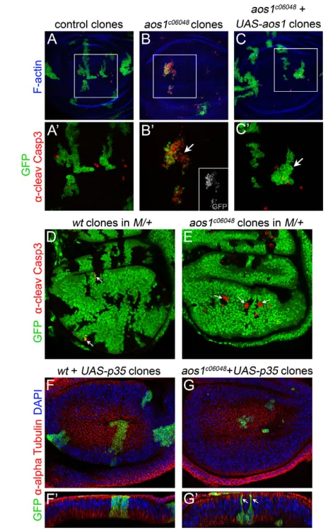
To determine the cellular function of aos1required for imaginal disc development, we induced GFP-positive mutant clones using the MARCM technique (Lee and Luo, 2001). Control clones formed irregular but contiguous patches with negligible levels of cell death as indicated by anti-cleaved Caspase 3 antibody staining (Fig. 2A,A). Three days after induction, aos1c06048 mutant clones in the wing imaginal disc were small and fragmented, with high levels of cell death (Fig. 2B,B). We confirmed the clone growth defect by conventional twin spot analysis (supplementary material Fig. S2B). This growth phenotype was a specific consequence of the absence of Aos1, as clone lethality was rescued by simultaneous expression of aos1cDNA (Fig. 2C,C). We next used the Minute technique to test whether mutant cell lethality was due to cell competition, a phenomenon where slow-dividing cells are killed by their wild-type neighbors (Morata and Ripoll, 1975). Still, induction of aos1mutant cells in a slow-dividing Minutebackground failed
D
E
V
E
LO
P
M
E
N
to rescue the growth or viability of the mutant cell clones (Fig. 2D,E). These experiments have two possible interpretations: either (1) cell proliferation is severely and specifically blocked within 72 hours of losing aos1, resulting in competitive clone death even in a M/+ background; or (2) within 72 hours of losing aos1, mutant clone cells are generally inviable owing to the pleiotropic effects of defective SUMOylation.
Aos1 is required for G2/M progression in imaginal disc cells
[image:4.612.54.469.59.449.2]In order to analyze cell cycle phasing in aos1c06048mutant clones, we blocked apoptosis via expression of p35, a baculoviral caspase inhibitor (Clem et al., 1991). This rescued cell viability but not proliferation, as mutant clone cell numbers were still reduced compared with controls (Fig. 2F,G). The rescued mutant cells Fig. 1. Loss of aos1disrupts cell proliferation but not larval cell homeostasis. (A)Schematic representation of the SUMOylation pathway. A SUMO precursor molecule is cleaved into a mature form that is transferred to target proteins by an enzymatic cascade that includes an E1 SUMO activating enzyme (Aos1/Uba2), an E2 SUMO conjugation enzyme (Ubc9) and an E3 SUMO ligase. SUMO modification of substrates is reversible and deconjugation is facilitated by SUMO-specific proteases. (B)aos1c06048homozygous mutant late third instar larvae occasionally exhibited
melanotic tumors of various sizes (arrows). There is a variation in size of larvae shown, but on average aos1mutant larvae grow slightly larger than wild type (average length of aos1mutant animals is 5.02 mm versus 4.56 mm in wild type). (C,D)Late third instar larvae expressing a Histone2Av-RFP fusion protein. Overall larval growth is similar between aos1c06048heterozygous (C) and aos1c06048homozygous (D) animals. However, imaginal
disc complexes (distinguished by high nuclear density in red) are missing in the homozygous aos1c06048mutant larvae (white arrows). Higher
magnification images show that only the fat body is present in the anterior terminus of the mutant larvae (white arrowheads). (E)aos1gene model.
{PB}Aos1c06048is located in the 5UTR of aos1. Primers F and R indicate the location of the DNA amplicon used to verify the precise excision of the
transposable element in various fly strains rescued by mobilization. The genomic region that was used to rescue aos1mutants is indicated. (F)Agarose gel showing the amplified larval genomic DNA from wild-type controls, aos1c06048/aos1c06048and selected homozygous excisions. The
primers used (shown in E) flank the PBac insertion therefore there is no amplification product from aos1c06048genomic DNA. By contrast, the
phenotypically reverted excision lines produce the correct band, verifying the precise excision of the PBac element. Sequencing of the amplified DNA products confirmed the precise excision (supplementary material Fig. S1). (G)In situ hybridization. In egg chambers, an antisense RNA probe detects aos1transcript in the nurse cells and oocyte. aos1is ubiquitously expressed in imaginal discs. In the CNS, there is strong expression in the optic lobes, which are significantly reduced in size in aos1homozygotes. The staining between the lobes is non-specific. We did not detect strong
aos1expression in trachea, epidermis or salivary glands of third instar larvae (data not shown).
D
E
V
E
LO
P
M
E
N
appeared to be significantly larger than control cells and were partially extruded from the epithelial layer, leaving only one or two thin membranous processes attached to the apical epithelial surface (Fig. 2F,G; supplementary material Fig. S3). This could be an aos1-related defect or, alternatively, may reflect an intermediate stage of the apoptotic extrusion process, similar to what has been reported in dying vertebrate cells (Rosenblatt et al., 2001).
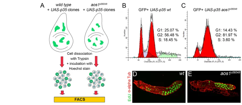
Based on the reduced size of aos1mutant clones, it is likely that only a few cell divisions occurred following loss of aos1 (Fig. 2G). To measure effects on cell cycle progression, we dissociated wing imaginal discs containing GFP-positive clones and analyzed their cell cycle phasing using fluorescence-activated cell sorting (FACS; Fig. 3A). Based on DNA content, FACS analysis showed that aos1c06048 mutant cells accumulated in G2/M (Fig. 3B,C). The average size of these cells was larger than the wild-type controls, but this was attributable to the high percentage of cells in G2/M, which are larger than the cells in G1. Importantly, the cell cycle profile of P35-expressing GFP+control clones did not differ significantly from
GFP-negative controls without P35, indicating that p35 expression alone did not interfere with cell cycle progression (data not shown). The accumulation of aos1c06048 mutant cells in G2/M is consistent with the hypothesis that aos1 is required for mitotic division, but not necessary for the endocycles of DNA replication that drive larval cell growth and consequently growth of the larval body. To test this, we analyzed DNA synthesis in the salivary glands, which execute numerous endoreplication cycles during larval development (Edgar and Orr-Weaver, 2001). Strikingly, in early third instar larvae, aos1c06048mutant salivary gland cells incorporated EdU in a manner similar to controls (Fig. 3D,E). This confirms that, despite a severe and early block in imaginal disc cell proliferation in aos1 homozygotes, S-phase entry and DNA synthesis were not affected.
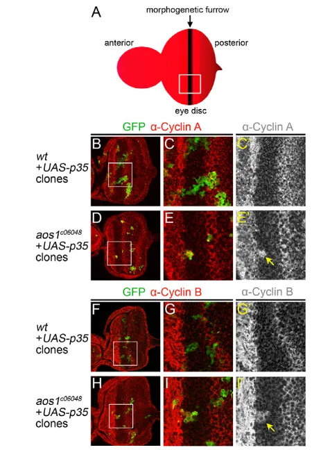
To verify the results from FACS analysis, we directly assayed the levels of mitotic cyclins in aos1 mutant clones. For these experiments, we took advantage of the fact that the eye disc features a distinct population of cells arrested in G1 within the morphogenetic furrow (Wolff and Ready, 1993). As a result, cyclins A and B are expressed in dividing cells on both sides of the G1-arrested population, but not within the furrow itself (Fig. 4A). By contrast, all surviving aos1c06048mutant clones recovered in the furrow had high levels of Cyclin A (Fig. 4B-E; n15 clones). Similar results were obtained for Cyclin B, where half of mutant clones in the furrow exhibited elevated Cyclin B levels (Fig. 4F-I; n6/12 clones). Together, the presence of cyclins A and B in aos1c06048mutant cells confirms that aos1mutant cells accumulate at G2/M. Furthermore, these results indicate that the limiting step for progression through mitosis in the mutant cells is not a lack of mitotic cyclin accumulation.
aos1 null alleles phenocopy aos1c06048
Our initial phenotypic analysis defined a clear requirement for Aos1 in imaginal disc cell proliferation, but found no apparent function in the non-dividing larval cells. This implies a primary requirement for SUMOylation in mitosis, along with a lesser or negligible role in other processes, including DNA replication. To rule out the possibility that aos1c06048is simply a hypomorphic allele that provides sufficient Aos1 for all functions besides mitosis, we next generated aos1-null alleles via homologous recombination (Gong and Golic, 2003). We designed an ‘ends-out’ targeting construct to delete the first and most of the second exon of aos1 (Fig. 5A), along with the ATG and some of the 5 UTR, eliminating the aos1 open reading frame. Using this strategy, we Fig. 2. Severe and cell-autonomous proliferation defects in
aos1c06048clones. (A-C) GFP-positive MARCM clones in the wing disc,
stained with anti-cleaved Caspase-3 (anti-CC3) to mark apoptotic cells (red) and phalloidin to counterstain F-Actin (blue). (A)Control clones, (B)
aos1c06048mutant clones and (C) aos1c06048mutant clones expressing an aos1cDNA rescue construct. White squares indicate the magnifications in A, Band C. (A)Control clones grow normally and apoptotic cells are randomly distributed. (B)By contrast, aos1c06048mutant clone
growth is severely perturbed and high levels of apoptosis are observed in clones in the wing pouch (white arrow). (C)aos1c06048mutant cell
lethality is rescued by expression of UAS-aos1. The rescued clones (white arrow) exhibit only random cell death, similar to the control clones in A. (D,E)GFP-negative mitotic clones induced in a Minute/+
genetic background, stained with -cleaved Caspase3 to label apoptotic cells (red). (D)Although there is minimal anti-CC3 staining in the negative control clones, we observed frequent apoptosis in GFP-expressing M/+cells at the border of wild-type clones (white arrows). This is expected as M/+cells are outcompeted by wild-type clone cells. (E)By contrast, even in the slow-growing M/+ background, aos1c06048
mutant clones are reduced in size and positive for anti-CC3 (white arrows). (F,G)GFP-positive MARCM clones expressing p35in the wing disc, stained with anti--Tubulin (red) and DAPI (blue). (F)Control clones. (G)Expression of p35in aos1c06048mutant cells inhibited cell
death. Mutant clones were nevertheless smaller than controls generated at the same time point (F). (F,G) xzcross-sectional view of the wing discs shown in F and G, respectively. GFP-expressing wild-type cells in F are attached to both the apical and basal surfaces of the epithelial monolayer. By contrast, the aos1c06048mutant clone cells (G) detached
from the apical surface, leaving only thin filament-like processes
attached to the apical epithelial surface (white arrows).
D
E
V
E
LO
P
M
E
N
[image:5.612.52.284.59.434.2]obtained two homozygous lethal lesions (aos1N1and aos1N2) that failed to complement aos1c06048. Strikingly, the homozygotes of both new alleles exhibited loss of the imaginal discs and severely reduced optic lobes without any obvious effect on larval growth (Fig. 5B-I). Sequencing confirmed the genomic deletion of aos1in both alleles, and one copy of the aos1genomic construct (Fig. 1E) was sufficient to rescue the lethality and growth defects in all the homozygous larvae (n50 aos1N1and 53 aos1N2third instar larvae). Taking advantage of these new alleles, we repeated the EdU incorporation assay, and found that aos1N1and aos1N2homozygous salivary gland cells still exhibit levels of DNA synthesis comparable with controls (data not shown). At a minimum, these results indicate that there is a much stronger requirement for Aos1 in imaginal disc cell proliferation than in the processes of larval cell growth or survival. The only clear phenotypic discrepancy between the new alleles and aos1c06048was that homozygous null animals died prior to puparium formation after a prolonged larval wandering stage (Fig. 5J). This is consistent with the reported requirement for SUMOylation in ecdysone synthesis and hence metamorphosis (Talamillo et al., 2008), and also constitutes evidence that aos1c06048 may provide some residual activity. Nevertheless, Aos1 protein levels in late larvae were reduced to undetectable levels in all genotypes when analyzed by western blot (Fig. 5K; supplementary material Fig. S4A). We conclude that during Drosophilalarval development, Aos1 is primarily required for G2/M progression during cell proliferation and is dispensable for many other processes, including larval cell survival, growth and DNA replication. This raises two unexpected questions: is SUMOylation itself only essential for mitosis or could there be some minimal E1 enzyme activity independent of Aos1?
uba2mutants exhibit the same phenotype as aos1
The E1 SUMO-activating complex is thought to require the presence of Aos1 and Uba2 subunits in a 1:1 ratio (Long and Griffith, 2000). Although there is a single homolog for each gene
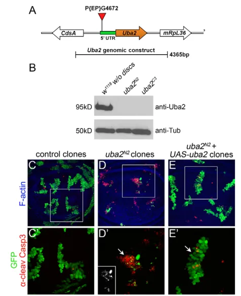
in the Drosophilagenome, the lack of more pleiotropic effects in aos1mutants could be explained by a partially redundant protein that interacts with Uba2 to maintain sufficient SUMOylation for all functions except mitosis. To test this hypothesis, we mutagenized uba2by imprecise excision of P{EP}G4672, a homozygous viable p-element insertion in the 5UTR (Fig. 6A). Following excision, we recovered uba2C3and uba2N2, independent recessive lethal lines that failed to complement deficiencies spanning the uba2region (1/56 and 0/58 expected transheterozygotes survived, respectively). Intriguingly, homozygosity for either uba2allele allowed normal larval growth and DNA replication, but resulted in early pupal lethality, with missing imaginal discs and severely reduced optic lobes (data not shown). Confirming that these effects were due to loss of uba2, the pupal lethality of both alleles was rescued by the presence of one copy of a 4.4 kb uba2genomic construct (Fig. 6A; n40 and 76 for uba2N2and uba2C3, respectively). Indeed, western blots showed that Uba2 protein was eliminated in both mutants (Fig. 6B; supplementary material Fig. S4B).
[image:6.612.60.485.58.242.2]In sum, the results above indicate that uba2C3and uba2N2are both strong alleles of uba2, and display identical phenotypes to loss-of-function alleles for its heterodimeric partner Aos1. Similarly, we found that uba2N2mutant clones exhibited the same phenotype as aos1clones, namely reduced growth and cell death (Fig. 6C-D; supplementary material Fig. S2D). This phenotype was fully rescued by expression of the uba2cDNA (Fig. 6E,E). In order to exclude the possibility that Aos1 and Uba2 might be individually sufficient to mediate basal E1 SUMO activity, we also generated aos1, uba2 double mutant animals. aos1N1, uba2C3 or aos1N1, uba2N2 homozygous animals displayed the same essential phenotype as their respective single mutants: normal larval growth with missing imaginal discs, reduced optic lobes and lethality in the late third instar (data not shown). We conclude that the phenotype of aos1-null mutant animals was not enhanced by disruption of uba2. Thus, even in the absence of both SUMO E1 components, the only visible phenotype was in mitotic proliferation.
Fig. 3. Aos1 is required for G2/M progression. (A)Schematic representation of the experimental flow. (B,C)Cell cycle profiles of GFP-positive, p35-expressing discs cells after FACS analysis. The percentage of cells with replicated DNA (G2/M) is significantly increased in aos1c06048mutant
cells (C) compared with that in wild-type cells (B); the percentages of mutant cells in G1 and S phase are reduced, respectively. The cell cycle profile of GFP-negative cells from discs containing wild-type or aos1c06048mutant mitotic clones resembles that of wild-type GFP-positive cells shown in B,
indicating that cell cycle phasing is not affected by p35expression. (D,E)Salivary glands from wild-type (D) and aos1c06048mutant (E) larvae stained
with anti--Tubulin (red), after a short incubation with EdU (green). Incorporation of EdU in DNA of aos1c06048mutant salivary glands is similar to
that of controls. The size of the salivary glands in both wild-type and mutant animals is variable as there are salivary glands of wild-type larvae that are significantly smaller and salivary glands of aos1mutant larvae that are same size or bigger than some wild type. These variations are probably caused by differences in crowding and subsequently on the nutrient availability for each larva.
D
E
V
E
LO
P
M
E
N
Global SUMOylation is severely reduced/ eliminated in E1 mutant animals
The experiments presented thus far indicate that either SUMOylation is only required for cell division, or that an alternative mechanism for SUMO activation can maintain basal SUMOylation in the absence of canonical E1 activity. To test whether there is still SUMOylation in the E1 mutants, we monitored global SUMOylation in selected third instar larval tissues for each of several genotypes. As SUMOylation is a highly unstable modification, we performed protein extraction in the presence of NEM (N-Ethylmaleimide), which inhibits the de-conjugating activity of SUMO proteases (Suzuki et al., 1999). Western blots of protein extracts with anti-SUMO antibody (Long and Griffith, 2000) showed that the global SUMOylation in aos1, uba2and aos1, uba2double mutants was nearly eliminated when compared with wild-type animals (Fig. 7A). Consistent with a failure to conjugate SUMO to substrates in the E1 complex mutants, we also observed that the free, unconjugated SUMO (~15
kDa) was visibly increased (Fig. 7A). We did not observe a major difference in SUMOylation levels between the different mutant genotypes; all of them exhibited a similarly strong reduction in bulk SUMOylation (Fig. 7A).
[image:7.612.51.276.58.382.2]To test whether the residual high molecular weight antibody signal observed in the mutant extracts reflected bona fide SUMOylation, we performed an additional experiment in which the SUMOylation levels were monitored in the presence and absence of NEM. We reasoned that if the residual signal reflected actual SUMOylated substrates, then it should be detectable with an Fig. 4. Accumulation of cyclins A and B inaos1c06048mutant cells.
[image:7.612.313.546.58.443.2](A)Diagram of an eye-antennal disc. (B-E) GFP-positive, p35-expressing MARCM clones in eye discs stained with -Cyclin A (red). (B,C,C) Wild-type control clones. The square in B (white) indicates magnification in C and C. Outside of the furrow, GFP-positive clone cells express Cyclin A (C,C). However, clone cells inside the furrow do not contain Cyclin A, similar to their GFP-negative neighbors (C). (D,E,E) aos1mutant cells in the furrow contain high levels of Cyclin A (yellow arrow in E) in contrast with the wild-type clone cells in C,C. (F-I’) Eye disc clones stained with -Cyclin B (red). (F,G,G) In wild-type control clones, the GFP-positive cells inside the furrow do not contain Cyclin B (G). (H,I,I) Similar to the observation for Cyclin A, GFP-positive aos1mutant cells inside the furrow contain high levels of Cyclin B (yellow arrow in I).
Fig. 5. Generation and characterization of aos1null alleles.
(A)Strategy for partial deletion of aos1and its replacement with the
mini whitegene in aos1N1and aos1N2. (B-E)Third instar larvae
expressing the Histone2Av-RFP fusion protein. In wild-type animals, imaginal disc complexes were visible in red owing to high nuclear density (B). By contrast, imaginal discs were missing in aos1c06048, aos1N1and aos1N2mutant larvae in C-E, respectively. (F-I)
Phalloidin-stained CNS preps from third instar larvae of the genotypes indicated above. The optic lobes were severely reduced in aos1mutant larvae (G-I) compared with controls (F). (J)Time course for puparium formation in
aos1c06048,aos1N1andaos1N2mutants. Four days after egg lay, ~60
larvae of each genotype were transferred in a new vial and puparium formation was monitored. (K)According to western blots, Aos1 protein was reduced beyond detection in aos1N1, aos1N2and aos1c06048
animals.
D
E
V
E
LO
P
M
E
N
independent antibody, and also sensitive to the absence of NEM. Indeed, western blot analysis with a second anti-SUMO antibody (Smith et al., 2004) showed a dramatic difference in SUMOylation levels between control and aos1mutants in the presence of NEM (Fig. 7B). In the absence of NEM, however, SUMO-conjugated proteins of high molecular weight were eliminated from aos1c06048 and aos1N1just as in controls, and the corresponding levels of free SUMO increased (Fig. 7C, longer exposure of 7B). These results clearly indicate that some substrate SUMOylation persists, even in aos1-null larvae (Fig. 7C), leading to two main conclusions. First, although SUMOylation is severely reduced in aos1, uba2and aos1, uba2double mutants, larval cells can still survive. Second, the presence of some residual SUMOylation in aos1-null mutants suggests that either maternal Aos1 or SUMO conjugation can persist over a period of several days, or that zygotic SUMOylation can take place independently of Aos1; perhaps this basal level of SUMOylation is sufficient to sustain all cellular processes short of mitotic proliferation.
SUMO pathway knockdown selectively disrupts mitotic cells in the eye disc
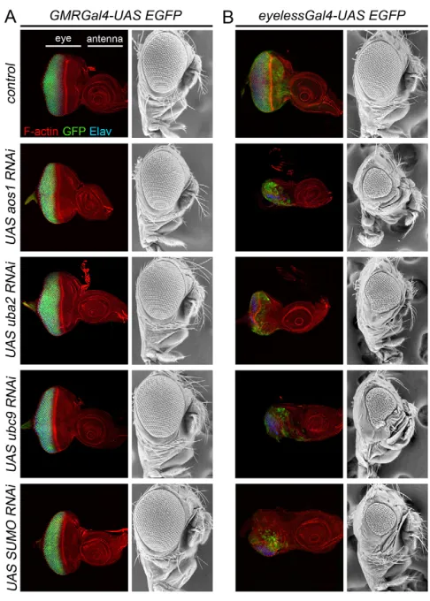
One of the most striking implications of the results presented thus far is that in vivo, SUMO E1 activity is strictly required for mitotic proliferation in the imaginal discs but is dispensable for growth, survival and homeostasis in non-dividing larval cells. To determine whether high levels of SUMOylation are also dispensible in post-mitotic imaginal cells, we compared the effects of knocking-down SUMO pathway components in proliferating and non-proliferating cell populations of the eye imaginal disc. We used the GMR-Gal4 line to drive RNAi expression in post-mitotic cells (behind the morphogenetic furrow), and eyeless-Gal4 to drive RNAi expression in both dividing and differentiated cells. Consistent with our earlier results, knocking down either aos1or uba2in post-mitotic cells had no effect on eye disc development, and no visible effect on differentiation of the adult eye (Fig. 8A). Under the control of eyeless-Gal4, however, expression of the very same RNAi constructs produced a dramatic reduction of imaginal disc size and severely reduced adult eyes (Fig. 8B). In many cases, head formation was abolished and adult flies never eclosed (data not shown). We observed similar phenotypes following RNAi knockdown of Ubc9 (the SUMO E2) and even SUMO itself (Fig. 8), indicating that disruption of cell proliferation but not cell viability, identity or differentiation is a common consequence of reduction in SUMOylation. As the adult eye morphology did not change when SUMO pathway components were knocked down in cells behind the furrow, we infer that the second mitotic wave (Wolff and Ready, 1991) was still completed by GMR-Gal4 -expressing mutant cells, perhaps because the expression of RNAi only initiates after exit from the furrow. Taken together, these results indicate that, in vivo, high levels of SUMOylation are primarily required for cell division and are less important for cell survival or homeostasis in non-dividing cells.
DISCUSSION
The E1 SUMO-activating enzyme controls cell cycle progression
In the present study, we show that aos1and uba2mutant animals exhibit late third instar/early pupal lethality and a severe disruption of imaginal disc and optic lobe development. Mosaic analysis of the aos1mutation in discs revealed that the mutant cells undergo apoptosis (Fig. 2), but when apoptosis is blocked, the aos1mutant imaginal disc cells arrest in G2/M (Fig. 3). The disrupted cell cycle of aos1 mutant disc cells could be the cause of the apoptosis observed in mutant clones (Fig. 2). However, it has recently been shown that SUMOylation regulates the JNK pathway as well, through Hipk kinase (Huang et al., 2011). This suggests that the apoptosis of SUMOylation-deficient cells is probably a synergistic result of many different causes.
[image:8.612.53.291.58.350.2]The accumulation of SUMOylation-deficient cells in G2/M has been observed both in yeast and in zebrafish upon Ubc9 depletion (Seufert et al., 1995; Nowak and Hammerschmidt, 2006). However, it is less clear what causes the cell cycle arrest. We tested activation of Cdk1 as defined by the presence of mitotic cyclins and found that both Cyclin A and Cyclin B were present in aos1 mutant cells (Fig. 4). Similarly, the mitotic cyclins are stabilized in yeast ubc9 (E2) mutants (Seufert et al., 1995). The effect of SUMOylation in degradation of mitotic cyclins could be direct, but so far there is no evidence that cyclins are modified by SUMO. By contrast, it has been reported that Pds1 (Securin) is also stabilized in smt3or ubc9mutant yeast cells (Dieckhoff et al., 2004). Pds1 is an APC (anaphase promoting complex) substrate like the mitotic Fig. 6. Novel alleles of uba2recapitulate theaos1phenotype.
(A)An uba2gene model, indicating the insertion of P{EP}G4672 in the 5 UTR. (B)Analysis of Uba2 protein levels by western blot. In both
uba2N2and uba2C3mutant animals, Uba2 protein levels were reduced
beyond detection. Blotting with anti -Tubulin was used for a loading control. (C-E)GFP-positive MARCM clones in wing discs, stained with -CC3 (red) and phalloidin (blue). (C)Control clones. (D)uba2N2mutant
clones. (E)uba2N2mutant clones rescued with UAS-uba2. (C-E)
Magnification of clones indicated by white boxes in C-E. (C)Control clones were intact and exhibited minimal anti-CC3 staining (red). (D)By contrast, uba2N2mutant clones in the wing pouch exhibited
frequent apoptosis (white arrow). Thus, similar to aos1c06048mutant
disc cells, uba2N2mutant clones were reduced in size. (E)uba2N2
mutant cell lethality was rescued by the expression of UAS-uba2. Rescued clones (white arrow) exhibited only sporadic apoptosis, similar to controls.
D
E
V
E
LO
P
M
E
N
cyclins, and the same study shows that more APC substrates are stabilized in SUMOylation-deficient cells. Thus, it seems that lack of SUMOylation affects APC-mediated proteolysis. However, it is not known whether any of the APC subunits are modified by SUMO, or if other factors that regulate APC activity require SUMOylation. In addition, we did not observe any evidence of metaphase arrest in aos1mutant disc cells, which indicates that there must be earlier defects in these cells apart from the inefficient degradation of substrates by the APC. Another possibility is that SUMOylation regulates activation of Cdk1 for mitotic entry by controlling its phosphorylation through the competitive functions of String phosphatase and Wee1 kinase. In yeast cells, the Wee1 homolog is a target of SUMOylation, and SUMO modification affects its protein stability (Simpson-Lavy and Brandeis, 2010). In addition, several other proteins that are known to have a role in mitosis are SUMOylation targets in Drosophila (Nie et al., 2009). Among these, Polo kinase is required for spindle formation, Topoisomerase 2 has a role in chromosome segregation and PP2A functions at multiple stages of the cell cycle (Nie et al., 2009).
In zebrafish ubc9mutants, a small percentage of mutant cells undergo an additional round of DNA replication, resulting in 8N DNA content (Nowak and Hammerschmidt, 2006). In yeast uba2 mutants, it is also reported that about 10% of affected cells have two buds (Dohmen et al., 1995). Hence, upon blockage of progression through mitosis, it seems that some mutant cells can
re-enter the cell cycle and become polyploid. These observations indicate that the rest of the cell cycle (but not mitosis) can be completed when SUMOylation is reduced or eliminated. We also found that endoreplication (S-phase progression) was not affected in larval cells of aos1mutant animals (Fig. 3). Collectively, our observations indicate that SUMOylation is strictly required for G2/M progression in vivo, with lesser or negligible requirements in G1/S.
[image:9.612.53.277.58.445.2]Our findings contrast with the observation that dsRNA knock down of SUMO in Drosophila S2 cells causes a G1/S arrest. Indeed, proteins required for the G1/S transition (Cdc2c/Cdk2) or DNA synthesis (RFC2, PCNA) are SUMOylation targets (Nie et al., 2009). This may indicate that SUMOylation is required for G1/S progression under certain conditions, although our data indicate that the demand for SUMOylation in G2/M progression is substantially higher in vivo. The fact that mutations in SUMOylation enzymes, such as Ubc9 in zebrafish (Nowak and Hammerschmidt, 2006) and yeast (Seufert et al., 1995), and Aos1 in Drosophila(this work), cause arrest in G2/M could be due to the fact that SUMOylation declines gradually in mutant cells as the enzyme is progressively consumed. A few E1 or E2 enzyme molecules could plausibly catalyze many SUMO conjugation reactions. As a result, cells would arrest when they reached the higher threshold of SUMOylation required for G2/M progression. By contrast, knockdown of SUMO itself in S2 cells (Nie et al., Fig. 7. SUMOylation is severely reduced but not completely eliminated in SUMO E1 mutant animals. (A)Western blot analysis monitoring global SUMOylation levels in wild-type control (w1118), aos1c06048, aos1N1, uba2N2and aos1N1,uba2N2mutant third instar
larvae. SUMOylated proteins and unconjugated SUMO were visualized with -SUMO1(Long and Griffith, 2000). SUMO-conjugated proteins
appeared as a high molecular weight smear in wild-type samples. However, in all mutant larvae, SUMO-conjugated protein levels were dramatically reduced, whereas the unconjugated (free) SUMO in the mutant samples was increased compared with that in the wild-type
without discssample (arrow). In wild-type animals that maintain the imaginal discs, there was also more unconjugated SUMO and probably more SUMOylated proteins. Furthermore, there was no significant difference in the SUMOylation pattern between aos1mutants, uba2
mutants and aos1, uba2double mutants. Blotting with anti--Tubulin was used as a loading control. (B,C)Western blot analysis of global SUMOylation levels in the presence or absence of NEM (inhibitor of SUMO proteases). SUMOylated proteins and free SUMO were visualized with -SUMO2(Smith et al., 2004). (C)A longer exposure of
the western blot in B. Comparison of SUMOylation levels between wild-type, aos1c06048and aos1N1samples that were prepared in presence of
NEM shows that global SUMOylation levels in aos1c06048and aos1N2
mutant animals were greatly reduced, with only a faint background smear remaining (B). However, in samples that were prepared in the absence of NEM, SUMO-conjugated proteins of high molecular weight (~90 kDa-250 kDa) are almost completely lost from all genotypes (C). Accordingly, the free SUMO is increased in NEM-negative samples compared with the NEM-positive samples of each genotype (black arrow in C). Thus, even in aos1-null larvae (aos1N1) there was residual
global SUMOylation. The protein bands that appear to be insensitive to NEM presence or absence could represent either very stable SUMO-conjugated proteins or simply non-specific bands.
D
E
V
E
LO
P
M
E
N
2009) may cause a more sudden drop in SUMOylation levels below the threshold required for G1/S progression and thus the cells arrest in G1/S.
A requirement for aos1in cell cycle progression can explain the developmental defects in the highly proliferative imaginal discs and optic lobes of aos1mutant larvae. Furthermore, these are tissues where the aos1gene is most highly expressed (Fig. 1G), reflecting the need for high levels of SUMOylation in dividing cells. Likewise, early embryonic divisions coincide with high levels of maternal aos1(Fig. 1G) and the rest of the components of SUMO conjugation pathway (Hashiyama et al., 2009). Maternal deposition of aos1 can thus explain the survival of zygotic null mutants through embryogenesis. Indeed, upon induction of aos1c06048 germline clones, we found that embryos generally failed to hatch (n401/473 embryos), similar to the results obtained for smt3(Nie et al., 2009). Germ line clones of the aos1N1-null allele failed to produce any eggs, probably owing to very early defects in oogenesis (data not shown).
One interesting aspect of the aos1phenotype was developmental delay and the formation of sporadic melanotic tumors. The formation of melanotic tumors has also been observed in lwr mutant larvae (Chiu et al., 2005; Huang et al., 2005), which indicates this may be a common consequence of disrupted SUMOylation. Melanotic tumors in lwr4-3/lwr5 and lwr5/lwr14 mutant larvae are caused by misregulation of the Toll/Dorsal pathway, which results in overproliferation of hemocytes and increased lamellocyte differentiation (Chiu et al., 2005). However, in lwr mutant animals with a stronger allelic combination (lwr4-3/lwr14), increased lamellocyte differentiation was observed without an increase in the total number of circulating hemocytes (Chiu et al., 2005). Melanotic tumor formation could still take place in these animals but with lower penetrance. Thus, melanotic tumors are not necessarily the result of hemocyte overproliferation, which is consistent with our findings that disrupted SUMOylation results in defects in cell cycle progression. Instead, an abnormal increase in lamellocyte differentiation due to Toll pathway misregulation could be the cause of the mild melanotic tumor formation observed in aos1c06048mutant larvae. Indeed, an increase in circulating lamellocytes was recently reported for aos1c06048 mutant larvae (Kalamarz et al., 2012). Nevertheless, we also cannot exclude the possibility that hemocytes in aos1mutant larvae could still be able to perform a few rounds of division before their maternal Aos1 protein is consumed and mitosis is blocked.
Aos1 and Uba2 appear to be dispensable in non-proliferating cells
In this study, we found that larval tissues comprising non-diving, endoreplicating cells were not overtly affected by Aos1 depletion. In fact, the aos1and uba2mutant phenotypes resembled those of several mutations that inhibit cell cycle progression and cause late third instar larval/early pupal lethality and defects in imaginal disc development (Gatti and Baker, 1989). This implies, unexpectedly, that aos1and uba2primarily function in cell cycle progression. We therefore monitored global SUMOylation levels in our mutants, and found that SUMOylation is dramatically reduced in aos1, uba2 and double mutant larvae, but not completely eliminated (Fig. 7). The remaining SUMOylation in aos1-null mutants could potentially be attributed to the function of residual maternal Aos1. However, we were not able to detect the presence of Aos1 by western blot. Another possibility is that the residual SUMOylation is a result of decreased deSUMOylation in aos1mutant animals in response to disrupted SUMO conjugation, and this leads to some persistently SUMO-modified substrates. An alternative explanation could be that the SUMO-activating function occurred independently of Aos1/Uba2, possibly by an E1 complex primarily assigned to another Ubiquitin-like molecule with low affinity for SUMO. However, there is no evidence so far of an E1 other than Aos1/Uba2 that can activate SUMO. In sum, our evidence suggests that endoreplicating larval cells have minimal requirements for SUMO conjugation and the residual SUMOylation observed in zygotic E1 mutants is sufficient for their survival and growth. Consistent with these findings, endoreplicating trophoblastic cells in ubc9 mutant mice embryos are viable and morphologically normal (Nacerddine et al., 2005).
[image:10.612.52.295.58.398.2]Supporting the idea of quantitatively lower requirements for SUMOylation outside of cell division, we found that RNAi-mediated disruption of aos1,uba2,ubc9and smt3 (SUMO) had no discernible effect on post-mitotic cells in the eye disc. In proliferating cells of the same tissue, however, the same constructs produced severe developmental defects (Fig. 8). Similarly, no Fig. 8. RNAi-knockdown of SUMO pathway components in the
developing eye disrupts dividing but not differentiated cells.
(A)GMR-Gal4drives expression of GFP in post-mitotic cells of the eye disc. The discs are stained with phalloidin (red) and -Elav (blue). Expression of RNAi against aos1and uba2(E1 enzyme components),
ubc9(E2 enzyme) and sumoin post-mitotic cells under the control of
GMRdriver had no effect on survival, patterning or differentiation of cells in the disc, and thus a normal adult eye is formed. (B)eyeless-Gal4
drives expression in both differentiating and dividing cells. Under the control of eyeless-Gal4, expression of the RNAi constructs against SUMO pathway components caused severe growth defects in the eye disc and consequently a severely reduced adult eye.
D
E
V
E
LO
P
M
E
N
obvious phenotype was observed when we used Elav-Gal4 to knock down aos1and uba2in all differentiated neurons of the entire animal (data not shown). Thus, as with endoreplicating larval cells, survival and homeostasis of differentiated cells in imaginal discs was not affected by reduction of SUMOylation. Overall these results imply that minimal SUMOylation levels may be sufficient to mediate non-mitotic functions of the pathway.
A reduced requirement for SUMOylation in non-dividing larval cells can explain the late third instar/early pupal lethality of aos1 and uba2mutants. However, the semi class of lwr(E2) mutants (Epps and Tanda, 1998) and smt3mutants (Nie et al., 2009) both die much earlier. In the case of E2 enzyme, lwr13/lwr13 homozygotes and lwr4-3/lwr13and lwr5/lwr13trans-heterozygotes both exhibit lethality at the third instar larval stage (Sun et al., 2003). lwr13is also molecular null as it deletes the entire lwrlocus (Sun et al., 2003). In the smt3 mutant case, lethality at earlier stages than the aos1 and uba2 mutants could be due to differential perdurance of the respective maternal proteins. An alternative explanation could be that there is an Aos1/Uba2-independent SUMO-activating mechanism that facilitates low level SUMOylation in aos1and uba2mutants, but cannot be useful upon elimination of SUMO in smt3mutants. Combined with previous observations in Drosophilaand other organisms, our results suggest that robust SUMOylation is essential for cell division but is required less for the cellular processes that mediate survival and homeostasis. Importantly, the selective requirement for SUMOylation in dividing cells could suggest a potential new avenue for therapeutic intervention in proliferative disease.
Acknowledgements
We thank Leslie Griffith, Albert Courey, Rosa Barrio, Ting Xie, Ali Shilatifard, Norbert Perrimon and Gary Struhl for kindly providing antibodies, fly stocks and plasmids. We also thank Jeff Haug for analyzing our FACS data, and Scott Hawley, Robb Krumlauf, Kausik Si, Laurence Florens and Ting Xie for advice and suggestions throughout the course of research.
Funding
This study was supported by the Stowers Institute for Medical Research.
Competing interests statement
The authors declare no competing financial interests.
Supplementary material
Supplementary material available online at
http://dev.biologists.org/lookup/suppl/doi:10.1242/dev.082974/-/DC1
References
Bischof, J., Maeda, R. K., Hediger, M., Karch, F. and Basler, K.(2007). An optimized transgenesis system for Drosophila using germ-line-specific phiC31 integrases. Proc. Natl. Acad. Sci. USA104, 3312-3317.
Bossis, G. and Melchior, F.(2006). SUMO: regulating the regulator. Cell Div.1, 13.
Bryant, P. J. and Schmidt, O.(1990). The genetic control of cell proliferation in Drosophila imaginal discs. J. Cell Sci.13 Suppl., 169-189.
Chiu, H., Ring, B. C., Sorrentino, R. P., Kalamarz, M., Garza, D. and Govind, S.(2005). dUbc9 negatively regulates the Toll-NF-kappa B pathways in larval hematopoiesis and drosomycin activation in Drosophila. Dev. Biol. 288, 60-72.
Clem, R. J., Fechheimer, M. and Miller, L. K.(1991). Prevention of apoptosis by a baculovirus gene during infection of insect cells. Science254, 1388-1390.
Dasso, M.(2008). Emerging roles of the SUMO pathway in mitosis. Cell Div.3, 5.
Dieckhoff, P., Bolte, M., Sancak, Y., Braus, G. H. and Irniger, S.(2004). Smt3/SUMO and Ubc9 are required for efficient APC/C-mediated proteolysis in budding yeast. Mol. Microbiol.51, 1375-1387.
Dohmen, R. J., Stappen, R., McGrath, J. P., Forrova, H., Kolarov, J., Goffeau, A. and Varshavsky, A.(1995). An essential yeast gene encoding a homolog of ubiquitin-activating enzyme. J. Biol. Chem.270, 18099-18109.
Edgar, B. A. and O’Farrell, P. H.(1990). The three postblastoderm cell cycles of Drosophila embryogenesis are regulated in G2 by string. Cell62, 469-480.
Edgar, B. A. and Orr-Weaver, T. L.(2001). Endoreplication cell cycles: more for less. Cell105, 297-306.
Epps, J. L. and Tanda, S.(1998). The Drosophila semushi mutation blocks nuclear import of bicoid during embryogenesis.Curr. Biol. 8, 1277-1280.
Foe, V. E., Odell, G. M. and Edgar, B. A.(1993). Mitosis and morphogenesis in the Drosophila embryo: point and counterpoint. In The Development of Drosophila melanogaster, pp. 149-300. Cold Spring Harbor, NY: Cold Spring Harbor Laboratory Press
Gatti, M. and Baker, B. S.(1989). Genes controlling essential cell-cycle functions in Drosophila melanogaster. Genes Dev.3, 438-453.
Geiss-Friedlander, R. and Melchior, F.(2007). Concepts in sumoylation: a decade on.Nat. Rev. Mol. Cell Biol. 8, 947-956.
Golic, K. G.(1991). Site-specific recombination between homologous chromosomes in Drosophila. Science252, 958-961.
Gong, W. J. and Golic, K. G.(2003). Ends-out, or replacement, gene targeting in Drosophila. Proc. Natl. Acad. Sci. USA100, 2556-2561.
Hashiyama, K., Shigenobu, S. and Kobayashi, S.(2009). Expression of genes involved in sumoylation in the Drosophila germline. Gene Expr. Patterns9, 50-53.
Hay, R. T.(2005). SUMO: a history of modification. Mol. Cell 18, 1-12.
Huang, H., Du, G., Chen, H., Liang, X., Li, C., Zhu, N., Xue, L., Ma, J. and Jiao, R.(2011). Drosophila Smt3 negatively regulates JNK signaling through sequestering Hipk in the nucleus. Development138, 2477-2485.
Huang, L., Ohsako, S. and Tanda, S.(2005). The lesswright mutation activates Rel-related proteins, leading to overproduction of larval hemocytes in Drosophila melanogaster. Dev. Biol. 280, 407-420.
Johnson, E. S.(2004). Protein modification by SUMO. Annu. Rev. Biochem.73, 355-382.
Johnson, E. S. and Gupta, A. A.(2001). An E3-like factor that promotes SUMO conjugation to the yeast septins. Cell106, 735-744.
Johnson, E. S., Schwienhorst, I., Dohmen, R. J. and Blobel, G.(1997). The ubiquitin-like protein Smt3p is activated for conjugation to other proteins by an Aos1p/Uba2p heterodimer. EMBO J.16, 5509-5519.
Kagey, M. H., Melhuish, T. A. and Wotton, D.(2003). The polycomb protein Pc2 is a SUMO E3. Cell113, 127-137.
Kagey, M. H., Melhuish, T. A., Powers, S. E. and Wotton, D.(2005). Multiple activities contribute to Pc2 E3 function. EMBO J.24, 108-119.
Kalamarz, M. E., Paddibhatla, I., Nadar, C. and Govind, S.(2012). Sumoylation is tumor-suppressive and confers proliferative quiescence to hematopoietic progenitors in Drosophila melanogaster larvae. Biol. Open1, 161-172.
Kirsh, O., Seeler, J. S., Pichler, A., Gast, A., Muller, S., Miska, E., Mathieu, M., Harel-Bellan, A., Kouzarides, T., Melchior, F. et al.(2002). The SUMO E3 ligase RanBP2 promotes modification of the HDAC4 deacetylase. EMBO J.21, 2682-2691.
Lee, T. and Luo, L.(2001). Mosaic analysis with a repressible cell marker (MARCM) for Drosophila neural development. Trends Neurosci.24, 251-254.
Long, X. and Griffith, L. C.(2000). Identification and characterization of a SUMO-1 conjugation system that modifies neuronal calcium/calmodulin-dependent protein kinase II in Drosophila melanogaster. J. Biol. Chem.275, 40765-40776.
Markstein, M., Pitsouli, C., Villalta, C., Celniker, S. E. and Perrimon, N.
(2008). Exploiting position effects and the gypsy retrovirus insulator to engineer precisely expressed transgenes. Nat. Genet. 40, 476-483.
Meulmeester, E. and Melchior, F.(2008). Cell biology: SUMO. Nature452, 709-711.
Morata, G. and Ripoll, P.(1975). Minutes: mutants of drosophila autonomously affecting cell division rate. Dev. Biol. 42, 211-221.
Mukhopadhyay, D. and Dasso, M.(2007). Modification in reverse: the SUMO proteases. Trends Biochem. Sci. 32, 286-295.
Nacerddine, K., Lehembre, F., Bhaumik, M., Artus, J., Cohen-Tannoudji, M., Babinet, C., Pandolfi, P. P. and Dejean, A.(2005). The SUMO pathway is essential for nuclear integrity and chromosome segregation in mice. Dev. Cell9, 769-779.
Nie, M., Xie, Y., Loo, J. A. and Courey, A. J.(2009). Genetic and proteomic evidence for roles of Drosophila SUMO in cell cycle control, Ras signaling, and early pattern formation. PLoS ONE4, e5905.
Nowak, M. and Hammerschmidt, M.(2006). Ubc9 regulates mitosis and cell survival during zebrafish development. Mol. Biol. Cell 17, 5324-5336.
Pandey, R., Heidmann, S. and Lehner, C. F.(2005). Epithelial re-organization and dynamics of progression through mitosis in Drosophila separase complex mutants. J. Cell Sci.118, 733-742.
Pichler, A., Gast, A., Seeler, J. S., Dejean, A. and Melchior, F.(2002). The nucleoporin RanBP2 has SUMO1 E3 ligase activity. Cell108, 109-120.
Rosenblatt, J., Raff, M. C. and Cramer, L. P.(2001). An epithelial cell destined for apoptosis signals its neighbors to extrude it by an actin- and myosin-dependent mechanism.Curr. Biol. 11, 1847-1857.
Sanchez, J., Talamillo, A., Lopitz-Otsoa, F., Perez, C., Hjerpe, R., Sutherland, J. D., Herboso, L., Rodriguez, M. S. and Barrio, R.(2010). Sumoylation modulates the activity of Spalt-like proteins during wing development in Drosophila. J. Biol. Chem.285, 25841-25849.
Seufert, W., Futcher, B. and Jentsch, S.(1995). Role of a ubiquitin-conjugating enzyme in degradation of S- and M-phase cyclins. Nature373, 78-81.
Simpson-Lavy, K. J. and Brandeis, M.(2010). Cdk1 and SUMO regulate Swe1 stability. PLoS ONE5, e15089.
D
Smith, M., Bhaskar, V., Fernandez, J. and Courey, A. J.(2004). Drosophila Ulp1, a nuclear pore-associated SUMO protease, prevents accumulation of cytoplasmic SUMO conjugates. J. Biol. Chem.279, 43805-43814.
Sun, X., Huang, L., Van Doren, M. R. and Tanda, S.(2003). isolation of amorphic alleles of the lesswrightgene by P element-mediated male recombination in Drosophila melanogaster. Dros. Inf. Serv.86,79-83.
Suzuki, T., Ichiyama, A., Saitoh, H., Kawakami, T., Omata, M., Chung, C. H., Kimura, M., Shimbara, N. and Tanaka, K.(1999). A new 30-kDa ubiquitin-related SUMO-1 hydrolase from bovine brain. J. Biol. Chem.274, 31131-31134.
Takanaka, Y. and Courey, A. J.(2005). SUMO enhances vestigial function during wing morphogenesis. Mech. Dev.122, 1130-1137.
Talamillo, A., Sanchez, J., Cantera, R., Perez, C., Martin, D., Caminero, E. and Barrio, R.(2008). Smt3 is required for Drosophila melanogaster metamorphosis. Development135, 1659-1668.
Tatham, M. H., Kim, S., Jaffray, E., Song, J., Chen, Y. and Hay, R. T.(2005). Unique binding interactions among Ubc9, SUMO and RanBP2 reveal a mechanism for SUMO paralog selection.Nat. Struct. Mol. Biol. 12, 67-74.
Thibault, S. T., Singer, M. A., Miyazaki, W. Y., Milash, B., Dompe, N. A., Singh, C. M., Buchholz, R., Demsky, M., Fawcett, R., Francis-Lang, H. L. et al.(2004). A complementary transposon tool kit for Drosophila melanogaster using P and piggyBac. Nat. Genet. 36, 283-287.
Wolff, T. and Ready, D. F.(1991). The beginning of pattern formation in the Drosophila compound eye: the morphogenetic furrow and the second mitotic wave. Development113, 841-850.
Wolff, T. and Ready, D. F.(1993). Pattern formation in the Drosophilaretina. In The Development of Drosophila melanogaster (ed. M. Bate and A. Martinez-Arias), pp. 1277-1326. Cold Spring Harbor, NY: Cold Spring Harbor Laboratory Press.
