D
E
V
E
LO
P
M
E
N
T
INTRODUCTION
The central nervous system (CNS) develops from the neuroectoderm that is originally formed by a sheet of symmetrically dividing neuroepithelial cells. These evolve to transform into radial glia cells that specify into different lineages, subtypes that give rise to neurons or generate astroglia (Malatesta et al., 2000). Postnatally, the latter lineage of radial glia transforms into astrocytes (Doetsch, 2003; Gotz and Huttner, 2005). Specialized astrocytes in the subependymal zone (SEZ) of the lateral ventricle or in the subgranular zone of the hippocampus serve as slowly dividing neural stem cells (NSCs) in the adult CNS (Alvarez-Buylla et al., 2001). Factors that regulate the complex pathway of NSC development begin to emerge. In their site of origin, NSCs are surrounded by a particular cellular environment and exposed to differentiation signals such as Notch and Delta ligand-receptor interactions (Campos et al., 2006). In addition, NSCs of the mouse respond to fibroblast growth factor (FGF) and epidermal growth factor (EGF) and sequentially express the corresponding receptors FGFR and EGFR with ongoing maturation (Reynolds and Weiss, 1996). Furthermore, it has been shown that NSCs are located in a structured pericellular environment that comprises distinct extracellular matrix (ECM) constituents (Gates et al., 1995; Quinones-Hinojosa et al., 2006). Substantial evidence has been provided in recent years that complex ECM milieus are involved in the regulation of CNS development and plasticity and in the response to lesion. The neural ECM is composed both of glycoproteins, such as tenascin-C, and proteoglycans (Garwood et
al., 2001). The latter can be divided in heparan sulfate proteoglycans (HSPGs), with a preferential membrane association, and the chondroitin sulfate proteoglycans (CSPGs) that occur primarily as constituents of the pericellular space (Bandtlow and Zimmermann, 2000). HSPGs play an important role as accessory factors in signaling processes, e.g. by exposing growth factors such as bFGF (also known as FGF2 – Mouse Genome Informatics) to the FGFR. By comparison, less is known about the CSPGs. We have recently shown that the Mab-473HD epitope, a unique chondroitin sulfate (CS) structure, is expressed in the germinal layers during mouse forebrain development (von Holst et al., 2006), and the release of CSPGs from NSCs has been reported (Ida et al., 2006). The 473HD epitope, also known as the DSD-1 epitope (Faissner et al., 1994), is expressed on the CSPG phosphacan that represents a soluble, released splice variant encoded by the receptor protein tyrosine phosphatase  (Rptp) gene (the protein is also known as phosphacan, DSD-1-PG and PTPRZ1 – Mouse Genome Informatics). The large RPTP-receptor is expressed by NSCs and radial glia during development of the CNS and exposes the 473HD structure (Garwood et al., 1999). Interestingly, recent studies have proposed that CSPGs play important roles in the adult CNS as inhibitors of synaptic plasticity and of axonal regeneration in scar tissues (Bradbury et al., 2002; Pizzorusso et al., 2002). In the light of these findings, treatment strategies based on the application of chondroitinase ABC (ChABC), an enzyme that degrades the CS-glycosaminoglycan (CS-GAG) complement of CSPGs, have been developed (Houle et al., 2006). On the other hand, evidence has emerged that neural progenitor cells are recruited to various types of lesion in the adult CNS (Arvidsson et al., 2002; Magavi et al., 2000), which raises the question as to whether ChABC application might impact on NSC development in the lesioned tissue (Dobbertin et al., 2003). We have, therefore, systematically addressed the question of whether ChABC affects stem cell behavior. We show here that the selective elimination of CS-GAGs with ChABC, both in vivo and in vitro, reduces NSC proliferation and the differentiation of radial glia to neurons, whereas it favors the maturation of the gliogenic subtype
Chondroitin sulfate glycosaminoglycans control
proliferation, radial glia cell differentiation and
neurogenesis in neural stem/progenitor cells
Swetlana Sirko1,*, Alexander von Holst1,*, Andrea Wizenmann2, Magdalena Götz2,3and Andreas Faissner1,†
Although the local environment is known to regulate neural stem cell (NSC) maintenance in the central nervous system, little is known about the molecular identity of the signals involved. Chondroitin sulfate proteoglycans (CSPGs) are enriched in the growth environment of NSCs both during development and in the adult NSC niche. In order to gather insight into potential biological roles of CSPGs for NSCs, the enzyme chondroitinase ABC (ChABC) was used to selectively degrade the CSPG glycosaminoglycans. When NSCs from mouse E13 telencephalon were cultivated as neurospheres, treatment with ChABC resulted in diminished cell
proliferation and impaired neuronal differentiation, with a converse increase in astrocytes. The intrauterine injection of ChABC into the telencephalic ventricle at midneurogenesis caused a reduction in cell proliferation in the ventricular zone and a diminution of self-renewing radial glia, as revealed by the neurosphere-formation assay, and a reduction in neurogenesis. These observations suggest that CSPGs regulate neural stem/progenitor cell proliferation and intervene in fate decisions between the neuronal and glial lineage.
KEY WORDS: Chondroitinase ABC, Astrocyte differentiation, Stem cell niche, Proteoglycans
Development 134, 2727-2738 (2007) doi:10.1242/dev.02871
1Chair of Cell Morphology and Molecular Neurobiology, Ruhr-University Bochum,
Building NDEF 05/339, Universitaetsstrasse 150, D-44780 Bochum, Germany. 2
GSF-Institute for Stem Cell Research, Ingolstädter Landstraße 1, D-85764 Neuherberg, Germany. 3Institute of Physiology, University of Munich, Schillerstaße 46, D-80634
München, Germany.
*These authors contributed equally to this work
†Author for correspondence (e-mail: andreas.faissner@ruhr-uni-bochum.de)
D
E
V
E
LO
P
M
E
N
T
of radial glia and the formation of astrocytes. This implies a role of CS-GAGs in the regulation of growth and differentiation factors for NSCs.
MATERIALS AND METHODS Mice
All experiments were performed with embryos derived from timed pregnancies of the NMRI or C57/Bl6 mouse strains obtained from Charles River Laboratories (Sulzfeld, Germany). The age of the embryos was determined according to the staging criteria of Theiler, in which E13 corresponds to TS 21 (Bard et al., 1998). Experimental procedures were performed in accordance with the Society for Neuroscience and European Union guidelines and were approved by the animal care and utilization committees at the GSF and Ruhr University.
Immunological reagents
The following primary antibodies were used. Monoclonal antibodies: 473HD (rat IgM) (Faissner et al., 1994), RC2 (mouse IgM; Developmental Studies Hybridoma Bank, University of Iowa, USA), anti-E-cadherin (mouse IgG; Santa Cruz), anti-nestin (mouse IgG; Chemicon, Temecula, CA), anti-III-tubulin (mouse IgG; Sigma, St Louis, MO) and anti-BrdU (mouse IgG; Roche, Mannheim, Germany). Polyclonal antibodies (all rabbit) against: DSD-1-PG/phosphacan [referred to as pk-anti-phosphacan; Batch KAF 13(4); (Faissner et al., 1994)], GFAP (Dako), atypical PKC (aPKC; BD Science), phospho-histone H3 (PH3), laminin, brain-lipid-binding protein (BLBP) and GLAST (all Chemicon). Secondary antibodies: subclass-specific CY2- or CY3-coupled anti-mouse, anti-rat and anti-rabbit antibodies (all Dianova, Hamburg, Germany).
Neurosphere (Nsph) cultures
Pregnant animals were sacrificed by cervical dislocation. The embryos were removed and transferred to phosphate-buffered saline (PBS). Subsequently, the embryonic brains were dissected, the meninges, hippocampi and olfactory bulbs were removed, and the cerebral cortices (Cor) and the ganglionic eminence (GE) were prepared in minimal essential medium (MEM, Sigma). Tissues were enzymatically digested with 0.05% (w/v) trypsin-EDTA in HBSS (Invitrogen, Karlsruhe, Germany) for 10 minutes at 37°C to obtain E13 neural cell suspensions. Digestion was stopped by the addition of 1 ml ovomucoid [1 mg/ml trypsin inhibitor (Sigma), 50 g/ml BSA (Sigma), 40 g/ml DNaseI (Worthington), in L-15 medium (Sigma)]. After centrifugation for 5 minutes at 1000 rpm (212 g), the cell pellets were suspended in Nsph medium consisting of DMEM/F12 (1:1) with 0.2 mg/ml L-glutamine (all Sigma), 2% (v/v) B27, 100 U/ml penicillin, 100 g/ml streptomycin (all Invitrogen). Embryonic NSCs were cultured at 37°C, 6% CO2at a cell density of 105cells/ml (bulk culture) in Nsph medium in the
presence of EGF and bFGF, both at 20 ng/ml (Preprotech, Tebu, Germany), unless otherwise indicated. In general, bFGF-containing cultures were supplemented with 0.5 U/ml heparin (Sigma). In parallel sets, 50 mU/ml chondroitinase ABC (ChABC; EC 4.2.2.4; Sigma) or 50 mU/ml keratanase (Calbiochem, La Jolla, CA) were added. The formation of secondary Nsphs was assessed in clonal density assays by plating 2⫻104cells in 6 ml of
FGF-and EGF-containing medium. The formation of Nsphs was quantified after 7 days in vitro (div) by size-dependent (<50, 100, 150, 200 and >200 m in diameter) counting of Nsphs in the entire dish area (clonal density assays) or in ten randomly selected visual fields.
Immunocytochemistry
For immunocytochemical stainings, the acutely dissociated cells originating from forebrain tissues, from cortical and striatal Nsphs, or obtained by immunopanning using the Mab 473HD (von Holst et al., 2006), were plated in FCS-containing medium [1% (v/v), Seromed] at a density of 5000 cells/well in four-well dishes (Greiner) coated with 10 g/ml polyornithine (Sigma) and incubated in a humidified atmosphere with 6% (v/v) CO2at
37°C for 1 hour. Cell suspensions were allowed to adhere, washed twice for 5 minutes in Krebs-Ringer-Hepes buffer [KRH with 0.1% (w/v) BSA, pH 7.4] at room temperature (RT) and incubated for 20 minutes at RT with Mab 473HD diluted in KRH/A (1:250). After washing twice for 5 minutes in KRH, the cells were fixed with 4% (w/v) paraformaldehyde (PFA) in PBS
for 10 minutes at RT, washed twice with PBT1 [PBS containing 1% (w/v) BSA and 0.1% (w/v) Triton X-100] and incubated for 30 minutes at RT with antibodies against the intracellular epitopes RC2 (1:500), nestin (1:500), III-tubulin (1:300), BLBP (1:1000), GLAST (1:1000) and GFAP (1:250), all diluted in PBT1. After three further washes with PBS/A, the cells were incubated for 30 minutes at RT with specific fluorochrome-conjugated secondary antibodies to detect the various primary antibodies. The last incubation step included bisbenzimide (1:10,000; Sigma) to label cell nuclei. After final washing with PBS, specimens were mounted in PBS/glycerol (1:1) and viewed under an Axiophot II fluorescence microscope (Zeiss, Oberkochen, Germany) using UV epifluorescence. To identify differentiated cell types after the exposure of cultures to 50 mU/ml ChABC or 50 mU/ml keratanase, the same immunostaining protocol was carried out after 24 hours of incubation. For differentiation assays, individual Nsphs of 150-250 m diameter were transferred to four-well dishes (Greiner) sequentially coated with polyornithine (10 g/ml) and laminin-1 (10 g/ml; Tebu, Germany) and incubated in Nsph medium containing 1% (v/v) FCS (see above) for a further 6 days in a humidified atmosphere with 6% (v/v) CO2at 37°C. The
differentiated cell types were identified by immunocytochemistry using antibody markers as described in the previous section.
Intracerebroventricular injections (ICVIs) in utero
ICVI into telencephalic ventricles of E13 embryos in utero was performed with timed-pregnant C57/Bl6 mice. Animals were anesthetized by intraperitoneal injection of 0.1 ml per 10 g body weight of the narcotic mixture 0.5 mg/kg medetomidine, 5 mg/ml midazolam, 0.05 mg/kg fentanyl hexal. Uterine horns were exposed by midline laparotomy and the ICVIs were performed through the uterine wall at the anterior end of the embryonic forebrain using fine-pulled microcapillaries (borosilicate glass capillaries, 1.5⫻0.86 mm; gc150F-10, Harvard apparatus). ChABC (10 mU/l) was injected into the lateral ventricles of all embryos in one uterine horn. Keratanase (10 mU/l) or artificial cerebrospinal fluid (ACSF) controls were injected into the lateral ventricles of the embryos in the second uterine horn. Thereafter, the uterus was reinstated in its physiological site. The incisions of the abdominal muscle and skin were stitched with separate sutures. Finally, the anesthesia was reversed by intraperitoneal application of antisedate (2.5 mg/kg antisedan, 0.5 mg/kg flumacenil, 1.2 mg/kg naloxone), and animals were left to recover in a clean cage. Twenty-four hours after injection, pregnant mice were sacrificed by cervical dislocation.
Immunohistochemistry
The embryos were removed and transferred to PBS (pH 7.4). Subsequently, the capita were immersion fixed overnight in 4% (w/v) PFA in PBS at 4°C. Thereafter, tissues were cryoprotected overnight in 30% (w/v) sucrose in water, embedded in Tissue Freezing Medium (Jung, Nussloch, Germany) and frozen on dry ice. Frontal sections (12-14 m) were cut on a cryostat (Leica). For immunohistochemistry, slides were rehydrated in PBS with 1.7% (w/v) NaCl and 10% (v/v) normal goat serum (Dianova) for 1 hour at RT and then incubated overnight (4°C) with one of the following primary antibodies: 473HD (1:500), RC2 (1:500), anti-nestin (1:500), anti- III-tubulin (1:300), anti-BLBP (1:1000), anti-PH3 (1:100), anti-GLAST (1:1000), anti-E-cadherin (1:50), anti-aPKC(1:50) or anti-laminin (1:80). Subsequently, the sections were washed four times for 5 minutes in PBS and incubated with appropriate secondary antibodies [1:500 in PBS, 0.1% (w/v) BSA)] for 2 hours at RT. After incubation with the secondary antibodies, cell nuclei were labeled with bisbenzimide (diluted 1:10,000). After three further washes in PBS, the sections were mounted in Immumount (Thermo Electron, Dreieich, Germany). For cryosectioning, Nsphs were allowed to settle in 15 ml Falcon tubes for 10 minutes before the culture medium was removed and replaced with 4% (w/v) PFA in PBS for 40 minutes at RT. After fixation, the Nsphs were cryoprotected with 30% (w/v) sucrose for 4 hours at 4°C. Finally, Nsphs were embedded in Tissue Freezing Medium, sectioned at 14 m on a cryostat (Leica) and processed for immunohistochemistry as described for embryonic brain sections above.
BrdU pulse-labeling
D
E
V
E
LO
P
M
E
N
T
Nsphs were dissociated, the single-cell suspension was plated and individual cells were immunocytochemically stained 1 hour later, as described above and according to the supplier’s protocol (BrdU Labeling and Detection Kit I, Roche). For analysis of cell proliferation in vivo, the label was introduced by intraperitoneal injection of 10 mg BrdU/100 g body weight 1 hour prior to removal of the litter. After cryoprotection of embryonic tissues, cryosections were cut at 14-18 m, boiled for 5 minutes in 0.01 M citrate buffer (pH 6.0) and washed twice in PBS before incubation with anti-BrdU (1:20) at 4°C overnight. Primary antibodies were detected using appropriate secondary antibodies, as described above.
Microscopy
The immunolabeling experiments were analyzed using a fluorescence microscope equipped with UV epifluorescence (Axioplan 2 imaging, Zeiss). Images were captured with a digital camera and documented using the Axiovision 3.1 and/or 4.2 programs (AxioCamHRc, Zeiss). In some cases, confocal laser scanning microscopy was applied (LSM 510 meta, Zeiss). Standard phase-contrast images of living cells were taken using a digital camera (DP10, Olympus) on an inverted CK40 microscope (Olympus).
Counting and statistical analysis
The proportion of single- and double-immunolabeled cells collected from dissociated Nsphs or selectively isolated cells was determined by counting 200 to 400 individual cells per independent experiment and antibody. Statistical significance of differences (see figure legends) observed between distinct experimental groups was assessed using an unpaired, two-tailed Student’s t-test.
RESULTS
Digestion of chondroitin sulfates reduces
proliferation rates of embryonic forebrain neural stem/progenitor cells and impairs neurosphere formation
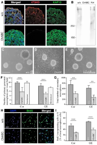
First, we enzymatically removed the CS-GAGs from the cell surface of telencephalic cell suspensions obtained from the ventral and dorsal forebrain at embryonic day (E) 13. Deglycanation was achieved with ChABC, which specifically cleaves the CS polymer close to its serine linkage in the core glycoprotein, leaving a carbohydrate stub that bears characteristics of the CS-GAG chain type, e.g. C0S, C4S or C6S (Sugahara et al., 2003). NSCs were grown as freely floating Nsphs in defined medium containing the growth factors EGF and FGF and were continuously exposed to the ChABC enzyme as added to the culture medium. The formation of Nsphs was monitored either in the presence or in the absence of ChABC. The ongoing digestion of CS-GAGs compromised the generation of Nsphs and caused a nearly complete settling of the Nsphs on the substrate of the culture dish, where a massive emigration of Nsph-derived cells was seen (Fig. 1). The efficiency of CS-GAG removal was assessed using antibodies 473HD and anti-phosphacan (KAF13), which detect a CS-GAG structure and the core glycoproteins of the Rptp gene products, respectively (Faissner et al., 1994). Whereas control cryosections of Nsphs displayed the 473HD epitope, its complete loss was observed when Nsphs were grown in the presence of ChABC. In this latter case, many cells still retained the core proteins of DSD-1-PG/RPTP- (Fig. 1A). Analogous results were obtained when Nsph extracts were examined by western blotting (Fig. 1B). Culture in the absence of enzyme or in the presence of keratanase, an enzyme that removes keratan sulfates but does not affect the 473HD epitope (Faissner et al., 1994), served as controls (Fig. 1).
The selective cleavage of the CS-GAGs decreased the number of Nsphs per visual field from 9.3±1.7 to 5.1±1.1 (n=10; P<0.001, unpaired t-test) in cortex-derived cultures and from 9.1±1.5 to 4.2±1.1 (n=10; P<0.001, unpaired t-test) in striatum-derived cultures
(Fig. 1F and see Fig. S1 in the supplementary material). Parallel experiments were performed with keratanase (50 mU/ml) instead of ChABC. The removal of keratan sulfates did not lead to substrate adherence (Fig. 1E) and the rate of Nsph formation was indistinguishable from the control (Fig. 1F). These findings suggested that CS-GAGs are required to maintain Nsphs in a free-floating state and might also influence neural stem/progenitor cell maintenance. To examine the role of CS-GAGs in Nsph formation and maintenance, an equal number of cells from ChABC-treated and control primary Nsphs were examined at clonal density (see methods) for the generation of secondary Nsphs. According to the view that only the self-renewing stem cells of the primary Nsphs would give rise to secondary Nsphs, this assay allows determination of the number of self-renewing stem cells present in the primary Nsph population. Using this approach, twice as many secondary Nsphs originated from cell suspensions derived from untreated primary cortical and striatal Nsphs, than from Nsphs that had been exposed to ChABC (Fig. 1G).
To further monitor whether not only self-renewal, but also proliferation in general, are affected, BrdU was added to the Nsph cultures 15 hours prior to fixation. The number of BrdU-positive cells decreased significantly in cortical or striatal Nsphs upon ChABC treatment (Fig. 1H,I). In conclusion, chondroitin sulfates promote proliferation and self-renewal of early telencephalic stem/progenitor cells grown in Nsph cultures.
D
E
V
E
LO
P
M
E
N
T
Digestion of chondroitin sulfates inhibits neurogenesis and increases gliogenesis
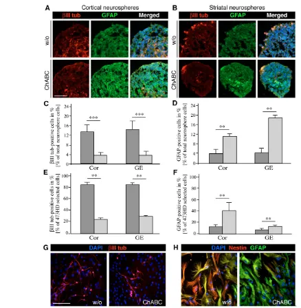
In order to examine how this altered precursor heterogeneity might affect the generation of specific cell types, we examined  III-tubulin-positive neurons and GFAP-III-tubulin-positive astrocytes in Nsphs. Cryosections of E13 cortical (Fig. 3A) or striatal (Fig. 3B) Nsphs kept in the presence of ChABC displayed an attenuated III-tubulin immunoreactivity, concomitant with an elevation of GFAP immunolabeling. The few differentiated cell types in Nsphs maintained under proliferating conditions are in line with previous findings (Campos et al., 2004; von Holst et al., 2006). A striking feature with regard to topography was that postmitotic neurons were located on the outer surface of control and treated striatal Nsphs rather than positioned in the core region, as had been reported previously for
cortical Nsphs (Campos et al., 2004). Quantitative assessment revealed a 4-fold reduction in the number of neurons amongst cortical or striatal Nsph cells (Fig. 3C), whereas the astroglial population was expanded 3- to 4-fold (Fig. 3D, Table 1). Taken together, these data suggest that chondroitin sulfates promote the proliferation or fate of neurogenic progenitors in this culture system.
[image:4.612.249.563.60.525.2]In order to examine a defined population of precursors in this culture system, we used immunopanning with Mab 473HD, which is known to enrich neurogenic neural stem/progenitor cells (von Holst et al., 2006). In the immunoselected cell populations obtained from cortical and striatal Nsphs, the 473HD epitope and the radial glia marker BLBP were coexpressed by up to 96% of the sorted cells, as previously shown. When this population was cultivated for 2 days in vitro in serum-containing medium in the presence of ChABC, Fig. 1. Cleavage of CS-GAGs reduces the
proliferation rate of mouse E13
telencephalic neural stem/progenitor cells and interferes with the generation of neurospheres. The efficacy of the CS-GAG removal after ChABC treatment of Nsph cultures was controlled by double immunofluorescence and western blot analyses. (A) Cryosections obtained from Nsphs grown for 7 days in vitro (div) in the absence (w/o, upper panels) or presence (lower panels) of 50 mU ChABC were immunostained with 473HD and pk-anti-phosphacan antibodies (KAF13) that detect the unique DSD-1 epitope and the proteins encoded by the Rptpgene, respectively. The 473HD epitope was strongly expressed in the outer cell layers of the Nsph under control conditions and was absent after ChABC treatment, which resulted in enhanced reactivity of the polyclonal antibodies with the RPTP-protein (KAF13). (B) Western blot analysis confirmed the efficient removal of the 473HD epitope by ChABC, which was not achieved by keratanase (Ker) treatment. (C-E) Phase-contrast micrographs documenting the formation of Nsphs grown in serum-free medium in the presence of EGF and bFGF (C) and upon addition of ChABC (D) or keratanase (E) after 5 div. ChABC treatment caused an almost complete settling of Nsphs on the culture substrate and subsequent cellular outgrowth. (F) Quantification of the numbers of cortical (Cor) and striatal (GE) Nsphs present per visual field in bulk cultures. Note the significant decrease in the number of primary Nsphs grown in the presence of ChABC in comparison with control (w/o) or keratanase-treated cultures. (G) The total number of secondary Nsphs generated from cortical and striatal control or ChABC-treated primary Nsphs was determined in clonal density assays. In comparison with control cultures (w/o), the number of secondary Nsphs obtained from primary ChABC-treated Nsphs appeared significantly reduced. (H) Photomicrographs of dissociated, BrdU-labeled primary control (w/o, upper panels) and ChABC-treated (lower panels) Nsphs that had received a 15-hour BrdU pulse. (I) The fraction of BrdU-incorporating cycling cells in H was
D
E
V
E
LO
P
M
E
N
T
neurogenesis was strongly compromised. Thus, the number of neurons originating from the 473HD-positive cell population dropped substantially in both cortex or striatum-derived Nsphs (Fig. 3E). In addition, a simultaneous preponderance of astroglial cell differentiation was obvious and more pronounced for cortical Nsph-derived 473HD-positive progenitors (Fig. 3F). These observations revealed that CS-GAGs are essential for the timely differentiation of the neurogenic 473HD/BLBP-positive radial glia into neurons.
The implication of CS-GAGs in neuronal versus glial fate decisions was further studied in the Nsph differentiation assay. Under these conditions also, ChABC exposure caused a dramatic decrease in differentiation into neurons (Fig. 3G) and a concomitant increase in the number of astrocytes that were defined as GFAP-positive and nestin-negative (Fig. 3H). The proportion of nestin- and  III-tubulin-positive cells was diminished by half after 6 days in vitro (div), whereas the number of GFAP-positive astroglia doubled during this time period (see Fig. S2 in the supplementary material). Analogous results were obtained for cortical and striatal Nsph differentiation
assays and were not apparent in the presence of keratanase. Taken together, the evidence collected in three independent lines of investigation concurs to support the conclusion that CS-GAGs are crucial components implicated in proliferation and lineage decisions of neural precursor cells in vitro. In the light of our findings, we propose that CS-GAGs might be part of the control machinery that delays the onset of gliogenesis and promotes self-renewal of stem cells and neurogenic precursors.
[image:5.612.155.556.61.310.2]Chondroitin sulfates are essential components for generation of the neurogenic precursor and their differentiation in the embryonic forebrain in vivo In order to ascertain the functional significance in vivo, ChABC, keratanase or artificial cerebrospinal fluid (ACSF) were injected into the cerebral ventricles of E13-14 embryos in utero (Grove et al., 1993). After 24 hours, the surviving embryos were fixed and analyzed. The intracerebroventricular injection (ICVI) of ACSF or keratanase did not notably perturb the histology of the developing Fig. 2. The elimination of
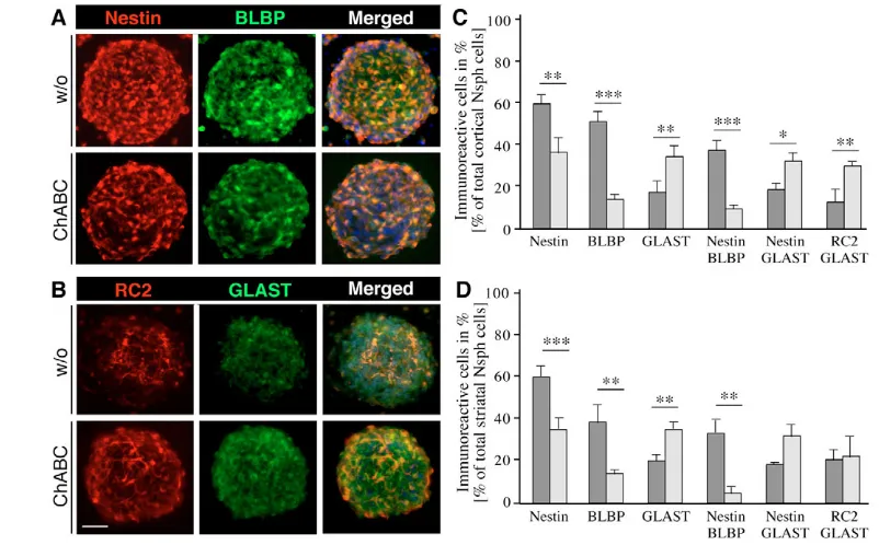
CS-GAGs modifies the composition of the radial glia precursor cell pool. (A,B) The distribution of precursor cells within mouse Nsphs grown in the absence or presence of ChABC was assessed by double immunofluorescence microscopy in cryosections stained for the precursor marker nestin and the radial glia markers BLBP (A), or the radial glia markers RC2 and GLAST (B). The degradation of CS-GAGs by ChABC reduced the number of nestin- and BLBP-positive precursor cells, whereas the number of RC2- and GLAST-positive radial glia increased. (C,D) These observations were quantified using acutely dissociated suspensions of control (dark
gray) and ChABC-treated (light gray) cortical (C) and striatal (D) Nsphs and plotted as percentage fractions. ChABC treatment of Nsphs led to a 3-to 4-fold reduction in the frequency of nestin/BLBP-positive cells in both the cortical and striatal Nsphs that was paralleled by a 2-fold increase in GLAST-positive cells, as compared with the control. *, P<0.05; **, P<0.01; ***, P<0.001. Scale bar: 50 m.
Table 1. Percentage of immunoreactive cells from cortical and striatal neurospheres grown for 7 div with or without ChABC Cor GE
Marker w/o ChABC P w/o ChABC P n
BrdU 51.5±6.0 27.4±2.5 <0.001 64.4±1.9 37.4±4.5 <0.01 5
Nestin 60.1±3.2 37.9±8.5 <0.01 60.7±4.0 35.5±5.0 <0.001 5
BLBP 51.6± 4.7 14.2±2.3 <0.01 38.8±4.9 14.2±1.9 n.s. 3
GLAST 17.7±5.2 35.0±4.0 <0.01 20.3±6.3 35.4±2.8 <0.01 3
Nestin/BLBP 37.9±4.4 9.7±1.6 n.s. 33.4±6.2 4.5±0.5 n.s. 3
Nestin/GLAST 19.0±2.8 32.7±3.6 n.s. 20.3±2.5 32.3±5.4 n.s. 3
III-tubulin 10.3±2.4 2.5±1.0 <0.01 8.8±1.6 2.0±0.6 <0.01 4
GFAP 3.8±0.7 10.8±1.0 <0.001 4.2±1.4 18.1±2.1 <0.001 4
The number of immunopositive cells was determined 1 hour after acute dissociation of the primary neurospheres grown under the control condition (w/o) or in the continuous presence of ChABC. Data are expressed as the relative number of immunopositive cells as a percentage (mean±s.d.) of the total neurosphere cell number counted.
n, Number of independent experiments.
[image:5.612.50.562.591.694.2]D
E
V
E
LO
P
M
E
N
T
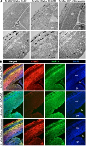
forebrain. By contrast, the elimination of CS-GAGs caused an apparent disorganization of the germinal regions, which appeared more loosely packed, rendering the distinction between ventricular zone (VZ) and subventricular zone (SVZ) almost impossible (Fig. 4A). In addition, a compaction of cells in the cortical plate could be distinguished after ChABC application, whereas the histological organization of the cortical marginal zone remained unchanged (Fig.
4A). Despite the obvious alterations in histology and cell morphology, only a few apoptotic cells were detectable in TUNEL stainings (see Fig. S3 in the supplementary material).
[image:6.612.54.492.54.502.2]Despite the histological alterations implying the effectiveness of the enzyme treatment, we examined the enzymatic activity of ChABC by double immunostaining with 473HD and anti-phosphacan (KAF13). The chondroitin sulfate-dependent 473HD Fig. 3. Interference with CS-GAGs impairs neurogenesis and simultaneously favors gliogenesis in vitro.The distribution of  III-tubulin-positive neurons and GFAP-III-tubulin-positive astrocytes was assessed in mouse Nsph cryosections by double immunohistochemistry. (A,B) Photomicrographs illustrating cortical (A) or striatal Nsphs (B) grown in the absence (w/o, upper panels) or presence (lower panels) of ChABC. The digestion of CS-GAGs by ChABC reduced III-tubulin immunoreactivity and enhanced GFAP immunoreactivity. (C,D) These observations were quantified using acutely dissociated suspensions of control (dark gray) and ChABC-treated (light gray) cortical (C) and striatal (D) Nsphs and plotted as percentage fractions. An apparent 4-fold reduction in the frequency of III-tubulin-positive neurons was recorded in both the cortical (Cor) and striatal (GE) ChABC-treated Nsphs that was accompanied by a 2-fold increase in GFAP-positive cells. (E,F) An analogous differentiation assay was performed using the 473HD-expressing subpopulation of radial glia immunopanned from acutely dissociated primary Nsphs. When the 473HD-immunoselected cells were cultivated for 2 days in vitro (div) in the presence of ChABC (light gray), the number of III-tubulin-positive neurons diminished 4-fold (E) and GFAP-expressing astrocytes doubled in number both for cortical and striatal Nsphs (F), as compared with control conditions (dark gray). (G,H) The
D
E
V
E
LO
P
M
E
N
T
epitope and core proteins of RPTP-were coexpressed on radially oriented cells in the germinal layers, in the cortical plate and in the marginal zone as reported previously (von Holst et al., 2006) (Fig. 4B). Upon ICVI of ChABC, many neuroepithelial cells analyzed 1 day after injection retained the core proteins of DSD-1-PG/RPTP-, as demonstrated by staining with the anti-DSD-1-PG/phosphacan antibody. Most importantly, the 473HD immunoreactivity almost completely vanished, with the exception of the marginal zone, demonstrating that the enzyme diffuses from the ventricle far into the brain parenchyma (Fig. 4B).
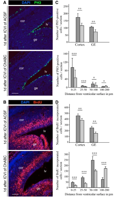
Given the effective removal of the CS epitope in vivo, we next examined proliferation and cell fate after this intervention. Using a combination of immunostaining with an antibody specific for phospho-histone H3 (PH3) and BrdU labeling 1 hour prior to sacrifice, cycling cells in M or S phases could be identified at the expected ventricular and abventricular positions (Fig. 5A,B). For
quantitative analysis, immunolabeled cells in the cortical and striatal germinal zones were counted in forebrain sections. In the case of ChABC-injected embryos, a reduction in PH3-positive cells by 30% was observed in both the cortex and the ganglionic eminence (GE) (Fig. 5C). Similarly, a significant reduction in the number of BrdU-labeled cells was observed both in the cortical and GE neuroepithelium after ChABC treatment (Fig. 5D).
[image:7.612.51.331.62.538.2]In order to gain insight into VZ and SVZ precursor subtypes, the distribution of the PH3- and BrdU-labeled cells was assessed with reference to their distance from the ventricular surface (Fig. 5C,D). Following ChABC application, a substantial decrease in PH3- and BrdU-labeled cells was apparent in the area close to the ventricular surface; this was not seen following injection of ACSF. This result suggested that ChABC exerted its effect mostly on radial glia precursors with access to the ventricle, whereas basal precursors dividing at abventricular positions were less affected. Thus, ChABC
Fig. 4. Injection of ChABC modifies the architecture of the ventricular zone.(A) Phase-contrast photomicrographs of E14.5 mouse forebrain cryosections obtained 1 day after intracerebroventricular injection (ICVI) of ACSF (left panels), ChABC (middle panels) or keratanase (right panels). The upper panels show low-power DIC images, and the lower panels are high-power images of histological stainings. Cryosections of control- or keratanase-injected embryos appeared normal with a clearly defined tissue stratification. By contrast, a
conspicuous disorganization of the germinal regions (VZ and SVZ) and an accumulation of cells in the normally positioned cortical plate was observed after injection of ChABC (middle panels). Note that the outer layers of the marginal region appeared comparable to those of the control (i.e. after ACSF injection). (B) The efficiency of ChABC injection was controlled by immunostaining with Mab 473HD. Photomicrographs of cryosections from embryos injected with ACSF (upper panels), ChABC (middle panels) or keratanase (lower panels). The 473HD epitope was almost completely absent after ChABC injection, whereas the reaction of the polyclonal antibodies with the RPTP-protein cores (KAF13) was unaltered. The 473HD and KAF13 stainings appeared unaltered by keratanase. Cell nuclei counterstained with bisbenzimide are shown in blue (DAPI). Cor, cortex; cp, cortical plate; e, surface ectoderm; ge, ganglionic eminence; imz, intermediate zone; lv, lateral ventricle; m, marginal zone; sep, septum; svz,
D
E
V
E
LO
P
M
E
N
T
treatment seems to affect cells that undergo interkinetic nuclear migration and possess an apical process with contact to the ventricle. In the light of the importance of cell polarity for subsequent neurogenic cell division patterns (Cappello et al., 2006), we also examined the distribution of E-cadherin (also known as cadherin 1 – Mouse Genome Informatics) and aPKC(also known as PRKCI) as apical markers and of laminin as a basolateral marker. No difference in E-cadherin and aPKC staining was found between ChABC- and control-injected telencephali (see Fig. S4 in the supplementary material).
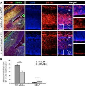
In order to further assess the impact of ChABC treatment on lineage decisions, the forebrain NSC population was examined for the expression of the neural precursor markers nestin, BLBP (Fig. 6A) and GLAST (Fig. 6B). The degradation of CS-GAGs in vivo decreased the nestin and BLBP immunoreactivity, whereas a higher number of cortical precursors were immunopositive for GLAST (Fig. 6A,B), consistent with the data obtained in vitro. To better quantify the role of CS-GAGs in the proliferation and differentiation behavior of neurogenic radial glia, the antigenic profiles of acutely dissociated cell populations from forebrain tissue 24 hours after injection of ACSF (control) or ChABC were determined. Interestingly, the suspensions obtained from treated and control brains contained comparable absolute cell numbers (see Table S1 in the supplementary material). A significant decrease in nestin- and BLBP-positive precursors was apparent in ChABC-injected telencephali, whereas the number of GLAST-immunoreactive precursors increased slightly (Fig. 6C). Hence, analogous to the situation documented in vitro, a radial glia subtype seemed attenuated, supporting the concept that CS-GAGs might intervene in cell fate choices. In order to explore this possibility further, the neuronal marker III-tubulin was examined in forebrain cryosections obtained from ACSF- or ChABC-injected animals using confocal microscopy (Fig. 7A). The elimination of CS-GAGs with ChABC caused a substantial reduction in  III-tubulin-positive neurons in the cortex and GE that was confined to the germinal layers, suggesting that newborn immature neurons were preferentially affected. Furthermore, acutely dissociated forebrain cell populations from ChABC-injected embryos contained significantly fewer III-tubulin-positive neurons (Fig. 7B). Thus, these antigenic profiles, recorded 1 day after ICVI of ChABC or ACSF, faithfully reflected the observations obtained by the immunohistochemical analysis of forebrain sections.
In the light of the significance of CS-GAGs for NSC proliferation reported earlier, we also examined the potential of forebrain tissues to generate Nsphs after ChABC or ACSF treatment. Cell suspensions were cultivated in the presence of EGF and bFGF for 7 days in vitro and emerging Nsphs were counted. When ChABC had been injected into the forebrain, the number of Nsph-forming cells was significantly reduced to half the yield upon ACSF injection (Fig. 6D). Taken together, the analysis of CS-GAG removal in vivo confirmed the importance of this specific class of carbohydrates for proliferation, cell fate decisions and neurogenesis of neural stem/progenitor cells.
DISCUSSION
[image:8.612.52.297.61.478.2]Here we demonstrated the crucial role of chondroitin sulfate proteoglycans (CSPGs) for proliferation, self-renewal and cell fate of NSCs in vitro and in vivo. Removal of chondroitin sulfates (CS-GAGs) severely impairs Nsph formation, self-renewal and the generation of their neuronal progeny. Strikingly similar effects were observed upon removal of CS-GAGs in the developing cerebral cortex in vivo. Thus, our data implicate CSPGs in two key functions of NSCs in development – self-renewal and neurogenesis.
Fig. 5. CS-GAGs are required for proliferation of neural precursor cells during forebrain development.(A,B) Photomicrographs of immunohistochemical stainings of E14.5 mouse forebrain cryosections 1 day after ventricular injection with ACSF control (upper panels) or ChABC (lower panels). Cell nuclei were counterstained with
D
E
V
E
LO
P
M
E
N
T
Chondroitin sulfates and radial glia fate
Neuronal precursors resemble NSCs in key aspects, namely their asymmetric mode of cell division (typically generating a postmitotic neuron and replicating a neuroepithelial or radial glial precursor cell) and their capacity to generate neurons (Gotz and Huttner, 2005). Indeed, during development, neuroepithelial and radial glial cells share many cell biological and molecular features with adult NSCs.
[image:9.612.50.375.60.325.2]Both have astroglial properties, and similar molecular determinants influence neurogenesis and proliferation (Alvarez-Buylla et al., 2001). For example, PAX6 promotes neurogenesis in the developing cerebral cortex (Heins et al., 2002), in embryonic and adult Nsph cells and in adult SEZ cells in vivo (Hack et al., 2005; Hack et al., 2004). Conversely, overactivation of Notch signaling promotes proliferation and maintains radial glia and NSCs during
Fig. 6. CS-GAGs are essential components for neurogenic precursor cells in vivo.
(A,B) Photomicrographs of immunohistochemical stainings of E14.5 mouse forebrain cryosections 1 day after ventricular injection with ACSF control (upper panels) or ChABC (lower panels). The cortical precursor pool was examined for coexpression of BLBP and nestin (A) and for GLAST expression (B). Following ChABC treatment, immunoreactivities for nestin and BLBP significantly decreased, whereas more cells were GLAST-positive, in comparison with the situation following ACSF injection. Cell nuclei were counterstained with bisbenzimide and are shown in blue (DAPI). (C,D) Single-cell suspensions obtained from embryos injected with ACSF (dark gray) or ChABC (light gray) were plated and immunopositive cells determined after 2 hours. (C) ChABC treatment caused a 2-fold reduction in the number of nestin- and BLBP-positive cells, whereas GLAST-positive cells increased. (D). The relative fractions of cortical (Cor) and striatal (GE) Nsphs grown from single-cell suspension were reduced following ChABC (light gray) injection. Numbers are plotted with reference to the control-injected telencephali, taken as 100% (dark gray). *,P<0.05; **, P<0.01. Scale bar: 100 m.
Fig. 7. CS-GAGs are necessary for neuronal differentiation of precursors in vivo.
[image:9.612.50.349.435.739.2]D
E
V
E
LO
P
M
E
N
T
development (Gaiano et al., 2000; Hitoshi et al., 2002). Intriguingly, Notch is expressed in a subset of radial glial cells during development and in adult NSCs, but not in most of the adult parenchymal astroglia, and positively regulates BLBP (Anthony et al., 2005; Hartfuss et al., 2001) (for a review, see Mori et al., 2005). Here, we were able to show that removal of CS-GAGs reduced the number of BLBP- and nestin-positive radial glial cells, while increasing the GLAST- and RC2-positive fraction. These subtype alterations upon ChABC treatment were virtually indistinguishable in Nsph cells in vitro and in radial glial cells in vivo. Moreover, these subtype modifications correlated with reduced proliferation and a decrease in neurogenesis in vitro and in vivo, suggesting that the nestin- and BLBP-positive radial glial cells represent a proliferative, self-renewing, neurogenic precursor population (Anthony et al., 2004; Hartfuss et al., 2001). This is consistent with Notch signaling regulating BLBP and cell proliferation (Anthony et al., 2005; Hatakeyama et al., 2004). Conversely, reduced proliferation and increased gliogenesis is accompanied by upregulation of GLAST, a molecule that persists in adult parenchymal astrocytes (Mori et al., 2006; Shibata et al., 1997). These results underline the functional relevance of radial glial subtypes with regard to proliferation and cell fate. Most excitingly, however, our results show the profound role of CSPGs in governing precursor subtypes, cell proliferation and cell fate in vitro and in vivo. Indeed, the key role of modifications of signaling molecules in the ECM is becoming ever clearer (Carulli et al., 2005; Deakin and Lyon, 1999; McLaughlin et al., 2003), although little is known about the role of chondroitin sulfates in general, and especially in the regulation of neural stem/progenitor cell fate.
Potential molecular mechanisms of CSPGs in neurogenesis and proliferation
How could CSPGs exert their crucial role in mediating radial glia and NSC proliferation and neurogenesis, as discovered here? Notably, the experiments in vivo revealed a clear effect on radial glial cell proliferation and neurogenesis, whereas the proliferation of basal precursors dividing at abventricular positions was less affected, despite the penetration of the enzyme deep into the cortical parenchyma. This is consistent with the finding that radial glia cell polarity was not notably affected by ChABC treatment, as deduced from the unaltered expression of the apical markers aPKC and E-cadherin. Indeed, when polarity is compromised by the conditional deletion of the Rho GTPase CDC42, an enhanced rate of basal mitoses and a decrease in apical divisions result (Cappello et al., 2006). In our case, basal progenitors seemed unperturbed and the cellular polarity conserved, with reduced proliferation confined to the apical compartment. These data further support the notion that the pathway(s) affected is predominantly active in the self-renewing radial glia/precursor cells, because basal precursors do not self-renew (Haubensak et al., 2004; Miyata et al., 2004; Noctor et al., 2004). Our interpretation is in agreement with the decrease in BLBP immunoreactivity that occurs prominently in radial glial cells, but not in basal precursors.
BLBP is regulated by Notch signaling that interacts with FGF and EGF signaling (Gaiano et al., 2000; Hasson et al., 2005; Yoon et al., 2004). Firstly, Notch signaling might be affected by ChABC activity. Moreover, EGF in conjunction with Notch signaling promotes radial glia formation (Gregg and Weiss, 2003). CSPGs bind various growth factors that might impinge on the proliferation rates of NSCs, and the implication of CSPGs in stem cell proliferation has recently been documented in the nematode
(Mizuguchi et al., 2003; Sugahara et al., 2003). Thus, CS-dependent GAG motifs may serve as docking sites for growth factors and thereby modulate responsiveness to bFGF in embryonic neural stem/progenitor cells (Deepa et al., 2002; Penc et al., 1998). CSPGs might intervene in the bFGF- or EGF-dependent signaling pathways, and thereby foster NSC and radial glia proliferation. This could be effected either through the manner in which CS-GAGs bind and store factors in the pericellular environment (Properzi et al., 2005), or by their serving as cis-acting co-factors for growth factor receptors, analogous to the role played by heparan sulfate proteoglycans with respect to the FGFR (Bandtlow and Zimmermann, 2000).
Indeed, the growth factors pleiotrophin and midkine, which have been associated with the proliferation of NSCs, are also secreted into Nsph-conditioned medium and strongly interact with the CS-GAGs of the CSPG phosphacan (Deepa et al., 2002; Furuta et al., 2004; Hienola et al., 2004). The 473HD epitope is expressed on the CSPG phosphacan, an isoform of the transmembrane-based receptor protein tyrosine phosphatase (RPTP)-/(Faissner et al., 1994; Garwood et al., 1999). Selected RPTP-/ isoforms are highly expressed in VZ in the embryonic brain and in the stem cell niches of the adult CNS, and hence also represent potential targets of ChABC treatment (Engel et al., 1996). The RPTP-/receptor is a potential ligand of the ECM glycoprotein tenascin-C, which interacts with the CSPG phosphacan (Milev et al., 1994). Tenascin-C is highly enriched in the VZ of the developing brain (Gotz et al., 1997) and is downregulated in the mature CNS, with the exception of areas of ongoing precursor cell proliferation, for example the SEZ (Gates et al., 1995). Deletion of Tncinterferes with NSC maturation by impairing the bFGF-stimulated acquisition of the EGFR on NSCs (Garcion et al., 2004). Notably, tenascin-C expression is virtually lost in the cerebral cortex of PAX6 mutant mice, concomitant with a decrease in neurogenesis from radial glia cells (Gotz et al., 1998; Stoykova et al., 1997). Interestingly, tenascin-C and sulfation of the ECM influence the canonical Wnt signaling pathway (Kakinuma et al., 2004), a well-known regulator of stem cell fate (Reya and Clevers, 2005). Taken together, the profound effect of ChABC treatment is likely to be due to effects on a multitude of signaling pathways.
D
E
V
E
LO
P
M
E
N
T
ConclusionThe present study suggests a role for CSPGs in stem cell biology. How CSPGs are integrated into the complex interplay of pericellular determinants of cell fate remains to be investigated in detail. However, the identification of the pivotal role of CS-GAGs in regulating stem cell proliferation and neurogenesis represents an important step forward in identifying key factors of the local environment that regulate stem cell fate and neurogenesis. Indeed, previous transplantation experiments have highlighted the local environment as the key determinant of adult neurogenesis. Glial cells isolated from non-neurogenic regions of the adult CNS generate neurons when transplanted into a neurogenic environment (Shihabuddin et al., 2000), whereas neurogenic precursors isolated from the adult SEZ do not succeed in generating neurons outside their niche (Lim et al., 2000). Thus, a better understanding of the NSC niche is of the utmost importance in the context of employing NSCs for repair processes (Scadden, 2006). Our work demonstrates for the first time that complex CS-GAG carbohydrates play a pivotal role in the orchestration of the NSC micromilieu.
The authors thank Nicole Haubst for help with the initial injection experiments. Monoclonal antibody RC2 developed by M. Yamamoto was obtained from the Developmental Studies Hybridoma Bank developed under the auspices of the NICHD and maintained by The University of Iowa, Department of Biological Sciences, Iowa City, IA 52242. The authors acknowledge grant support from the Federal Ministry of Research and Technology (BMBF, Stem Cells for Therapies of the CNS, 01GN0504) and the German Research Council (DFG, SPP 1109). M.G. is funded by the DFG, BMBF and FORNEUROCELL.
Supplementary material
Supplementary material for this article is available at http://dev.biologists.org/cgi/content/full/134/15/2727/DC1
References
Alvarez-Buylla, A., Garcia-Verdugo, J. M. and Tramontin, A. D.(2001). A unified hypothesis on the lineage of neural stem cells.Nat. Rev. Neurosci.2, 287-293.
Anthony, T. E., Klein, C., Fishell, G. and Heintz, N.(2004). Radial glia serve as neuronal progenitors in all regions of the central nervous system. Neuron41, 881-890.
Anthony, T. E., Mason, H. A., Gridley, T., Fishell, G. and Heintz, N.(2005). Brain lipid-binding protein is a direct target of Notch signaling in radial glial cells. Genes Dev.19, 1028-1033.
Arvidsson, A., Collin, T., Kirik, D., Kokaia, Z. and Lindvall, O.(2002). Neuronal replacement from endogenous precursors in the adult brain after stroke.Nat. Med.8, 963-970.
Bandtlow, C. E. and Zimmermann, D. R.(2000). Proteoglycans in the developing brain: new conceptual insights for old proteins. Physiol. Rev.80, 1267-1290.
Bard, J. L., Kaufman, M. H., Dubreuil, C., Brune, R. M., Burger, A., Baldock, R. A. and Davidson, D. R.(1998). An internet-accessible database of mouse developmental anatomy based on a systematic nomenclature. Mech. Dev.74, 111-120.
Bernreuther, C., Dihne, M., Johann, V., Schiefer, J., Cui, Y., Hargus, G., Schmid, J. S., Xu, J., Kosinski, C. M. and Schachner, M.(2006). Neural cell adhesion molecule L1-transfected embryonic stem cells promote functional recovery after excitotoxic lesion of the mouse striatum. J. Neurosci. 26, 11532-11539.
Bradbury, E. J., Moon, L. D., Popat, R. J., King, V. R., Bennett, G. S., Patel, P. N., Fawcett, J. W. and McMahon, S. B.(2002). Chondroitinase ABC promotes functional recovery after spinal cord injury. Nature416, 636-640.
Campos, L. S., Leone, D. P., Relvas, J. B., Brakebusch, C., Fassler, R., Suter, U. and ffrench-Constant, C.(2004). Beta1 integrins activate a MAPK signalling pathway in neural stem cells that contributes to their maintenance. Development131, 3433-3444.
Campos, L. S., Decker, L., Taylor, V. and Skarnes, W.(2006). Notch, epidermal growth factor receptor, and beta1-integrin pathways are coordinated in neural stem cells. J. Biol. Chem.281, 5300-5309.
Cappello, S., Attardo, A., Wu, X., Iwasato, T., Itohara, S., Wilsch-Brauninger, M., Eilken, H. M., Rieger, M. A., Schroeder, T. T., Huttner, W. B. et al. (2006). The Rho-GTPase cdc42 regulates neural progenitor fate at the apical surface.Nat. Neurosci.9, 1099-1107.
Carulli, D., Laabs, T., Geller, H. M. and Fawcett, J. W.(2005). Chondroitin sulfate proteoglycans in neural development and regeneration.Curr. Opin. Neurobiol.15, 116-120.
Chanas-Sacre, G., Thiry, M., Pirard, S., Rogister, B., Moonen, G., Mbebi, C., Verdiere-Sahuque, M. and Leprince, P.(2000). A 295-kDA intermediate filament-associated protein in radial glia and developing muscle cells in vivo and in vitro. Dev. Dyn.219, 514-525.
Chen, J., Bernreuther, C., Dihne, M. and Schachner, M.(2005). Cell adhesion molecule L1-transfected embryonic stem cells with enhanced survival support regrowth of corticospinal tract axons in mice after spinal cord injury. J. Neurotrauma22, 896-906.
Colognato, H., ffrench-Constant, C. and Feltri, M. L.(2005). Human diseases reveal novel roles for neural laminins. Trends Neurosci.28, 480-486. D’Amour, K. A. and Gage, F. H.(2003). Genetic and functional differences
between multipotent neural and pluripotent embryonic stem cells. Proc. Natl. Acad. Sci. USA100Suppl. 1, 11866-11872.
Deakin, J. A. and Lyon, M.(1999). Differential regulation of hepatocyte growth factor/scatter factor by cell surface proteoglycans and free glycosaminoglycan chains. J. Cell Sci. 112, 1999-2009.
Deepa, S. S., Umehara, Y., Higashiyama, S., Itoh, N. and Sugahara, K.(2002). Specific molecular interactions of oversulfated chondroitin sulfate E with various heparin-binding growth factors. Implications as a physiological binding partner in the brain and other tissues. J. Biol. Chem.277, 43707-43716.
Dobbertin, A., Rhodes, K. E., Garwood, J., Properzi, F., Heck, N., Rogers, J. H., Fawcett, J. W. and Faissner, A.(2003). Regulation of
RPTPbeta/phosphacan expression and glycosaminoglycan epitopes in injured brain and cytokine-treated glia. Mol. Cell. Neurosci.24, 951-971.
Doetsch, F.(2003). The glial identity of neural stem cells.Nat. Neurosci.6, 1127-1134.
Engel, M., Maurel, P., Margolis, R. U. and Margolis, R. K.(1996). Chondroitin sulfate proteoglycans in the developing central nervous system. I. cellular sites of synthesis of neurocan and phosphacan. J. Comp. Neurol.366, 34-43. Faissner, A., Clement, A., Lochter, A., Streit, A., Mandl, C. and Schachner, M.
(1994). Isolation of a neural chondroitin sulfate proteoglycan with neurite outgrowth promoting properties.J. Cell Biol. 126, 783-799.
Feng, L., Hatten, M. E. and Heintz, N.(1994). Brain lipid-binding protein (BLBP): a novel signaling system in the developing mammalian CNS. Neuron12, 895-908.
Furuta, M., Shiraishi, T., Okamoto, H., Mineta, T., Tabuchi, K. and Shiwa, M. (2004). Identification of pleiotrophin in conditioned medium secreted from neural stem cells by SELDI-TOF and SELDI-tandem mass spectrometry. Brain Res. Dev. Brain Res.152, 189-197.
Gaiano, N., Nye, J. S. and Fishell, G.(2000). Radial glial identity is promoted by Notch1 signaling in the murine forebrain. Neuron26, 395-404.
Garcion, E., Halilagic, A., Faissner, A. and ffrench-Constant, C.(2004). Generation of an environmental niche for neural stem cell development by the extracellular matrix molecule tenascin C. Development131, 3423-3432. Garwood, J., Schnadelbach, O., Clement, A., Schutte, K., Bach, A. and
Faissner, A.(1999). DSD-1-proteoglycan is the mouse homolog of phosphacan and displays opposing effects on neurite outgrowth dependent on neuronal lineage. J. Neurosci. 19, 3888-3899.
Garwood, J., Rigato, F., Heck, N. and Faissner, A.(2001). Tenascin
glycoproteins and the complementary ligand DSD-1-PG/phosphacan–structuring the neural extracellular matrix during development and repair. Restor. Neurol. Neurosci.19, 51-64.
Gates, M. A., Thomas, L. B., Howard, E. M., Laywell, E. D., Sajin, B., Faissner, A., Gotz, B., Silver, J. and Steindler, D. A.(1995). Cell and molecular analysis of the developing and adult mouse subventricular zone of the cerebral hemispheres. J. Comp. Neurol.361, 249-266.
Gotz, M. and Huttner, W. B.(2005). The cell biology of neurogenesis.Nat. Rev. Mol. Cell Biol. 6, 777-788.
Gotz, M., Bolz, J., Joester, A. and Faissner, A.(1997). Tenascin-C synthesis and influence on axonal growth during rat cortical development. Eur. J. Neurosci. 9, 496-506.
Gotz, M., Stoykova, A. and Gruss, P.(1998). Pax6 controls radial glia differentiation in the cerebral cortex. Neuron21, 1031-1044.
Gregg, C. and Weiss, S.(2003). Generation of functional radial glial cells by embryonic and adult forebrain neural stem cells. J. Neurosci. 23, 11587-11601. Grove, E. A., Williams, B. P., Li, D. Q., Hajihosseini, M., Friedrich, A. and
Price, J.(1993). Multiple restricted lineages in the embryonic rat cerebral cortex. Development117, 553-561.
Hack, M. A., Sugimori, M., Lundberg, C., Nakafuku, M. and Gotz, M.(2004). Regionalization and fate specification in neurospheres: the role of Olig2 and Pax6. Mol. Cell. Neurosci.25, 664-678.
Hack, M. A., Saghatelyan, A., de Chevigny, A., Pfeifer, A., Ashery-Padan, R., Lledo, P. M. and Gotz, M.(2005). Neuronal fate determinants of adult olfactory bulb neurogenesis.Nat. Neurosci.8, 865-872.
Hartfuss, E., Galli, R., Heins, N. and Gotz, M.(2001). Characterization of CNS precursor subtypes and radial glia. Dev. Biol. 229, 15-30.
D
E
V
E
LO
P
M
E
N
T
Courey, A. J. and Paroush, Z.(2005). EGFR signaling attenuates Groucho-dependent repression to antagonize Notch transcriptional output. Nat. Genet. 37, 101-105.
Hatakeyama, J., Bessho, Y., Katoh, K., Ookawara, S., Fujioka, M., Guillemot, F. and Kageyama, R.(2004). Hes genes regulate size, shape and histogenesis of the nervous system by control of the timing of neural stem cell differentiation. Development131, 5539-5550.
Haubensak, W., Attardo, A., Denk, W. and Huttner, W. B.(2004). Neurons arise in the basal neuroepithelium of the early mammalian telencephalon: a major site of neurogenesis. Proc. Natl. Acad. Sci. USA101, 3196-3201. Heins, N., Malatesta, P., Cecconi, F., Nakafuku, M., Tucker, K. L., Hack, M. A.,
Chapouton, P., Barde, Y. A. and Gotz, M.(2002). Glial cells generate neurons: the role of the transcription factor Pax6.Nat. Neurosci.5, 308-315.
Hienola, A., Pekkanen, M., Raulo, E., Vanttola, P. and Rauvala, H.(2004). HB-GAM inhibits proliferation and enhances differentiation of neural stem cells. Mol. Cell. Neurosci.26, 75-88.
Hitoshi, S., Alexson, T., Tropepe, V., Donoviel, D., Elia, A. J., Nye, J. S., Conlon, R. A., Mak, T. W., Bernstein, A. and van der Kooy, D.(2002). Notch pathway molecules are essential for the maintenance, but not the generation, of mammalian neural stem cells. Genes Dev.16, 846-858.
Houle, J. D., Tom, V. J., Mayes, D., Wagoner, G., Phillips, N. and Silver, J. (2006). Combining an autologous peripheral nervous system “bridge” and matrix modification by chondroitinase allows robust, functional regeneration beyond a hemisection lesion of the adult rat spinal cord. J. Neurosci. 26, 7405-7415. Ida, M., Shuo, T., Hirano, K., Tokita, Y., Nakanishi, K., Matsui, F., Aono, S.,
Fujita, H., Fujiwara, Y., Kaji, T. et al.(2006). Identification and functions of chondroitin sulfate in the milieu of neural stem cells. J. Biol. Chem.281, 5982-5991.
Kakinuma, Y., Saito, F., Osawa, S. and Miura, M.(2004). A mechanism of impaired mobility of oligodendrocyte progenitor cells by tenascin C through modification of wnt signaling. FEBS Lett.568, 60-64.
Lendahl, U., Zimmerman, L. B. and McKay, R. D.(1990). CNS stem cells express a new class of intermediate filament protein. Cell60, 585-595.
Leone, D. P., Relvas, J. B., Campos, L. S., Hemmi, S., Brakebusch, C., Fassler, R., Ffrench-Constant, C. and Suter, U.(2005). Regulation of neural progenitor proliferation and survival by beta1 integrins. J. Cell Sci. 118, 2589-2599. Lim, D. A., Tramontin, A. D., Trevejo, J. M., Herrera, D. G., Garcia-Verdugo, J.
M. and Alvarez-Buylla, A.(2000). Noggin antagonizes BMP signaling to create a niche for adult neurogenesis. Neuron28, 713-726.
Magavi, S. S., Leavitt, B. R. and Macklis, J. D.(2000). Induction of neurogenesis in the neocortex of adult mice. Nature405, 951-955.
Malatesta, P., Hartfuss, E. and Gotz, M.(2000). Isolation of radial glial cells by fluorescent-activated cell sorting reveals a neuronal lineage. Development127, 5253-5263.
McLaughlin, D., Karlsson, F., Tian, N., Pratt, T., Bullock, S. L., Wilson, V. A., Price, D. J. and Mason, J. O.(2003). Specific modification of heparan sulphate is required for normal cerebral cortical development. Mech. Dev.120, 1481-1488.
Milev, P., Friedlander, D. R., Sakurai, T., Karthikeyan, L., Flad, M., Margolis, R. K., Grumet, M. and Margolis, R. U.(1994). Interactions of the chondroitin sulfate proteoglycan phosphacan, the extracellular domain of a receptor-type protein tyrosine phosphatase, with neurons, glia, and neural cell adhesion molecules.J. Cell Biol. 127, 1703-1715.
Miyata, T., Kawaguchi, A., Saito, K., Kawano, M., Muto, T. and Ogawa, M. (2004). Asymmetric production of surface-dividing and non-surface-dividing cortical progenitor cells. Development131, 3133-3145.
Mizuguchi, S., Uyama, T., Kitagawa, H., Nomura, K. H., Dejima, K., Gengyo-Ando, K., Mitani, S., Sugahara, K. and Nomura, K.(2003). Chondroitin proteoglycans are involved in cell division of Caenorhabditis elegans. Nature 423, 443-448.
Mori, T., Buffo, A. and Gotz, M.(2005). The novel roles of glial cells revisited: the contribution of radial glia and astrocytes to neurogenesis.Curr. Top. Dev. Biol. 69, 67-99.
Mori, T., Tanaka, K., Buffo, A., Wurst, W., Kuhn, R. and Gotz, M.(2006). Inducible gene deletion in astroglia and radial glia – a valuable tool for functional and lineage analysis. Glia54, 21-34.
Noctor, S. C., Martinez-Cerdeno, V., Ivic, L. and Kriegstein, A. R.(2004). Cortical neurons arise in symmetric and asymmetric division zones and migrate through specific phases.Nat. Neurosci.7, 136-144.
Penc, S. F., Pomahac, B., Winkler, T., Dorschner, R. A., Eriksson, E., Herndon, M. and Gallo, R. L.(1998). Dermatan sulfate released after injury is a potent promoter of fibroblast growth factor-2 function. J. Biol. Chem.273, 28116-28121.
Pizzorusso, T., Medini, P., Berardi, N., Chierzi, S., Fawcett, J. W. and Maffei, L.(2002). Reactivation of ocular dominance plasticity in the adult visual cortex. Science298, 1248-1251.
Properzi, F., Carulli, D., Asher, R. A., Muir, E., Camargo, L. M., van Kuppevelt, T. H., ten Dam, G. B., Furukawa, Y., Mikami, T., Sugahara, K. et al.(2005). Chondroitin 6-sulphate synthesis is up-regulated in injured CNS, induced by injury-related cytokines and enhanced in axon-growth inhibitory glia. Eur. J. Neurosci. 21, 378-390.
Quinones-Hinojosa, A., Sanai, N., Soriano-Navarro, M., Gonzalez-Perez, O., Mirzadeh, Z., Gil-Perotin, S., Romero-Rodriguez, R., Berger, M. S., Garcia-Verdugo, J. M. and Alvarez-Buylla, A.(2006). Cellular composition and cytoarchitecture of the adult human subventricular zone: a niche of neural stem cells. J. Comp. Neurol.494, 415-434.
Reya, T. and Clevers, H.(2005). Wnt signalling in stem cells and cancer. Nature 434, 843-850.
Reynolds, B. A. and Weiss, S.(1996). Clonal and population analyses demonstrate that an EGF-responsive mammalian embryonic CNS precursor is a stem cell. Dev. Biol. 175, 1-13.
Scadden, D. T.(2006). The stem-cell niche as an entity of action. Nature441, 1075-1079.
Shibata, T., Yamada, K., Watanabe, M., Ikenaka, K., Wada, K., Tanaka, K. and Inoue, Y.(1997). Glutamate transporter GLAST is expressed in the radial glia-astrocyte lineage of developing mouse spinal cord. J. Neurosci. 17, 9212-9219. Shihabuddin, L. S., Horner, P. J., Ray, J. and Gage, F. H.(2000). Adult spinal cord stem cells generate neurons after transplantation in the adult dentate gyrus. J. Neurosci. 20, 8727-8735.
Stoykova, A., Gotz, M., Gruss, P. and Price, J.(1997). Pax6-dependent regulation of adhesive patterning, R-cadherin expression and boundary formation in developing forebrain. Development124, 3765-3777. Sugahara, K., Mikami, T., Uyama, T., Mizuguchi, S., Nomura, K. and
Kitagawa, H.(2003). Recent advances in the structural biology of chondroitin sulfate and dermatan sulfate.Curr. Opin. Struct. Biol. 13, 612-620.
von Holst, A., Sirko, S. and Faissner, A.(2006). The unique 473HD-chondroitinsulfate epitope is expressed by radial glia and involved in neural precursor cell proliferation. J. Neurosci.26, 4082-4094.