http://dx.doi.org/10.4236/wjns.2016.62014
How to cite this paper: Jimenez-Vazquez, O.H. and Nagore, N. (2016) Neuroendoscopy in Epilepsy Surgery. World Journal of Neuroscience, 6, 114-118. http://dx.doi.org/10.4236/wjns.2016.62014
Neuroendoscopy in Epilepsy Surgery
Oscar Humberto Jimenez-Vazquez
1, Norma Nagore
21Department of Neurosurgery, State General Hospital, Morelia, México 2Nagore, Norma, Facultad de QFB, UMSNH, Morelia, México
Received 27 January 2016; accepted 3 May 2016; published 6 May 2016
Copyright © 2016 by author and Scientific Research Publishing Inc.
This work is licensed under the Creative Commons Attribution International License (CC BY). http://creativecommons.org/licenses/by/4.0/
Abstract
Epilepsy surgery has constantly evolved in various fields of knowledge. Surgical criteria have shifted from standard procedures to individualized forms of treatment, depending on physiologi-cal tests and specific imagenology findings in an individual patient. New instruments and applica-tions based upon older instruments have been described in the treatment of epilepsy surgery, in-cluding the use of endoscopes. Frequent indications of neuroendoscopy in epilepsy surgery have been mostly to assist in open procedures, particularly when fluid-filled spaces are present within the surgical field, such as cystic parasites, tumors, arachnoid, or other types of cysts. Other indica-tions certainly include cases of temporal lobe epilepsy, where ventricular exploration precedes intraventricular electrode placing as a tool to localize epileptogenic zones. Although described several years ago, there has been a recent trend in performing endoscopy-assisted section of the corpus-callosum in patients with generalized seizures. As neurosurgical instruments and tech-niques continue their progress, endoscopy will be included more frequently as part of the arma-mentarium in epilepsy surgery.
Keywords
Neuroendoscopy, Epilepsy Surgery, Intracranial Cysts
1. Introduction
Endoscopy has been used as well in the treatment of various types of intracranial or intra-spinal pathologies, or as an aid to obtain accurate diagnosis [2] [3]. Recently, epilepsy-associated pathologies, such as congenital cysts, cystic or solid tumors, parasites, abscesses, and blood-clots, have been included among endoscopy indica-tions, demonstrating its usefulness particularly in limited settings [2].
2. Endoscopy-Assisted Epilepsy Surgery Indications
According to our review of the world literature, endoscopy-assisted epilepsy surgery has been indicated in the following settings.
Endoscopic-assisted temporal lobe epilepsy surgery. Intraventricular electrode placement has not been regu-larly used for the study of epilepsy surgery candidates, but some patients with temporal lobe epilepsy may bene-fit from this procedure, as previously mentioned by us [4] and others [5]. Endoscopic ventricle exploration fol-lowed by intraventricular electrode placing as part of accurate epileptic foci localization could be useful, so that affected brain tissue may then be resected without excessive manipulation of the brain. Intraventricular elec-trode-placement has been described by various authors as an alternative to determine with more precision the epileptiogenic zone in certain cases of temporal lobe epilepsy. Either intraventricular individual electrodes, or electrode arrays, in coordination with cortical surface electrodes have facilitatedthe localization and subsequent resection of the epileptogenic zone.
Endoscopy-assisted corpus callosotomy. Surgical section of the corpus callosum is an old procedure that has been used for tract interruption, and therefore epilepsy focus isolation. It is still useful in cases where tissue re-section is not indicated, particularly when multiple ictal foci are present and lie in eloquent areas of the cortex, or in cases of generalized epilepsy syndromes. Traditionally, the extent of the callosotomy is determined by a postoperative MRI, or even as an intraoperative study, which may only be possible in few hospitals world-wide. In order to determine the extent of the corpus-callosotomy in the intraoperative period, endoscope-assisted oper-ations have been an alternative. This procedure was previously performed and documented by our group in five patients [6], taking into account intracranial anatomical references obtained from preoperative image studies. Subsequent publications describe this procedure in accordance to intraoperative endoscopic images [7] and MR images, as well as with intraoperative MRI and neuronavigation [8]-[10]. These authors have also described the use of neuroendoscopy in minimally invasive hemispherotomy, both as an individual procedure, or in addition to the corpus-callosotomy.
Endoscopy-assisted intratumoral cyst exploration. Epilepsy associated cystic tumors may be explored with
the endoscope, allowing clear images on the anatomy, vascularization, cystic content, and other features that might be helpful in selecting tumor sites where tissue samples may be taken from. Several authors [11] [12] have associated endoscopic images with the quality of the samples to obtain reliable histological diagnosis, hence an appropriate treatment. Eight of our cases underwent endoscopic sampling of cystic tumors, and in two, intracavitary electrode placement, with electrographic recordings from selected tumor areas were performed to increase sampling accuracy (Figure 1). This procedure was based upon the hypothesis that neoplastic activity in malignant tumors may cause local neuronal excitability, abnormal transmitter release, changes in blood-brain barrier, and other factors that may derive in network hyperexcitability [13]. Histopathological diagnosis has successfully confirmed the quality of the areas sampled, although a larger number of cases are needed to statis-tically demonstrate this hypothesis.
Endoscopy-based exploration and resection of deep brain intraparenchymal cystic lesions. Cases with
mul-tiple subcortical brain cysts of unknown etiology, have been explored and resected under endoscopic visualiza-tion. According to their characteristics, we have identified viable cysticerci and subsequent endoscopical extrac-tion has been achieved (Figure 2).
Neurocysticercosis is the most frequent parasitic disease in the central and peripheral nervous system. It is considered endemic in various underdeveloped countries where efficient sanitary policies and procedures are not efficiently implemented. Neurocysticercosis may be diagnosed with variable precision criteria, where image studies and epidemiological data are considered essential [14]. However, these criteria may be absent in a sig-nificant number of cases, particularly when studies are performed with old and outdated scanners, which may be the case of many medical facilities in those countries. The use of endoscopy in this context, increase accurate diagnosis in neurocysticercosis, proving its invaluable importance in this setting [15].
116
[image:3.595.219.406.82.270.2]Figure 1. Endoscopic view of a cystic neoplasm. Selected areas were sampled for histological analy-sis.
Figure 2. Intraventricuar cyst diagnosed endoscop-ically as a viable cysticercus (white area).
[image:3.595.219.408.315.509.2] [image:3.595.222.405.552.682.2]117
small percentage of arachnoid cysts (15%). They have been considered benign intracranial lesions that may cause multiple signs and symptoms, including epilepsy. The treatment options for these lesions have been con-troversial and diverse, ranging from simple observation to various surgical procedures, including endoscopic cyst fenestrations to the basal cisterns or ventricles. This procedure may be considered the treatment of choice for many surgeons, since it is associated with 0% of complications [16]-[18], compared to the second procedure of choice, which is the craniotomy cyst decompression, with a 17% rate of complications, being the most fre-quent the formation of subdural clots [19].
3. Discussion
Surgical endoscope procedures have coupled a relatively low cost of the equipment, with a high yield of results, making this instrument an important component of the surgical armamentarium. High definition image studies have proved essential to determine which patients may be suitable candidates for surgical epilepsy procedures [20] [21]. Such image studies, as well as specialized surgical equipment, may not be available in public hospitals from developing countries. To overcome the lack of diagnostic technological resources, endoscopy-associated direct visualization and real-time intraoperative diagnosis has been performed [2].
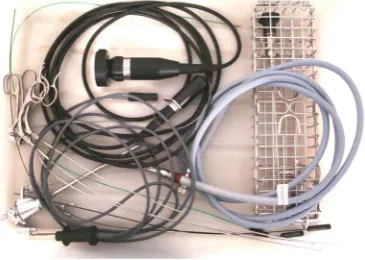
Diagnostic endoscopic observations may then be followed by intraoperative adjustment to the original surgic-al plan in up to thirty five percent of the procedures, thus, immediate therapeutic changes may be applied at the time of the surgical intervention [2]. Such modifications have been associated with a reduction in the number of surgical interventions, with significant decrease in morbidity and mortality risks, as well as hospital costs. Sur-gical protocols before intervention in patients harboring a variety of diseases, with clear indication for endos-copic procedures, follow a similar course as in conventional procedures. Patients must be admitted to a second or third level hospital, where thorough image, laboratory, and functional studies must be obtained, including con- ventional or specialized Computed Tomography (CT) scans and 1.5 tesla Magnetic Resonance Images (MRI), as well as functional scans, depending on its availability. Additional preoperative screening include video-electro- encephalograms, blood-cell count, and coagulation tests, cardiovascular risk evaluation, as well as psychometric and psychological interviews for testing memory, attention, psychiatric data and signed consent. Staff discus-sions based on the results, are crucial to determine the best approach for every individual. Flexible endoscopes may be used, although 6 mm external diameter rigid endoscope with Hopkins view system and three working channels1, in our experience, is preferred (Figure 3).
4. Conclusion
Endoscopy may represent a useful instrument in the currently diagnosis and treatment of various intracranial cystic lesions, as well as in the assistance of open intracranial procedures as an aid in the treatment of lesions associated with epilepsy. More experience is required to determine its value in the long term.
References
[1] Nowell, M., Miserocchi, A., McEvoy, A.W. and Duncan, J.S. (2014) Advances in Epilepsy Surgery. Journal of Neu-rology, Neurosurgery & Psychiatry, 84, 1273-1279. http://dx.doi.org/10.1136/jnnp-2013-307069
[2] Jimenez-Vazquez, O.H. and Nagore, N. (2008) The Impact of Neuroendoscopy in the Emergency Setting—A Retros-pective Study of Imaging, Intraoperative Findings, and Surgical Outcome in 55 Patients. Clinical Neurology and Neu-rosurgery, 10, 539-543. http://dx.doi.org/10.1016/j.clineuro.2008.02.019
[3] Bauer, B.L., Hellwig, D., Sweet, W.H. and Schmidek, H.H. (1995) Surgical Management of Intracranial Arachnoid, Suprasellar, and Rathke’s Cleft Cyst. In: Schmidek, H.H. and Sweet, W.H., Eds., Operative Neurosurgical Techniques, WS Saunders Company, 579-597.
[4] Jiménez, O., Leal, R. and Nagore, N. (2002) Minimally Invasive Electrodiagnostic Monitoring in Epilepsy Surgery.
British Journal of Neurosurgery, 16, 498-500. http://dx.doi.org/10.1080/0268869021000030339
[5] Sontag, J.K., Abou-Khalil, B. and Konrad, P.E. (2003) Intraventricular Monitoring for Temporal Lobe Epilepsy: Re-port on Technique and Initial Results in Eight Patients. Journal of Neurology, Neurosurgery & Psychiatry, 74, 561- 565. http://dx.doi.org/10.1136/jnnp.74.5.561
[6] Jimenez-Vazquez, O.H. (2012) Endoscopic Intracranial Imaging, Neuroimaging—Methods. In: Prof. Peter Bright (Ed.),
118
InTech. http://www.intechopen.com/books/neuroimaging-methods/endoscopic-intracranial-imaging
[7] Sood, S., Marupudi, N.I., Asano, E., Haridas, A. and Ham, S.D. (2015) Endoscopic Corpus Callosotomy and Hemis-pherotomy. Journal of Neurosurgery: Pediatrics, 6, 681-686. http://dx.doi.org/10.3171/2015.5.PEDS1531
[8] Chandra, S.P., Kurvale, N.S., Chibber, S.S., Banerji, J., Dwivedi, R., Garg, A., Bal, C., Tripathi, M., Sarkar, C. and Tripathi, M. (2016) Endoscopic-Assisted (through a Mini Craniotomy) Corpus Callosotomy Combined with Anterior, Hippocampal, and Posterior Commisurotomy in Lennox-Gastaut Syndrome: A Pilot Study to Establish Its Safety and Efficacy. Neurosurgery, 78, 743-751. http://dx.doi.org/10.1227/NEU.0000000000001060
[9] Chandra, P.S., Kurwale, N., Garg, A., Dwivedi, R., Malviya, S.V. and Tripathi, M. (2015) Endosocopy-Assisted Inter-hemispheric Transcallosal Hemispherotomy: Preliminary Description of a Novel Technique. Neurosurgery, 76, 485- 494. http://dx.doi.org/10.1227/NEU.0000000000000675
[10] Chandra, S.P. and Tripathi, M. (2015) Endosocopic Epilepsy Surgery: Emergence of a New Procedure. Neurology In-dia, 63, 571-582. http://dx.doi.org/10.4103/0028-3886.162056
[11] Akmal, S. and Flint, G. (2013) Endoscopic Biopsy of Cystic Intracerebral Tumours. British Journal of Neurosurgery, 27, 355-358. http://dx.doi.org/10.3109/02688697.2012.739217
[12] Jiménez-Vázquez, O.H. (2014) Endoscopic Imaging of Intracranial Tumour Cavities. British Journal of Neurosurgery, 28, 434. http://dx.doi.org/10.3109/02688697.2014.915011
[13] Campbell, S.L., Buckingham, S.C. and Sontheimer, H. (2012) Human Glioma Cells Induce Hyperexcitability in Cor-tical Networks. Epilepsia, 53, 1360-1370. http://dx.doi.org/10.1111/j.1528-1167.2012.03557.x
[14] Nash, T.E. and García, H.H. (2011) Diagnosis and Treatment of Neurocysticercosis. Nature Reviews Neurology, 7, 584-594. http://dx.doi.org/10.1038/nrneurol.2011.135
[15] Jiménez-Vazquez, O.H. and Nagore, N. (2008) Role of Neuroendoscopy in the Treatment of Large Viable Cysticerci in the Brain Parenchyma. British Journal of Neurosurgery, 22, 682-683. http://dx.doi.org/10.1080/02688690801942080
[16] Tamburrini, G., D’Angelo, L., Paternoster, G., Massimi, L., Caldarelli, M. and Di Rocco, C. (2007) Endoscopic Man-agement of Intra and Paravetricular CSF Cysts. Child’s Nervous System, 23, 645-651.
http://dx.doi.org/10.1007/s00381-007-0327-4
[17] Raju, S., Sharma, R.S., Moningi, S. and Momin, J. (2015) Neuroendoscopy for Intracranial Arachnoid Cysts in Infants: Therapeutic Considerations. Journal of Neurological Surgery, Part A: Central European Neurosurgery, Epub ahead of print.
[18] González-García, L., Ros-López, B., Ibáñez-Botella, G., Romero-Moreno, L., Martín-Gallego, A. and Arráez-Sánchez, M.A. (2015) Neuroendoscopic Treatment for Hydrocephalus Associated to Midline Arachnoid Cysts in a Series of Nine Pediatric Patients. Minerva Pediatrica, Epub Head of Print.
[19] Helland, C.A. and Wester, K. (2007) A Population Based Study of Intracranial Arachnoid Cysts: Clinical and Neuroi-maging Outcomes Following Surgical Cyst Decompression in Adults. Journal of Neurology, Neurosurgery & Psy-chiatry, 78, 1129-1135. http://dx.doi.org/10.1136/jnnp.2006.107995
[20] Kuzniecky, R.I. (2005) Neuroimaging of Epilepsy: Therapeutic Implications. NeuroRx, 2, 384-393.
http://dx.doi.org/10.1602/neurorx.2.2.384
[21] Pittau, F., Grouiller, F., Spinelli, L., Seeck, M., Michel, C.M. and Vulliemoz, S. (2014) The Role of Functional Neu-roimaging in Pre-Surgical Epilepsy Evaluation. Frontiers in Neurology, 5, 1-16.