Chronic Aspergillosis
A. Oliva,aP. Flori,b,cC. Hennequin,d,e,f,gJ.-C. Dubus,hM. Reynaud-Gaubert,iD. Charpin,jJ. M. Vergnon,k,lP. Gay,kA. Colly,b R. Piarroux,a,mH. Pelloux,n S. Ranquea,m
Aix Marseille University, IP-TPT UMR MD3, Marseille, Francea
; Service des Agents Infectieux et Hygiène, CHU de Saint-Etienne, Saint-Etienne, Franceb
; Université Jean
Monnet, EA 3064-GIMAP, Saint-Etienne, Francec
; Sorbonne Universités, UPMC Univ. Paris 06, CR7, Centre d’Immunologie et des Maladies Infectieuses (CIMI-Paris), Paris,
Franced
; INSERM, U1135, CIMI-Paris, Paris, Francee
; CNRS, ERL 8255, CIMI-Paris, Paris, Francef
; AP-HP, Hôpital St. Antoine, Service de Parasitologie-Mycologie, Paris, Franceg
;
Pediatric Pulmonology Unit and Cystic Fibrosis Centre, APHM, CHU Timone, Marseille, Franceh
; Department of Pulmonary Disease and Lung Transplantation, Competence
Centre for Rare Pulmonary Diseases, APHM North Hospital, Aix Marseille University, Marseille, Francei
; Clinique des Bronches, Allergie et Sommeil, Hôpital Nord et INSERM
U1067 CNRS UMR1067, Marseille, Francej
; Service de Pneumologie et d’Oncologie Thoracique, CHU de Saint-Etienne, Saint-Etienne, Francek
; Université Jean Monnet, EA
4624-LINA, Saint-Etienne, Francel
; Parasitology-Mycology, APHM, CHU Timone, Marseille, Francem
; Parasitologie-Mycologie, Département des Agents Infectieux, CHU
Grenoble and Université Grenoble Alpes, Grenoble, Francen
Immunoprecipitin detection (IPD) is the current reference confirmatory technique for anti-Aspergillusantibody detection; how-ever, the lack of standardization is a critical drawback of this assay. In this study, we evaluated the performance of theAspergillus Western blot (Asp-WB) IgG kit (LDBio Diagnostics, Lyon, France), a recently commercialized immunoblot assay for the diagno-sis of various clinical presentations of chronic aspergillodiagno-sis. Three hundred eight serum samples from 158 patients with aspergil-losissensu lato(s.l.) were analyzed. More specifically, 267 serum samples were derived from patients withAspergillusdisease, including 89 cases of chronic pulmonary aspergillosis, 10 of aspergilloma, and 32 of allergic bronchopulmonary aspergillosis, while 41 samples were from patients withAspergilluscolonization, including 15 cystic fibrosis (CF) and 12 non-CF patients. For blood donor controls, theAsp-WB specificity was 94%, while the kit displayed a sensitivity for the aspergillosis s.l. diagnosis of 88.6%, with a diagnostic odds ratio (DOR) of 119 (95% confidence interval [CI], 57 to 251). The DOR values were 185.22 (95% CI,78.79 to 435.45) and 43.74 (95% CI, 15.65 to 122.20) for the diagnosis ofAspergillusdisease andAspergilluscolonization, re-spectively. Among the patients, the sensitivities of theAsp-WB in the diagnosis ofAspergilluscolonization were 100% and 41.7% in CF and non-CF patients, respectively. TheAsp-WB yielded fewer false-negative results than did IPD. In conclusion, the Asp-WB kit performed well for the diagnosis of various clinical presentations of aspergillosis in nonimmunocompromised pa-tients, with an enhanced standardization and a higher sensitivity than with IPD, which is the current reference method.
T
he various clinical presentations of aspergillosis primarily depend on host immune system efficiency (1,2). One of the most frequent presentations is chronic pulmonary aspergillosis (CPA). This disease affects approximately 3 million, mostly immunocompetent, individuals worldwide, with a 20% to 33% mortality rate during the initial treatment and a 50% 5-year mortality rate (3, 4). It has been estimated that CPA causes approximately 450,000 deaths per year (seehttp://www.life-worldwide .org/fungal-diseases/chronic-pulmonary-aspergillosis/). However, the aspergillosis diagnosis relies on a body of evidence, including nonspecific clinical signs, radiological findings, and biological di-agnostic test results. Therefore, the accurate diagnosis of aspergil-losis currently remains a challenging issue in clinical mycology (1, 2,5,6). Among the diagnostic tests, serological assays that aim to detect antibodies directed against the infecting fungus in host serum are particularly relevant for the diagnosis of CPA inimmu-nocompetent patients (5, 7). Although enzyme-linked
immu-nosorbent assays (ELISAs) are widely used to detect specific
anti-Aspergillus antibodies, immunoprecipitin detection (IPD) is currently considered the reference assay (5,6, 8, 9). However, several drawbacks associated with IPD have also been described, including protracted results, extended turnaround times, and poor standardization, thereby limiting the possibility of result comparison (6,8). The interlaboratory reproducibility, as IPD procedures differ between laboratories, and intralaboratory in-terreader reproducibility of IPD results are relatively poor (6,8).
Therefore, the disparity between the limited performance of ref-erence diagnostic assays in the clinic and the severity of CPA is striking.
A new assay for aspergillosis diagnosis based on immunoblot-ting technology, theAspergillusWestern blot IgG kit (Asp-WB), has recently been commercialized (LDBio Diagnostics, Lyon, France). Commercial Western blot tests are commonly used for the diagnosis of infectious diseases, such as toxoplasmosis and HIV (10,11); however, such tests for aspergillosis diagnosis were unavailable until recently. In contrast to IPD, Western blot assays are convenient, standardized, automatable, and easy to use and interpret. In this study, we evaluated the performance of the
Received19 September 2014Returned for modification16 October 2014
Accepted1 November 2014
Accepted manuscript posted online12 November 2014
CitationOliva A, Flori P, Hennequin C, Dubus J-C, Reynaud-Gaubert M, Charpin D,
Vergnon JM, Gay P, Colly A, Piarroux R, Pelloux H, Ranque S. 2015. Evaluation of theAspergillusWestern blot IgG kit for diagnosis of chronic aspergillosis. J Clin Microbiol 53:248 –254.doi:10.1128/JCM.02690-14.
Editor:E. Munson
Address correspondence to S. Ranque, stephane.ranque@ap-hm.fr. Copyright © 2015, American Society for Microbiology. All Rights Reserved. doi:10.1128/JCM.02690-14
on May 16, 2020 by guest
http://jcm.asm.org/
Asp-WB kit regarding the diagnosis of different presentations of pulmonary aspergillosis in nonimmunocompromised patients.
(This study’s results were presented in part at the 23rd Euro-pean Congress of Clinical Microbiology and Infectious Diseases [ECCMID], 27 to 30 April 2010, Berlin, Germany.)
MATERIALS AND METHODS
Patients and sera.Sera were collected from a random sample of
indepen-dent randomly selected blood donors (considered healthy controls) and patients complying with the inclusion criteria of the various clinical pre-sentations of aspergillosis, as detailed inTable 1. Patient sera included in the study were stored at⫺20°C in the mycology laboratories at four French university hospitals, located in St. Etienne (St. E), Grenoble (G), Marseille (M), and Saint Antoine, Paris (St. A). The patients who matched with the case definition criteria were included (as detailed inTable 1).
Case definitions.Patient classification served to assess the test
diag-nostic indices for each clinically relevant subpopulation. Therefore, asper-gillosissensu lato(s.l.) patients were divided into one of two groups, the
Aspergillusdisease orAspergilluscolonization group, based on clinical, radiological, mycological, and serological criteria (Table 1). These criteria are a combination of those used in each of the participating centers (12– 14) and those described in the literature (1,2,15). The first group, referred to as theAspergillusdisease group, was further subdivided into the CPA, uncomplicated aspergilloma, or allergic bronchopulmonary aspergillosis
(ABPA) group. The second group, referred to asAspergilluscolonization, was further subdivided according to the cystic fibrosis (CF) status of the patient.
Serological analyses. (i) Immunoprecipitin detection test.IPD was
performed on samples from aspergillosis s.l. patients according to the routine procedures in each participating center; the immunoelectropho-resis assays were performed usingAspergillus fumigatusantigen, with ei-ther an in-house antigen (16) for G or a commercialized antigen by Bio-Rad (France) for M and St. E or Microgen bioproduct (United Kingdom) for St. A.
(ii)AspergillusWestern blot IgG kit.Each serum was tested using the
Asp-WB IgG kit (LDBio Diagnostics, Lyon, France) according to the man-ufacturer’s instructions. Briefly, 1.2 ml of sample buffer was dispensed into each channel of an incubation tray. Strips were placed into the incu-bation tray, and 10l of serum was dispensed following the distribution plan and incubated for 90 min under agitation. For each test, a positive control supplied by the manufacturer was used. After 3 washes with the wash buffer diluted 1/10, 1.2 ml of IgG conjugate was distributed into each channel, and the strips were incubated for 60 min. After another wash step, 1.2 ml of substrate was dispensed and incubated for 60 min, depend-ing on the strip coloration. Each strip was left to dry at ambient temper-ature for at least 15 min. The result was then compared with the positive control and interpreted as either positive or negative according to the manufacturer’s recommendations. Four protein bands at 16, 18 to 20, 22, and 30 kDa have been shown to be specific forAspergillussensitization. TheAsp-WB assay was considered positive when at least two of these bands were present (Fig. 1A). TheAsp-WB global intensity was also quan-tified by summing the intensity results of each specific band, which was scored from 1 to 4 (Fig. 1B).Asp-WB intensity was then categorized as weak (⬍2), moderate (2 to 4), high (5 to 10), and very high (⬎10). Each
[image:2.585.41.286.85.421.2]Asp-WB test was performed in duplicate and read by two experts. A panel of eight quality-control serum samples (highly positive human serum
TABLE 1Details of the diagnostic criteria for various clinical presentations of aspergillosis included in the study
Groupa Criteria
Aspergillusdisease
CPA (i) Abnormal radiological/CT scan images (ii) Alteration of the patient’s general state (iii)Aspergillussp.-positive culture (⫾3 mo)
from respiratory sample or biopsy specimen and/orAspergillus fumigatus
precipitin IgG
Aspergilloma (i) Radiological/CT scan Monad’s sign (ii) No deterioration of the patient’s general
state
(iii)Aspergillussp.-positive culture (⫾3 mo) from respiratory sample or biopsy specimen and/orAspergillus fumigatus
precipitin IgG
ABPAb (i) Degradation of respiratory functions
(ii) Global IgE level ofⱖ1,000 kIU/liter (iii)Aspergillus-specific IgE level ofⱖ0.7
kIU/liter
(iv) Global IgE level ofⱖ500 kIU/liter (v)Aspergillussp.-positive culture (⫾3 mo)
and/orAspergillus fumigatusprecipitin IgG and/or an abnormal radiological status
Colonization
Aspergilluscolonization (i) TwoAspergillussp.-positive cultures from respiratory samples collected betweenⱖ10 days apart andⱕ6 mo apart (ii) NoAspergillusdisease criterion
Control
Blood donors Randomly selected blood donor sera
a
TheAspergillusdisease andAspergilluscolonization groups are included in the aspergillosissensu latogroup. CPA, chronic pulmonary aspergillosis; ABPA, allergic bronchopulmonary aspergillosis; CT, computed tomography.
bCriteria i, ii, and iii or i, iii, iv, and v are mandatory.
FIG 1Asp-WB results. (A) C⫹, positive control; values on next to the arrows indicate the mass (in kDa) of the specific bands. NC, negative con-trol. (B) Quantification ofAsp-WB band intensity in a positive assay. The intensities of four specific bands were 2, 4, 3, and 3, respectively, yielding a total intensity of 12.
on May 16, 2020 by guest
http://jcm.asm.org/
[image:2.585.316.523.416.676.2]sample diluted in buffer with preservative), showing different specific profiles, was tested to control inter- and intralot reproducibility.
Statistical analyses. (i)Asp-WB diagnostic indices.Blood donor sera
were used to determineAsp-WB specificity. As some patients provided multiple serum samples,Asp-WB sensitivity and other diagnostic indices were calculated considering either the result for a patient or a serum sam-ple, for all aspergillosis s.l. patients, and for each group of patients. In the patient-based analysis, the test was considered positive if at least one of the patient’s sera yielded a positive result; the denominator was the number of patients included. The serum-based analysis did not allow for intrapatient correlation, and the result of each serum was considered independent; the denominator was the number of samples included. The complementary diagnostic indices, including the diagnostic odds ratio (DOR) and the Yule Q coefficient (interpreted as null [0], negligible [0.01 to 0.09], light [0.10 to 0.29], moderate [0.30 to 0.49], strong [0.50 to 0.69], and very strong [0.70 to 1]) were calculated for aspergillosis s.l.,Aspergillusdisease, andAspergilluscolonization.
(ii)Asp-WB and IPD agreement.Asp-WB result patterns were
ana-lyzed, and concordance with IPD was evaluated. The concordance be-tween theAsp-WB and IPD results was assessed via global agreement, and Cohen’s kappa coefficient was used to estimate the strength of agreement. Cohen’s kappa coefficient was interpreted as slight (0 to 0.20), fair (0.21 to 0.40), moderate (0.41 to 0.60), substantial (0.61 to 0.80), and almost per-fect (0.81 to 1) agreement. The proportions ofAsp-WB band detection and intensity were determined for each IPD band profile. To produce semiquantitative WB results, theAsp-WB results were categorized as neg-ative, weak positive, or strong positive (Table 2), by combining both the number and intensity data of the specific bands. The correlation between the semiquantitativeAsp-WB results and the number of IPD bands was measured using Spearman’s rank correlation coefficient.
RESULTS
Patients and sera.A panel of 212 independent blood donor serum samples and 308 serum samples from 158 patients (as defined in Table 1) was analyzed. The mean number of samples per patient was 1.95 (ranging from 1 to 4). Depending on their clinical char-acteristics, each patient was categorized into one of the various clinical presentations of aspergillosis defined inTable 1. Accord-ingly, 267 serum samples were derived from 131 patients with
Aspergillusdisease, including 197 from 89 patients with CPA, 13 from 10 patients with aspergilloma, and 57 from 32 patients with ABPA. The 41 serum samples from patients withAspergillus col-onization included 18 from 15 CF patients and 23 from 12 non-CF patients.
Asp-WB diagnostic indices.Thirteen of the 212 blood donor control serum samples tested positive (8 and 5 were classified as weak and strong positive, respectively); therefore, the specific-ity of theAsp-WB assay was 93.9% (95% confidence interval [CI],89.7% to 96.7%) for this group. The otherAsp-WB
diagnos-tic indices were calculated for the patient- and serum-based anal-yses.
Patient-based analysis.TheAsp-WB diagnostic indices con-sidering patient diagnosis are shown inTable 3. The sensitivity of theAsp-WB aspergillosis s.l. diagnosis was 88.6%. TheAsp-WB sensitivity was 91.6% and 74.1% in the CPA andAspergillus colo-nization groups, respectively.Asp-WB sensitivity in the diagnosis of patients withAspergillusdisease reached 90.0%, 91.0%, and 93.8% for the diagnoses aspergilloma, CPA, and ABPA, respec-tively. For patients withAspergilluscolonization, the sensitivity of
Asp-WB was 41.7% and 100% in non-CF and CF patients, respec-tively. The Yule Q coefficient ranged from 0.96 to 0.99, and the DOR ranged from 43.7 (95% CI, 15.7 to 122.2 ) to 185.2 (95% CI, 79.8 to 435.5), thereby indicating that theAsp-WB kit exhibited good diagnostic performance (Table 3).
Serum-based analysis.The results of the serum-based analysis are given inTable 3. Overall, they were similar to those of the patient-based analysis, although theAsp-WB diagnostic indices were higher in the serum-based analysis than in the patient-based analysis.
Asp-WB and IPD agreement.Importantly, IPD results were available for the patients but not for the healthy controls. Further-more, although IPD results were included in the patient diagnostic criteria, it is worth noting that, in both patient- and serum-based analyses, the true-positive rate was higher forAsp-WB than for IPD for the diagnosis of aspergillosis s.l.,Aspergillusdisease, and
Aspergilluscolonization (Table 3). The agreement betweenAsp-WB and IPD ranged from 76% to 86%, and the Cohen’s kappa coeffi-cient indicated a slight to moderate concordance between the two assays depending on the patient diagnosis in both the patient- and the serum-based analyses (Table 3).
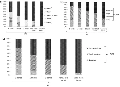
Asp-WB banding pattern analysis. At least three specific
Asp-WB bands were observed in 84% of the aspergillosis s.l. serum WB profiles. Compared with IPD (Fig. 2A), the number of bands increased with the number of IPD bands so that 100% of the sera with at least 6 precipitin lines displayed at least 3Asp-WB bands. The same trend was observed when considering the global
Asp-WB band intensity (Fig. 2B); 94% of the sera with at least 3 precipitin lines had high or very highAsp-WB banding pattern intensities. To interpret theAsp-WB results in a semiquantitative manner, theAsp-WB results were classified into three catego-ries, depending on intensity and band number, as described in Materials and Methods. Among the aspergillosis s.l. serum samples, 28 were classified as negative, 96 were classified as weak positive, and 176 were classified as strong positive. In
agreement with the findings described above, the Asp-WB
semiquantitative results correlated (r ⫽ 0.77) with the IPD banding pattern, as depicted inFig. 2C.
DISCUSSION
[image:3.585.39.288.78.156.2]The main findings of the study were the relatively high sensitivity of the detection of specific bands with the evaluatedAspergillus -specific IgG Western blot detection kit for the diagnosis of asper-gillosis in nonimmunocompromised patients and the lack of cor-relation between any particular banding feature and clinical presentations of aspergillosis. A major advantage of using a com-mercialized kit is the uniformity of reagents. Indeed, the standard-ization of diagnostic tests is critical when epidemiological studies and therapeutic trials evaluating diagnostic or therapeutic strate-gies for aspergillosis treatment require multicenter enrollment. TABLE 2Details of semiquantification of theAsp-WB results
No. of bands
Resultsa
Negative Weak positive Strong positive
0–1 NA NA NA
2 NA 2ⱕIⱕ4 4⬍I
3 NA 3ⱕIⱕ6 6⬍I
4 NA 4ⱕIⱕ8 8⬍I
aResults are from the combination of the number of 16-, 18- to 20-, 22-, and 30-kDa
Aspergillus-specific bands detected and the sum of each detected band’s intensity (I) relative to the positive control, as illustrated inFig. 1B. NA, not available.
on May 16, 2020 by guest
http://jcm.asm.org/
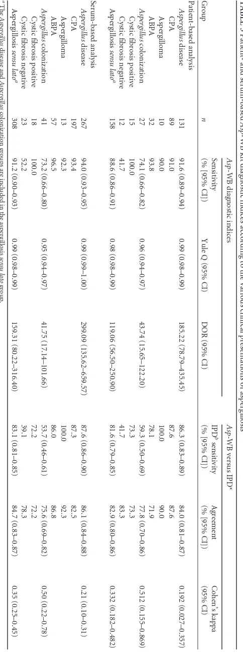
TABLE 3 Patient-and serum-based Asp -WB kit diagnostic indices according to the various clinical presentations of aspergillosis Group n Asp -WB diagnostic indices Asp -WB versus IPD* Sensitivity (% [95% CI]) Yule Q (95% CI) DOR (95% CI) IPD b sensitivity (% [95% CI]) Agreement (% [95% CI]) Cohen’s kappa (95% CI) Patient-based analysis Aspergillus disease 131 91.6 (0.89–0.94) 0.99 (0.98–0.99) 185.22 (78.79–435.45) 86.3 (0.83–0.89) 84.0 (0.81–0.87) 0.192 (0.027–0.357) CPA 89 91.0 87.6 87.6 Aspergilloma 10 90.0 100.0 90.0 ABPA 32 93.8 78.1 71.9 Aspergillus colonization 27 74.1 (0.66–0.82) 0.96 (0.94–0.97) 43.74 (15.65–122.20) 59.3 (0.50–0.69) 77.8 (0.70–0.86) 0.512 (0.155–0.869) Cystic fibrosis positive 15 100.0 73.3 73.3 Cystic fibrosis negative 12 41.7 41.7 83.3 Aspergillosis sensu lato a 158 88.6 (0.86–0.91) 0.98 (0.98–0.99) 119.06 (56.50–250.90) 81.6 (0.79–0.85) 82.9 (0.80–0.86) 0.332 (0.182–0.482) Serum-based analysis Aspergillus disease 267 94.0 (0.93–0.95) 0.99 (0.99–1.00) 299.09 (135.62–659.57) 87.6 (0.86–0.90) 86.1 (0.84–0.88) 0.21 (0.10–0.31) CPA 197 93.4 87.3 82.5 Aspergilloma 13 92.3 100.0 92.3 ABPA 57 96.5 86.0 86.8 Aspergillus colonization 41 73.2 (0.66–0.80) 0.95 (0.94–0.97) 41.75 (17.14–101.66) 53.7 (0.46–0.61) 75.6 (0.69–0.82) 0.50 (0.22–0.78) Cystic fibrosis positive 18 100.0 72.2 72.2 Cystic fibrosis negative 23 52.2 39.1 78.3 Aspergillosis sensu lato a 308 91.2 (0.90–0.93) 0.99 (0.98–0.99) 159.31 (80.22–316.40) 83.1 (0.81–0.85) 84.7 (0.83–0.87) 0.35 (0.25–0.45) a The Aspergillus disease and Aspergillus colonization groups are included in the aspergillosis sensu lato group. b IPD, immunoprecipitin detection assay.
on May 16, 2020 by guest
http://jcm.asm.org/
[image:4.585.170.412.66.724.2]Furthermore, the turnaround time for results is shorter with
Asp-WB assays (⬃4 h) than with IPD assays (⬃24 to 36 h). The sensitivity and diagnostic performance of theAsp-WB assay was highlighted in both the serum- and the patient-based analyses, as evaluated by the DOR and Yule Q coefficient values. The slight concordance between theAsp-WB and IPD results (Table 3) may be at least partly explained by the heterogeneity of the IPD assays used in each of the four centers. Our objective was to evaluate the diagnostic indices of this WB test for the diagnosis of various presentations of aspergillosis and not to compare the WB with the immunoprecipitin test. Yet, in patients with various clinical pre-sentations of aspergillosis, it was noticeable that, although IPD results were included in the case definition criteria, there were fewer false-negative results with theAsp-WB assay than with the IPD assay. One exception concerned the diagnosis of aspergilloma (Table 3), in which all patients had a positive IPD result and one had a false-negativeAsp-WB result with only one intense (grade 3 on an intensity scale of 4) specific 18- to 20-kDaAsp-WB band (data not shown).
The comparison of the present findings with those of other clinical studies was limited due to the novelty of this Western blot-based test. However, some limitations of the current study are outlined. (i) Controlling for misclassification bias was
ham-pered by the composite diagnostic criteria of aspergillosis that we used (Table 1), as no efficient gold standard is currently available. Therefore, sera from some patients infected by a microorganism other than Aspergillusmight have been included in the study, which may explain at least some of the negativeAsp-WB results obtained for patients diagnosed with aspergillosis. (ii) The retro-spective study design did not allow us to calculate the predictive values of theAsp-WB assay. (iii) Blood donors were considered healthy, although no data were available concerning their poten-tial exposure toAspergillusfungi. Therefore, the underlying rea-son behind positiveAsp-WB results obtained for some controls remains unclear. The choice to use blood donors as controls was based on the difficulty in ruling out the diagnosis of aspergillosis in at-risk patients due to the relatively poor sensitivity of the current diagnostic criteria. (iv) Cross-reactions of theAsp-WB assay were not evaluated because, according to the study design, all included patients were infected or colonized by anAspergillussp. alone or in combination with other fungi. (v) Specific banding patterns ac-cording toAspergillusspecies were not evaluated, asA. fumigatus
was isolated in all patients, either alone or in combination with anotherAspergillusspecies (data not shown).
Assessments of band numbers and intensities in the semiquan-titative interpretation of WB results have been used for the diag-FIG 2(A) Repartition ofAsp-WB banding profile according to immunoprecipitin (IPD) band number. (B) Repartition ofAsp-WB global intensity according to IPD banding profile. (C) Repartition ofAsp-WB categories (combining band numbers and intensities) according to IPD banding profile.
on May 16, 2020 by guest
http://jcm.asm.org/
[image:5.585.44.538.63.422.2]nosis of various infectious diseases, including HIV (17), Lyme borreliosis (18), andHelicobacter pyloricarriage (19). While nei-ther a particularAsp-WB banding pattern nor a semiquantitative
Asp-WB result was significantly associated with any of the asper-gillosis clinical presentations (data not shown), we found a signif-icant correlation between semiquantitativeAsp-WB results and the IPD band number. Using a semiquantitative interpretation might therefore facilitate the transition from semiquantitative IPD analysis toAsp-WB assay in the clinical practice.
AsAspergilluscolonization is considered a pathway to infec-tion, the management of clinically asymptomatic patients with
Aspergillussp. colonization remains a matter of debate. In line with this hypothesis, it has been demonstrated that persistent col-onization can induce an antibody response, and according to some authors, this seroconversion should prompt the reinforce-ment of patient monitoring and/or the start of antifungal therapy (20–22). The primary interest of including colonized patients in this evaluation is that they are typically those in whomAspergillus
serology is performed. In contrast to effect in CF patients (21, 23–25), little is known concerning the impact ofAspergillus colo-nization in non-CF patients. Despite the relatively small sample size, we observed a striking difference inAsp-WB sensitivity, with 100% and 42% in CF and non-CF patients, respectively. The spec-ificity ofAsp-WB to detectAspergilluscolonization in CF patients was not determined because we did not study noncolonized CF patients. Our data show that in CF patients, theAsp-WB assay is positive inAspergillusdisease and inAspergilluscolonization. Fur-ther research is required to determine wheFur-therAspergillus-specific IgG apparition is an early marker of aspergillosis onset.
In conclusion, this novel Western blot assay designed to detect anti-Aspergillus antibodies may be useful for the diagnosis of aspergillosis in immunocompetent patients. Its sensitivity was higher than that of the IPD assay (the current reference in
anti-Aspergillusantibody detection assays), as highlighted by nonover-lapping 95% CI (Table 3). Further prospective studies are re-quired to gain further insight into the clinical significance of
Asp-WB results in diagnosing the various aspergillosis clinical pre-sentations and monitoring patients.
ACKNOWLEDGMENTS
We are grateful to B. Michel, A. Forticaux, and D. Petkova for English language editing and the LDBio Diagnostics team for technical assistance. A.O. is a Ph.D. student employee at LDBio Diagnostics. A.C. is a tech-nician employee in part at LDBio Diagnostics.
This study was sponsored by LDBio Diagnostics. LDBio Diagnostics participated in the study design but did not interfere with the analyses or conclusions reported herein.
Aspergillus fumigatusantigen and diverse parasite antigens that are sold to LDBio Diagnostics are produced at the institution where P.F. is currently employed.Toxoplasma gondiiantigen that is sold to LDBio Di-agnostics is produced at the institution where S.R. and R.P. are currently employed.
C.H. received a research grant from Bio-Rad.
REFERENCES
1.Sherif R, Segal BH.2010. Pulmonary aspergillosis: clinical presentation, diagnostic tests, management and complications. Curr Opin Pulm Med
16:242–250.http://dx.doi.org/10.1097/MCP.0b013e328337d6de. 2.Zmeili OS, Soubani AO.2007. Pulmonary aspergillosis: a clinical update.
QJM100:317–334.http://dx.doi.org/10.1093/qjmed/hcm035.
3.Nam H-S, Jeon K, Um S-W, Suh GY, Chung MP, Kim H, Kwon OJ, Koh W-J. 2010. Clinical characteristics and treatment outcomes of
chronic necrotizing pulmonary aspergillosis: a review of 43 cases. Int J Infect Dis14:e479 – e482.http://dx.doi.org/10.1016/j.ijid.2009.07.011. 4.Jewkes J, Kay PH, Paneth M, Citron KM.1983. Pulmonary
aspergil-loma: analysis of prognosis in relation to haemoptysis and survey of treat-ment. Thorax38:572–578.http://dx.doi.org/10.1136/thx.38.8.572. 5.Kurup VP.2005.Aspergillusantigens: which are important? Med Mycol
43(Suppl 1):S189 –S196.http://dx.doi.org/10.1080/13693780500064763. 6.Persat F.2012.Aspergillusserology, from yesterday to today for
tomor-row. J Mycol Med 22:72– 82. (In French.)http://dx.doi.org/10.1016/j .mycmed.2012.01.004.
7.Van Toorenenbergen AW.2012. Between-laboratory quality control of au-tomated analysis of IgG antibodies againstAspergillus fumigatus. Diagn Mi-crobiol Infect Dis 74:278 –281. http://dx.doi.org/10.1016/j.diagmicrobio .2012.07.002.
8.Kauffmann HF, De Vries K.1980. Antibodies againstAspergillus fumiga-tusII: identification and quantification by means of crossed immunoelec-trophoresis. Int Arch Allergy Appl Immunol62:265–275.http://dx.doi .org/10.1159/000232522.
9.Baxter CG, Denning DW, Jones AM, Todd A, Moore CB, Richardson MD.2013. Performance of twoAspergillusIgG EIA assays compared with the precipitin test in chronic and allergic aspergillosis. Clin Microbiol Infect19:E197–E204.http://dx.doi.org/10.1111/1469-0691.12133. 10. Franck J, Garin YJ-F, Dumon H.2008. LDBio-Toxo II
immunoglob-ulin G Western blot confirmatory test for anti-toxoplasma antibody detection. J Clin Microbiol46:2334 –2338.http://dx.doi.org/10.1128 /JCM.00182-08.
11. Guan M.2007. Frequency, causes, and new challenges of indeterminate results in Western blot confirmatory testing for antibodies to human im-munodeficiency virus. Clin Vaccine Immunol14:649 – 659.http://dx.doi .org/10.1128/CVI.00393-06.
12. Fourneret-Vivier A, Lebeau B, Mallaret MR, Brenier-Pinchart MP, Brion JP, Pinel C, Garban F, Pison C, Hamidfar R, Plantaz D, Pelloux H, Grillot R.2006. Hospital-wide prospective mandatory surveillance of invasive aspergillosis in a French teaching hospital (2000 –2002). J Hosp Infect62:22–28.http://dx.doi.org/10.1016/j.jhin.2005.06.013.
13. Jubin V, Ranque S, Stremler Le Bel N, Sarles J, Dubus J-C.2010. Risk factors forAspergilluscolonization and allergic bronchopulmonary asper-gillosis in children with cystic fibrosis. Pediatr Pulmonol45:764 –771.
http://dx.doi.org/10.1002/ppul.21240.
14. Garnaud C, Brenier-Pinchart M-P, Thiebaut-Bertrand A, Hamidfar R, Quesada J-L, Bosseray A, Lebeau B, Mallaret M-R, Maubon D, Saint-Raymond C, Pinel C, Hincky V, Plantaz D, Cornet M, Pelloux H.2012. Seven-year surveillance of nosocomial invasive aspergillosis in a French University Hospital. J Infect65:559 –567.http://dx.doi.org/10.1016/j.jinf .2012.08.006.
15. Izumikawa K, Tashiro T, Tashiro M, Takazono T, Kosai K, Morinaga Y, Kurihara S, Nakamura S, Imamura Y, Miyazaki T, Tsukamoto M, Kakeya H, Hayashi T, Yanagihara K, Nagayasu T, Kohno S. 2014. Pathogenesis and clinical features of chronic pulmonary aspergillosis: is it possible to distinguish CNPA and CCPA clinically? J Infect Chemother
20:208 –212.http://dx.doi.org/10.1016/j.jiac.2013.10.016.
16. Fricker-Hidalgo H, Coltey B, Llerena C, Renversez J-C, Grillot R, Pin I, Pelloux H, Pinel C.2010. Recombinant allergens combined with biolog-ical markers in the diagnosis of allergic bronchopulmonary aspergillosis in cystic fibrosis patients. Clin Vaccine Immunol17:1330 –1336.http://dx .doi.org/10.1128/CVI.00200-10.
17. Burke DS, Redfield RR, Putman P, Alexander SS.1987. Variations in Western blot banding patterns of human T-cell lymphotropic virus type III/lymphadenopathy-associated virus. J Clin Microbiol25:81– 84. 18. Zoller L, Burkard S, Schafer H.1991. Validity of Western immunoblot
band patterns in the serodiagnosis of Lyme borreliosis. J Clin Microbiol
29:174 –182.
19. Simán JH, Engstrand L, Berglund G, Florén C-H, Forsgren A.2005. Evaluation of Western blot CagA seropositivity inHelicobacter pylori -seropositive and -seronegative subjects. Clin Diagn Lab Immunol12:304 – 309.http://dx.doi.org/10.1128/CDLI.12.2.304-309.2005.
20. Barberan J, Alcazar B, Malmierca E, Garcia de la Llana F, Dorca J, Del Castillo D, Villena V, Hernandez-Febles M, Garcia-Perez F-J, Granizo J-J, Gimenez M-J, Aguilar L, ASP Investigator Group.2012. Repeated
Aspergillusisolation in respiratory samples from non-immunocompro-mised patients not selected based on clinical diagnoses: colonisation or infection? BMC Infect Dis12:295.http://dx.doi.org/10.1186/1471-2334 -12-295.
on May 16, 2020 by guest
http://jcm.asm.org/
21. Bardana EJ, Jr, Sobti KL, Cianciulli FD, Noonan MJ.1975.Aspergillus
antibody in patients with cystic fibrosis. Am J Dis Child129:1164 –1167. 22. Tashiro T, Izumikawa K, Tashiro M, Takazono T, Morinaga Y,
Yamamoto K, Imamura Y, Miyazaki T, Seki M, Kakeya H, Yamamoto Y, Yanagihara K, Yasuoka A, Kohno S.2011. Diagnostic significance of
Aspergillusspecies isolated from respiratory samples in an adult pneumol-ogy ward. Med Mycol49:581–587.http://dx.doi.org/10.3109/13693786 .2010.548084.
23. Fillaux J, Brémont F, Murris M, Cassaing S, Tétu L, Segonds C, Pipy B, Magnaval JF.2014.Aspergillussensitization or carriage in cystic fibrosis
patients. Pediatr Infect Dis J33:680 – 686.http://dx.doi.org/10.1097/INF .0000000000000231.
24. Speirs JJ, Van der Ent CK, Beekman JM.2012. Effects of Aspergillus fumigatuscolonization on lung function in cystic fibrosis. Curr Opin Pulm Med18:632– 638.http://dx.doi.org/10.1097/MCP.0b013e328358d50b. 25. De Vrankrijker AMM, Van der Ent CK, Van Berkhout FT, Stellato
RK, Willems RJL, Bonten MJM, Wolfs TFW.2011.Aspergillus fumiga-tuscolonization in cystic fibrosis: implications for lung function? Clin Microbiol Infect 17:1381–1386. http://dx.doi.org/10.1111/j.1469-0691 .2010.03429.x.