Serovar Typhi Isolates
Lay Ching Chai1,2, Boon Hong Kong1,2, Omar Ismail Elemfareji1,2, Kwai Lin Thong1,2*
1Institute of Biological Sciences, Faculty of Science, University of Malaya, Kuala Lumpur, Malaysia,2Laboratory of Biomedical Science and Molecular Microbiology, Institute of Graduate Studies, University of Malaya, Kuala Lumpur, Malaysia
Abstract
Background:Salmonella entericaserovar Typhi (S.Typhi) is strictly a human intracellular pathogen. It causes acute systemic (typhoid fever) and chronic infections that result in long-term asymptomatic human carriage.S. Typhi displays diverse disease manifestations in human infection and exhibits high clonality. The principal factors underlying the unique lifestyle of
S.Typhi in its human host during acute and chronic infections remain largely unknown and are therefore the main objective of this study.
Methodology/Principal Findings:To obtain insight into the intracellular lifestyle ofS.Typhi, a high-throughput phenotypic microarray was employed to characterise the catabolic capacity of 190 carbon sources inS.Typhi strains. The success of this study lies in the carefully selected library of S. Typhi strains, including strains from two geographically distinct areas oftyphoid endemicity, an asymptomatic human carrier, clinical stools and blood samples and sewage-contaminated rivers. An extremely low carbon catabolic capacity (27% of 190 carbon substrates) was observed among the strains. The carbon catabolic profiles appeared to suggest thatS.Typhi strains survived well on carbon subtrates that are found abundantly in the human body but not in others. The strains could not utilise plant-associated carbon substrates. In addition, a -glycerolphosphate, glycerol, L-serine, pyruvate and lactate served as better carbon sources to monosaccharides in theS.
Typhi strains tested.
Conclusion:The carbon catabolic profiles suggest thatS. Typhi could survive and persist well in the nutrient depleted metabolic niches in the human host but not in the environment outside of the host. These findings serve as caveats for future studies to understand how carbon catabolism relates to the pathogenesis and transmission of this pathogen.
Citation:Chai LC, Kong BH, Elemfareji OI, Thong KL (2012) Variable Carbon Catabolism amongSalmonella entericaSerovar Typhi Isolates. PLoS ONE 7(5): e36201. doi:10.1371/journal.pone.0036201
Editor:Dipshikha Chakravortty, Indian Institute of Science, India
ReceivedDecember 12, 2011;AcceptedApril 3, 2012;PublishedMay 25, 2012
Copyright:ß2012 Chai et al. This is an open-access article distributed under the terms of the Creative Commons Attribution License, which permits unrestricted use, distribution, and reproduction in any medium, provided the original author and source are credited.
Funding:The work was supported by grant from University of Malaya UMC/HIR/MOHE/02 [A000002–50001]. The funders had no role in study design, data collection and analysis, decision to publish, or preparation of the manuscript.
Competing Interests:The authors have declared that no competing interests exist.
* E-mail: thongkl@um.edu.my
Introduction
TheSalmonella entericaserovar Typhi (S. Typhi) is an important human intracellular pathogen of global importance, infecting as many as 21.7 million people and killing 217,000 people annually [1]. The pathogen causes typhoid fever in humans and is more prevalent in less developed countries in South Central and Southeast Asia with poor sanitation and unsafe water and food supply [1]. The key risk factors of the disease include consumption of contaminated water and food and contact with human carriers. In Malaysia, healthy human carriers were detected in Kelantan, which is a hotspot for typhoid fever in Malaysia [2].S. Typhi is rarely detected in water and food. Unlike other serovars of Salmonella, which invade only the mucosal surface of the intestines, S. Typhi has evolved the ability to spread to deeper tissues, including the liver, spleen, bone marrow and gallbladder, in which it persists, multiplies and disseminates in the urine and faeces [3]. S. Typhi is exclusively adapted to infect human host.
Despite the unique pathology and epidemiologic characteristics ofS. Typhi, it has received less attention globally as compared with other Salmonella serovars, such as S. Typhimurium and S.
En-teritidis [4]. As the world continues to focus on many recent findings concerning the pathogenic and adaptive mechanisms of foodborneS. Typhimurium andS. Enteritidis, the principal factors underlying the unique epidemiological pattern and disease manifestation of this virulent, human-restricted, intracellular pathogen,S. Typhi, remains intruiging. In recent years, increasing evidence has implicated carbon catabolism as a virulence determinant of human pathogens [5-7]. The ability of pathogenic bacteria to metabolize various nutrients, especially carbon sources, is essential for the invasion, growth, survival and colonisation in intestinal and extra-intestinal sites in their hosts. To successfully colonise and persist in the various niches within the host during the course of infection, bacterial pathogens need to adjust and adapt their metabolic activity to the local nutrient availability. None-theless, as compared with the growing knowledge of molecular bacterial virulence and pathogenesis, research on pathogenic bacterial metabolism and persistence in the human host has progressed very little [6].
other living organisms; able to cause both acute and chronic infection, displaying various disease manifestations; and able to transform the human host into long-term asymptomatic carriage in the environment with periodical dissemination via urine and faeces. Therefore, it is important to understand the intracellular lifestyle of this unique pathogen. To do so, we employed high-throughput phenotypic microarray analysis to characterise the carbon metabolic capacity ofS.Typhi in the human host.
The novelty of this study is derived from the interesting and diverse background of each of theS.Typhi strains included in this work. These S. Typhi strains were carefully selected to include strains from Malaysia and Chile to determine whether there were any metabolic differences between the two areas of typhoid endemicity which are distantly separated. To explore our hypothesis that the metabolic capacity of the strains isolated from stool develops an adaptive persistence mechanism in the liver or gallbladder during chronic infection, these stool strains will differ from the strains isolated from the blood during acute systemic infection in the human host. A bacterial strain originated from a healthy human carrier was included in this study to contrast with the strains of transient chronic infection. The occurrence of S. Typhi in the environment is extremely rare, even though it is generally accepted that the pathogen could be transmitted via contaminated water and foods. To examine whether transient persistence in the sewage-contaminated water requires a special metabolic capacity, we have included a strain isolated from sewage-contaminated river in Chile during the 1983 Chilean Typhoidal outbreak [8].
Results and Discussion
Phenotypic Characteristics
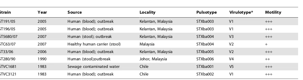
The eight uniqueS. Typhi strains studied in this work were characterised into six pulsotypes based on pulsed-field gel electrophoresis, which was performed previously in the laboratory (unpublished data). Of these strains, ST191/05 and ST196/05, both from the Kelantan outbreak in 2005, shared a similar pulsotype. Similarly, strains ST5680/07, from another outbreak in 2007, and STC63/07 were isolated from a healthy human carrier in the same locality, were and found to display identical pulsotypes. This observation led to the speculation that the chronic human carriage acts as the pathogen reservoir in this hyperendemic state. However, the virulotyping of 22 virulence and virulence-associated genes clearly distinguished the outbreak strain ST5680/07 from the carrier strain, STC63/07, which was also grouped together with ST33/06 (Table 1). Further analysis of motility and carbon catabolism revealed divergent responses for these two strains, therefore suggesting that ST5680/07 and STC63/07 are two unique strains. However, strains ST191/05 and ST196/05 with similar pulsotype also shared similar virulotypes, motility characteristic and carbon catabolic profiles. Undoubtedly, ST191/05 and ST196/05 possibly originated from the same ancestral strain.
Fimbrial genes are located in the chromosome and on the plasmid. TheagfA,agfC,lpfA,lpfC,sefC, andsefD andpefA genes are important for adherence to different sites of the host cells; any loss of these fimbrial genes will decrease the ability ofSalmonellafor adherence in the host cells [9], [10]. The S. Typhi strains tested lacked plasmids (data not shown); thus, it was not surprising that thespvBandspvC,pefA,lpfA andlpfC genes, which are located on the virulence plasmid, were absent. Nevertheless, variations among the strains were observed in the aggregative fimbriae gene (agfC), the SEF14 fimbriae genes (sefC and sefD) and the genes involved in the intra-macrophage survivability (mgtC). The strains
ST190/07 and ST196/07, derived from the 2007 typhoid outbreak, and strain STVC3121, derived from Chilean typhoid outbreak, possessed 17 out of 22 virulence and virulence-associated genes tested. The fimbriae genes,sefC andsefD, were not detected in strains ST5680/07 and ST280/90. In addition, mgtC gene was absent in strain ST280/90. The aggregative fimbriae gene (agfC) was detected in all strains except the Chilean environmental strain, STVC1681. The virulotype profiles are shown in Table 1.
We have observed remarkably little motility in the carrier strain, STC63/07, while strain ST5680/07 demonstrated rapid swarm-ing on the agar surface (Table 1). The low motility rate was also recorded in the Johor strain, ST280/90, which was isolated from stool (Table 1). In fact, motility had been identified as a bacterial virulence factor that aids in the gut colonisation to initiate infection in the human host. The observation of a relatively weaker motility capacity in the chronic carrier strain suggests that motility might not be essential for the long-term persistence in the human host. This postulation requires further investigation to better understand the adaptive life style of S.Typhi within the human host.
Limited Carbon Catabolism Capacity amongS.Typhi Strains
S. Typhimurium LT-2, including mucic acid. However, not all compounds used byS.Typhimurium (about 15 compounds) [11] could serve as the sole carbon sources forS.Typhi. Perhaps, the capability to catabolise a wider range of carbon sources is an essential property for bacterial survival in a broader host range or environment. Nonetheless, this hypothesis requires further inves-tigation.
S.Typhi strain ST280/90 demonstrated extremely low carbon metabolic activity. Virulotyping revealed ST280/90 as a variant strain lacking mgtCgene.mgtC encodes for a 25-kDa protein of unknown function. InS. Typhimurium [14] andS. Typhi [15], the experimental evidence suggested that mgtC participates in the adaptation to low-Mg2+ environments, supporting bacterial invasion and proliferation in macrophages. MgtC is a virulence factor that plays a vital role that is not supplied by any other bacterial factor located inSalmonellaPathogenicity Island-3 (SPI-3) [15]. The related studies have shown that the Salmonella mutant lackingmgtCgrew significantly less than the wild type at 10mM of MgCl2whereas no difference was observed between the mutant and wild type at 10 mM Mg2+[15]. Therefore, we suspected that the concentration of MgCl2 (50mM) in the Biolog inoculating fluid, IF-0 [16], might be too low to promote maximum growth of the variant strain. To clarify the speculation, we cultivated the variant strain, ST280/90, in M9 minimal media supplemented with glucose and 10mM, 50mM and 100mM of MgCl2. We observed no growth in all the three MgCl2concentrations tested (data not shown). Despite its poor growth in most carbon sources, we find it rather intriguing that ST280/90 showed comparable growth as with the other seven strains in glycerol, a-methyl-D-galactoside, 2-deoxyadenosine, adenosine and L-serine (Figure 1). Further investigation is needed to find out how these carbon sources compensate for the lack ofmgtC gene, particularly theS. Typhi strain.
S.Typhi Showed Active Respiration on Nucleosides,a -glycerolphosphate, Glycerol, L-serine, Pyruvate and Lactate
S.Typhi strains were able to uptake and metabolise most of the monosaccharides tested, such as glucose, fructose, D-galactose and D-glucose-1-phosphate. However, relative to the respiration rate on nucleosides,a-glycerolphosphate, glycerol,
L-serine, pyruvate and lactate, monosaccharides supported only moderate respiration in S. Typhi strains (Figure 1). It was not surprising that nucleosides were rapidly catabolised byS.Typhi strains, as numerous studies have reported that nucleosides are excellent carbon sources in E. coli and Salmonella [17],[18]. We believe that the active metabolism of nucleosides supports the rapid proliferation of S. Typhi during invasive infection of the bloodstream. Human blood serum is abundant in various metabolites, including various amino acids and nucelosides [19], [20]. The efficient uptake and catabolism of nucleosides during the infection process provides sufficient substrates for biosynthesis of nucleotide bases required for DNA synthesis and repair of damaged DNA induced by the host’s reactive oxygen intermediates during infection [21].
Alpha-glycerolphosphate and glycerol are abundant in the liver and kidneys. In the human body, glycerol is rapidly adsorbed in the intestines and stomach and distributed over the extracellular space [22]. Glycerol is phosphorylated to a -glycerophosphate by glycerol kinase, predominantly in the liver (80–90%) and kidneys (10–20%) [22]. Glycerol, L-serine, pyruvate and lactate serve as the substrate or end product of gluconeogenesis and glycolysis, which also occurs predominantly in the liver and to a lesser extend in the cortex of kidney. Therefore, the observation of the relatively active metabolism of these carbon sources was highly speculated to be associated to adaptive mechanisms acquired byS. Typhi for the colonisation and long-term persistence in the human host, specifically in the liver, during chronic infection in the human host. The availability of a-glycerophosphate, glycerol, L-serine, pyruvate and lactate in the cortex of the kidney suggested the possible transient colonisation of S.Typhi in human kidneys and causes the dissemination of S. Typhi via urine. In fact, it has been known for years that the typhoid human carriers disseminate the pathogen through faeces and urine. This observation will be investigated more in depth in a future study.
Distinct Carbon Catabolism Profile between Strains Isolated from Human Stools and Blood
[image:3.612.66.562.79.211.2]PCA analysis exhibited clearly distinct carbon catabolic activity between the two clusters ofS.Typhi strains: ST191/05, ST196/05 and ST33/06 as compared with ST5680/07, ST280/90 and Table 1.Salmonella entericaserovar Typhi strains used in this study.
Strain Year Source Locality Pulsotype Virulotype* Motility
ST191/05 2005 Human (blood); outbreak Kelantan, Malaysia STXba003 V1 +++
ST196/05 2005 Human (blood); outbreak Kelantan, Malaysia STXba003 V1 +++
ST5680/07 2007 Human (stool); outbreak Kelantan, Malaysia STXba004 V3 +++
STC63/07 2007 Healthy human carrier (stool) Malaysia STXba004 V2 +
ST33/06 2006 Human (blood); outbreak Kelantan, Malaysia STXba005 V2 +++
ST280/90 1990 Human (stool);outbreak Johor, Malaysia STXba006 V4 ++
STVC1681 1983 Sewage contaminated water Chile STXba001 V5 +++
STVC3121 1983 Human (blood); outbreak Chile STXba002 V1 +++
*Virulence genes profiles among 8S.Typhi strains tested.
(V) :agfA-agfC-cdtB-invA-iroN-lpfA-lpfC-mgtC-misL-orfL-pefA-pipD-prgH-sefC-sefD-sifA-sitC-sopB-sopE-spiC-spvB-spvC. (V1):agfA-agfC-cdtB-invA-iroN- -mgtC-misL-orfL- -pipD-prgH-sefC-sefD-sifA-sitC-sopB-sopE-spiC-.
STC63/07 (P.0.05) (Table 3). The differences observed appeared to be associated with the origin of the strains. The strains ST191/ 05, ST196/05 and ST33/06 were all isolated from human blood samples, sharing a similarity of 86.7% to 91.2% in their carbon catabolic profile whereas the catabolic activity among the strains obtained from stool samples (ST280/90, ST5680/07 and STC63/07) was more diverse, with a lower similarity level, which ranged from 82.5% to 87.6%. Although the clinical Chilean strain STVC3121 was also isolated from human blood, it was catabolically unique from both clusters and from the environmental Chilean strain (STVC1681). Notably, all the three strains derived from the stool samples had a narrower range in carbon utilisation and were comparatively weaker in their catabolic activity (P,0.05; Table 3), specifically in the catabolism of uridine and D-glucose-6-phosphate. The motility test and growth in rich medium confirmed the findings discussed above, as both analyses demonstrated lower motility and delayed growth in S. Typhi strains from stools. The ability to catabolise glucose-6-phosphate has been associated with the intracellular survival and virulence of Salmonellae in mice [23]. This observation was indeed intriguing, as S. Typhi strains isolated from stool and blood actually represented two different stages in human infection and colonisation niches within the human body. S.Typhi is able to invade the intestinal wall and replicate within macrophages and infected phagocytes [24]. The
replication of the bacteria within macrophages in the liver and spleen [25], [26] resulted in the release of the pathogen into the bloodstream [27]. The pathogen later invades the gallbladder and leads to bacterial shedding in the urine and faeces in the chronic carriage of infected individuals [28], [29]. It is possible that S. Typhi acquires different metabolic activity and pheno-types for colonisation and persistence in these two different niches, liver and spleen, which disseminate S. Typhi into the blood stream and the gallbladder, which releasesS. Typhi in the urine and faeces. However, we could not entirely rule out the possibility that the strains could be grouped simply by the predominant strain in a given time and place. That is, the strains from 2005 and 2006 from Kelantan could simply be fortuitously similar whereas those strains isolated from 2007 would be similar; these strains would also be similar to those isolated in 1990 and the Chilean strains would comprise their own group. This grouping could be addressed through the study of more strains, preferably from the same outbreak, isolated both from blood and stools. A comprehensive study would address the phenotypic differences in S. Typhi.
High Divergence in Carboxylic Acids Catabolism
[image:4.612.67.557.77.425.2]Another notable finding in this study was the observation of great divergence in the catabolism of amino acids, fatty acids and other carboxylic acids. While less variation was recorded in the Table 2.Catabolism of 190 carbon substrates bySalmonella entericaserovar Typhi strains.
Carbon substrate
*No. of carbon substrates catabolised by all strains
*No. of carbon substrates catabolised by at least a strain
*No. of carbon substrates not catabolised
*No. of carbon substrates tested
Sugars and derivatives Monosaccharide 9(45) 1(5) 10(50) 20
Disaccharide 2(22) 7(78) 9
Oligosaccharide 1(10) 9(90) 10
Polysaccharide 1(17) 5(83) 6
Sugar alcohol 4(22) 14(78) 18
Amino sugar 3(38) 5(63) 8
Deoxy sugar 4(100) 4
Aldaric acid 1(20) 4(80) 5
Aldonic acid 1(50) 1(50) 2
Uronic acid 3(38) 5(63) 8
Glycoside 1(11) 8(89) 9
Carboxylic acids and derivatives Monocarboxylic acid 1(11) 2(22) 6(67) 9
Dicarboxylic acid 11(100) 11
Tricarboxylic acid 1(33) 2(67) 3
Keto acid 1(11) 1(11) 7(78) 9
Fatty acid 1(14) 6(86) 7
Amino acid and peptide 5(15) 6(18) 22(67) 33
Amide 3(100) 3
Ketone 3(100) 3
Lactone and ester 2(40) 3(60) 5
Polysorbate surfactant 3(100) 3
Nucleic acid Nucleotide 5(100) 5
TOTAL 39(21) 13(7) 138(73) 190
*Number of substrate catabolised by all strains or some strains or not able to be catabolised by any strains (percentage of substrate catabolised by all strains or some strains or not able to be catabolised by any strains).
metabolism of sugars, one strain ofS.Typhi was unable to grow on these six amino acids (proline, D-alanine, alanine, L-glutamic acid, L-asparagine and L-threonine) and three short-chain fatty acids (propionic acid, ketobutyric acid and a-hydroxybutyric acid). The phenotypic microarray analyses revealed that both the carrier (STC63/07) and the Johor strain (ST280/90) lacked the catabolic pathways to uptake and assimilate most of the amino acids and fatty acids stated above. Studies have shown that the ability to catabolise amino acids in Salmonella is essential for the virulence in mice [30–33]. In addition, the catabolism of C2 substrates (e.g., fatty acids) plays a crucial role inSalmonella metabolism in-vivo as the mutants of S.
Typhimurium that had lost their ability to grow on C2 substrates are attenuated in mice [34]. The ability to metabolise on fatty acids might also play a role in the early stages of infection, when the bacteria reside within the lipid-rich mucus covering the intestinal epithelium [35]. Metabolism of short chain fatty acids might also be involved in the signaling required for expression of virulence genes or the induced detoxification of these com-pounds, which can have antimicrobial effects at high concentra-tions [35–37]. Additional work is needed to determine how the ability to catabolise carbon substrates relates to the virulence of S. Typhiin-vivo.
Figure 1. Cluster analysis based on the carbon catabolism profile of the eight Salmonella enterica serovar Typhi strains. The dendrogram and catabolic profiles show 52 active carbon substrates catabolised by the eight strains ofS.Typhi tested. The cluster analysis was performed with simple matching similarity based on the catabolic profile of 190 carbon substrates and the dendrogram was built with unweighted paired group of arithmetic mean (UPGMA).
[image:6.612.58.504.262.677.2]doi:10.1371/journal.pone.0036201.g001
Figure 2. Three-dimensional principle component analysis (PCA) of eight Salmonella enterica serovar Typhi strains. A three-dimensional principle component analysis (PCA) of the eightS.Typhi strains was calculated from the corrected average area under the bacterial growth curve. For each carbon well, the average area under the bacterial growth curve was corrected to zero if the Tmax–Tminvalue was lesser than
the threshold value of 50.
Conclusion and Future Directions
In conclusion, the findings obtained in this study have revealed interesting variations in the carbon catabolism among theS.Typhi strains of a different background. The observation was indeed astonishing, as carbon catabolism is one of the earliest means used by the scientists to distinguish the bacteria, and is an essential property of bacterial survival and persistence in its specific niches of habitat. The diversity observed in the carbon catabolism profiles among this set of diverseS. Typhi strains has suggested the possible involvement of various metabolic pathways that might be related to the virulence and pathogenesis of this host-restricted human pathogen. The findings obtained in this study would serve as an important foundation for future genomic and transcriptomic studies to understand the relationship of carbon metabolism to the pathogen-esis and transmission of this human-host-adapted pathogen. The questions concerning the diversity of carbon catabolism provide caveats for the future study of the pathogenicity ofS. Typhi.
Materials and Methods
Media and Strains Used
Eight clinical strains ofS.Typhi from our culture library were selected for the present work (Table 1). All strains, except STVC1681 and STVC3121 from the 1983 typhoid outbreak in Chile [38] were of local origin. Four S. Typhi strains, namely ST191/05, ST196/05, ST33/06 and ST5680/07, were isolated during the typhoid outbreaks in the northeast state of Peninsular Malaysia, Kelantan, which was a major typhoid hotspot from 2005 to 2007. The former two strains were isolated from blood samples in 2005 and were genetically identical in terms of PFGE subtyping; ST33/06 was isolated from a blood sample in 2006, and ST5680/07 was isolated from a stool sample in 2007. In 2007, a survey was performed in Kelantan to detect the presence of potential typhoid carrier. Strain STC63/07 was isolated from the stool sample of a healthy adult in Kelantan. The pulsotype of STC63/07 was indistinguishable from strain ST5680/07, which was isolated from the 2007 Kelantan typhoid outbreak (unpub-lished data).S. Typhi strain ST280/90 originated from the stool sample of a victim of another typhoid outbreak in Malaysia, Johor, in 1990 and was also included in the study [38]. All S. Typhi
strains were cultured and maintained on Luria-Bertani (LB) medium.
Motility Test
The motility tests were performed as previously described [6]. Briefly, bacteria were grown on LB agar, transferred with an inoculation needle to the centre of an LB agar plate containing 0.3% agar and incubated for 24 hours at 37uC. The plates were measured for the colony diameter at 6, 12, 18 and 24 hours. The motility test was performed in triplicate.
Virulotyping
Five multiplex PCRs were used to amplify the 22 genes:agfA, sefC andsefD [10];cdtB,iroN,lpfC,pefA,prgH,sifA,sitC,sopB and spvB [39]; misL, orfL, pipD,sopE andspiC [40]; invA [41]; mgtC [42];agfC [43];spvC [44]; andlpfA [10]. These genes are involved in adhesion, invasion, intracellular survival, systemic infection, toxin production and Mg2+ and iron uptake. The amplification was performed in a 25ml reaction mixture that included 100 ng of DNA template, 3 mM MgCl2, 0.75 U of TaqDNA Polymerase (Promega Inc., Madison, WI, U.S.A.), 1X PCR buffer, 400mM dNTPs mix and 0.4mM of each primer. The amplification was carried out in a Mastercycler Gradient (Eppendorf, U.S.A.) with the following cycling conditions: 95uC for 5 min, followed by 30 cycles of 94uC for 30 sec, 56.3uC for 30 sec, 72uC for 2 min, and a final cycle of 72uC for 10 min.
Phenotypic Microarray
[image:7.612.57.557.88.263.2]The eight strains ofS.Typhi were assayed for the carbon utilisation on Phenotype Microarray (PM) (Biolog) microplates PM1 and PM2, and the catabolism capacity of 190 different carbon substrates was tested. The PM technology uses the irreversible reduction of tetrazolium violet to formazan as a reporter of active metabolism [16]. The reduction of the dye causes the formation of a purple colour that is recorded by a charge coupled-device camera every 15 min for the duration of the incubation period. All procedures were performed as indicated by the manufacturer. First, S.Typhi isolates were streaked on LB agar and incubated at 37uC for 18 hours. Five to ten single colonies of each of the isolates were carefully picked up with a Table 3.Carbon catabolic activity amongSalmonella entericaserovar Typhi strains isolated from human blood, stool and sewage-contaminated water.
Origin Strain
No. of carbon
substrates utilised Catabolism rate Catabolism activity*
Range Average
Standard deviation
Blood{
ST191/05 51 24–173 96 35 4919
ST196/05 50 26–160 90 33 4503
ST33/06 52 31–141 79 27 4097
STVC3121 49 24–248 90 32 4418
Stool{
STC63/07 40 12–139 74 32 2940
ST280/90 43 3–138 56 33 2392
ST5680/07 51 28–155 74 33 3753
Sewage contaminated water STVC1681 51 15–151 88 31 4482
TOTAL 48 3–248 81 32 3938
*Catabolism activity = No. of carbon sources utilized6mean of catabolism rate.
{
Catabolism activity between strains of blood origin and stool origin were significantly different at P-value of 0.013 (exclude the environmental strain VC1681) and 0.029 (include strain VC1681).
moistened cotton swab and resuspended in 15 ml of Biolog inoculating fluid IF-0 until a cell density of 85% transmittance was reached. Subsequently, 1% Biolog dye A (vol/vol) was added to the cell suspension, and 100ml of the mixture was loaded into each well on PM microplate PM1 and PM2. All PM microplates were incubated at 37uC in an OmniLog reader, and the colour changes in the wells were monitored automatically every 15 min. The readings were recorded for 24 hours, and the data were analysed using OminoLog PM software, which generated a time course kinetic curve for tetrazolium colour development. The option of A1 zero was selected during data processing to deduct the background signal with reference to the A1 negative control well in each plate. Each strain was analysed in duplicate, and the results were checked for consistency.
Statistical Analysis of PM Results
For each well, the averaged product of the average area under the kinetic growth curve and the difference (Tmax–Tmin) of the kinetic data obtained in duplicate experiments were used to assay for positive growth under various PM conditions. The values of average area under the kinetic growth curve for all negative wells were analysed to obtain the background value so that a threshold can be determined to offset the average area obtained for each of the carbon substrates. A threshold value of 50 was set. The PM conditions exceeding a threshold value of 50 were further screened at the Tmax–Tminvalue. Double screening for both the average area and the differences ensured that only wells with an increasing signal and a sufficient total area represent a positive growth condition. The numerical values of the average area for positive PM conditions were used for further analysis with BioNumerics software (Applied Math, Kortrijk, Belgium).
For principal component analysis (PCA), the PM data were filtered using the corrected average growth area as a parameter and subsequently processed with BioNumerics software. For each
well, the average growth was corrected to zero if the difference was less than the threshold value of 50. Clustering analysis was performed to group the strains based on the metabolic diversity with the simple matching similarity, and the clustering was based on UPGMA.
Supporting Information
Table S1 Standardized value of the average area under the kinetic growth curve (catabolic rate) for each of the fifty-two carbon substrates catabolised by theS. Typhi strains.
[image:8.612.309.553.434.737.2](PDF)
Table S2 One-hundred and ninety various carbon sources included in the carbon metabolic profiling of the eightS. Typhi strains. Fifty-two substrates were utilized by theS. Typhi strains tested while the others were not supportive of growth ofS. Typhi. (PDF)
Acknowledgments
We thank Adjunct Professor Dr. Niyaz Ahmed for proofreading the manuscript and Professor Dr. Son Radu for the use of the Biolog equipments.
The work was performed at Laboratory of Biomedical Science and Molecular Microbiology, Institute of Graduate Studies, University of Malaya.
Author Contributions
Conceived and designed the experiments: KLT LCC. Performed the experiments: LCC KLT BHK OIE. Analyzed the data: LCC KLT. Contributed reagents/materials/analysis tools: KLT. Wrote the paper: KLT LCC.
References
1. Crump J, Mintz E (2010) Global trends in typhoid and paratyphoid fever. Clin Infect Dis 50: 241–246.
2. Ismail A (2011) Early detection of cases and carriers of typhoid and other food borne diseases. Malaysian J Pub Health Med 11: 4.
3. Parry CM, Hien TT, Dougan G (2002) Typhoid fever. N Engl J Med 347(22): 1770–1782.
4. Galanis ED M, Wong LF, Patrick ME, Binsztein N, Cieslik A, et al. (2006) Web-based surveillance and globalSalmonelladistribution, 2000–2002. Emerg Infect Dis. 12: 381–388.
5. Chang D-E, SmalleyDJ, Tucker DL, Leatham MP, Norris WE, et al. (2004) Carbon nutrition ofEscherichia coliin the mouse intestine. Proceeding of the Natl Acad Sci USA 101: 7427–7432.
6. Eylert E, Scha¨r J, Mertins S, Stoll R, Bacher A, et al. (2008) Carbon metabolism of Listeria monocytogenes growing inside macrophages. Mol Microbiol 69: 1008–1017.
7. Eisenreich W, Dandekar T, Heesemann J, Goebel W (2010) Carbon metabolism of intracellular bacterial pathogens and possible links to virulence. Nature Rev Microbiol 401–412.
8. Thong KL, Cordano AM, Yassin RM, Pang T (1996) Molecular analysis of environmental and human isolates ofSalmonella typhi. Appl Env Microbiol 62: 271–274.
9. Ba¨umler AJ, Gilde AJ, Tsolis RM, van der Velden AWM, Ahmer BM, et al. (1997) Contribution of horizontal gene transfer and deletion events to the development of distinctive patterns of fimbrial operons during evolution of
Salmonellaserotypes. J Bacteriol 179: 317–322.
10. Ba¨umler AJ, Heffron F (1995) Identification and sequence analyses of lpfABCDE, a fimbrial putative operon ofSalmonellatyphimurium. J Bacteriol 177: 2087–2097.
11. Gutnick D, Calvo JM, Klopotowski T, Ames BN (1969) Compounds which serve as the sole source of carbon or nitrogen forSalmonellaTyphimurium LT-2. J Bacteriol 100: 215–219.
12. Sternfeld L, Saunders F (1938) The fermentation of mucic acid by some interstina; bacteria. J Bacteriol 36: 53–56.
13. Sternfeld L, Saunders F (1937) The utilization of various sugars and their derivatives by bacteria. Jour. Amer Chem Soc 59: 2653–2658.
14. Blanc-Potard AB, Groisman EA (1997) TheSalmonella selC locus contains a pathogenicity island mediating intramacrophage survival. Embo J 16: 5376–5385.
15. Retamal P, Castillo-Ruiz M, Mora GC (2009) Characterization of mgtC, a virulent factor of Salmonella enterica serovar Typhi. PLoS ONE 4: e5551. doi:10.1371/journal.pone.0005551.
16. Bochner BR, Gadzinski P, Panomitros E (2001) Phenotype microarrays for high-throughput phenotypic testing and assay of gene function. Genome Res 11: 1246–1255.
17. Jensen KF, Nygaard P (1975) Purine-nucleoside phosphorylase from Escherichia coli andSalmonella typhimurium: purification and some properties. Eur J Biochem 51: 253–265.
18. Hammer-Jespersen K (1983) Nucleoside catabolism. In Metabolism of Nucleotides, Nucleosides and Nucleobases in Microorganisms, pp. 203-258. Edited by A. Munch-Petersen. New York: Academic Press.
19. Simmonds RJ, Harkness RA (1981) High-performance liquid chromatographic methods for base and nucleoside analysis in extracellular fluids and in cells. J Chromatogr 226: 369–381.
20. Aguilo A, Castano E, Tauler P, Guix MP, Serra N, et al. (2000) Participation of blood cells in the changes of blood amino acid concentrations during maximal exercise. J Nutr Biochem 11: 81–86.
21. Samant S, Lee H, Ghassemi M, Chen J, Cook JL, et al. (2008) Nucleotide biosynthesis Is critical for growth of bacteria in human blood. PLoS Pathog 4(2): e37. doi:10.1371/journal.ppat.0040037.
22. Lin ECC (1977) Glycerol utilization and its regulation in mammals. Ann Rev Biochem 46: 765–795.
23. Lundberg BE, Wolf RE, Dinauer MC, Xu Y, Fang FC (1999) Glucose-6-phosphate dehydrogenase is required forSalmonella typhimuriumvirulence and resistance to reactive oxygen and nitrogen intermediates. Infect Immun 67: 436–438.
24. Vazquez-Torres A, Jones-Carson J, Ba¨umler AJ, Falkow S, Valdivia R, et al. (1999) Extraintestinal dissemination ofSalmonellaby CD18-expressing phago-cytes. Nature 410: 804–808.
26. Salcedo SP, Noursadeghi M, Cohen J, Holden DW (2001) Intracellular replication of Salmonella typhimurium strains in specific subsets of splenic macrophages in vivo. Cell Microbiol 3: 587–589.
27. Sheppard M, Webb C, Heath F, Mallows V, Emilianus R, et al. (2003) Dynamics of bacterial growth and distribution within the liver during Salmonella infection. Cell Microbiol 5: 593–600.
28. Monack DM, Mueller A, Falkow S (2004) Persistent bacterial infections: the interface of the pathogen and the host immune system. Nat Rev Microbiol 2: 747–765.
29. Munoz-Elias E, McKinney JD (2006) Carbon metabolism of intracellular bacteria. Cell Microbiol 8: 10–22.
30. Hoiseth SK, Stocker BA (1981) Aromatic-dependentSalmonella typhimuriumare non-virulent and effective as live vaccines. Nature 291: 238–239.
31. Fields PI, Swanson RV, Haidaris CG, Heffron F (1986) Mutants ofSalmonella typhimuriumthat cannot survive within the macrophage are avirulent. Proc Natl Acad Sci USA 83: 5189–5193.
32. McFarland WC, Stocker BA (1987) Effect of different purine auxotrophic mutations on mouse-virulence of a Vi-positive strain ofSalmonellaDublin and of two strains ofSalmonella typhimurium. Microb Pathog 3: 129–141.
33. O’Callaghan D, Maskell D, Liew FY, Easmon CS, Dougan G (1988) Characterization f aromatic- and purine-dependent Salmonella typhimurium: attenuation, persistence, and ability to induce protective immunity in BALB/c mice. Infect Immun 56: 419–423.
34. Utley M, Franklin DP, Krogfelt KA, Laux DC, Cohen PS (1998) ASalmonella typhimurium mutant unable to utilize fatty acids and citrate is avirulent and immunogenic in mice. FEMS Microbiol Lett 163: 129–134.
35. Lawhon SD, Maurer R, Suyemoto M, Altier C (2002) Intestinal short-chain fatty acids alterSalmonella typhimuriuminvasion gene expression and virulence through BarA/sirA. Mol Microbiol 46: 1451–1464.
36. Horswill AR, Dudding AR, Escalante-Semerena JC (2001) Studies of propionate toxicity inSalmonella entericaidentify 2-methylcitrate as a potent inhibitor of cell growth. J Biol Chem 276: 19094–19101.
37. Brock M, Buckel W (2004) On the mechanism of action of the antifungal agent propionate. Eur J Biochem 271: 3227–3241.
38. Thong KL, Cheong YM, Puthucheary S, Koh CL, Pang T (1994) Epidemiologic analysis of sporadic Salmonella typhi isolates and those from outbreaks by pulsed-field gel electrophoresis. J Clin Microbiol 32: 1135–1141. 39. Skyberg J, Logue C, Nolan L (2006) Virulence genotyping ofSalmonellaspp. with
multiplex PCR. Avian Dis 50: 77–81.
40. Hughes L, Shopland S, Wigley P, Bradon H, Leatherbarrow A, et al. (2008) Characterization ofSalmonella entericaserotype Typhimurium isolates from wild birds in northern England from 2005–2006. BMC Vet Res 4: 4.
41. Rahn K, De Grandis S, Clarke R, Mcewen S, Gala´n J, et al. (1992) Amplification of aninvA gene sequence ofSalmonella typhimuriumby polymerase chain reaction as a specific method of detection ofSalmonella. Mol Cell Probes 6: 271–279.
42. Soto SM, Rodriguez I, Rodicio MR, Vila J, Mendoza MC (2006) Detection of virulence determinants in clinical strains ofSalmonella entericaserovar Enteritidis and mapping on macro restriction profiles. J Med Microbiol 55: 365–373. 43. Gibson DL, White AP, Rajotte CM, Kay WW (2007) AgfC and AgfE facilitate
extracellular thin aggregative fimbriae synthesis in Salmonella Enteritidis. Microbiol 153: 1131–1140.