Technology (IJRASET)
Content Based Image Retrieval for Lung CT Slices
using Texture Features
Anita Titus1, M. Bruntha2
Department of Electronics and Communication Engineering Easwari Engineering College, Chennai, India
Abstract—Content Based Image Retrieval (CBIR) is used to retrieve the relevant CT slices from the database based on the query CT slice. In the proposed method, the database consists of diseased CT slices of Cavitary TB. The processes involved in the proposed method include preprocessing, segmentation, ROI extraction and slice retrieval. The lung CT slice is segmented using adaptive thresholding and the diseased regions are extracted from the segmented CT slice by Harris corner point detection. The combination of Block Difference of Inverse Probabilities (BDIP) and Tamura feature extraction methods are proposed for the extracted diseased region and the segmented lung. The Euclidean distance measures the similarity between the features of the query CT slice and the database CT slices. Depending on the distance measures, CT slices are retrieved from the database. The performance of the proposed feature extraction methods are calculated using precision and recall parameters for better retrieval of CT slices.
Keywords—CBIR, lung segmentation, ROI extraction, BDIP and Tamura features, similarity measure, precision and recall
I. INTRODUCTION
Recent advances of technology in digital imaging and digital storage devices make it possible to easily generate, transmit, manipulate and store large number of digital images and documents. As a result of advances in the new digital image sensor technologies, the volume of digital images produced by scientific, educational, medical, and industrial increased dramatically. CBIR is the application of computer vision techniques to the image retrieval problem, that is, the problem of searching for digital images in large databases. "Content-based" means that the search analyzes the contents of the image rather than the metadata such as keywords, tags, or descriptions associated with the image. The term "content" in this context refers to colors, shapes, textures, or any other feature that can be derived from the image [1].
The CBIR system comprises of multiple inter-dependent tasks performed by various phases. Inter-tuning of all the phases of the retrieval system is inevitable for over all good results. For development of a real time CBIR system, feature processing time and query response time should be optimized. A better performance can be achieved if feature-dimensionality and space complexity of the algorithms are optimized [2]. The CBIR system extracts the features automatically from the images and the similarity of the images is found by computing the distance between the images.
II. RELATED WORK
Gandhani et al. [3] in their work proposed the combined features of shape, color and texture for the natural image database. The color features are extracted using color histogram technique. The shape features are extracted using Canny Edge Detection. The texture features are extracted using Block Difference of Inverse Probabilities (BDIP) and Block Based Local Correlation (BVLC). This method improved the retrieval performance.
Titus et al. [4] in their work proposed the algorithm for the extraction of affected regions by pleural effusion and pneumothorax in lung CT slices. The proposed method used ten texture features for both types of diseased CT slices. This proposed algorithm resulted in high accuracy for both the affected CT slices.
Titus et al. [5] in their work proposed the algorithm for the extraction of regions in Computer Aided Diagnosis. The training database follows PSO optimized wavelet feature extraction and the testing database follows wavelet feature extraction methods. This proposed work provides the exact match of different lung tissue patters using SVM classifier.
Technology (IJRASET)
Yogamangalam et al. [7] in their work explains the classification of segmentation. The classification is based on region, edge, threshold, and model. The results of segmented images are compared for better segmentation.
Khandave et al. [8] in their work algorithm for the extraction of natural images. This work involves Gray Level Co-occurrence matrix (GLCM) and Color Co-occurrence matrix (CCM) to extract the texture feature. The HSV color space is used to extract the color feature. The image retrieval using these integrated features gives better and accurate results than image retrieval using single feature.
Suhasini et al. [9] in their work a simple color-based search in is performed using color histograms and compares with the color histograms of different images using the Euclidean Distance Equation. With segmentation the performance of this system is found to be increased especially using color feature.
Comparing with the works discussed in the literature, the proposed work is different in the following aspects: The Harris Corner Detection provides the best result of regions, the combined texture feature helps in better retrieval of lung CT slices.
III. PROPOSED METHODOLOGY
The architecture of the proposed system is described below in the following sequence: preprocessing, segmentation, ROI extraction, feature extraction and slice retrieval.
Preprocessing is the initial step in image processing which is used to provide the image in the required format [10]. The preprocessing system provides resized and converted gray scale CT slice based on the 2D Gaussian filter. In order to extract the diseased features of the CT slices, the segmentation of the lung is necessary. In the proposed CBIR system, the segmentation is used to segment the left and right lung based on the adaptive thresholding. In adaptive thresholding, the removal of gray scale pixels is done with the help of global threshold [11] - [12]. ROI Extraction plays a major role of detecting the diseased regions in the segmented CT slices which helps in the retrieval process. This process is done by Harris corner point detection in the proposed method. The proposed CBIR system contains the combination of different feature extraction methods for the extraction of features from ROI. The features are extracted using the combined texture features named BDIP and Tamara.
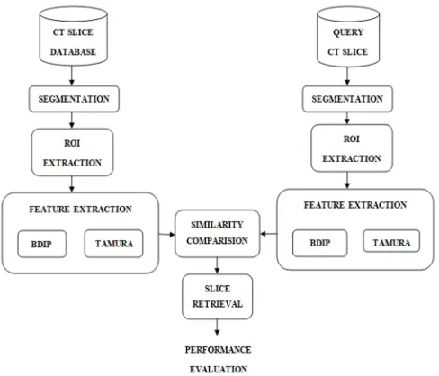
[image:3.612.184.428.408.617.2]The architecture of the proposed CBIR for lung CT slices is given in the Fig. 1. The process involved in the proposed system is explained in the following sessions.
Fig. 1 Architecture of proposed CBIR system
A. Preprocessing
Input:
Query lung CT slice Process Logic:
Technology (IJRASET)
Step 2: Convert the RGB CT slice to gray level.
Step 3: Apply 2D Gaussian filter to the gray scale CT slice using Eq. (1),
( , ) = 1
2 (1)
Where ( , ) is the gaussian function in two dimentions, x is the distance from the origin in the horizontal axis, y is the distance from the origin in the vertical axis, and σ is the standard deviation of the Gaussian distribution.
Output:
Resized and filtered gray scale lung CT slice.
B. Segmentation
Input:
Resized and filtered gray scale lung CT slice. Process Logic:
Step 1: Compute the global threshold which is an appropriate threshold for the conversion of gray scale to binary. Step 2: Convert the preprocessed lung CT slice which is in gray scale into binary using the computed global threshold. Step 3: Specify the pixel of the required intensity.
Step 4: Remove all the components less than the specified pixel.
Step 5: Perform morphological closing for binary CT slice using the created morphological structuring element. Output:
Segmented left and right lung.
C. ROI Extraction
Input:
Segmented left and right lung. Process Logic:
Step 1: Compute x and y derivatives of the segmented CT slice using Eq. (2),
= ∗ , = ∗ (2)
where is the Gaussian window function over horizontal axis, is the Gaussian window function over vertical axis, is the image.
Step 2: Compute the product of derivatives at every pixel using Eq. (3),
= ∗ , = ∗ , = ∗ (3)
Step 3: Compute the overall product of derivatives at every pixel using Eq. (4),
= ∗ , = ∗ , = ∗ ∗ (4)
Step 4: Determine the matrix H(x,y) at every pixel using Eq. (5),
( , ) = ( , ) ( , )
( , ) ( , ) (5)
Step 5: Compute the trace of the matrix using Eq. (6),
( ) = ( , ) + ( , ) (6) Step 6: Compute the region at each pixel using Eq. (7),
= ( )− ( ) (7) where is the empirical constant
Output:
Affected regions with diseases
D. Feature Extraction
Input:
Technology (IJRASET)
Step 1: Divide the regions into blocks
Step 2: Compute the BDIP feature by using the Eq. (8)
= −∑( , )∈ ( , )
max ( , )∈ ( , )
(8)
where denotes the block of size M x M, ( , ) is the value at a pixel (i,j) in the region I. Step 3: Compute the average of neighborhood of size 2 x2 at every pixel point by Eq. (9)
( , ) = ( , ) 2⁄ (9)
where ( , ) is the gray-level at ( , )
Step 4: Compute the differences between pairs of averages at every pixel in both horizontal and vertical direction by Eq. (10) and (11),
, ( , )= | ( + 2 , )− ( −2 , )| (10)
, ( , )= | ( , + 2 )− ( , −2 )| (11)
Step 5: Compute the best size at each pixel by Eq. (12)
( , ) = 2 (12)
where k maximizes E in either direction
Step 6: Compute the average of over the CT slice be the coarseness measure by Eq. (13),
= 1
( , ) (13)
where and are the width and height of the image
Step 7: Compute the contrast of CT slice with = ⁄ where is the fourth moment of the mean and is the variance by Eq. (14),
= ⁄( ) ⁄ (14)
Step 8: Compute the directionality of the CT slice by Eq. (15)
= 1− . . (∅ − ∅ )
∅∈
. (∅) (15)
where is the number of peaks, ∅ is the peak position of , is the range of peak between valleys, is the normalizing factor related to quantizing levels of ∅, ∅ is the quantized direction code, ( ) = ( )⁄∑ ( ),where =0,1,…, −1, ( ) is the number of points in the region
Output:
Extracted features
E. Slice Retrieval
Input:
Extracted features Process Logic:
Step 1: Compute the metric coefficients as the distance between the feature Step 2: Compute the distance using the Eq. (16),
( , ) = [( − ) ( − )] ⁄ (16)
where ( , ) is the Euclidean distance, is the symmetric metric as = ( ), is the metric coefficients Step 3: Retrieve the slices from the database having the highest rank of distance
Output:
Retrieved Slices
Technology (IJRASET)
Input:
Retrieved Slices Process Logic:
Step 1: Note down the number of slices retrieved Step 2: Note down the relevant slices from the retrieval Step 3: Compute the precision by Eq. (17)
=
+ (17)
Step 4: Compute the recall by Eq. (18)
=
+ (18)
Step 5: Compute the accuracy of the system by Eq. (19)
= +
+ + + (19)
Output:
Precision, Recall and Accuracy
IV. RESULTS AND DISCUSSION
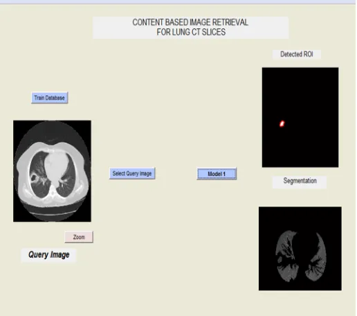
[image:6.612.178.436.368.596.2]The proposed system was tested with 106 CT slices including the Cavitary TB and the normal CT slices. The Segmented lung and the ROI extracted for the given query CT slice is given in the Fig. 2.
Technology (IJRASET)
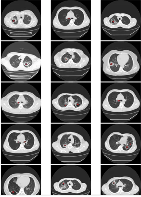
Fig. 3 Collection of the retrieval slices of proposed system
The resulted collection of retrieved CT slices for the given query slice is given in the Fig. 3.
V. CONCLUSION
The CBIR system implemented is based on the effective use of texture information within the slices to retrieve relevant CT slices for the query lung CT slice. The combined feature based methods implemented using BDIP and Tamura methods gave the accuracy of 85%, while the precision and recall of the system is 89% and 80%. This work has been extended by combined texture and shape features and the effective method is found for better retrieval of lung CT slices.
REFERENCES
[1] Gandhani, S. Bhujade, R. And Sinhal, A. (2013) ‘An Improved And Efficient Implementation Of CBIR System Based On Combined Features’, Third International Conference on Computational Intelligence and Information Technology pp.353-359.
[2] Fu, X. Li, Y. Harrison, R. and Belkasim, S. ‘Content-based Image Retrieval Using Gabor-Zernike Features’, International Conference on Pattern Recognition, vol.2, pp.417-420, 2006.
[3] Xie, S. Shan, S. Chen, X. and Chen, J. ‘Fusing Local Patterns of Gabor Magnitude and Phase for Face Recognition’, IEEE Transactions On Image Processing, Vol. 19, No. 5, pp.1349-1361, 2010.
[4] Prasad, B. and Krishna, A. ‘Statistical Texture Feature-based Retrieval and Performance Evaluation of CT Brain Images’, International Conference on Electronics Computer Technology, Vol. 2, pp. 289-293, 2011.
[5] Lee, Y. Hao, S. Lin, S. and Yi Li, S. ‘Image Retrieval by Region Of Interest Motif Co-occurence Matrix’, IEEE International Symposium on Intelligent Signal Processing and Communication Systems, pp. 270-274, 2012.
[5] Chuctaya, H. Portugal, C. Beltran, C. Gutierrez, J. Lopez, C. and Tupac, Y. ‘M-CBIR: A medical content-based image retrieval system using metric data-structures’, International Conference of the Chilean Computer Science Society, pp. 135-141, 2011.
Technology (IJRASET)
899-903, 2008.[7] Prakash, K. and Prasad, S. ‘HSV Color Motif Co-Occurrence Matrix for Content based Image Retrieval’, International Journal of Computer Applications, Vol. 48, No. 16, pp. 8-16, 2012.
[8] Khandave, V. and Mishra, N. ‘CBIR By Integration of Color and Texture Features’, International Journal of Recent Development in Engineering and Technology, Vol. 2, No. 1, pp. 50-55, 2014.
[9] Akakin, H.C. and Gurcan, M.N. ‘Content - Based Microscopic Image Retrieval System for Multi-Image Queries’, IEEE Trans. on Information Technology in Biomedicine, Vol.16, No.4, pp.758–769, 2012.
[10] Hirwane, R. ‘Fundamental of Content Based Image Retrieval’, International Journal of Computer Science and Information Technologies, Vol.3, No.1, pp.3260– 3263, 2012.
[11] Yogamangalam, R. and Karthikeyan, B. ‘Segmentation Techniques Comparison in Image Processing’, International Journal of Engineering and Technology, Vol. 5, No. 1, pp.307–313, 2013.