Laboratory Diagnosis of Lassa Fever
Vanessa Raabe,aJeffrey Koehlerb
Divisions of Adult and Pediatric Infectious Diseases, Emory University, Atlanta, Georgia, USAa; Diagnostic Systems Division, United States Army Medical Research Institute of Infectious Diseases, Fort Detrick, Maryland, USAb
ABSTRACT Lassa virus remains an important cause of illness in West Africa and
among the travelers returning from this region with an acute febrile illness. The symptoms of Lassa fever can be nonspecific and mimic those of other endemic in-fections, especially early in illness, making a clinical diagnosis difficult; therefore, lab-oratory testing is needed to confirm the diagnosis. An early identification of Lassa fever is crucial for maximizing the benefit of available antiviral therapy, as treatment efficacy rapidly decreases following the clinical onset of the disease. This minireview provides an overview of the currently available diagnostic tests for Lassa fever and their strengths and weaknesses.
KEYWORDS Lassa fever
L
assa virus, an arenavirus first isolated in 1969 in Jos, Nigeria (1), is the cause of Lassa fever, an acute viral illness that affects 100,000 to 300,000 persons per year based on 1970s estimates (2). Lassa fever is endemic in regions of West Africa, including Guinea, Liberia, Nigeria, and Sierra Leone, but cases have been exported to other countries by infected travelers. The natural reservoir for Lassa virus is the African soft-furred rat (Mastomys natalensis), which may be found throughout West Africa. The virus is transmitted to humans via direct contact with or the inhalation or ingestion of infected rat excreta or person to person via contact with infected body secretions (3). Lassa fever presents with nonspecific symptoms similar to many other endemic illnesses in West Africa, making it difficult to diagnose clinically; therefore, laboratory testing is needed to confirm the diagnosis (4). The availability of laboratory testing has been limited by the designation of Lassa virus as a category A pathogen by the National Institute of Allergy and Infectious Diseases (5). Biosafety level 4 (BSL-4) precautions are recommended for handling potentially infectious specimens (6). In 2014, the World Health Organization issued a call for early diagnostic tests for Lassa fever (7). This article provides a brief review of the challenges of identifying Lassa fever and the different diagnostic tests available for Lassa fever along with their strengths and weaknesses.CLINICAL PRESENTATION AND TREATMENT
Illness in humans develops within 3 weeks after infection with Lassa virus (3, 4). The initial symptoms of Lassa fever are nonspecific and may include fever, malaise, head-ache, sore throat, myalgia, cough, chest pain, abdominal pain, nausea, vomiting, and diarrhea (4, 8, 9). In most cases, symptoms are mild; however, severe illness complicated by abnormal bleeding, generalized edema, respiratory distress, hypotension, protein-uria, transaminitis, deafness, encephalopathy, and/or hypotension develops in approx-imately 20% of cases (3, 4, 8, 9). Although the overall fatality rate from Lassa fever is low (2, 10), it is 15 to 20% among patients who are hospitalized (3, 11). Higher fatality rates have been reported during outbreaks and among pregnant women, particularly in the third trimester of pregnancy (12). Treatment with ribavirin lowers the fatality risk to less than 5% if started in patients during the first 6 days of illness, but the beneficial effect on fatality is diminished if ribavirin is started later in the course of illness (13).
Accepted manuscript posted online12 April 2017
CitationRaabe V, Koehler J. 2017. Laboratory diagnosis of Lassa fever. J Clin Microbiol 55:1629 –1637.https://doi.org/10.1128/JCM .00170-17.
EditorColleen Suzanne Kraft, Emory University
Copyright© 2017 American Society for Microbiology.All Rights Reserved.
Address correspondence to Vanessa Raabe, vanessa.raabe@emory.edu.
crossm
on May 16, 2020 by guest
http://jcm.asm.org/
LASSA VIRUS DIAGNOSTIC CHALLENGES
One significant challenge in West Africa is differentiating between etiologies of febrile illness with similar initial clinical presentations, including malaria, influenza, dengue, yellow fever, and Lassa fever, with limited laboratory facility and reagent availabilities. Empirical treatment for presumed malaria or bacterial infection is often trialed, and Lassa fever is only suspected when a patient fails to improve with antima-larial and antibiotic therapy. This diagnostic delay leads to delayed patient isolation, an increased potential for transmission to family members and health care workers, and delayed initiation of ribavirin therapy, thereby decreasing its beneficial effect. Further highlighting the challenges of appropriate diagnostics is the emergence of Ebola virus in West Africa. A recent study found 60 to 70% of the patients with blood samples submitted to the Lassa Diagnostic Laboratory in Kenema, Sierra Leone, in the years prior to the Ebola virus outbreak were negative for malaria and Lassa virus, and there was serological evidence of Ebola and Marburg virus infections (14, 15). Correctly identifying the cause of an acute febrile illness in West Africa in an actionable time frame requires validated, rapid region-appropriate diagnostic assays.
Given the risk of person-to-person virus spread via bodily fluids, laboratory staff should be aware of the risk of Lassa virus when processing potentially infectious specimens. Poor sample storage and handling may pose a safety hazard to laboratory staff as well as decrease the sensitivity of diagnostic assays. The World Health Organi-zation guidelines for the collection, storage, and handling of specimens for Ebola virus testing should be followed when testing for Lassa virus (16–18). BSL-4 precautions are recommended when handling specimens which may contain infectious Lassa virus (6); however, the availability of such high-containment laboratories is limited worldwide. If BSL-4 precautions are not available, samples may be handled in a class II or III biosafety cabinet or inactivated to allow safe handling of specimens under BSL-2 precautions (16, 19).
While there are multiple methods for viral inactivation in the literature, different methods are appropriate depending on the intended downstream testing (e.g., mo-lecular or immunological pathogen detection, clinical laboratory tests, etc.). Chemical inactivation using solutions containing guanidine salts (e.g., TRIzol, Triton X-100, and buffer AVL combined with ethanol) is well documented, is effective with multiple pathogens, and is commonly used (20–22). Inactivation can be achieved by heating a blood specimen to 60°C for 60 min (23), although inactivation at 56°C for 30 min has been reported (24). Depending on the sample matrix and the specific pathogen, heat exposure alone may not result in complete inactivation; the use of chemical denatur-izing solutions in combination with heating to provide more complete inactivation is recommended (19). Gamma irradiation is also used to inactivate Lassa virus in liquid and dried samples (23, 25–27). Since the required absorbed radiation dose for success-ful viral inactivation varies depending on the temperature of the sample (25), empirical sample safety testing is required to confirm inactivation.
The high-containment safety requirements complicate Lassa virus assay develop-ment and validation studies. Many assay reagents need to be generated under BSL-4 conditions. Synthetic nucleic acids and recombinant proteins are more commonly being used as assay components, but assay validation with mock clinical samples still requires viral materials generated under BSL-4 conditions.
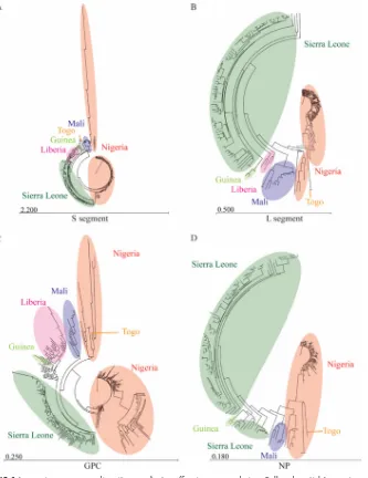
The development of appropriate diagnostic assays is further complicated by signif-icant Lassa virus diversity. The high nucleotide and amino acid diversity of Lassa virus isolates sequenced across West Africa (Fig. 1) can result in false-negative results if the primer/probe or antibody pairs do not bind to the target sufficiently. For example, a commonly used reverse transcriptase PCR (RT-PCR) assay (28) was redesigned when false negatives were identified due to primer-template mismatches (29). Furthermore, an NCBI protein BLAST analysis of the Lassa virus Josiah strain showed that glycoprotein ([GPC] NP_694870) and nucleoprotein ([NP] NP_694869.1) varied in percent identity from 91 to 99% and 86 to 99%, respectively, with full-length protein sequences of the
on May 16, 2020 by guest
http://jcm.asm.org/
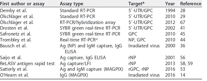
other Lassa virus protein sequences in GenBank (30). For example, Emmerich and colleagues evaluated the anti-Lassa virus antibody response in a human sample set from West Africa by immunofluorescence assay (IFA) and reverse enzyme-linked im-munosorbent assays (ELISAs) using several different Lassa virus strains (31). The authors found differing antibody responses depending on the virus strain used; stronger antibody responses were observed with local Lassa virus strains (31). Reassortant virus ML29, containing the L RNA segment of Mopeia virus and the S RNA segment of Lassa virus (Josiah strain), provides protection when injected into guinea pigs against dis-tantly related strains of Lassa virus from Nigeria; ML29 could potentially serve as a broadly cross-reacting reagent for assay development (32, 33). Table 1 highlights a selection of Lassa virus assays found in the literature.
However, we are currently unaware of any Lassa virus diagnostic validation studies demonstrating assay performance using viruses isolated across West Africa that cover the wide diversity of Lassa genetic variation possible. In the absence of a single, timely, FIG 1Lassa virus sequence diversity complicates efficacious assay design. Full and partial Lassa virus nucleic acid sequences were acquired from GenBank for the S RNA segment (A) and L RNA segment (B), and amino acid sequences for glycoprotein (GPC) (C) and nucleoprotein (NP) (D) were generated. Sequences were aligned, and phylogenetic trees (neighbor joining, Jukes-Cantor) were generated using CLC Genomics Workbench. Highlighted sections illustrate the geographic distribution and variability of Lassa virus.
on May 16, 2020 by guest
http://jcm.asm.org/
[image:3.585.45.378.69.501.2]pan-Lassa virus diagnostic assay, one future strategy could be the designing and validating of assays based on geographic region, as Lassa virus diversity generally clusters with geographic location (Fig. 1) (34). While ideal for use in specific countries/ regions, this approach, in the context of exported cases of Lassa fever from multiple countries where it is endemic, would require many validated assays being available for accurate diagnosis.
VIRAL CULTURE
Given the diagnostic challenges due to Lassa virus diversity, viral isolation in cell culture remains the “gold standard” for the diagnosis of Lassa fever, although RT-PCR and immunoassays have become commonly used assays for a clinically actionable diagnosis. Viremia is often present at the time of presentation to medical care and declines after the sixth day of illness in patients who ultimately survived their infection, whereas it may persist until death in fatal cases (35). Culturing is performed by the inoculation of sample specimens suspected of containing the virus in Vero E6 cells at 37°C (36). A positive result may yield a cell cytopathic effect; however, a second method of detection, such as RT-PCR, viral antigen detection, or electron microscopy, must be used to confirm the identity of the virus (37). Lassa virus may be cultured from blood, throat swabs, urine, and cerebrospinal fluid samples from patients, although the detection of virus in throat swabs and urine is inconsistent among patients with serum viremia (35, 38, 39). The viral culture may be positive from organ samples (liver, spleen, lung, kidney, heart, and placenta) at autopsy in cases of fatal infections (35).
Viral culture allows for detection that is independent of genetic variations between strains and allows further characterization of the virus if desired. Viral culture also allows for the quantification of viremia, which may provide additional virus characterization information, as viremia with 10350% tissue culture infective doses (TCID
50)/ml has a fatality odds ratio of 3.7 compared with viremia with less than 103TCID
50/ml (35). Viral culture is neither rapid, taking at least several days to produce results, nor widely available due to the need for BSL-4 precautions to handle live viral specimens, which limits its utility for the early diagnosis of Lassa infection.
NUCLEIC ACID DETECTION METHODS
[image:4.585.41.373.83.213.2]Real-time RT-PCR is a commonly used diagnostic approach for infectious diseases due to the high specificity and sensitivity of the method and has become a clinical standard for Lassa fever diagnosis. Coupled with automated sample processing and 96-well-plate-based thermocyclers, large numbers of samples can be tested quickly and inexpensively. PCR assays may be able to detect virus for a longer duration and earlier in illness compared with detection by viral culture (40) and may be performed on chemically inactivated specimens. The use of cycle threshold values with quantitative RT-PCR may assist with gross estimates of viremia (41) using appropriately selected positive-control material for standard curve generation. Depending on the primers and TABLE 1Selected assays for detecting Lassa virus
First author or assay Assay type Targetb Year Reference
Demby et al. Standard RT-PCR 5=-UTR/GPC 1994 28
Ölschläger et al. Standard RT-PCR 5=-UTR/GPC 2010 29
Ölschläger et al. RT-PCR/hybridization array 5=-UTR/GPC 2012 67
Drosten et al. SYBR green real-time RT-PCR 5=-UTR/GPC 2002 41
Safronetz et al. SYBR green real-time RT-PCR GPC 2010 45
Trombley et al. Real-time RT-PCRa NP, GPC 2010 44
Bausch et al. Ag (NP) and IgM capture, IgG
ELISA
Irradiated virus 2000 36
Saijo et al. Ag capture, IgG ELISA rNP 2001 56
ReLASV antigen rapid test Ag capture/LFI rNP 2013 58, 59
Satterly et al. Ag and IgM capture (MAGPIX) rGPC, rNP 2016 53
O’Hearn et al. IgG (MAGPIX) Irradiated virus 2016 14
aMultiple assays are used to detect different Lassa strains.
brNP, recombinant nucleoprotein; rGPC, recombinant glycoprotein complex.
on May 16, 2020 by guest
http://jcm.asm.org/
virus strain used, the 95% probability limit of detection estimates with RT-PCR vary from 1,237 to 4,290 RNA copies/ml (29, 41, 42). However, with highly diverse pathogens such as Lassa virus, genetic diversity can be problematic for nucleic acid-based assays, as even a single nucleotide variant in one of the primers can have a significant negative impact on assay sensitivity depending on the location of the nucleotide variant (43).
Multiple real-time RT-PCR assays are published in the literature for Lassa virus (41, 44–46). For example, Safronetz and colleagues initially detected Lassa virus circulating in Mali using a SYBR green real-time RT-PCR assay targeting the Lassa virus S RNA segment (45), and Trombley and colleagues developed multiple probe-based Lassa virus assays due to strain diversity (44). However, standard RT-PCR assays are commonly used (28, 29, 46) due to their ease of use and the decreased specificity for probe-based real-time RT-PCR. Probe-based real-time RT-PCR (two primers and a probe) introduces the possibility of probe mismatches due to the high degree of Lassa virus diversity, potentially increasing the false-negative rate compared with that from RT-PCR using only two primers. While assays using DNA binding dyes (e.g., SYBR green real-time RT-PCR) are generally avoided due to the increased background observed due to primer, dimer, and target mispriming, such assays are advantageous when probe design is challenging or not possible (e.g., due to high diversity).
As additional sample testing and sequencing information becomes available, mis-matches have been identified using established assays, necessitating assay redesign to improve performance. For example, Ölschläger and colleagues redesigned a commonly used standard RT-PCR assay for Lassa virus after identifying decreased assay sensitivity due to sequence variants for the reverse primer (29). This new RT-PCR assay is widely used for screening samples for Lassa virus and performed well in an external quality assessment study conducted by the European Network for Diagnostics of Imported Viral Diseases (46). Multiplex panels to simultaneously detect a multitude of viruses that can produce hemorrhagic fever syndromes, including Lassa and Ebola viruses, using RT-PCR alone or in combination with either enzyme hybridization or ligase detection reactions have also been developed (47–49).
ANTIGEN AND ANTIBODY DETECTION ASSAYS
Given the high diversity of the Lassa virus genome and the austere laboratory conditions where Lassa fever is endemic, antigen- and antibody-based assays are attractive alternatives to the high specificity and technical requirements of PCR assays. Antibody/antigen binding is generally less specific than primer/probe hybridization, allowing for greater flexibility in detecting diverse pathogens. Antigen detection relies on using specific antibodies against components of the Lassa virus to detect viral antigens in blood specimens. Initial assays detected nonspecific Lassa virus antigens with polyclonal antibodies, whereas more recent ELISAs target the Lassa virus nucleo-protein antigen. A diagnosis based on the detection of the relatively conserved Lassa nucleoprotein antigen (Fig. 1) may decrease the variability of test efficacy between genetically diverse viral strains in comparison to that from nucleic acid-based assays.
Lassa virus nucleoprotein antigen is detectable in patients with Lassa fever during the first week of illness and wanes during the second week in temporal association with the rise of detectable immunoglobulins (36). Increased levels of antigenemia have been found in fatal cases of Lassa fever compared with those in nonfatal cases (50). The short duration of antigenemia makes the detection of Lassa virus antigen more specific to acute Lassa virus infection than detection with antibody assays. Antigen detection assays may diagnose Lassa fever earlier during illness than antibody assays, as anti-bodies frequently may not be detectable until the second week of illness (36). However, Lassa virus antigen levels may become undetectable despite persistent viremia (51), and so a negative antigen test during an acute illness does not rule out Lassa fever.
Multiple antigen and IgM capture ELISAs have been developed using inactivated virus (36, 52–54); however this approach is limited to BSL-4 capable facilities. The use of recombinant antigens allows for improved assay development and access (50, 55–57). A lateral flow assay for Lassa virus nucleoprotein (ReLASV) is one type of rapid
on May 16, 2020 by guest
http://jcm.asm.org/
diagnostic test that could be used for point-of-care testing. Following initial develop-ment and testing efforts (50, 57), this assay received the CE mark in 2013, although the assay has not been approved by the U.S. Federal Drug Administration. According to the product insert, the test yields results in 15 to 25 min and has 85% sensitivity and 99% specificity using confirmed Lassa virus-positive blood samples (Lassa virus positive by RT-PCR and IgM negative by ELISA) (58). In one study from Kenema Government Hospital in Sierra Leone, the use of ReLASV identified 95% of acute Lassa fever cases (defined as RT-PCR positive, increasing IgM titers, or IgM positive with IgG seroconver-sion), while missed cases were associated with resolving disease or mild disease with low levels of viremia (59).
Ideally, a diagnostic assay would not only detect Lassa virus infection but would also screen for multiple other pathogens with similar clinical presentations endemic in West Africa at the same time. Satterly and colleagues recently described transitioning Lassa and Ebola virus antigen- and IgM-based ELISAs onto a MAGPIX system (53) that uses individually labeled magnetic beads to detect multiple targets in a single assay. This assay has lower limits of detection for Lassa virus nucleoprotein and IgM than tradi-tional ELISAs (53). The same group also developed and tested a multiplex MAGPIX IgG assay for a wide spectrum of hemorrhagic fever viruses, including alphaviruses, arena-viruses, flaviarena-viruses, and filoviruses (14). Further development of multiplex MAGPIX assays, including testing for Lassa virus antigen and common endemic diseases such as malaria, would assist with the diagnosis and clinical management of suspected Lassa fever cases, especially in scenarios of coinfection with Lassa virus and bacterial or parasitic organisms where multiple therapeutic modalities may be indicated.
Delays in a patient seeking medical care following disease onset could negatively impact direct pathogen detection with a nucleoprotein detection assay, and diagnosis by Lassa virus-specific IgM may be more appropriate. Lassa IgM usually becomes detectable during the second week of infection (36), although it may be detectable within 4 days of onset of illness in some patients. A lack of an antibody response has been reported in some fatal cases of Lassa fever (60). Lassa virus IgG levels rise later than IgM levels, having a mean time to detection of 25.6 days after symptom onset (36), although positive IgG titers have occasionally been detected in patients with acute Lassa fever within the first few days of illness (35, 59).
Historically, antibody detection was conducted using immunofluorescence assays (IFAs) (4, 13, 36); however, IFAs have been replaced over time with ELISAs due to their ease of use, their increased sensitivity and specificity, and reduced interobserver variation in readings (36, 54). Estimates of the sensitivity of IgM detection for diagnos-ing Lassa fever compared with that of RT-PCR range from 55% to 72% (36, 61, 62). Antibody assays have been used to diagnose Lassa fever in those with a clinically consistent illness based on either detectable levels of Lassa virus antibody in a serum sample collected during illness or a rise in Lassa antibody titers (2, 63). However, Lassa virus IgM titers remain elevated for months to years following an acute infection (50). One study found that 28% of healthy hosts sampled from a region where the virus is endemic without a recent preceding febrile illness had detectable levels of Lassa virus IgM, suggesting that IgM positivity alone may be insufficient to diagnose Lassa fever in persons residing in regions where it is endemic (50). In regions where Lassa virus is endemic, a positive nucleoprotein antigen test, a rise in antibody titers between acute-and convalescent-phase serum, or the development of a new positive IgG titer in combination with a positive IgM titer may more accurately reflect acute Lassa fever than a single positive IgM test. While the detection of a new positive IgG titer combined with an IgM response in the correct clinical setting may support a diagnosis of Lassa fever, the detection of a positive IgG response alone is insufficient to make a diagnosis. Lassa IgG titers may persist for decades (64), and seroprevalence studies in regions of endemicity have shown 4 to 55% of healthy individuals living in areas where the virus is endemic have detectable Lassa virus IgG titers (14, 52, 55, 65, 66).
Bausch and colleagues conducted a direct comparison of acute Lassa fever cases by testing Lassa virus-positive samples identified by virus isolation and RT-PCR using IgM,
on May 16, 2020 by guest
http://jcm.asm.org/
IgG, and antigen ELISAs and IFAs (36). Based on the onset of clinical symptoms, Lassa fever patients were generally antigen positive and IgM negative in the first week of clinical disease, were IgM positive and antigen negative in the second week, and were IgG positive around week 3 (36). The authors concluded that virus isolation is the most sensitive but clinically impractical diagnostic tool, and the combination of antigen capture assays and IgM ELISA was the best for diagnosis throughout the clinical disease course. However, direct pathogen detection allows the earliest diagnosis (when IgM is likely negative) and increases the chances of instituting ribavirin treatment in the first few days of illness when it is most efficacious.
SUMMARY AND CONCLUSIONS
Accurate and rapid diagnosis of Lassa fever is especially challenging due to the nonspecific clinical presentation, the high degree of Lassa virus genetic diversity observed in West Africa, and the biosafety concerns regarding laboratory testing for high consequence pathogens. While there are many diagnostic assays for Lassa virus, there currently is no timely, validated pan-Lassa virus assay available to both capture the diversity among viral strains and provide a diagnosis at any time point during the clinical course of illness.
Viral culture remains the “gold standard” for Lassa fever diagnosis across the diversity of Lassa strains but requires a clinically nonactionable amount of time and BSL-4 precautions to perform. Nucleic acid-based assays have become the clinical diagnostic standard and may be performed rapidly on inactivated specimens under BSL-2 conditions but may have false-negative results due to the high degree of genetic diversity among viruses. Viral antigen assays may provide a rapid diagnosis early on during illness but may miss the diagnosis at later stages once the antigenemia phase has resolved. The detection of a new IgM antibody response can diagnose Lassa fever but may miss the diagnosis during the first week of illness, may be falsely negative in severe infections where patients are unable to mount a serological response, and may remain positive for a prolonged period potentially causing false-positive results. A rise in baseline antibody titers between acute- and convalescent-phase serum or a positive IgM accompanied by the development of a new positive IgG response may be more indicative of acute Lassa fever in regions where it is endemic than a single positive IgM titer.
Overall, the appropriate diagnosis of Lassa fever will likely require a combination of a clinically compatible presentation along with serological and molecular diagnostic assays. Having a rapid, point-of-care multiplex test that can diagnose Lassa fever as well as other high consequence pathogens, such as Ebola virus, would accelerate accurate diagnosis, patient isolation, and efficacious therapy. As work for a Lassa virus vaccine and therapeutics moves toward clinical studies, having well-validated diagnostic assays available will be a necessity to ensure appropriate patient enrollment and countermea-sure performance. Future directions for research in Lassa fever diagnostics should include assay improvement to increase detection across the genetically diverse spec-trum of Lassa virus strains, assay validation to demonstrate efficacy across geographic regions and viral lineages, point-of-care diagnostic development and field validation, and content expansion of multiplex assays to distinguish Lassa fever from other diseases with similar clinical presentations.
ACKNOWLEDGMENTS
Opinions, interpretations, conclusions, and recommendations are those of the au-thors and are not necessarily endorsed by the U.S. Army.
This effort was funded in part by Defense Threat Reduction Agency (DTRA) through the JSTO-CBD project 11467651.
REFERENCES
1. Buckley SM, Casals J. 1970. Lassa fever, a new virus disease of man from West Africa. 3. Isolation and characterization of the virus. Am J Trop Med Hyg 19:680 – 691.
2. McCormick JB, Webb PA, Krebs JW, Johnson KM, Smith ES. 1987. A prospective study of the epidemiology and ecology of Lassa fever. J Infect Dis 155:437– 444.https://doi.org/10.1093/infdis/155.3.437.
on May 16, 2020 by guest
http://jcm.asm.org/
3. Centers for Disease Control and Prevention. Lassa fever. Centers for Disease Control and Prevention, Atlanta, GA.http://www.cdc.gov/vhf/ lassa/pdf/factsheet.pdf. Accessed 2 November 2016.
4. McCormick JB, King IJ, Webb PA, Johnson KM, O’Sullivan R, Smith ES, Trippel S, Tong TC. 1987. A case-control study of the clinical diagnosis and course of Lassa fever. J Infect Dis 155:445– 455.https://doi.org/10 .1093/infdis/155.3.445.
5. National Institute of Allergy and Infectious Diseases. 26 October 2016. NIAID emerging infectious diseases/pathogens. National Institute of Allergy and Infectious Diseases, Bethesda, MD.https://www.niaid.nih .gov/research/emerging-infectious-diseases-pathogens. Accessed 29 Oc-tober 2016.
6. Centers for Disease Control and Prevention. 1988. Management of pa-tients with suspected viral hemorrhagic fever. Centers for Disease Con-trol and Prevention, Atlanta, GA.http://www.cdc.gov/MMWR/preview/ mmwrhtml/00037085.htm.
7. World Health Organization. 2016. WHO calls for early diagnostic tests for Lassa fever. World Health Organization, Geneva, Switzerland.http:// www.who.int/csr/disease/lassafever/early-diagnostic-lassa-fever/en/. Ac-cessed 20 October 2016.
8. World Health Organization. March 2016. Lassa fever. World Health Organization, Geneva, Switzerland.http://www.who.int/mediacentre/ factsheets/fs179/en/. Accessed 20 October 2016.
9. Frame JD, Baldwin JM, Jr, Gocke DJ, Troup JM. 1970. Lassa fever, a new virus disease of man from West Africa. I. Clinical description and path-ological findings. Am J Trop Med Hyg 19:670 – 676.
10. Richmond JK, Baglole DJ. 2003. Lassa fever: epidemiology, clinical fea-tures, and social consequences. BMJ 327:1271–1275.https://doi.org/10 .1136/bmj.327.7426.1271.
11. McCormick JB, Walker DH, King IJ, Webb PA, Elliott LH, Whitfield SG, Johnson KM. 1986. Lassa virus hepatitis: a study of fatal Lassa fever in humans. Am J Trop Med Hyg 35:401– 407.
12. Price ME, Fisher-Hoch SP, Craven RB, McCormick JB. 1988. A prospective study of maternal and fetal outcome in acute Lassa fever infection during pregnancy. BMJ 297:584 –587.https://doi.org/10.1136/bmj.297 .6648.584.
13. McCormick JB, King IJ, Webb PA, Scribner CL, Craven RB, Johnson KM, Elliott LH, Belmont-Williams R. 1986. Lassa fever. Effective therapy with ribavirin. N Engl J Med 314:20 –26. https://doi.org/10.1056/ NEJM198601023140104.
14. O’Hearn AE, Voorhees MA, Fetterer DP, Wauquier N, Coomber MR, Bangura J, Fair JN, Gonzalez JP, Schoepp RJ. 2016. Serosurveillance of viral pathogens circulating in West Africa. Virol J 13:163.https://doi.org/ 10.1186/s12985-016-0621-4.
15. Schoepp RJ, Rossi CA, Khan SH, Goba A, Fair JN. 2014. Undiagnosed acute viral febrile illnesses, Sierra Leone. Emerg Infect Dis 20:1176 –1182.
https://doi.org/10.3201/eid2007.131265.
16. World Health Organization. 2014. Interim guideline: laboratory diagnosis of Ebola virus disease. World Health Organization, Geneva, Switzerland. 17. World Health Organization. 2014. Interim guidance: how to safely ship human blood samples from suspected Ebola cases within a country by road, rail and sea. World Health Organization, Geneva, Switzerland. 18. World Health Organization. 2014. How to safely collect blood samples by
phlebotomy from patients suspected to be infected with Ebola. World Health Organization, Geneva, Switzerland.
19. Pan American Health Organization, World Health Organization. 2015. General procedures for inactivation of potentially infectious samples with Ebola virus and other highly pathogenic viral agents. Regional Office for the Americas of the World Health Organization, Washing-ton, DC.
20. Blow JA, Dohm DJ, Negley DL, Mores CN. 2004. Virus inactivation by nucleic acid extraction reagents. J Virol Methods 119:195–198.https:// doi.org/10.1016/j.jviromet.2004.03.015.
21. Haddock E, Feldmann F, Feldmann H. 2016. Effective chemical inactiva-tion of Ebola virus. Emerg Infect Dis 22:1292–1294.https://doi.org/10 .3201/eid2207.160233.
22. Smither SJ, Weller SA, Phelps A, Eastaugh L, Ngugi S, O’Brien LM, Steward J, Lonsdale SG, Lever MS. 2015. Buffer AVL alone does not inactivate Ebola virus in a representative clinical sample type. J Clin Microbiol 53:3148 –3154.https://doi.org/10.1128/JCM.01449-15. 23. Mitchell SW, McCormick JB. 1984. Physicochemical inactivation of Lassa,
Ebola, and Marburg viruses and effect on clinical laboratory analyses. J Clin Microbiol 20:486 – 489.
24. King AMQ, Lefkowitz E, Adams MJ, Carstens EB (ed). 2012. Family
-Arenaviridae, p 715–723, Virus taxonomy. Ninth report of the Interna-tional Committee on Taxonomy of Viruses. Elsevier, Philadelphia, PA. 25. Elliott LH, McCormick JB, Johnson KM. 1982. Inactivation of Lassa,
Mar-burg, and Ebola viruses by gamma irradiation. J Clin Microbiol 16: 704 –708.
26. Sagripanti JL, Lytle CD. 2011. Sensitivity to ultraviolet radiation of Lassa, vaccinia, and Ebola viruses dried on surfaces. Arch Virol 156:489 – 494.
https://doi.org/10.1007/s00705-010-0847-1.
27. Lytle CD, Sagripanti JL. 2005. Predicted inactivation of viruses of rele-vance to biodefense by solar radiation. J Virol 79:14244 –14252.https:// doi.org/10.1128/JVI.79.22.14244-14252.2005.
28. Demby AH, Chamberlain J, Brown DW, Clegg CS. 1994. Early diagnosis of Lassa fever by reverse transcription-PCR. J Clin Microbiol 32:2898 –2903. 29. Olschlager S, Lelke M, Emmerich P, Panning M, Drosten C, Hass M, Asogun D, Ehichioya D, Omilabu S, Gunther S. 2010. Improved de-tection of Lassa virus by reverse transcription-PCR targeting the 5=
region of S RNA. J Clin Microbiol 48:2009 –2013.https://doi.org/10 .1128/JCM.02351-09.
30. NCBI Resource Coordinators. 2016. Database resources of the National Center for Biotechnology Information. Nucleic Acids Res 44:D7–D19.
https://doi.org/10.1093/nar/gkv1290.
31. Emmerich P, Gunther S, Schmitz H. 2008. Strain-specific antibody re-sponse to Lassa virus in the local population of West Africa. J Clin Virol 42:40 – 44.https://doi.org/10.1016/j.jcv.2007.11.019.
32. Lukashevich IS, Carrion R, Jr, Salvato MS, Mansfield K, Brasky K, Zapata J, Cairo C, Goicochea M, Hoosien GE, Ticer A, Bryant J, Davis H, Hamma-mieh R, Mayda M, Jett M, Patterson J. 2008. Safety, immunogenicity, and efficacy of the ML29 reassortant vaccine for Lassa fever in small non-human primates. Vaccine 26:5246 –5254. https://doi.org/10.1016/j .vaccine.2008.07.057.
33. Carrion R, Jr, Patterson JL, Johnson C, Gonzales M, Moreira CR, Ticer A, Brasky K, Hubbard GB, Moshkoff D, Zapata J, Salvato MS, Lukashevich IS. 2007. A ML29 reassortant virus protects guinea pigs against a distantly related Nigerian strain of Lassa virus and can provide sterilizing immu-nity. Vaccine 25:4093– 4102. https://doi.org/10.1016/j.vaccine.2007.02 .038.
34. Bowen MD, Rollin PE, Ksiazek TG, Hustad HL, Bausch DG, Demby AH, Bajani MD, Peters CJ, Nichol ST. 2000. Genetic diversity among Lassa virus strains. J Virol 74:6992–7004.https://doi.org/10.1128/JVI.74.15.6992 -7004.2000.
35. Johnson KM, McCormick JB, Webb PA, Smith ES, Elliott LH, King IJ. 1987. Clinical virology of Lassa fever in hospitalized patients. J Infect Dis 155:456 – 464.https://doi.org/10.1093/infdis/155.3.456.
36. Bausch DG, Rollin PE, Demby AH, Coulibaly M, Kanu J, Conteh AS, Wagoner KD, McMullan LK, Bowen MD, Peters CJ, Ksiazek TG. 2000. Diagnosis and clinical virology of Lassa fever as evaluated by enzyme-linked immunosorbent assay, indirect fluorescent-antibody test, and virus isolation. J Clin Microbiol 38:2670 –2677.
37. Drosten C, Kummerer BM, Schmitz H, Gunther S. 2003. Molecular diag-nostics of viral hemorrhagic fevers. Antiviral Res 57:61– 87.https://doi .org/10.1016/S0166-3542(02)00201-2.
38. Gunther S, Weisner B, Roth A, Grewing T, Asper M, Drosten C, Emmerich P, Petersen J, Wilczek M, Schmitz H. 2001. Lassa fever encephalopathy: Lassa virus in cerebrospinal fluid but not in serum. J Infect Dis 184: 345–349.https://doi.org/10.1086/322033.
39. Monath TP, Casals J. 1975. Diagnosis of Lassa fever and the isolation and management of patients. Bull World Health Organ 52:707–715. 40. Trappier SG, Conaty AL, Farrar BB, Auperin DD, McCormick JB,
Fisher-Hoch SP. 1993. Evaluation of the polymerase chain reaction for diagnosis of Lassa virus infection. Am J Trop Med Hyg 49:214 –221.
41. Drosten C, Gottig S, Schilling S, Asper M, Panning M, Schmitz H, Gunther S. 2002. Rapid detection and quantification of RNA of Ebola and Marburg viruses, Lassa virus, Crimean-Congo hemorrhagic fever virus, Rift Valley fever virus, dengue virus, and yellow fever virus by real-time reverse transcription-PCR. J Clin Microbiol 40:2323–2330. https://doi.org/10 .1128/JCM.40.7.2323-2330.2002.
42. Vieth S, Drosten C, Lenz O, Vincent M, Omilabu S, Hass M, Becker-Ziaja B, ter Meulen J, Nichol ST, Schmitz H, Gunther S. 2007. RT-PCR assay for detection of Lassa virus and related Old World arenaviruses targeting the L gene. Trans R Soc Trop Med Hyg 101:1253–1264.https://doi.org/ 10.1016/j.trstmh.2005.03.018.
43. Stadhouders R, Pas SD, Anber J, Voermans J, Mes TH, Schutten M. 2010. The effect of primer-template mismatches on the detection and
on May 16, 2020 by guest
http://jcm.asm.org/
tification of nucleic acids using the 5= nuclease assay. J Mol Diagn 12:109 –117.https://doi.org/10.2353/jmoldx.2010.090035.
44. Trombley AR, Wachter L, Garrison J, Buckley-Beason VA, Jahrling J, Hensley LE, Schoepp RJ, Norwood DA, Goba A, Fair JN, Kulesh DA. 2010. Comprehensive panel of real-time TaqMan polymerase chain reaction assays for detection and absolute quantification of filoviruses, arenavi-ruses, and New World hantaviruses. Am J Trop Med Hyg 82:954 –960.
https://doi.org/10.4269/ajtmh.2010.09-0636.
45. Safronetz D, Lopez JE, Sogoba N, Traore SF, Raffel SJ, Fischer ER, Ebihara H, Branco L, Garry RF, Schwan TG, Feldmann H. 2010. Detection of Lassa virus, Mali. Emerg Infect Dis 16:1123–1126. https://doi.org/10.3201/ eid1607.100146.
46. Nikisins S, Rieger T, Patel P, Muller R, Gunther S, Niedrig M. 2015. International external quality assessment study for molecular detection of Lassa virus. PLoS Negl Trop Dis 9:e0003793.https://doi.org/10.1371/ journal.pntd.0003793.
47. Pang Z, Li A, Li J, Qu J, He C, Zhang S, Li C, Zhang Q, Liang M, Li D. 2014. Comprehensive multiplex one-step real-time TaqMan qRT-PCR assays for detection and quantification of hemorrhagic fever viruses. PLoS One 9:e95635.https://doi.org/10.1371/journal.pone.0095635.
48. Das S, Rundell MS, Mirza AH, Pingle MR, Shigyo K, Garrison AR, Paragas J, Smith SK, Olson VA, Larone DH, Spitzer ED, Barany F, Golightly LM. 2015. A multiplex PCR/LDR assay for the simultaneous identification of category A infectious pathogens: agents of viral hemorrhagic fever and variola virus. PLoS One 10:e0138484. https://doi.org/10.1371/journal .pone.0138484.
49. He J, Kraft AJ, Fan J, Van Dyke M, Wang L, Bose ME, Khanna M, Metallo JA, Henrickson KJ. 2009. Simultaneous detection of CDC category “A” DNA and RNA bioterrorism agents by use of multiplex PCR & RT-PCR enzyme hybridization assays. Viruses 1:441– 459. https://doi.org/10 .3390/v1030441.
50. Branco LM, Grove JN, Boisen ML, Shaffer JG, Goba A, Fullah M, Momoh M, Grant DS, Garry RF. 2011. Emerging trends in Lassa fever: redefining the role of immunoglobulin M and inflammation in diagnosing acute infection. Virol J 8:478.https://doi.org/10.1186/1743-422X-8-478. 51. Jahrling PB, Niklasson BS, McCormick JB. 1985. Early diagnosis of human
Lassa fever by ELISA detection of antigen and antibody. Lancet i:250 –252.
52. Bausch DG, Demby AH, Coulibaly M, Kanu J, Goba A, Bah A, Conde N, Wurtzel HL, Cavallaro KF, Lloyd E, Baldet FB, Cisse SD, Fofona D, Savane IK, Tolno RT, Mahy B, Wagoner KD, Ksiazek TG, Peters CJ, Rollin PE. 2001. Lassa fever in Guinea: I. Epidemiology of human disease and clinical observations. Vector Borne Zoonotic Dis 1:269 –281.https://doi.org/10 .1089/15303660160025903.
53. Satterly NG, Voorhees MA, Ames AD, Schoepp RJ. 2017. Comparison of MagPix assays and enzyme-linked immunosorbent assay for the detec-tion of hemorrhagic fever viruses. J Clin Microbiol 55:68 –78.https://doi .org/10.1128/JCM.01693-16.
54. Emmerich P, Thome-Bolduan C, Drosten C, Gunther S, Ban E, Sawinsky I, Schmitz H. 2006. Reverse ELISA for IgG and IgM antibodies to detect Lassa virus infections in Africa. J Clin Virol 37:277–281.https://doi.org/ 10.1016/j.jcv.2006.08.015.
55. Lukashevich IS, Clegg JC, Sidibe K. 1993. Lassa virus activity in Guinea: distribution of human antiviral antibody defined using enzyme-linked immunosorbent assay with recombinant antigen. J Med Virol 40: 210 –217.https://doi.org/10.1002/jmv.1890400308.
56. Saijo M, Georges-Courbot MC, Marianneau P, Romanowski V, Fukushi S, Mizutani T, Georges AJ, Kurata T, Kurane I, Morikawa S. 2007. Develop-ment of recombinant nucleoprotein-based diagnostic systems for Lassa fever. Clin Vaccine Immunol 14:1182–1189.https://doi.org/10.1128/CVI .00101-07.
57. Grove JN, Branco LM, Boisen ML, Muncy IJ, Henderson LA, Schieffellin JS, Robinson JE, Bangura JJ, Fonnie M, Schoepp RJ, Hensley LE, Seisay A, Fair JN, Garry RF. 2011. Capacity building permitting comprehensive moni-toring of a severe case of Lassa hemorrhagic fever in Sierra Leone with a positive outcome: case report. Virol J 8:314.https://doi.org/10.1186/ 1743-422X-8-314.
58. Corgenix, Inc. 2015. ReLASV antigen rapid test package insert-IVD. Cor-genix, Inc., St. Ingbert, Germany.
59. Shaffer JG, Grant DS, Schieffelin JS, Boisen ML, Goba A, Hartnett JN, Levy DC, Yenni RE, Moses LM, Fullah M, Momoh M, Fonnie M, Fonnie R, Kanneh L, Koroma VJ, Kargbo K, Ottomassathien D, Muncy IJ, Jones AB, Illick MM, Kulakosky PC, Haislip AM, Bishop CM, Elliot DH, Brown BL, Zhu H, Hastie KM, Andersen KG, Gire SK, Tabrizi S, Tariyal R, Stremlau M, Matschiner A, Sampey DB, Spence JS, Cross RW, Geisbert JB, Folarin OA, Happi CT, Pitts KR, Geske FJ, Geisbert TW, Saphire EO, Robinson JE, Wilson RB, Sabeti PC, Henderson LA, Khan SH, Bausch DG, Branco LM, Garry RF, Viral Hemorrhagic Fever Consortium. 2014. Lassa fever in post-conflict Sierra Leone. PLoS Negl Trop Dis 8:e2748.https://doi.org/ 10.1371/journal.pntd.0002748.
60. Wulff H, Johnson KM. 1979. Immunoglobulin M and G responses mea-sured by immunofluorescence in patients with Lassa or Marburg virus infections. Bull World Health Organ 57:631– 635.
61. Ibekwe TS, Nwegbu MM, Asogun D, Adomeh DI, Okokhere PO. 2012. The sensitivity and specificity of Lassa virus IgM by ELISA as screening tool at early phase of Lassa fever infection. Niger Med J 53:196 –199.https:// doi.org/10.4103/0300-1652.107552.
62. Ter Meulen J, Koulemou K, Wittekindt T, Windisch K, Strigl S, Conde S, Schmitz H. 1998. Detection of Lassa virus antinucleoprotein globulin G (IgG) and IgM antibodies by a simple recombinant immuno-blot assay for field use. J Clin Microbiol 36:3143–3148.
63. Wulff H, Lange JV. 1975. Indirect immunofluorescence for the diagnosis of Lassa fever infection. Bull World Health Organ 52:429 – 436. 64. Bond N, Schieffelin JS, Moses LM, Bennett AJ, Bausch DG. 2013. A
historical look at the first reported cases of Lassa fever: IgG antibodies 40 years after acute infection. Am J Trop Med Hyg 88:241–244.https://doi .org/10.4269/ajtmh.12-0466.
65. Fukushi S, Tani H, Yoshikawa T, Saijo M, Morikawa S. 2012. Serological assays based on recombinant viral proteins for the diagnosis of arena-virus hemorrhagic fevers. Viruses 4:2097–2114.https://doi.org/10.3390/ v4102097.
66. Kerneis S, Koivogui L, Magassouba N, Koulemou K, Lewis R, Aplogan A, Grais RF, Guerin PJ, Fichet-Calvet E. 2009. Prevalence and risk factors of Lassa seropositivity in inhabitants of the forest region of Guinea: a cross-sectional study. PLoS Negl Trop Dis 3:e548. https://doi.org/10 .1371/journal.pntd.0000548.
67. Olschlager S, Gunther S. 2012. Rapid and specific detection of Lassa virus by reverse transcription-PCR coupled with oligonucleotide array hybrid-ization. J Clin Microbiol 50:2496 –2499. https://doi.org/10.1128/JCM .00998-12.