CD36: a class B scavenger receptor involved in
angiogenesis, atherosclerosis, inflammation,
and lipid metabolism
Maria Febbraio, … , David P. Hajjar, Roy L. Silverstein
J Clin Invest.
2001;
108(6)
:785-791.
https://doi.org/10.1172/JCI14006
.
CD36, identified more than a quarter of a century ago as a platelet integral membrane
glycoprotein (glycoprotein IV), was until recently best known as a receptor for
thrombospondin-1 (TSP-1). TSP-1 is found in ECMs and platelet
a
granules, and it
participates in cell attachment, motility, and proliferation, as well as in modulation of
protease activity, TGF-
b
activation, neurite outgrowth, and angiogenesis (1). Initially, this
receptor-ligand pair was shown to mediate interactions between platelets and monocytes,
tumor cells, and matrix. Since then, CD36 has been implicated in multiple biological
processes that define it as a multiligand scavenger receptor (see ref. 2 for review). These
ligands appear remarkably diverse: In addition to TSP-1, they include long-chain fatty acids,
modified LDL, retinal photoreceptor outer segments, Plasmodium falciparum
malaria-parasitized erythrocytes, sickle erythrocytes, anionic phospholipids, apoptotic cells, and
collagens I and IV. The biology of CD36 can be broadly divided in terms of functions that it
mediates with or without TSP-1, but it is probable that it acts in concert with other proteins,
such as fatty acid–binding proteins, caveola-associated proteins, integrins, cytoskeletal
proteins, and signaling molecules, to effect its diverse functions. Molecular properties CD36
belongs to the class B scavenger receptor family, which includes the receptor for selective
cholesteryl ester uptake, scavenger receptor class B type I (SR-BI), and lysosomal integral
membrane protein II (LIMP-II). […]
Perspective
Find the latest version:
PERSPECTIVE SERIES
Monty Krieger and David M. Stern, Series Editors
Multiligand receptors
CD36, identified more than a quarter of a century ago as a platelet integral membrane glycoprotein (glyco-protein IV), was until recently best known as a recep-tor for thrombospondin-1 (TSP-1). TSP-1 is found in ECMs and platelet α granules, and it participates in cell attachment, motility, and proliferation, as well as in modulation of protease activity, TGF-βactivation, neurite outgrowth, and angiogenesis (1). Initially, this receptor-ligand pair was shown to mediate interac-tions between platelets and monocytes, tumor cells, and matrix. Since then, CD36 has been implicated in multiple biological processes that define it as a multi-ligand scavenger receptor (see ref. 2 for review). These ligands appear remarkably diverse: In addition to TSP-1, they include long-chain fatty acids, modified LDL, retinal photoreceptor outer segments, Plasmodi-um falciparPlasmodi-ummalaria-parasitized erythrocytes, sickle erythrocytes, anionic phospholipids, apoptotic cells, and collagens I and IV. The biology of CD36 can be broadly divided in terms of functions that it mediates with or without TSP-1, but it is probable that it acts in concert with other proteins, such as fatty acid–binding proteins, caveola-associated proteins, integrins, cytoskeletal proteins, and signaling molecules, to effect its diverse functions.
Molecular properties
CD36 belongs to the class B scavenger receptor fami-ly, which includes the receptor for selective cholesteryl ester uptake, scavenger receptor class B type I (SR-BI), and lysosomal integral membrane protein II (LIMP-II). The nucleotide sequence of the human cDNA predicts a protein of 471 amino acids and a molecular weight of 53 kDa (3). The protein is heavi-ly N-linked gheavi-lycosylated, a modification that may pro-vide proteins in this family some protection from degradation in proteinase-rich environments such as the lysosome and areas of inflammation or tissue damage. Within the carboxy-terminal segment of
CD36 is a region of 27 hydrophobic amino acids cor-responding to a transmembrane domain. The amino-terminus has an uncleaved signal peptide, which is probably a second membrane-spanning domain. The predicted structure orients most of the protein extra-cellularly, except for two short (9–13 amino acids) cytoplasmic tails which can be palmitoylated (4). Asch et al. report that the phosphorylation state of the extracellular threonine 92 may determine the relative binding affinity of CD36 for TSP-1 and collagen (5). A well-defined human blood group polymorphism, Naka, is carried by platelet CD36 (6). The null pheno-type (Naka-negative) occurs with high frequency in African, Japanese, and other Asian populations, and this is perhaps related to the role of CD36 in malaria pathogenesis. Affected individuals may lack platelet (type II deficiency) or both platelet and monocyte CD36 (type I). The genetic basis of the Nakanegative phenotype is under study by several groups.
CD36 protein sequence is highly conserved between the cloned human and rodent proteins. There are two CD36-family homologs in Drosophila, Croquemort, and epithelial membrane protein (emp) (7, 8). Croquemort
(“catcher of death”) has 23% homology to human CD36 at the amino acid level and is expressed on macrophages and hemocytes, where it is essential for phagocytosis of apoptotic corpses. Emp, which is expressed in precursor cells for adult epidermal struc-tures, has 32% homology to human CD36 and 34% homology to human LIMP-II. There are three predict-ed CD36-family homologs in the genome of Caenorhab-ditis elegans.Study of the function of CD36 in these organisms provides insight into its primary human role and may also elucidate how this protein evolved to acquire its broad ligand specificity.
Structure-function relationships
The use of glutathione-S-transferase human CD36 fusion proteins and synthetic peptides has provided
CD36: a class B scavenger receptor involved in angiogenesis,
atherosclerosis, inflammation, and lipid metabolism
Maria Febbraio,
1,2David P. Hajjar,
2,3and Roy L. Silverstein
1,2 1Department of Medicine, Division of Hematology—Medical Oncology,2Center of Vascular Biology, and
3Department of Pathology and Department of Biochemistry, Weill Medical College of Cornell University, 1300 York Avenue, New York, New York, USA
Address correspondence to: Maria Febbraio, Department of Medicine, Division of Hematology—Medical Oncology, Weill Medical College of Cornell University, 1300 York Avenue, Box 113, New York, New York 10021, USA. Phone: (212) 746-2068; Fax: (212) 746-8866; E-mail: mjfebbra@med.cornell.edu.
some basic structure-function information. Amino acids 93–120 define a minimal high-affinity binding site for TSP-1 that may be modulated by a down-stream sequence (amino acids 139–155). Binding of oxidized LDL (oxLDL) has been mapped to a major domain at amino acids 120–155 and a second region with less but significant affinity at amino acids 28–93. The site for binding of apoptotic cells and P. falci-parum–infected erythrocytes is proposed to lie between amino acids 155 and 183, but this conclusion is based solely on mAb inhibition. Antibodies to this immunodominant epitope block all known CD36 function, including TSP-1 binding, making it diffi-cult to interpret these results unequivocally.
Our laboratory determined that the TSP-1 binding site of CD36 is contained within a highly conserved sequence in CD36 family members that we termed the CLESH (CD36 LIMP-II Emp sequence homology) domain (9), which is also found in other proteins. Interestingly, the type I repeat of TSP-1, the domain containing the CD36 binding site, is also found in many other proteins. Characterizing expression pro-files of these domains may identify potential receptor-ligand pairs. For example, we noted that the gp120 envelope protein of HIV contains a CLESH domain and showed that it binds TSP-1 (10). This knowledge may directly relate to the inhibitory effect on HIV infectivity of saliva, which contains a large concentra-tion of TSP-1, and it may be exploited in the design of drugs to inhibit HIV transmission.
Distribution and function of CD36
CD36 colocalizes with caveolin-1 in specialized plasma membrane microdomains known as caveolae (11). These cholesterol- and sphingolipid-enriched structures may serve to concentrate signaling molecules and facilitate the integration of signaling cascades. For example, in platelets, in addition to CD36, src, and the src-related kinase lyn, also colocalize in these domains (12). Multi-ple lines of evidence indicate that caveolae serve an inte-gral role in the trafficking of cholesterol in cells. Uitten-bogaard et al. have shown that oxLDL depletes endothelial cell caveolae of cholesterol, resulting in dis-placement of endothelial nitric oxide synthase and an altered response to acetylcholine (13). These disruptive effects of oxLDL are mediated by CD36 and can be blocked by interaction of SR-BI with HDL, which pre-vents the cholesterol depletion of caveolae. Recently, Lee et al. reported that CD36 and other dually acylated pro-teins that are targeted to caveolae can inhibit phospho-rylation of caveolin-1 (14), suggesting a novel lipid-based regulatory mechanism that may affect downstream sig-naling events. Thus, there are three potential roles for CD36 in these domains: as a receptor that targets spe-cific ligands to these structures, as a signaling molecule, and as a regulator of caveolar function.
CD36 expression is broad and includes microvascular (but not large vessel) endothelium, adipocytes, skeletal muscle, dendritic cells, epithelia of the retina, breast, and intestine, smooth muscle cells, and hematopoietic cells, including erythroid precursors, platelets, mono-cytes/macrophages, and megakaryocytes. Evidence
would suggest that scavenger function related to innate immunity is the most ancient role of CD36, but two other important activities have emerged: fatty acid transport and regulation of angiogenesis.
Uptake of modified lipoproteins: CD36 in atherogenesis
An important pathologic function of scavenger recep-tors, related to macrophage foam cell formation and the pathogenesis of atherosclerosis, is recognition and inter-nalization of oxidatively modified LDL. In 1993, Ende-mann et al. first identified CD36 as a potential oxLDL receptor (15). Unlike macrophage scavenger receptor A types I and II (SR-AI/II), CD36 binds LDL that has been exposed to “minimally” oxidizing conditions. Later work by Podrez et al. showed that, again in contrast to SR-AI/II, CD36 can recognize LDL modified by the myeloperoxidase–hydrogen peroxide–nitrite system of phagocytic cells (MPO-oxLDL), which may have more physiological relevance than copper-oxidized or acety-lated LDL (16). The same authors have also shown that MPO-oxLDL–dependent foam cell formation can be inhibited by as much as 80% with mAb’s against CD36 (17). Data from Naka-negative individuals further sup-port a role for CD36 in foam cell formation: Incubation of CD36-deficient monocytes/macrophages with oxLDL results in only 40–60% as much oxLDL binding, inter-nalization, and cholesterol ester accumulation as is seen in CD36-expressing cells. The most compelling data sup-porting a critical role for CD36 in foam cell formation and atherosclerosis are from studies of a CD36-null mouse engineered by our laboratory. Macrophages iso-lated from these animals are profoundly defective in uptake of oxLDL and foam cell formation. We have found that breeding the CD36 deficiency onto a proatherogenic apoE-null background yields animals that are significantly protected from lesion development. Animals fed a Western diet showed a >70% reduction in aortic lesion size and distribution (18).
Clearance of apoptotic cells: role in infection, immunity, and retina homeostasis
macrophages with apoptotic cells leads to increased expression of inhibitory mediators of inflammation and decreased LPS-mediated secretion of proinflam-matory mediators. Some of these effects can be blocked with antibodies against TSP-1 or can be repro-duced by substituting ligation of CD36 for coincuba-tion with apoptotic cells. These findings suggest that signals mediated by CD36 ligation are responsible for these anti-inflammatory effects, and they reveal an interesting parallel between monocyte/macrophage and endothelial CD36 signaling: in both circum-stances agonist-induced activation pathways (bFGF or LPS) are downmodulated.
Two interesting variations of apoptotic cell clearance, photoreceptor segment catabolism and antigen “cross-priming” (see below), are also mediated by CD36. The retinal pigment epithelium (RPE) envelops photore-ceptor rod outer segments (ROSs), which are shed daily and must be phagocytosed and degraded to maintain normal vision. ROS membranes are composed of rhodopsin and phospholipids, including anionic phos-pholipids, which have been shown to be CD36 ligands. Our studies suggest that CD36 is at least one of the receptors involved in ROS recognition and internaliza-tion by RPE (22), another being the integrin αvβ5. Thus, as in the uptake of apoptotic cells, photoreceptor seg-ment phagocytosis involves CD36, an αvintegrin, and the ligand phosphatidylserine.
Recognition and internalization by dendritic cells (DCs) of apoptotic cells that result from malignant transformation or viral infection can lead to “cross-priming” — presentation of tumor or viral antigens to CD8+T cells in the context of MHC-I antigen. Albert et al. have shown that the recognition and uptake of apop-totic cells by immature dendritic cells and subsequent cross-priming are mediated by CD36 and αvβ5(23). This process bears striking resemblance to the function of CD36 in macrophages but results in antigen represen-tation instead of degradation. These studies support the hypothesis of Krieger and colleagues that scavenger receptors originated from the innate immune system as recognition molecules for foreign antigens (24).
Interaction with modified erythrocytes: possible role in the pathogenesis of malaria and sickle cell disease
The pathophysiological event that distinguishes P. falci-parummalaria is adherence of parasitized erythrocytes to the microvasculature of key organs, especially in the brain, a process known as cytoadherence. CD36-depend-ent cytoadhesion, a property of almost all P. falciparum
malaria isolates, involves recognition of membrane changes induced by parasitic infection (25). Interesting-ly, two studies have found little cerebral expression of CD36, and thus its role in P. falciparummorbidity has been questioned. It has been suggested that cytoadhe-sion can also sequester parasitized erythrocytes and allow their escape from immune surveillance. In Africa, there is a high frequency of mutation in the CD36gene; the most common mutation is predicted to cause pre-mature truncation of the protein upstream of the report-ed binding site for parasitizreport-ed erythrocytes. Aitman et al.
tested the hypothesis that CD36 deficiency correlates with less severe malaria, as defined by cerebral involve-ment (26). They found, in fact, the opposite: Those patients with CD36 deficiency had more severe malaria, suggesting that expression of macrophage CD36 pro-vides an important clearance mechanism for parasitized erythrocytes and prevents overwhelming infection. Sim-ilarly, McGilvray et al. recently showed that macrophage CD36 mediates nonopsonic phagocytosis of P. falci-parum–infected erythrocytes (27). Phagocytosis is inde-pendent of αvβ3and TSP-1 and is probably mediated by CD36 recognition of a parasite-encoded ligand with homology to TSP-1. Although these studies support the hypothesis that scavenger receptors originally func-tioned as part of the innate immune system, the ques-tion of why CD36 deficiency has been maintained in Africa and Asia remains unanswered.
CD36 may also recognize alterations in erythrocyte membranes resulting from hemoglobin S, another mutation prevalent in African populations. The slow-er transit time of sickle cells promotes adhesive intslow-er- inter-actions involving multiple receptor/ligand complex-es, including CD36-TSP-1, contributing to the vascular pathobiology of the disease (28). TSP-1 may act as a bridging molecule between CD36 on the retic-ulocyte surface and CD36 on the endothelium to bring about adhesion. Additionally, sickle erythro-cytes are characterized by membrane asymmetry, a hallmark of apoptotic cells, which may further pro-mote recognition by CD36. Interestingly, heterozy-gosity for hemoglobin S protects against P. falciparum
malaria because the parasites cannot invade reticulo-cytes. The persistence of both these mutations at high frequency in Africa may result from complex evolu-tionary pressures related to malaria.
CD36 as a transporter of long-chain fatty acids
Studying rat adipocytes, Abumrad identified CD36 as a fatty acid translocase (FAT) necessary for the trans-port of long-chain fatty acids (LCFAs) (29). Protein-facilitated transport of LCFAs has been controversial, because data suggest that they can diffuse across cell membranes. However, molecules such as glucose can also freely diffuse, but regulated transport by protein mediators has been unquestionably established. Part of the contentiousness relates to the use of the word “transporter” or “translocator,” which may imply a classic energy-dependent receptor mediated process. Although a mechanism has not been defined, and it is not known whether CD36 affects this function alone or with other proteins in a complex, the experimental data support a role for CD36 in regulated LCFA uptake in a variety of tissues. Recent studies on CD36 trans-genic and null mice have confirmed a physiologic role for fatty acid transporters.
CD36-null mice had decreased adipocyte and oxidative mus-cle transport of LCFAs, increased levels of cholesterol, triacylglycerol and fatty acids, and fasting hypo-glycemia (31). These changes reflect an impact on energy utilization from LCFAs and suggest an impor-tant role for CD36 in lipoprotein and lipid homeosta-sis. Further in vivo support has come from studies of the spontaneous hypertensive rat (SHR), which has defective adipocyte transport of LCFAs and a type 2 diabetes syndrome and was shown to have a mutant
CD36gene (32). Finally, studies in humans have also provided support for a physiological role for CD36 in LCFA translocation. Lack of uptake of a LCFA analog in heart correlates with the Nakanegative phenotype, and some studies show a correlation with diabetes or cardiomyopathy (33). These latter reports remain con-troversial because of the small cohort sizes.
CD36 and the antiangiogenic activity of TSP-1
The work of Bouck and colleagues identified TSP-1 and derived fragments as potent endogenous inhibitors of tumor angiogenesis, effective against virtually all angio-genic inducers including bFGF and VEGF (34). Further study demonstrated that the domain of TSP-1 respon-sible for this angiostatic effect lies within the type I repeat, suggesting that CD36 may be involved. Our lab-oratory subsequently confirmed that CD36 mediates the antiangiogenic signal of TSP-1 (35). In recent stud-ies, Simantov et al. showed that this interaction can be modulated by another CLESH-containing protein, his-tidine-rich glycoprotein (36). With Bouck and col-leagues, we have begun to delineate the pathway by
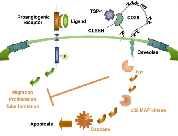
which TSP-1/CD36 exerts an antiangiogenic response (37). A model of this pathway is shown in Figure 1. Studies in several cell types have shown that CD36 physically associates with nonreceptor protein tyrosine kinases fyn, lyn, and yes. Exposure of microvascular endo-thelial cells (ECs) to TSP-1 leads to recruitment of fynto a CD36 mem-brane complex with activation of the kinase and subsequent down-stream activation of p38 mitogen-activated protein kinase. Studies with cells from null animals and pharmacological and immunologic inhibitors have shown that the antiangiogenic signal mediated by CD36 and TSP-1 is dependent on both of these kinases. Furthermore, the antiangiogenic activity of TSP-1 is linked to its ability to induce EC apoptosis via caspase-3–like effec-tors. CD36 signaling can lead to programmed cell death in other cell types: Rusinol et al. showed that incubation of CD36-transfected Chinese hamster ovary cells with oxLDL resulted in a dose- and time-dependent induction of apoptosis, which did not occur in nontransfected cells (38). As shown in the model, we suggest that the CD36/TSP-1 antiangiogenic signal provides a “switch” that diverts a growth factor–mediated proangiogenic response to an antiangiogenic, proapoptotic re-sponse. Unraveling this antagonistic pathway and comparing it with those of other angiogenic inhibitors may reveal a common ele-ment which would provide a powerful therapeutic tar-get that can be exploited in the treatment of tumors, diabetic retinopathy, and other diseases in which angio-genesis is a component.
CD36 ligands
[image:5.576.61.365.50.282.2]Scavenger receptors have high affinities for modified lipoproteins (see Platt and Gordon [ref. 39] and Krieger [ref. 40], both in this Perspective series). In the class B family, nothing is known of the ligands for LIMP-II, but those for CD36 and SR-BI have been well defined. SR-BI and CD36 have both been reported to bind native and modified lipoproteins, but it is interesting to note how their functions have diverged in the course of evolution. The interaction between SR-BI and HDL has become exquisitely refined to allow for selective lipid uptake and lipid exchange between cells and HDL (41). In contrast, CD36 has emerged as a specific recep-tor for transport of LCFAs. Part of this refinement of function may pertain to their respective tissue expres-sion patterns. While SR-BI is predominantly expressed in steroidogenic tissues and liver, CD36 is expressed in tissues with high LCFA storage or oxidative capacity. Additionally, the expression of CD36 on macrophages, RPE, and DCs has possibly resulted in retention of
Figure 1
some primitive scavenger functions, such as uptake of apoptotic cells. An extension of this role is recognition of sickle and parasitized erythrocytes, which have fea-tures in common with apoptotic cells. The ability of CD36 to cooperate in cross-priming in DCs shows evo-lution of the innate function of simple recognition of pathogen-induced apoptotic cells or host cells rendered “foreign” by mutation. Whether recognition of oxLDL is a further evolution of the capacity to recognize lig-ands common to apoptotic cells remains an open ques-tion, but the pathology that has emerged from this interaction is largely attributable to the Western diet.
Regulation of CD36 expression: identification of a proatherogenic feed-forward loop
Our laboratory and others have demonstrated that CD36 expression is highly regulated in monocytes and that it can be upregulated at the transcriptional level by adhesion, M-CSF, GM-CSF, native and modified LDLs, cellular cholesterol, and IL-4. CD36 is downregulated by corticosteroids, TGF-β1, HDL, and LPS (42–44), consis-tent with the idea that CD36 expression correlates with monocyte maturation and is downregulated during inflammation. A critical regulator of CD36 expression is the nuclear hormone receptor PPARγ (45, 46). The human CD36gene contains a PPARγ-responsive element shown to be functional in macrophages. Ligands for PPARγ (which include lipids derived from oxLDL, 9- and 13-hydroxyoctadecadienoic acid [HODE], prostaglandin J2 [PGJ2], and the thiazolidinedione class of
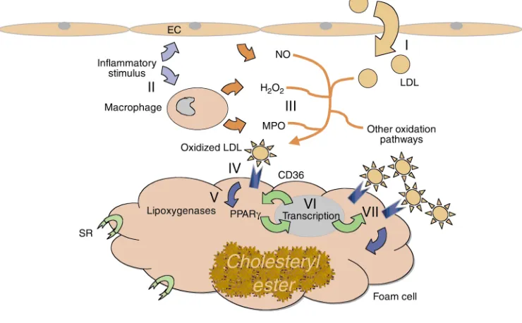
antidiabet-ic drugs) increase CD36 expression. The induction of CD36 by IL-4 in macrophages was recently shown by Huang et al. to be mediated by activation of intracellular lipoxygenases and generation of the PPARγligand PGJ2 (47). Downregulation of CD36 by TGF-βwas observed to be related to phosphorylation and inactivation of PPARγ. The relationship between CD36 expression and PPARγ, as well as the ability of CD36 to participate in macrophage internalization of potential lipid ligands for PPARγ, led Evans and colleagues to postulate a proatherogenic feed-forward loop in the vessel wall (46). Figure 2 shows a model for this pathway, in which macrophages and LDL particles become entrapped in the vessel wall as a result of a proatherogenic injury (48). Inflammatory stimuli then lead to production of reac-tive oxygen and nitrogen metabolites, generating epi-topes on LDL that are recognized by CD36. This leads to internalization of modified LDL, generation of intracel-lular PPARγ ligands, translocation of active PPARγ tran-scriptional activator complexes to the nucleus, and upregulation of PPARγ target genes, including PPARγ
itself and CD36. The increased expression of CD36 on the cell surface leads to further internalization of modi-fied LDL and eventual foam cell formation.
In heart and skeletal muscle, CD36 is regulated by plasma triacylglycerol and fatty acid levels and energy requirements. Cardiac CD36 expression is also respon-sive to an agonist of PPARα but not PPARγ, which reflects the cellular distribution of these factors, and establishes a pattern of regulation of CD36 by this
fam-Figure 2
[image:6.576.110.480.403.632.2]ily. During adipocyte development, CD36 expression increases as the cells become competent to take up and store LCFAs and may be essential to maturation (49). Adipogenesis is accompanied by expression of several members of the PPAR family, including PPARγ2 and PPARα (δ), which may regulate CD36 expression. Expression of CD36 and other fatty acid transporters is also regulated in response to environmental cues, such as plasma insulin and glucose, LPS, and cytokines.
Possible link to insulin resistance
The expression and regulation of CD36 in adipose tissue, skeletal muscle, and heart and its role as a translocator of LCFAs position it as a potential mediator of energy metabolism and may suggest an indirect role in glucose uptake and utilization. This hypothesis is supported by work with a diabetic strain of SHR by Aitman et al. (32), who showed that these animals express a mutant CD36
gene. Replacement of the mutant allele with the wild-type version in a congenic strain reverses the fatty acid trans-port defect and corrects the diabetic phenotype. Further support for a role for CD36 in glucose metabolism comes from studies showing a higher prevalence of type 2 dia-betes in Naka-negative patients. Interestingly, CD36-null mice are not diabetic, perhaps reflecting species differ-ences in glucose and fatty acid metabolism or a difference in genetic modifiers. However, CD36-null mice do have altered lipid and glucose metabolism and provide a model to dissect the interrelationship between CD36, fatty acid and glucose uptake and utilization. Interest-ingly, Li et al. reported that treatment of LDL recep-tor–deficient mice with thiazolidinediones improves insulin sensitivity in male (but not female) mice. In spite of the impact these drugs have on macrophage CD36 expression, they appear to decrease atherosclerosis in male mice (50). It may be that insulin resistance has the greatest proatherogenic effect in this model and that cor-rection of this metabolic feature counters any negative effect due to potential upregulation of macrophage CD36. This study therefore underscores the complexity of the regulation of CD36 in different tissues and in response to different environmental cues.
Conclusion
Work in the last 25 years has established CD36 as a multiligand receptor involved in cellular adhesion; fatty acid and lipid transport, utilization, and storage; antigen presentation; and clearance of apoptotic cells and shed photoreceptors. The challenge for the future will be to understand the mechanisms by which it effects these diverse functions and to design thera-peutic strategies that can alter the course of the dis-eases with which it is associated, including atheroscle-rosis, diabetes, cardiomyopathy, obesity, blindness, sickle cell anemia, and malaria.
Acknowledgments
Reference space limitations preclude recognition of many colleagues who have contributed to our knowledge of CD36; we wish to acknowledge them here. We wish to thank members of the Silverstein laboratory, especially David Lennon and Ronit Simantov, and David Stern for
critical reading of this manuscript and insightful sug-gestions. The studies summarized here were supported in part by grants from the NIH (EY-10967, HL-42540, HL-58559, POHL-56987), the Silbermann Fellowship, and the Dorothy Rodbell Cohen Foundation.
1. Adams, J.C., Tucker, R.P., and Lawler, J. 1995. The thrombospondin gene fam-ily. R.G. Landes. Austin, Texas, USA.
2. Silverstein, R.L., and Febbraio, M. 2000. CD36 and atherosclerosis. Curr. Opin. Lipidol. 11:483–491.
3. Oquendo, P., Hundt, E., Lawler, J., and Seed, B. 1989. CD36 directly mediates cytoadherence of Plasmodium falciparum parasitized ery-throcytes. Cell. 58:95–101.
4. Gruarin, P., et al. 2000. CD36 is a ditopic glycoprotein with the N-ter-minal domain implicated in intracellular transport. Biochem. Biophys. Res. Commun. 275:446–454.
5. Asch, A.S., et al. 1993. Analysis of CD36 binding domains: ligand speci-ficity controlled by dephosphorylation of an ectodomain. Science.
262:1436–1440.
6. Yamamoto, N., et al. 1990. A platelet membrane glycoprotein (GP) defi-ciency in healthy blood donors: Naka- platelets lack detectable GPIV (CD36). Blood. 76:1698–1703.
7. Franc, N.C., Dimarcq, J.L., Lagueux, M., Hoffmann, J., and Ezekowitz, R.A. 1996. Croquemort, a novel Drosophila hemocyte/macrophage receptor that recognizes apoptotic cells. Immunity. 4:431–443. 8. Hart, K., and Wilcox, M. 1993. A Drosophila gene encoding an epithelial
membrane protein with homology to CD36/LIMP II. J. Mol. Biol.
234:249–253.
9. Crombie, R., and Silverstein, R.L. 1998. Lysosomal integral membrane protein II binds thrombospondin-1. Structure-function homology with the cell adhesion molecule CD36 defines a conserved recognition motif.
J. Biol. Chem. 273:4855–4863.
10. Crombie, R., et al. 1998. Identification of a CD36-related throm-bospondin 1-binding domain in HIV-1 envelope glycoprotein gp120: relationship to HIV-1-specific inhibitory factors in human saliva. J. Exp. Med. 187:25–35.
11. Lisanti, M.P., et al. 1994. Characterization of caveolin-rich membrane domains isolated from an endothelial-rich source: implications for human disease. J. Cell Biol. 126:111–126.
12. Dorahy, D.J., Lincz, L.F., Meldrum, C.J., and Burns, G.F. 1996. Biochem-ical isolation of a membrane microdomain from resting platelets high-ly enriched in the plasma membrane ghigh-lycoprotein CD36. Biochem. J.
319:67–72.
13. Uittenbogaard, A., Shaul, P.W., Yuhanna, I.S., Blair, A., and Smart, E.J. 2000. High density lipoprotein prevents oxidized low density lipopro-tein-induced inhibition of endothelial nitric-oxide synthase localization and activation in caveolae. J. Biol. Chem. 275:11278–11283.
14. Lee, H., et al. 2001. Palmitoylation of caveolin-1 at a single site (Cys-156) controls its coupling to the c-Src tyrosine kinase. J. Biol. Chem. In press. Published July 12, 2001 as 10.1074/jbc.M104530200.
15. Endemann, G., et al. 1993. CD36 is a receptor for oxidized low density lipoprotein. J. Biol. Chem. 268:11811–11816.
16. Podrez, E.A., Schmitt, D., Hoff, H.F., and Hazen, S.L. 1999. Myeloperox-idase-generated reactive nitrogen species convert LDL into an athero-genic form in vitro. J. Clin. Invest. 103:1547–1560.
17. Podrez, E.A., et al. 2000. Macrophage scavenger receptor CD36 is the major receptor for LDL modified by monocyte-generated reactive nitro-gen species. 105:1095–1108.
18. Febbraio, M., et al. 2000. Targeted disruption of the class B scavenger receptor CD36 protects against atherosclerotic lesion development in mice. J. Clin. Invest. 105:1049–1056.
19. Savill, J., Hogg, N., and Haslett, C. 1991. Macrophage vitronectin recep-tor, CD36, and thrombospondin cooperate in recognition of neu-trophils undergoing programmed cell death. Chest. 99:6S–7S. 20. Bird, D.A., et al. 1999. Receptors for oxidized low-density lipoprotein on
elicited mouse peritoneal macrophages can recognize both the modified lipid moieties and the modified protein moieties: implications with respect to macrophage recognition of apoptotic cells. Proc. Natl. Acad. Sci. USA. 96:6347–6352.
21. Chang, M.K., et al. 1999. Monoclonal antibodies against oxidized low-density lipoprotein bind to apoptotic cells and inhibit their phagocyto-sis by elicited macrophages: evidence that oxidation-specific epitopes mediate macrophage recognition. Proc. Natl. Acad. Sci. USA.
96:6353–6358.
22. Ryeom, S.W., Sparrow, J.R., and Silverstein, R.L. 1996. CD36 participates in the phagocytosis of rod outer segments by retinal pigment epitheli-um. J. Cell Sci. 109:387–395.
24. Krieger, M. 1997. The other side of scavenger receptors: pattern recogni-tion for host defense. Curr. Opin. Lipidol. 8:275–280.
25. Ockenhouse, C.F., et al. 1991. Molecular basis of sequestration in severe and uncomplicated Plasmodium falciparum malaria: differential adhesion of infected erythrocytes to CD36 and ICAM-1. J. Infect. Dis. 164:163–169. 26. Aitman, T.J., et al. 2000. Malaria susceptibility and CD36 mutation.
Nature. 405:1015–1016.
27. McGilvray, I.D., Serghides, L., Kapus, A., Rotstein, O.D., and Kain, K.C. 2000. Nonopsonic monocyte/macrophage phagocytosis of plasmodium falciparum-parasitized erythrocytes: a role for CD36 in malarial clear-ance. Blood. 96:3231–3240.
28. Sugihara, K., Sugihara, T., Mohandas, N., and Hebbel, R.P. 1992. Throm-bospondin mediates adherence of CD36+ sickle reticulocytes to endothelial cells. Blood. 80:2634–2642.
29. Abumrad, N.A., el-Maghrabi, M.R., Amri, E.Z., Lopez, E., and Grimaldi, P.A. 1993. Cloning of a rat adipocyte membrane protein implicated in binding or transport of long-chain fatty acids that is induced during preadipocyte differentiation. Homology with human CD36. J. Biol. Chem.
268:17665–17668.
30. Ibrahimi, A., et al. 1999. Muscle-specific overexpression of FAT/CD36 enhances fatty acid oxidation by contracting muscle, reduces plasma triglycerides and fatty acids, and increases plasma glucose and insulin.
J. Biol. Chem. 274:26761–26766.
31. Febbraio, M., et al. 1999. A null mutation in murine CD36 reveals an important role in fatty acid and lipoprotein metabolism. J. Biol. Chem.
274:19055–19062.
32. Aitman, T.J., et al. 1999. Identification of Cd36 (Fat) as an insulin-resist-ance gene causing defective fatty acid and glucose metabolism in hyper-tensive rats. Nat. Genet. 21:76–83.
33. Hwang, E.H., et al. 1998. Absent myocardial iodine-123-BMIPP uptake and platelet/monocyte CD36 deficiency. J. Nucl. Med. 39:1681–1684. 34. Good, D.J., et al. 1990. A tumor suppressor-dependent inhibitor of
angio-genesis is immunologically and functionally indistinguishable from a fragment of thrombospondin. Proc. Natl. Acad. Sci. USA. 87:6624–6628. 35. Dawson, D.W., et al. 1997. CD36 mediates the in vitro inhibitory effects
of thrombospondin-1 on endothelial cells. J. Cell. Biol. 138:707–717. 36. Simantov, R., et al. 2001. Histidine rich glycoprotein inhibits the
antian-giogenic effect of thrombospondin-1. J. Clin. Invest. 107:45–52. 37. Jimenez, B., et al. 2000. Signals leading to apoptosis-dependent
inhibi-tion of neovascularizainhibi-tion by thrombospondin-1. Nat. Med. 6:41–48.
38. Rusinol, A.E., et al. 2000. Isolation of a somatic cell mutant resistant to the induction of apoptosis by oxidized low density lipoprotein. J. Biol. Chem. 275:7296–7303.
39. Platt, N., and Gordon, S. 2001. Is the class A macrophage scavenger receptor (SR-A) multifunctional? — The mouse’s tale. J. Clin. Invest.
108:649–654.
40. Krieger, M. 2001. Scavenger receptor class B type I is a multiligand HDL receptor that influences diverse physiologic systems. J. Clin. Invest.
108:793–797.
41. Acton, S., et al. 1996. Identification of scavenger receptor SR-BI as a high density lipoprotein receptor. Science. 271:518–520.
42. Huh, H.Y., Pearce, S.F., Yesner, L.M., Schindler, J.L., and Silverstein, R.L. 1996. Regulated expression of CD36 during monocyte-to-macrophage differentiation: potential role of CD36 in foam cell formation. Blood.
87:2020–2028.
43. Han, J., et al. 2000. Transforming growth factor-beta1 (TGF-beta1) and TGF-beta2 decrease expression of CD36, the type B scavenger receptor, through mitogen-activated protein kinase phosphorylation of peroxisome proliferator-activated receptor-gamma. J. Biol. Chem. 275:1241–1246. 44. Feng, J., et al. 2000. Induction of CD36 expression by oxidized LDL and
IL-4 by a common signaling pathway dependent on protein kinase C and PPAR-gamma. J. Lipid Res. 41:688–696.
45. Nagy, L., Tontonoz, P., Alvarez, J.G., Chen, H., and Evans, R.M. 1998. Oxi-dized LDL regulates macrophage gene expression through ligand acti-vation of PPAR gamma. Cell. 93:229–240.
46. Tontonoz, P., Nagy, L., Alvarez, J.G., Thomazy, V.A., and Evans, R.M. 1998. PPARgamma promotes monocyte/macrophage differentiation and uptake of oxidized LDL. Cell. 93:241–252.
47. Huang, J.T., et al. 1999. Interleukin-4-dependent production of PPAR-gamma ligands in macrophages by 12/15-lipoxygenase. Nature.
400:378–382.
48. Steinberg, D. 1997. Lewis A. Conner Memorial Lecture. Oxidative mod-ification of LDL and atherogenesis. Circulation. 95:1062–1071. 49. Sfeir, Z., Ibrahimi, A., Amri, E., Grimaldi, P., and Abumrad, N. 1997.
Reg-ulation of FAT/CD36 gene expression: further evidence in support of a role of the protein in fatty acid binding/transport. Prostaglandins Leukot. Essent. Fatty Acids. 57:17–21.