Verotoxin and ricin have novel effects on
preproendothelin-1 expression but fail to modify
nitric oxide synthase (ecNOS) expression and
NO production in vascular endothelium.
M M Bitzan, … , J Lin, P A Marsden
J Clin Invest.
1998;
101(2)
:372-382.
https://doi.org/10.1172/JCI522
.
Interaction of bipartite Escherichia coli O157-derived verotoxins (VTs) 1 and 2 (Shiga toxin
1 and 2) with vascular endothelium is believed to play a central role in the pathogenesis of
the thrombotic microangiopathy and ischemic lesions characteristic of hemolytic uremic
syndrome and of E. coli O157-associated hemorrhagic colitis. We defined the effects of VTs
on the expression of potent endothelial cell-derived regulators of vascular wall function,
namely endothelin-1 (ET-1) and nitric oxide (NO). In quiescent bovine aortic endothelial
cells, both VT1 and VT2, but not receptor-binding VT B-subunit which lacks N-glycosidase
activity, induced concentration-dependent (0.1-10 nM) increases in steady state preproET-1
mRNA transcript levels, an effect that was maximal at 12-24 h. Metabolic-labeling
experiments indicated that VTs increased preproET-1 mRNA transcript levels at
concentrations that had trivial effects on nascent DNA, RNA, and protein synthesis. In
contrast to preproET-1, endothelin converting enzyme-1 and endothelial constitutive NO
synthase mRNA transcript levels remained unchanged. Consistent with these findings, VTs
failed to modulate immunoreactive endothelial constitutive NO synthase expression and
basal and calcium-dependent L-[14C]arginine to L-[14C]citrulline conversion or the NO
chemiluminescence signal. The plant-derived toxin ricin, which shows a similar molecular
mechanism of enzymatic ribosomal modification to VTs, caused comparable effects on
these endothelial vasomediators and metabolite incorporation, at 3 log orders lower
concentrations. Nuclear transcription and actinomycin D chase experiments indicated that
[…]
Research Article
Find the latest version:
J. Clin. Invest.
© The American Society for Clinical Investigation, Inc. 0021-9738/98/01/0372/11 $2.00
Volume 101, Number 2, January 1998, 372–382 http://www.jci.org
Verotoxin and Ricin Have Novel Effects on Preproendothelin-1 Expression but
Fail to Modify Nitric Oxide Synthase (ecNOS) Expression and NO Production in
Vascular Endothelium
Martin M. Bitzan, Yang Wang, Janie Lin, and Philip A. Marsden
Division of Nephrology, Department of Medicine, St. Michael’s Hospital and University of Toronto, Toronto, Ontario, Canada M5S 1A8
Abstract
Interaction of bipartite
Escherichia coli
O157-derived
vero-toxins (VTs) 1 and 2 (Shiga toxin 1 and 2) with vascular
en-dothelium is believed to play a central role in the
pathogen-esis of the thrombotic microangiopathy and ischemic lesions
characteristic of hemolytic uremic syndrome and of
E. coli
O157-associated hemorrhagic colitis. We defined the effects
of VTs on the expression of potent endothelial cell–derived
regulators of vascular wall function, namely endothelin-1
(ET-1) and nitric oxide (NO). In quiescent bovine aortic
en-dothelial cells, both VT1 and VT2, but not receptor-binding
VT B-subunit which lacks
N
-glycosidase activity, induced
concentration-dependent (0.1–10 nM) increases in steady
state preproET-1 mRNA transcript levels, an effect that
was maximal at 12–24 h. Metabolic-labeling experiments
indicated that VTs increased preproET-1 mRNA transcript
levels at concentrations that had trivial effects on nascent
DNA, RNA, and protein synthesis. In contrast to preproET-1,
endothelin converting enzyme-1 and endothelial
constitu-tive NO synthase mRNA transcript levels remained
un-changed. Consistent with these findings, VTs failed to
modu-late immunoreactive endothelial constitutive NO synthase
expression and basal and calcium-dependent
L-[
14C]argi-nine to
L-[
14C]citrulline conversion or the NO
chemilu-minescence signal. The plant-derived toxin ricin, which
shows a similar molecular mechanism of enzymatic
riboso-mal modification to VTs, caused comparable effects on
these endothelial vasomediators and metabolite
incorpora-tion, at 3 log orders lower concentrations. Nuclear
tran-scription and actinomycin D chase experiments indicated
that VTs stabilize labile preproET-1 mRNA transcripts in
endothelial cells. Therefore, VTs potently increase select
mRNA transcript levels in endothelial cells at
concentra-tions of toxins that have minimal effects on protein
synthe-sis. Perturbed expression of endothelial-derived
vasomedia-tors may play a pathophysiologic role in the microvascular
dysfunction that is the hallmark of hemolytic uremic
syn-drome and hemorrhagic colitis. (
J. Clin. Invest.
1998. 101:
372–382.) Key words: hemolytic uremic syndrome
•endo-thelin-1
•nitric oxide synthase
•mRNA
•endothelin-con-verting enzyme-1
Introduction
Verotoxins (VTs)1 (Shiga toxins) comprise a family of
struc-turally related, bipartite protein exotoxins produced by Es-cherichia coli, especially serotype O157:H7 (1). VT1 and VT2 have been implicated by epidemiologic, microbiologic, and cel-lular biology studies in the pathogenesis of hemorrhagic colitis and hemolytic uremic syndrome (HUS) in humans (2–4). VT2e has been identified as the cause of systemic edema dis-ease of weaning piglets (5). Characteristic histopathologic and pathophysiologic features of VT-associated HUS include throm-botic microangiopathy, focal areas of ischemia, and, in some settings, profound hypertension (6–9). Though there is abun-dant evidence of platelet–microvascular endothelial interac-tion, perturbation of the fibrinolytic cascade, and arachidonic acid metabolism (7, 10, 11), the precise disease mechanisms re-main enigmatic (4, 12).
VTs bind with high affinity to the membrane-anchored glycosphingolipids globotriosylceramide (Gb3) and globotet-raosylceramide (Gb4). Glycolipid binding is mediated by the lec-tin-like, pentameric B subunit of VTs (13). Retrograde intra-cellular transport of endocytosed VT to the endoplasmic reticulum via the Golgi apparatus (14) is a prerequisite for the protein-synthesis–inhibiting action of the toxins (1). The VT A subunit displays structural and functional homology with the A subunit of ricin, a heterodimeric lectin derived from the seeds of the castor oil plant (Ricinus communis) (15, 16). The 32-kD ricin A chain is linked by a disulfide bond to a single B chain of similar size which mediates membrane receptor binding and internalization (14, 15). This contrasts with the noncovalent, 1:5 stoichiometric relationship of the A:B subunits in VTs (1). The A subunits of VT and ricin possess intrinsic N-glycosidase ac-tivity. In cell-free systems they both hydrolyze a specific ade-nine residue at position 4324 of the 28S ribosomal RNA of the 60S mammalian ribosomal subunit. This posttranscriptional modification leaves the phosphodiester bond intact, but effec-tively interferes with the binding and coordinated function of eukaryotic elongation factors EF-1 and -2 (1, 16, 17).
We hypothesized that endothelial cell activation plays an important role in the pathophysiology of VT-associated dis-ease, specifically HUS. A paucity of data is available on the endothelial genes implicated in the pathobiology of
endothe-Address correspondence to Dr. Philip A. Marsden, University of Toronto, Medical Sciences Bldg., Rm. 7358, 1 King’s College Circle, Toronto, ON, Canada M5S 1A8. Phone: 978-2441; FAX: 416-978-8765; E-mail: p.marsden@utoronto.ca
Received for publication 5 May 1997 and accepted in revised form 14 November 1997.
lial dysfunction in VT-associated HUS. Endothelial-derived ni-tric oxide (NO) radical and the peptide endothelin-1 (ET-1) are major paracrine and autocrine mediators which, among others, regulate local blood flow and, in the case of NO, modu-late pmodu-latelet adhesion, aggregation, and degranulation (18, 19). We speculated that VT might perturb the expression of these vasomediators in vascular endothelium. Therefore, the purpose of this study was to define the effects of VTs and of ricin on the expression of these endothelial-derived vasomediators using a well characterized, robust endothelial culture model (20).
Methods
Materials.Cell culture reagents, dithiothreitol, and MMLV reverse transcriptase polymerases were from GIBCO BRL (Grand Island, NY). Bovine calf serum (low endotoxin) was purchased from Hy-clone Laboratories (Logan, UT). Cell culture plates were from Corn-ing Inc. (CornCorn-ing, NY). Ionomycin was from Calbiochem (La Jolla, CA). Deoxynucleotidetriphosphates were from Boehringer Mann-heim GmbH (MannMann-heim, Germany), restriction enzymes from New England Biolabs (Beverly, MA), RNases A and T from Ambion (Austin, TX), and DNase 1 and RNase inhibitor from Promega (Madison, WI). Guanidinium thiocyanate was from Fluka Biochemika (Buchs, Switzerland). L-[14C]arginine (sp act 300 Ci/mmol), Hybond1
nylon membranes, ET-1 radioimmunoassay kit (21 peptide-specific), and enhanced chemiluminescence substrate were from Amersham (Arlington Heights, IL). SepPak C18 cartridges were from Waters Millipore Corp. (Milford, MA). Protran nitrocellulose membranes were from Schleicher and Schuell (Keene, NH). Bradford method– based protein assay (protein assay kit II) and Dowex AG 50WX-8 cation exchange resin (Na1 form, 100–200 mesh) were from Bio-Rad (Hercules, CA). Deoxycytidine 59-[a-32P]triphosphate ([a-32P]dCTP;
3,000 Ci/mmol), uridine 59-[a-32P]triphosphate ([a-32P]UTP; 3,000 Ci/
mmol), L-[3,4,5-3H(N)]leucine ([3H]leucine; . 140 Ci/mmol), [methyl-3H]thymidine ([3H]thymidine; 70–90 Ci/mmol), [5,6-3H]uridine ([3
H]uri-dine; 35–50 Ci/mmol) were purchased from DuPont-NEN (Boston, MA). Mouse monoclonal IgG against human endothelial constitutive nitric oxide synthase (ecNOS) protein was from Transduction Labo-ratories (Lexington, KY). Horseradish-conjugated sheep anti–mouse IgG antibody was from Bio-Rad. Other reagents were from Sigma Chemical Co. (St. Louis, MO). VTs were purified to homogeneity from defined E. coli strains by sequential chromatography in the lab-oratories of Drs. M.A. Karmali (The Hospital for Sick Children, Tor-onto, Canada) and J.L. Brunton (The Toronto Hospital, TorTor-onto, Canada) (0.6–3.1 mg protein/ml, cytotoxic titers [Vero cells] z 1028
for the holotoxins, , 1022 for the pentameric VT1 B subunit) as
de-scribed previously (21–23). VT preparations contained negligible amounts of endotoxin (at or less than the detection limit of 0.05 en-dotoxin units/ml) (E-toxic assay; Sigma Chemical Co.) (24). Highly purified ricin was purchased from Sigma Chemical Co. (RCA60).
Toxin aliquots were stored at 2808C and diluted in serum-free me-dium immediately before experimentation.
Cell culture. Bovine endothelial cells were isolated from the tho-racic aorta of calves and phenotypically characterized as described previously (25). Single-clone, homogeneous bovine aortic endothelial cell (BAEC) cultures were serially passaged on 0.2% gelatin-coated 100-mm dishes after digestion with trypsin-EDTA (0.05%) and prop-agated in RPMI 1640 medium containing 15% bovine calf serum, penicillin G (100 U/ml), and streptomycin sulfate (100 mg/ml). For the majority of experiments, cells were used at passages 4 and 5. To examine the effect of VT on BAEC, 100-mm dishes were plated at a density of 5–10 3 105 cells per dish in 10 ml complete medium. 2 d
af-ter reaching confluence, generally 5 d afaf-ter splitting, complete me-dium was replaced by serum-free meme-dium, and cells were harvested 48 h later. Toxin was added at the indicated concentrations 24 h be-fore harvesting, unless stated otherwise.
Metabolite pulse labeling. Cells were grown 2 d after confluence in 24-well dishes containing 0.4 ml complete RPMI medium. The me-dium was changed to serum-free RPMI 24 h before toxin or vehicle was added. 1 h before harvest, 1 mCi of [3H]thymidine, [3H]uridine, or
[3H]leucine was added per well. Monolayers were rapidly washed
with ice-cold PBS to terminate isotope uptake, incubated with 0.5 ml of 15% TCA at 48C for 20 min, and rinsed twice with ice-cold H2O.
Cell-associated radioactivity was determined after solubilization with 0.1 M NaOH-0.1% SDS (wt/vol). Aliquots from each well were re-moved for scintillation counting. Data were normalized against vehi-cle-treated controls with no added toxin. Results of three indepen-dent experiments, each performed in triplicate, were expressed as arithmetic means6SEM.
Molecular cloning and cDNA probes.Random-primed, first strand complementary DNA (cDNA) was prepared from total cellular RNA isolated from BAEC using MMLV reverse transcriptase (26). A 744-bp DNA fragment for coding sequences of bovine endothelin converting enzyme-1 (ECE-1) was generated by PCR (sense primer BE-1: 59 GCT CCT GGC GGC GGC ATT GGT G 39; antisense primer BE-2: 59 TCC TGG GGG ATG GTG ATG TTG G 39), cloned into the pCRII vector and subjected to dideoxynucleotide sequence analysis. A BstXI restriction enzyme fragment of the cloned bovine ECE-1 cDNA corresponding to amino acids 58–305 was used as a probe. The 1.4-kb EcoRI restriction enzyme fragment of pBov ET-1 containing the open reading frame of bovine preproET-1 was described previ-ously (27). A 59-preproET-1 cDNA probe containing the first 360 bp (exons 1–3), and a 1 kb 39-preproET-1 probe containing the remain-ing sequences (exons 4–5) were obtained by digestion of pBovET-1 with NcoI and EcoRI. The 1.9-kb EcoRI restriction enzyme bovine ecNOS cDNA clone pBov39NOS corresponding to exons 15–26 of the human ecNOS gene has been described (28). A 551-bp 59 bovine ecNOS cDNA fragment corresponding to exons 1–4 of human ecNOS was generated by EcoRI/SfiI digestion of the 1785-bp EcoRI restriction enzyme clone pBov59NOS (28). A HindIII/XbaI restric-tion fragment of GAPDH (20) encompassing 547 bp of the human glyceraldehyde-3-phosphate dehydrogenase cDNA and a 0.75-kb BamH1/Sph1 restriction fragment of 18S ribosomal RNA cDNA (29) were used for normalization in Northern and actinomycin D chase experiments, respectively.
Northern blot analysis. Total cellular RNA was extracted using the guanidinothiocyanate-phenol-chloroform procedure, size-frac-tionated by electrophoresis through denaturing (1% agarose-0.66 M formaldehyde) gels, transferred onto nylon membranes, UV cross-linked, and hybridized using published methods (30). Gels were stained with ethidium bromide to verify loading of comparable amounts of intact RNA. Membranes were prehybridized at 428C in 50% deion-ized formamide, 63 SSPE, 53 Denhardt’s solution, 0.5% SDS, and 100 mg/ml denatured salmon sperm DNA (hybridization solution), for a minimum of 4 h. Gel-purified cDNA probes were labeled to a sp act of $ 109 cpm/mg with [a-32P]dCTP by the random primer labeling
method. Hybridization was carried out with 106 cpm/ml in fresh
hy-bridization solution at 428C for 18–24 h. Membranes were washed un-der high stringency conditions and monitored with a Geiger counter. Radioactive signals were recorded using a PhosphorImager (Molecu-lar Dynamics, Sunnyvale, CA) and quantitated using ImageQuant software 4.2 for Windows NT (Molecular Dynamics). In addition, conventional autoradiography was performed.
Nuclear run-on analysis. Studies were performed as described previously (20). Briefly, confluent VT-treated and untreated BAEC cultures were washed with sterile 10 mM PBS and lysed in situ with chilled lysis buffer (10 mM Tris-HCl [pH 7.9], 0.15 M NaCl, 1 mM EDTA, and 0.6% [vol/vol] Nonidet P-40) for 10 min on ice. Cell ly-sates were centrifuged for 5 min at 500 g at 48C, and the nuclear pellet resuspended in 75 ml of chilled nuclear buffer containing 0.3 M (NH4)2SO4, 100 mM Tris-HCl (pH 7.9), 4 mM MgCl2, 4 mM MnCl2,
0.2 M NaCl, 0.4 mM EDTA, 0.1 mM PMSF, and 40% (vol/vol) glyc-erol. 100 ml of 13 cold transcription buffer (0.3 M (NH4)2SO4, 100 mM
EDTA, and 0.1 mM PMSF) containing 0.2 mM DTT, 40 U RNasin, 0.2 mM ATP, CTP, and GTP, and 150 mCi [a-32P]UTP (3,000 Ci/
mmol) was added to the nuclear suspension, and incubated for 30 min at 288C. 20 U of RNase-free DNase 1 and 125 mg tRNA were added and incubated for 10 min at 378C, followed by digestion with protein-ase K at a final concentration of 300 mg/ml in buffer (10 mM Tris-HCl, pH 7.9, 10 mM EDTA, 0.5% SDS) for 30 min at 428C. Nuclear transcripts were then extracted as described above and resuspended at 2 3 106 cpm/ml in Northern hybridization buffer. Equal amounts
(1 mg) of gel-purified cDNA were denatured by boiling in 0.4 M NaOH-10-mM EDTA and neutralized with equal volumes of 2 M ammonium acetate, pH 7.0 (26), and slot-blotted onto nitrocellulose filters. The cDNA slot-blotted was identical to that used to generate the probes for Northern blot analysis. Hybridization was performed for 48–72 h at 428C in Northern hybridization buffer as detailed above. Hybridized filters were washed as described (20), and sub-jected to autoradiography using a PhosphorImager (Molecular namics) and quantitative densitometry assisted by the Molecular Dy-namics ImageQuant Software package.
Actinomycin D chase experiments. To assess the effect of VT on the half-life of preproET-1 mRNA transcripts, confluent BAEC monolayers were treated with VT at 5 nM for 20 h before addition of 10 mg/ml actinomycin D. Previous studies demonstrated that the in-corporation of [3H]uridine into TCA-insoluble material was inhibited
99.5% by this treatment (31). Total cellular RNA was extracted at 0, 15, 30, 60, 120, and 180 min after the addition of actinomycin D. Blots were reprobed with an 18S rRNA-cDNA probe to ensure equal load-ing. PreproET-1 mRNA transcript levels were normalized for 18S rRNA using ImageQuant (Molecular Dynamics) and mRNA decay rates determined according to the formula: Nt5N0e2lt. Results were
from three independent experiments.
Protein extraction and immunoblotting. Confluent BAEC mono-layers were lysed in situ with Laemmli buffer (62.5 mM Tris-HCl [pH 7.4], 2% [wt/vol] SDS, 10% [vol/vol] glycerol, and 0.72 M 2-mercapto-ethanol), and total cellular protein extracted as described previously (20). Cell lysates were cleared at 100,000 g for 30 min at 258C and stored at 2808C before analysis. The protein was quantitated using the modified Bradford method with bovine serum albumin as stan-dard. 20 mg of protein was electrophoresed under reducing and dena-turing conditions in 6% polyacrylamide gels. Equal loading was con-firmed by replicate gel staining with Coomassie blue. Proteins were transferred to nitrocellulose filters using an electroblotting apparatus (Bio-Rad). Filters were blocked overnight at 48C with 4% bovine se-rum albumin in Tris-buffered saline with 0.1% Tween 20, followed by a 2-h incubation at room temperature with a primary mouse mono-clonal IgG antibody to human ecNOS which also recognizes bovine ecNOS (20) at a concentration of 0.125 mg/ml. Bound primary anti-body was detected using horseradish-conjugated sheep anti–mouse IgG antibody diluted 1:20,000 for 1 h at room temperature followed by enhanced chemiluminescence substrate and brief exposure to au-toradiographic films.
Endothelin radioimmunoassay. Confluent BAECs were exposed to VT2 at various concentrations for 24 h in serum-free culture me-dium in 24-well dishes. Conditioned meme-dium (triplicate wells) was ex-tracted over SepPak C18 cartridges, eluted with methanol-H2O-acetic
acid (90:9.6:0.4), lyophilized, and stored at 2808C until analysis with a radioimmunoassay kit specific for ET 1–21 as described previously (20). Cross-reactivity with big ET-1 was , 0.4%. While the assay cross-reacts with ET-2 and ET-3, ET-1 is the only isoform known to be produced by vascular endothelial cells (32). Levels of immunore-active peptides are therefore taken to reflect production of ET-1. The intra- and interassay coefficients of variation averaged 6 and 10%, re-spectively. Cell numbers were assessed in representative wells using an automated cell counter (triplicate determinations).
Measurement of NO synthase activity. L-[14C]citrulline conversion
was determined using previously published methods with minor mod-ifications (33, 34). Briefly, confluent cell monolayers grown in 6-well dishes were treated with VT2 at 4 or 0.1 nM or vehicle for 24 h as
de-scribed above, gently rinsed with a physiologic salt solution (130 mM NaCl, 5 mM KCl, 1.5 mM CaCl2, 1 mM MgCl2, 25 mM Hepes [pH 7.4]
and 10 mM D-glucose) equilibrated in 1 ml of the same buffer for 30 min at 378C and labeled with z 0.5 3 106 cpm/ml TLC-purified L-[14C]arginine for 30 min. Cells were treated with 5 mM ionomycin or
vehicle for the final 20 min at 378C. The reaction was stopped with 4 ml of chilled 10 mM PBS/5 mM EDTA, pH 7.6. Cells were ex-tracted with 1 ml of 0.3 M HClO4 for 20 min at 48C. Extracts were
neutralized with 1 M K2CO3 and loaded onto a 1-ml wet bed volume
of Dowex AG 50WX-8 cation exchange resin followed by 2 ml of wa-ter. L-[14C]citrulline in the column effluent was quantitated by
scintil-lation counting. TLC resolution of fractions of the column effluents and quantitation with the PhosphorImager indicated that L-[14
C]cit-rulline was the only detectable product. Counts (cpm) eluted from each resin column were normalized to total protein content and cell number per well.
Measurement of NO release. Basal and calcium-stimulated NO
release was measured with chemiluminescence as described previ-ously (35, 36) with minor modifications. Confluent monolayers grown in 6-well dishes treated with 5 nM VT2 for 24 h were gently rinsed with PBS containing 1.5 mM CaCl2, and equilibrated in 0.6 ml of the
same buffer at 378C for 20 min before adding 5 mM ionomycin or ve-hicle. The conditioned buffer was removed at various time intervals (5–30 min) after the addition of ionomycin or vehicle and immedi-ately frozen for storage at 2808C. To measure cell-released NO/ NO22, a rapid-response, ozone chemiluminescence analyzer was used
(Model 280; Sievers, Boulder, CO). NO22 was reduced to NO in the
presence of glacial acetic acid and excess NaI, and purged from solu-tion with inert gas allowing mass-sensitive chemiluminescence detec-tion based on the reacdetec-tion of the NO radical with ozone (O3).
Manu-facturer-specified lower limit of sensitivity for this analyzer was z 1 pmol of NO22 or NO injected into the purge vessel. The amount of
released NO was derived from a standard curve of NaNO2 (0.1–100
mM). Inter- and intraassay coefficient of variation was , 12% (36).
Data analysis. Data are expressed as means6SEM obtained in at least three separate experiments, unless indicated otherwise. Statisti-cal analysis for paired comparisons was carried out using the Stu-dent’s t test, and one-way ANOVA for serial determinations. The level of significance was defined as P , 0.05.
Results
Effect of VTs on endothelial metabolism. Confluent
[image:4.612.315.558.589.678.2]monolay-ers of BAECs were treated with VT1 and VT2 for 24 h at toxin concentrations ranging from 0.01 to 100 nM. The incorpora-tion of [3H]thymidine, [3H]uridine, or [3H]leucine was utilized
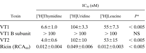
Table I. Concentration-dependent Effects of VTs and Ricin on Metabolite Incorporation Rates of BAECs
Toxin
IC50 (nM)
[3H]Thymidine [3H]Uridine [3H]Leucine P*
VT1 6.661.0 10463.3 5567.3 , 0.005 VT1 B subunit . 100 . 100 . 100 NS VT2 4.060.6 102610 53615 , 0.005 Ricin (RCA60) 0.01260.004 0.04960.006 0.01260.003 , 0.005
to assess overall rates of synthesis of nascent DNA, RNA, and peptides, respectively. As shown in Fig. 1, at the highest toxin concentrations, incorporation of all metabolites was signifi-cantly reduced. 50% inhibitory concentrations (IC50) are
sum-marized in Table I. For VT2, likely the more important VT in human disease based on epidemiological data (37, 38), IC50
values for [3H]thymidine, [3H]uridine, and [3H]leucine
aver-aged 4.060.6, 102610, and 53615 nM, respectively. As shown
in Fig. 1 and Table I, IC50 values for VT1 and VT2 on DNA,
RNA, and peptide synthesis rates were quantitatively different in that more robust effects on inhibition of DNA synthesis were evident. VT1 and VT2 inhibited [3H]thymidine
incorpo-ration at concentincorpo-rations that were 1 to 2 log orders lower than those that reduced [3H]leucine incorporation (P , 0.01 and
, 0.05, respectively). In addition, the rate of [3H]uridine
[image:5.612.56.535.52.332.2]incor-poration was slightly increased at VT1 and VT2 concentrations Figure 1. VT effects on global
indi-ces of endothelial cell metabolism. Confluent, serum-starved BAECs were exposed for 24 h to the indi-cated toxin concentrations and for 1 h to 1 mCi of [3H]thymidine,
[3H]uridine, or [3H]leucine.
Radio-activity incorporated into TCA-insoluble material was determined. Data have been normalized to vehi-cle-treated cells. Shown are the means6SEM, n 5 3 experiments, triplicate determinations. When er-ror bars are not present, the SE was too small to appear. * Values signifi-cantly different from vehicle-treated BAECs.
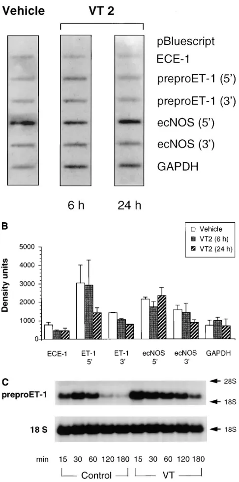
Figure 2. Time-dependent effect of VT2 on BAECs. Northern blot analysis (15 mg/lane) of total cellular RNA from BAECs treated with VT2 (8 nM) for the indicated time periods before harvest. Membranes were hybridized with [32P]dCTP-labeled bovine preproET-1, ecNOS and with
[image:5.612.65.547.460.673.2]up to 20 nM (Fig. 1). VT1 B subunit, which lacks N-glycosidase activity, failed to modify DNA, RNA, and peptide synthesis (Fig. 1), suggesting that the A subunit is responsible for the ob-served biologic effects. VT2e, which utilizes a different gly-cosphingolipid receptor, namely Gb4, was significantly less active in BAECs than VT1 and VT2 up to concentrations of 100 nM (results not shown).
VTs increase preproET-1 steady-state mRNA levels. The
effects of VT1 and VT2 on steady-state mRNA expression of preproET-1, ECE-1, and ecNOS in cultured endothelial cells was assessed by Northern analysis. Exposure to VT1 or VT2 induced a concentration- and time-dependent increase in 2.3-kb preproET-1 mRNA transcript levels in BAECs. Under these conditions, monolayers revealed little to no microscopic alter-ations compared with vehicle-treated controls. VT-induced in-creases in preproET-1 mRNA levels were evident after as early as 4 h, with maximal effects observed at 12–24 h (Fig. 2). Concentration–response relationships (0.001–10 nM) were as-sessed at 24 h and are shown in Fig. 3. The threshold
concen-tration for increases in preproET-1 mRNA level was 0.5 nM for both VT1 and VT2 (Fig. 3 B). Concentrations between 1 and 10 nM, used in various independent experiments, caused a 2–3-fold increase in steady-state preproET-1 mRNA levels over GAPDH signals (VT1, 2.660.3-fold increase, mean6SE,
n 5 7; VT2, 2.260.2-fold, n 5 8). These results indicate that in-creased preproET-1 transcript levels were observed at VT concentrations that had minor effects on overall protein syn-thesis and gene transcription (compare with Fig. 1). VT1 B subunit (1–10 nM) failed to change steady-state preproET-1 mRNA levels (Fig. 4).
Effect of VTs on ECE-1 and ecNOS mRNA expression.
VT1 and VT2 holotoxins, and the VT1 B subunit failed to modify steady-state mRNA levels for ECE-1 and ecNOS at varied time points and concentrations (Figs. 2 and 3). These negative findings were confirmed with multiple independent BAEC clones (n 5 6). Results of a representative experiment comparing the effects of relevant concentrations of VT1, VT2, and the VT1 B subunit on preproET-1 and ECE-1 mRNA transcript levels in BAECs are shown in Fig. 4.
Effect of VT2 on immunoreactive endothelin peptide secre-tion. Exposure of BAEC to 5 nM VT2 for 24 h increased the
amount of secreted immunoreactive peptide as assessed with a highly specific radioimmunoassay at 5 nM (Fig. 5).
Effect of VT2 on ecNOS protein expression. Total cellular
immunoreactive endothelial NOS protein in BAECs was as-sessed by Western blot studies. In protein extracts of cells un-der basal conditions a single immunoreactive band of z 135 kD was detected. Treatment of BAECs with VT2 (8 nM) for 24 or 48 h failed to have a significant effect on steady-state levels of immunoreactive ecNOS protein. A representative blot is shown in Fig. 6. Results were confirmed with six independent BAEC clones.
Measurement of NOS enzymatic activity.L-[14C]arginine to
L-[14C]citrulline conversion was determined using quantitative
[image:6.612.55.298.57.412.2]cation-exchange chromatography as described previously (34). Figure 3. Concentration-dependent effect of VT2 on BAECs.
North-ern blot analysis of total cellular RNA (15 mg/lane) from BAECs ex-posed for 24 h to the indicated concentrations of VT1. Blots were hy-bridized with [32P]dCTP-labeled preproET-1 cDNA, stripped, and
reprobed for ecNOS and GAPDH. (A) A representative blot is shown from one of four independent experiments with VT1. (B) Densitometric analysis of blots from four independent experiments normalized to GAPDH and displayed as fold increase over baseline. *Values significantly different from control BAECs.
Figure 4. Comparative Northern blot analysis of total cellular RNA
[image:6.612.320.554.487.679.2]Fig. 7 A shows the effect of VT2 (24 h; 0.1 and 4 nM) on BAECs L-[14C]citrulline formation in the absence and
pres-ence of the calcium ionophore ionomycin (5 mM, 20 min).
L-[14C]citrulline formation increased in BAECs in response to
ionomycin consistent with the observation that constitutive NOS activity in BAECs is calcium and calmodulin-dependent. Rates of L-[14C]citrulline formation were not modified by VT2
in the absence or presence of ionomycin.
Effect of VT2 on NO chemiluminescence signal. NO
re-lease from BAEC monolayers was determined as [NO 1 NO22] using a chemiluminescence detection method.
Ionomy-cin (5 mM) treatment increased the chemiluminescence signal approximately threefold above vehicle-treated cells. Vehicle-and ionophore-stimulated release of NO/NO22 from BAECs
was not altered by VT2 (24 h, 5 nM, n 5 3) (Fig. 7 B).
Nuclear run-on studies. To define the molecular
mecha-nism underlying the observed increase in preproET-1 steady-state mRNA levels after VT treatment, nuclear run-on studies were performed in BAECs exposed to VT2 (5 nM) for 6 or 24 h. cDNA probes reflecting 59 and 39 regions of the transcription units were used to assess potential effects on polymerase pro-cessivity (transcriptional arrest) (39) after VT treatment. Al-though hybridization of labeled, nascent nuclear RNA to 39 -cDNA probes for preproET-1 and ecNOS yielded weaker
sig-nals than hybridization to the 59 probes of the same cDNA, no appreciable differences in the rate of transcription of prepro-ET-1, ecNOS, and ECE-1 mRNA, or in polymerase processiv-ity were noted between different treatment conditions (Fig. 8,
A and B). Identical results were obtained when BAECs were
[image:7.612.317.556.61.453.2]exposed to equimolar concentrations of VT1 (not shown). Figure 5. Measurement
of immunoreactive en-dothelin in the condi-tioned medium of BAECs exposed to vehi-cle or VT2 at 5 nM for 24 h. Data represent mean6SE from one of two experiments (tripli-cate determinations).
Figure 6. Effect of VT2 on immunoreactive ecNOS protein levels in
[image:7.612.58.297.494.671.2]BAECs. BAECs were exposed to vehicle or VT2 (8 nM) for 24 or 48 h. Immunoblotting was performed using 20 mg/lane of total cellular pro-tein extract. Bovine immunoreactive propro-tein was detected with an mAb for ecNOS. Data are representative of four independent experi-ments using six different BAEC clones.
Figure 7. Effect of VT on NOS activity and NO/NO22 release from
BAECs. (A) L-[14C]arginine–L-[14C]citrulline conversion. Confluent
cell monolayers were exposed to vehicle or VT2 at the indicated con-centrations for 24 h. 1 h before harvesting, culture medium was re-placed by a physiologic salt solution containing 0.5 3 106 cpm/well L-[14C]arginine and then treated with vehicle or ionomycin (5 mM, 20
min). After extraction with ice-cold HClO4, L-[14C]arginine was
de-termined by quantitative cation-exchange chromatography. Bars represent the mean6SEM of three experiments (triplicate deter-minations). (B) Chemiluminescence determination of NO/NO22.
Confluent monolayers were treated with 5 nM VT2 for 24 h. After replacement of culture medium with a physiological salt solution ve-hicle or ionomycin (5 mM) was added for 20 min. NO/NO22 content
Actinomycin D chase experiments. BAECs were treated
with a potent transcriptional inhibitor, actinomycin D, allow-ing quantitation of the rate of preproET-1 mRNA decay. Fig. 8 C shows a representative Northern blot of one of three inde-pendent actinomycin D chase experiments. Confluent mono-layers, pretreated for 20 h with 5 nM VT or vehicle received actinomycin D (10 mg/ml) at time 0. Total cellular RNA was extracted 15–180 min after addition of actinomycin D. Analy-sis of densitometric data revealed that VT increased the half-life of preproET-1 mRNA in BAECs on average by 2.5-fold, from 3562 to 86623 min (n 5 3; P , 0.05). These findings, in concert with the nuclear run-on studies, indicated that the ob-served increase in preproET-1 mRNA transcript levels in vas-cular endothelial cells was not due to increased rates of tran-scription but solely due to changes in mRNA degradation: specifically, VTs prolong the mRNA half-life of preproET-1.
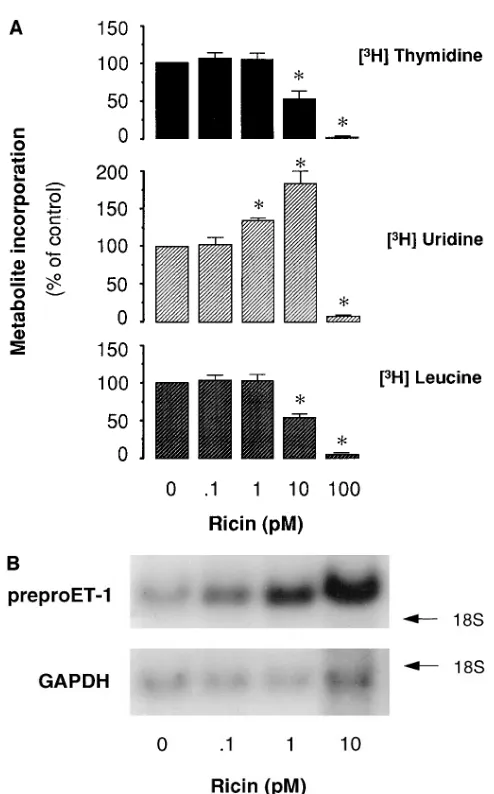
The effect of ricin on endothelial metabolism and preproET-1 steady-state mRNA levels. The plant protein toxin ricin shares
with VTs the ability, via its structurally related A subunit, to depurinate 28S rRNA at the identical site (16). However, VTs and ricin display differences in receptor specificity and intra-cellular transport (40). To gain further insight into the biologic effects of ribosome inactivating proteins in endothelial cells, we examined the effects of ricin on basal rates of DNA, RNA, and protein synthesis and on preproET-1 mRNA steady-state levels in BAECs. As shown in Table I, ricin inhibited [3
H]leu-cine incorporation at a concentration 3 log orders below those of VT1 and VT2 (IC50 1263 pM vs. . 50 nM). Furthermore,
ri-cin significantly elevated the incorporation of [3H]uridine at 10
pM (Fig. 9 A). Ricin increased preproET-1 mRNA transcript levels in a time- (not shown) and concentration-dependent manner (1.860.2-fold and 5.161.0-fold at 1 and 10 pM, respec-tively; n 5 8) (Fig. 9 B), and, similar to VT1 and VT2, failed to modify ecNOS and ECE-1 mRNA transcript levels (not shown).
Discussion
[image:8.612.56.299.70.560.2]In this study, we determined the effect of VTs on the expres-sion of the endothelial vasomediators, ET-1 and NO, and their key regulatory enzymes, ECE-1 and ecNOS. The major find-ing was the ability of VT1 and VT2 to increase preproET-1 steady-state mRNA levels in vascular endothelium in a time-and concentration-dependent manner. Importantly, the VT effect on preproET-1 mRNA levels was observed at concen-trations that had minor effects on global nascent peptide syn-thesis, and therefore does not appear to be a consequence of generalized ribosomal blockade. Furthermore, the concentra-tion of VT2 used did not diminish the release of immunoreac-tive endothelin in conditioned medium, but rather increased the amount of immunoreactive peptide. In contrast to pre-proET-1, mRNA transcript levels of the membrane-bound metalloprotease ECE-1, a key enzyme in the biosynthesis pathway of the endothelins, were not modified by VT1 or Figure 8. Molecular mechanism of VT-induced increases in
steady-state preproET-1 mRNA levels. (A) Effect of vehicle or VT2 (5 nM; 0, 6, 24 h) on the rate of endothelial gene transcription. Isolated cell nuclei were allowed to generate nuclear run-on products in the pres-ence of [32P]UTP and then hybridized to linear, immobilized cDNA
probes as indicated. PreproET-1 and ecNOS cDNA probes reflecting 39 and 59 regions of the transcription units were used to assess poly-merase processivity after VT treatment. There were no significant differences in signal strength between vehicle and VT2-treated BAECs. (B) Quantitative densitometry of the results of three inde-pendent nuclear run-on experiments. Data represent mean6SE from three independent experiments. (C) Representative Northern blot (15 mg/lane) analysis of the rate of disappearance of preproET-1 mRNA in BAECs after transcriptional blockade at time 0 with acti-nomycin D (10 mg/ml). Confluent monolayers were treated with VT (5 nM) or vehicle, 20 h before the addition of actinomycin D. Total cellular RNA was extracted at various time points after addition of
VT2. Our novel observation that VTs induce preproET-1 ex-pression in the absence of exogenous cytokines suggests that VT1 and VT2 can activate endothelial cells directly. We take these data to indicate that activation of endothelial cells by VTs is an important biological function of this family of toxins. We originally hypothesized that modulation of NO produc-tion may play a role in the pathogenesis of VT-induced throm-botic microangiopathy. Endothelial-derived NO is known to prevent platelet activation (18). For example, rats injected with LPS and the NOS inhibitor L-NAME evidence throm-botic microangiopathy in the kidney: glomerular capillary thrombosis was observed in . 50% of the glomeruli examined, whereas , 5% of the glomeruli were affected in rats receiving
LPS or L-NAME alone (41). That VTs may act, in part, by im-pairing ecNOS expression or activity was an attractive hypoth-esis. However, VT2 failed to modify steady-state ecNOS mRNA or immunoreactive protein levels and calcium-stimulated
L-[14C]arginine to L-[14C]citrulline conversion rates or NO/
NO22 release in BAECs under in vitro conditions.
To what extent are VT-induced increases in preproET-1 mRNA and protein relevant in the pathophysiological con-text? Increased preproET-1 may contribute directly or indi-rectly to the vasculopathy associated with VTs. Endothelins are produced by, bind to, and stimulate biologic processes in various tissues and cell types in an autocrine and paracrine fashion (32, 42). Relevant pathophysiological features of HUS and hemorrhagic colitis, such as severe hypertension (9, 43), and focal ischemia of renal cortex (6–8), gut mucosa (ischemic colitis, hemorrhage) (3, 7, 44), and the central nervous system (seizures, stroke) (45, 46) are compatible with VT-mediated production of this potent vasoconstrictor. Although the syn-thesis of paracrine and autocrine mediators is poorly reflected by their plasma or urine levels, especially in the setting of im-paired renal function, it is interesting to note that children with VT-associated HUS demonstrated increased urinary concen-trations of ET-1 (47). The renal vasculature appears to be ex-quisitely sensitive to ET-1. ET-1 can potently activate a cas-cade of signaling events in glomerular mesangial cells, that culminates in a contractile, proliferative, and secretory pheno-type (42). For instance, ET-1 may elicit autoinduction of ET-1 secretion in these cells (42, 48). Taken as a whole, a number of studies have implicated ET-1 in diverse aspects of acute renal failure. For example, studies utilizing ETA and ETB receptor
antagonists have demonstrated an important role for ET-1 in ischemia/reperfusion injury in the kidney, and acute cyclospo-rine nephrotoxicity, among others (42, 49, 50). Moreover, en-dothelin-mediated vasoconstriction can reduce cerebral blood flow causing ischemic neuronal injury and induce neurotoxic-ity (51). In the gut, ET-1 can induce ischemia and mucosal damage (52, 53), disturb ion transport, and elicit contractions of nonvascular smooth muscle, especially in the colon wall (54, 55). Under certain conditions ET-1 may also exert prothrom-botic effects (56). Future studies assessing expression of pre-proET-1 mRNA in animal models of verotoxemia are war-ranted in order to support the biologic significance of our in vitro findings.
The human preproET-1 gene has been used as a prototypic gene in defining the basic principles governing constitutive gene transcription in vascular endothelium. Reporter-pro-moter gene constructs have identified GATA-2 and AP1
trans-acting factors as quintessential in the functional activity
[image:9.612.54.301.51.448.2]of the proximal core ET-1 promoter (57). Superimposed upon these basal processes is the regulation of rates of transcription initiation by exogenous stimuli: shear stress, hypoxia, and in-flammatory cytokines, such as TNF-a, LPS, and IL-1, among others (32). Studies on the metabolic fate of preproET-1 mRNA indicate that the transcript is very labile, with a half-life of z 15–30 min (30). In this study, VT-induced preproET-1 mRNA transcript accumulation in BAECs was detectable within 4–8 h, and maximal after 12–24 h of VT exposure. Actino-mycin D chase experiments demonstrated that VTs increased the half-life of preproET-1 mRNA transcripts z 2.5-fold. Rates of preproET-1 gene transcription initiation and elonga-tion were not affected. We take these data to indicate that the VTs increase steady-state mRNA levels for preproET-1 by Figure 9. Ricin effect on BAECs. (A) Confluent cells were exposed
for 24 h to the indicated toxin concentrations, pulse-labeled for 1 h with [3H]thymidine, [3H]uridine, or [3H]leucine (1 mCi/0.5 ml
me-dium), and harvested as described in the legend to Fig. 1. Results from three independent triplicate experiments normalized to un-treated controls are shown (mean6SE). * Values significantly differ-ent from vehicle-treated BAECs. (B) Northern blot analysis of ricin concentration–response relationship measured at 24 h. Total RNA was extracted from BAECs monolayers, size-fractionated (7.5 mg/ lane), and hybridized with [32P]-labeled cDNA for preproET-1 or
modifying the biological fate of the mRNA. This is a novel finding as preproET-1 was thought previously to be regulated in endothelial cells solely at the level of transcription initiation (31, 32). Interestingly, Iwasaki et al. (48) reported recently that in rat glomerular mesangial cells ETB receptor–mediated
auto-induction of ET-1 involves both increased rates of transcrip-tion and mRNA stability of preproET-1.
Understanding the biologic fates of mRNA transcripts is an exciting and fast developing field. Rapid decay of labile mRNA species has been ascribed to the presence of AU-rich ribonu-cleotide sequences, especially the UUAUUUA(U/A)(U/A) nonamer consensus element in the 39 untranslated region (UTR) of mRNA transcripts (58, 59). Sequence inspection reveals that the human and bovine preproET-1 mRNAs contain sev-eral AU-rich elements. A nonconsensus, conserved nonamer sequence (AUAUUUAUA) (59) is present in the 39-UTR of human and bovine preproET-1. We posit that the molecular basis of VT-induced increases in the mRNA half-life of endo-thelial preproET-1 mRNA transcripts involves VT-induced changes in interactions between RNA-binding proteins and cis elements of these transcripts, with subsequent alterations of their decay rates (60, 61). An alternative hypothesis, that is not mutually exclusive, suggests that VTs modulate translation-dependent mRNA degradation pathways in a transcript-spe-cific fashion (61, 62). Clearly, further mechanistic insight into this novel effect of VTs on labile mRNAs is necessary. In this regard, preproET-1 will be a useful model. Recent studies on the effect of VTs on mouse peritoneal macrophages (63) and human peripheral blood monocytes (64, 65) indicate that VTs, at a concentration of 10 nM, enhanced the secretion of TNF-a and IL-1b via an LPS-independent pathway (64, 65). These cy-tokines are also encoded by labile mRNA transcripts (59). In the above studies, the monocytic cells were resistant to protein synthesis inhibition at the VT concentrations used. In mice transgenic for a TNF-a promoter chloramphenicol acetyltrans-ferase reporter gene, VT increased chloramphenicol acetyl-transferase activity in kidney homogenates (66). The design of this insertional transgene, which contained both 59-flanking se-quences and the complete TNF-a 39-UTR with its consensus nonamer AU-rich element, precludes comments with respect to the relative contributions of transcription or RNA stability. Taken together, it is plausible that VTs modulate the biologic fate of a unique population of mRNA species and that this phenomenon plays a prominent role in the pathophysiology of endothelial activation.
The extremely potent cytotoxicity of ricin for mammalian cells has been documented in a publicized homicide case (67). The therapeutic utility of ricin and, more recently, of VT1 in malignancies (15, 68) has stimulated interest in the specific pathways involved in ribosome-inactivating proteins and pro-tein–rRNA interaction (14, 17, 69, 70). Here we describe a model of endothelial activation by ricin. This toxin exhibited potent effects on endothelial phenotype at picomolar concen-trations, i.e., three log orders lower than VT1 and VT2. Ricin is efficiently internalized via clathrin-dependent and -indepen-dent receptor-cycling pathways utilizing b-D -galactopyrano-side and N-acetyl-D-galactosamine moieties of membrane gly-colipids and glycoproteins (69). The differential sensitivity of BAECs to the biologic effects of VTs and ricin may be related to the abundance of receptors required for the intracellular (retrograde) transport of these toxins (13, 14). Indeed, we found that BAECs contain significantly less Gb3 than Vero
cells (6.461.2 vs. 272 pmol/106 cells) and no detectable Gb4
(HPLC measurement of independent BAEC clones; data not shown). Nevertheless, both VT and ricin demonstrated a simi-lar increase in preproET-1 mRNA levels in BAECs at concen-trations that had marginal effects on [3H]leucine incorporation
(Figs. 1 and 9). In view of the similarity of the enzymatic action of VT and ricin A chains, our results suggest a major role for the A subunit in the observed biologic effects in endothelial cells.
Recent work indicates that ricin and certain classes of pep-tide synthesis inhibitors (e.g., anisomycin) activate stress-acti-vated protein kinases (or c-Jun NH2-terminal kinases) at
con-centrations that do not affect rates of global peptide synthesis (71). The relationship between these observations, as well as the signaling cascades relevant to the effects of VTs and ricin on overall rates of [3H]uridine and [3H]thymidine
incorpora-tion at sublethal concentraincorpora-tions remains to be determined. We posit that enzymatic modification of the A4324 residue of the
28S rRNA subunit by VTs or ricin elicits an intriguing change in cellular signaling cascades that impinges on the degradation pathway(s) of select mRNAs. This pathway is activated by toxin concentrations that are below those necessary for their well characterized inhibitory effects on nascent peptide syn-thesis. The pathway likely operates via cis-RNA elements in the preproET-1 transcripts, possibly the 39-UTR.
Taken together, these studies indicate that VTs and ricin possess novel regulatory and cell-activating effects at concen-trations that are below their inhibitory effects on de novo pep-tide synthesis. These effects may be of particular relevance for our understanding of the pathogenesis and pathophysiology of VT-mediated disease, especially HUS.
Acknowledgments
We thank M.A. Karmali for preparations of VT1 and VT2; C.A. Lingwood and G.J. Tyrrell for VT2e; J.L. Brunton for VT1 B subunit; K. Ludwig, M. Winkler, and D.J. Bast for assistance with toxin purifi-cation; B. Boyd for glycolipid measurements; and N. Zamel, P.E. Silkoff, and S. Dai for help with NO measurements.
M.M. Bitzan is the recipient of a Dyson Research Fellowship and Kidney Foundation of Canada Award. Y. Wang is the recipient of a Medical Research Council of Canada Fellowship Award. P.A. Mars-den is the recipient of a Medical Research Council of Canada Schol-arship and is supported by grant GR-13298 from the Medical Re-search Council of Canada.
References
1. O’Brien, A.D., V.L. Tesh, A. Donohue-Rolfe, M.P. Jackson, S. Olsnes, K. Sandvig, A.A. Lindberg, and G.T. Keusch. 1992. Shiga toxin: biochemistry, genetics, mode of action, and role in pathogenesis. Curr. Top. Microbiol. Immu-nol. 180:65–94.
2. Karmali, M.A., B.T. Steele, M. Petric, and C. Lim. 1983. Sporadic cases of hemolytic uremic syndrome associated with fecal cytotoxin and cytotoxin-producing Escherichia coli. Lancet. 1:619–620.
3. Riley, L.W., R.S. Remis, S.D. Helgerson, H.B. McGee, J.G. Wells, B.R. Davis, R.J. Hebert, E.S. Olcott, L.M. Johnson, N.T. Hargrett, et al. 1983. Hem-orrhagic colitis associated with a rare Escherichia coli serotype. N. Engl. J. Med. 308:681–685.
4. Tarr, P.I. 1995. Escherichia coli O157:H7. Clinical, diagnostic, and epide-miological aspects of human infection. Clin. Infect. Dis. 20:1–10.
5. Linggood, M.A., and J.M. Thompson. 1987. Verotoxin production among porcine strains of Escherichia coli and its association with oedema disease. J. Med. Microbiol. 25:359–362.
6. Habib, R., M. Levy, M.F. Gagnadoux, and M. Broyer. 1982. Prognosis of the hemolytic uremic syndrome in children. Adv. Nephrol. 11:99–128.
histopathology of the hemolytic uremic syndrome associated with Verocyto-toxin-producing Escherichia coli infections. Hum. Pathol. 19:1102–1108.
8. Argyle, J.C., R.J. Hogg, T.J. Pysher, F.G. Silva, and R.L. Siegler. 1990. A clinicopathological study of 24 children with hemolytic uremic syndrome. Pedi-atr. Nephrol. 4:52–58.
9. O’Brien, J.A., S.K. van Why, M.S. Keller, K.M. Gaudio, T.L. Kennedy, and N.J. Siegel. 1994. Altered renovascular resistance after spontaneous recov-ery from hemolytic uremic syndrome. Yale J. Biol. Med. 67:1–14.
10. Bergstein, J.M., M. Riley, and N.U. Bang. 1992. Role of plasminogen-activator inhibitor type 1 in the pathogenesis and outcome of the hemolytic-uremic syndrome. N. Engl. J. Med. 327:755–759.
11. Benigni, A., and G. Remuzzi. 1994. The role of eicosanoids in the patho-genesis of hemolytic uremic syndrome. Prostaglandins Leukot. Essent. Fatty Acids. 51:75–79.
12. Remuzzi, G., and P. Ruggenenti. 1995. The hemolytic uremic syndrome. Kidney Int. 47:2–19.
13. Lingwood, C.A. 1993. Verotoxins and their glycolipid receptors. Adv. Lipid Res. 25:189–211.
14. Sandvig, K., M. Ryd, O. Garred, E. Schweda, P.K. Holm, and B. van Deurs. 1994. Retrograde transport from the Golgi complex to the ER of both Shiga toxin and the nontoxic Shiga B-fragment is regulated by butyric acid and cAMP. J. Cell Biol. 126:53–64.
15. Lord, J.M., L.M. Roberts, and J.D. Robertus. 1994. Ricin: structure, mode of action, and some current applications. Fed. Am. Soc. Exp. Biol. J. 8: 201–208.
16. Furutani, M., K. Kashiwagi, K. Ito, Y. Endo, and K. Igarashi. 1992. Comparison of the modes of action of a Vero toxin (a Shiga-like toxin) from Es-cherichia coli, of ricin, and of alpha-sarcin. Arch. Biochem. Biophys. 293:140–146.
17. Wool, I.G., A. Glück, and Y. Endo. 1992. Ribotoxin recognition of ribo-somal RNA and a proposal for the mechanism of translocation. Trends Bio-chem. Sci. 17:266–269.
18. Radomski, M.W., R.M.J. Palmer, and S. Moncada. 1987. Endogenous nitric oxide inhibits human platelet adhesion to vascular endothelium. Lancet. 2:1057–1058.
19. Marsden, P.A., and B.M. Brenner. 1991. Nitric oxide and endothelins: novel autocrine/paracrine regulators of the circulation. Semin. Nephrol. 11:169–185. 20. Flowers, M.A., Y. Wang, R.J. Stewart, B. Patel, and P.A. Marsden. 1995. Reciprocal regulation of endothelin-1 and endothelial constitutive NOS in proliferating endothelial cells. Am. J. Physiol. 269:H1988–H1997.
21. Petric, M., M.A. Karmali, S. Richardson, and R. Cheung. 1987. Purifica-tion and biological properties of Escherichia coli verocytotoxin. FEMS Micro-biol. Lett. 41:63–68.
22. Downes, F.P., T.J. Barrett, J.H. Green, C.H. Aloisio, J.S. Spika, N.A. Strockbine, and I.K. Wachsmuth. 1988. Affinity purification and characteriza-tion of Shiga-like toxin II and produccharacteriza-tion of toxin-specific monoclonal antibod-ies. Infect. Immun. 56:1926–1933.
23. Ramotar, K., B. Boyd, G. Tyrrell, J. Gariepy, C. Lingwood, and J. Brun-ton. 1990. Characterization of Shiga-like toxin I B subunit purified from over-producing clones of the SLT-I B cistron. Biochem. J. 272:805–811.
24. Richardson, S.E., T.A. Rotman, V. Jay, C.R. Smith, L.E. Becker, M. Petric, N.R. Olivieri, and M.A. Karmali. 1992. Experimental verocytotoxemia in rabbits. Infect. Immun. 60:4154–4167.
25. Marsden, P.A., T.A. Brock, and B.J. Ballermann. 1990. Glomerular en-dothelial cells respond to calcium-mobilizing agonists with release of EDRF. Am J. Physiol. 258:F1295–F1303.
26. Sambrook, J., E.F. Fritsch, and T. Maniatis. 1989. Molecular Cloning: A Laboratory Manual. Cold Spring Harbor Laboratory Press, Cold Spring Har-bor, NY.
27. Lamas, S., T. Michel, T. Collins, B.M. Brenner, and P.A. Marsden. 1992. Effects of interferon-gamma on nitric oxide synthase activity and endothelin-1 production by vascular endothelial cells. J. Clin. Invest. 90:879–887.
28. Lamas, S., P.A. Marsden, G.K. Li, P. Tempst, and T. Michel. 1992. En-dothelial nitric oxide synthase: molecular cloning and characterization of a dis-tinct constitutive enzyme isoform. Proc. Natl. Acad. Sci. USA. 89:6348–6352.
29. Oberbäumer, I.A. 1992. Retroposons do jump: a B2 element recently in-tegrated in an 18S rDNA gene. Nucl. Acids Res. 20:671–677.
30. Marsden, P.A., D.M. Dorfman, T. Collins, B.M. Brenner, S.H. Orkin, and B.J. Ballermann. 1991. Regulated expression of endothelin 1 in glomerular capillary endothelial cells. Am. J. Physiol. 261:F117–F125.
31. Marsden, P.A., and B.M. Brenner. 1992. Transcriptional regulation of the endothelin-1 gene by TNF-a. Am. J. Physiol. 262:C854–C861.
32. Rubanyi, G.M., and M.A. Polokoff. 1994. Endothelins: molecular biol-ogy, biochemistry, pharmocolbiol-ogy, physiolbiol-ogy, and pathophysiology. Pharma-col. Rev. 46:325–415.
33. Bredt, D.S., and S.H. Snyder. 1989. Nitric oxide mediates glutamate-linked enhancement of cGMP levels in the cerebellum. Proc. Natl. Acad. Sci. USA. 86:9030–9033.
34. Lamas, S., T. Michel, B.M. Brenner, and P.A. Marsden. 1991. Nitric ox-ide synthesis in endothelial cells: evox-idence for a pathway inducible by TNF-alpha. Am. J. Physiol. 261:C634–C641.
35. Leone, A.M., P. Rhodes, V. Fürst, and S. Moncada. 1995. Techniques for the measurement of nitric oxide. Methods Mol. Biol. 41:285–299.
36. Silkoff, P.E., P.A. McClean, A.S. Slutsky, H.G. Furlott, E. Hoffstein, S. Wakita, K.R. Chapman, J.P. Szalai, and N. Zamel. 1997. Marked flow-depen-dence of exhaled nitric oxide using a new technique to exclude nasal nitric ox-ide. Am. J. Respir. Crit. Care Med. 155:260–267.
37. Ostroff, S.M., P.I. Tarr, M.A. Neill, J.H. Lewis, N. Hargrett-Bean, and J.M. Kobayashi. 1989. Toxin genotypes and plasmid profiles as determinants of systemic sequelae in Escherichia coli 0157:H7 infections. J. Infect. Dis. 160: 994–998.
38. Bitzan, M., K. Ludwig, M. Klemt, H. Koenig, J. Büren, and D.E. Müller-Wiefel. 1993. The role of Escherichia coli O157 infections in the classical (en-teropathic) haemolytic uraemic syndrome: results of a Central European, mul-ticentre study. Epidemiol. Infect. 110:183–196.
39. Biragyn, A., and S.A. Nedospasov. 1995. Lipopolysaccharide-induced expression of TNF-a gene in the macrophage cell line ANA-1 is regulated at the level of transcription processivity. J. Immunol. 155:674–683.
40. Sandvig, K., and B. van Deurs. 1994. Endocytosis and intracellular sort-ing of ricin and Shiga toxin. FEBS Lett. 346:99–102.
41. Shultz, P.J., and L. Raij. 1992. Endogenously synthesized nitric oxide prevents endotoxin-induced glomerular thrombosis. J. Clin. Invest. 90:1718– 1725.
42. Kohan, D.E. 1997. Endothelins in the normal and diseased kidney. Am. J. Kidney Dis. 29:2–26.
43. Siegler, R.L. 1995. The hemolytic uremic syndrome. Pediatr. Clin. N. Am. 42:1505–1529.
44. Sawaf, H., M.J. Sharp, K.J. Youn, P.A. Jewell, and A. Rabbani. 1978. Ischemic colitis and stricture after hemolytic-uremic syndrome. Pediatrics. 61: 315–316.
45. Crisp, D.E., R.L. Siegler, J.F. Bale, and J.A. Thompson. 1981. Hemor-rhagic cerebral infarction in the hemolytic-uremic syndrome. J. Pediatr. 99:273– 276.
46. Siegler, R.L. 1994. Spectrum of extrarenal involvement in postdiarrheal hemolytic-uremic syndrome. J. Pediatr. 125:511–518.
47. Siegler, R.L., S.S. Edwin, R.D. Christofferson, and M.D. Mitchell. 1991. Endothelin in the urine of children with the hemolytic uremic syndrome. Pedi-atrics. 88:1063–1066.
48. Iwasaki, S., T. Homma, Y. Matsuda, and V. Kon. 1995. Endothelin re-ceptor subtype B mediates autoinduction of endothelin-1 in rat mesangial cells. J. Biol. Chem. 270:6997–7003.
49. Gomez-Garre, D., R. Largo, X.H. Liu, S. Gutierrez, M.J. Lopez-Armada, I. Palacios, and J. Egido. 1996. An orally active ETA/ETB receptor antagonist ameliorates proteinuria and glomerular lesions in rats with proliferative nephri-tis. Kidney Int. 50:962–972.
50. Brooks, D.P. 1996. Role of endothelin in renal function and dysfunction. Clin. Exp. Pharmacol. Physiol. 23:345–348.
51. Reid, J.L., D. Dawson, and I.M. Macrae. 1995. Endothelin, cerebral ischaemia and infarction. Clin. Exp. Hypertens. 17:399–407.
52. Hof, R., A. Hof, and Y. Takiguchi. 1989. Massive regional differences in the vascular effects of endothelin. J. Hypertens. Suppl. 7:S274–S275.
53. Kurose, I., S. Miura, D. Fukumura, H. Tashiro, H. Imaeda, H. Shiozaki, M. Suematsu, H. Nagata, E. Sekizuka, and M. Tsuchiya. 1992. Role of platelet activating factor on the fibrinolytic activation in the pathogenesis of gastric mu-cosal damage induced by endothelin-1. Gut. 33:868–871.
54. Kuhn, M., M. Fuchs, F. Beck, S. Martin, J. Jahne, J. Klempnauer, V. Kaever, G. Rechkemmer, and W. Forssmann. 1996. Endothelin-1 potently stim-ulates chloride secretion and inhibits Na(1)-glucose absorption in human intes-tine in vitro. J. Physiol. (Lond.). 499:391–402.
55. Inagaki, H., A. Bishop, C. Escrig, J. Wharton, T. Allen-Mersh, and J. Polak. 1991. Localization of endothelinlike immunoreactivity and endothelin binding sites in human colon. Gastroenterology. 101:47–54.
56. Halim, A., N. Kanayama, E. el Maradny, K. Maehara, H. Masahiko, and T. Terao. 1994. Endothelin-1 increased immunoreactive von Willebrand factor in endothelial cells and induced micro thrombosis in rats. Thromb. Res. 76:71–76.
57. Kawana, M., M.E. Lee, E.E. Quertermous, and T. Quertermous. 1995. Cooperative interaction of GATA-2 and AP1 regulates transcription of the en-dothelin-1 gene. Mol. Cell. Biol. 15:4225–4231.
58. Zubiaga, A.M., J.G. Belasco, and M.E. Greenberg. 1995. The nonamer UUAUUUAUU is the key AU-rich sequence motif that mediates mRNA deg-radation. Mol. Cell. Biol. 15:2219–2230.
59. Lagnado, C.A., C.Y. Brown, and G.J. Goodall. 1994. AUUUA is not sufficient to promote poly(A) shortening and degradation of an mRNA: the functional sequence within AU-rich elements may be UUAUUUA(U/A)(U/ A). Mol. Cell. Biol. 14:7984–7995.
60. Gerez, L., G. Arad, S. Efrat, M. Ketinel, and R. Kaempfer. 1995. Post-transcriptional regulation of human interleukin-2 gene expression at processing of precursor transcripts. J. Biol. Chem. 270:19569–19575.
61. Ross, J. 1995. mRNA stability in mammalian cells. Microbiol. Rev. 59: 423–450.
64. Ramegowda, B., and V.L. Tesh. 1996. Differentiation-associated toxin receptor modulation, cytokine production, and sensitivity to Shiga-like toxins in human monocytes and monocytic cell lines. Infect. Immun. 64:1173–1180.
65. van Setten, P.A., L.A.H. Monnens, R.G.G. Verstraten, L.P.W.J. van den Heuvel, and V.W.M. van Hinsbergh. 1996. Effects of verocytotoxin-1 on nonadherent human monocytes: binding characteristics, protein synthesis, and induction of cytokine release. Blood. 88:174–183.
66. Harel, Y., M. Silva, B. Giroir, A. Weinberg, T.B. Cleary, and B. Beutler. 1993. A reporter transgene indicates renal-specific induction of tumor necrosis factor (TNF) by shiga-like toxin. Possible involvement of TNF in hemolytic uremic syndrome. J. Clin. Invest. 92:2110–2116.
67. Knight, B. 1979. Ricin—a potent homicidal poison. Br. Med. J. 278:350–351. 68. Farkas-Himsley, H., R. Hill, B. Rosen, S. Arab, and C.A. Lingwood.
1995. The bacterial colicin active against tumor cells in vitro and in vivo is vero-toxin 1. Proc. Natl. Acad. Sci. USA. 92:6996–7000.
69. Simpson, J.C., C. Dascher, L.M. Roberts, J.M. Lord, and W.E. Balch. 1995. Ricin cytotoxicity is sensitive to recycling between the endoplasmic retic-ulum and the Golgi complex. J. Biol. Chem. 270:20078–20083.
70. Vater, C.A., L.M. Bartle, J.D. Leszyk, J.M. Lambert, and V.S. Gold-macher. 1995. Ricin A chain can be chemically cross-linked to the mammalian ribosomal proteins L9 and L10e. J. Biol. Chem. 270:12933–12940.