
Regulatory functions of CD8
+
CD28
–
T cells in an
autoimmune disease model
Nader Najafian, … , Mohamed H. Sayegh, Samia J. Khoury
J Clin Invest.
2003;112(7):1037-1048. https://doi.org/10.1172/JCI17935.
CD8
+T cell depletion renders CD28-deficient mice susceptible to experimental
autoimmune encephalomyelitis (EAE). In addition, CD8
–/–CD28
–/–double-knockout mice
are susceptible to EAE. These findings suggest a role for CD8
+T cells in the resistance of
CD28-deficient mice to disease. Adoptive transfer of CD8
+CD28
–T cells into CD8
–/–mice
results in significant suppression of disease, while CD8
+CD28
+T cells demonstrate no
similar effect on the clinical course of EAE in the same recipients. In vitro, CD8
+CD28
–but
not CD8
+CD28
+T cells suppress IFN-
g
production of myelin oligodendrocyte glycoprotein–
specific CD4
+T cells. This suppression requires cell-to-cell contact and is dependent on
the presence of APCs. APCs cocultured with CD8
+CD28
–T cells become less efficient in
inducing a T cell–dependent immune response. Such interaction prevents upregulation of
costimulatory molecules by APCs, hence decreasing the delivery of these signals to CD4
+T cells. These are the first data establishing that regulatory CD8
+CD28
–T cells occur in
normal mice and play a critical role in disease resistance in CD28
–/–animals.
Article
Autoimmunity
Find the latest version:
Introduction
CD28-deficient mice are resistant to actively induced experimental autoimmune encephalomyelitis (EAE) (1–4). Interestingly, T cells from CD28–/–mice can be primed to antigen and can develop an effector/mem-ory phenotype (4). In an effort to explain the resist-ance to EAE induction, one study demonstrated a sig-nificantly decreased peptide-specific delayed-type hypersensitivity response in CD28–/–mice (3). Earlier studies found that CD28–/–T cells produce signifi-cantly less macrophage inflammatory protein-1α (MIP-1α) than WT T cells (5). Therefore, lack of clin-ical disease could be at least in part due to inefficient migration to the CNS. We have recently reported that CD28–/–mice have a significantly lower frequency of myelin oligodendrocyte glycoprotein–specific (MOG-specific) IFN-γ–producing cells than WT controls (4). Double immunization with MOG restored the disease susceptibility and was associated with significant
expansion of antigen-specific Th1 cells (4), consistent with the observation that increasing antigen load or the strength of signal 1 can overcome the requirement for costimulation (6).
The immune system has evolved several mechanisms to control the expansion and differentiation of acti-vated T cells, including anergy, death, and regulation (7). One level of control resides in the function of reg-ulatory cells including both CD4+(8) and CD8+T cell populations (9). Previous reports suggest a protective role of CD8+T cells in EAE, especially in preventing reinduction of disease or in prevention of disease relapses (10–12). CD8+T cells were reported to be essential for the protective effect of T cell vaccination, and they also participate in oral tolerance (13–16). On the other hand, CD8+T cells may be effector cells, con-tributing to disease in EAE (11, 17, 18). Therefore, CD8+T cells or subsets thereof may participate as both effectors and or regulators of immune responses. Indeed, recent studies have characterized CD8+CD28– class I–restricted regulatory T cells in humans in vitro (19–23). It is not known whether these cells exist in vivo in animal models and whether they function as regulatory cells in autoimmune disease such as EAE. In this study, we investigated the role of CD8+T cells in a model of chronic EAE and report the novel find-ing that a subset of CD8+T cells lacking CD28 expres-sion is responsible for the regulatory functions of CD8+T cells in both WT and CD28–/–mice. These reg-ulatory T cells, through a cell-cell contact mechanism, inhibit upregulation of costimulatory molecules on APCs and thus inhibit activation and clonal expansion of encephalitogenic CD4+Th1 cells.
Regulatory functions of CD8
+CD28
–T cells
in an autoimmune disease model
Nader Najafian,
1,2Tanuja Chitnis,
2,3Alan D. Salama,
1,2Bing Zhu,
3Christina Benou,
3Xueli Yuan,
1Michael R. Clarkson,
1Mohamed H. Sayegh,
1,2and Samia J. Khoury
31Laboratory of Immunogenetics and Transplantation, Brigham and Women’s Hospital, Boston, Massachusetts, USA 2Nephrology Division, Children’s Hospital, Harvard Medical School, Boston, Massachusetts, USA
3Center for Neurologic Diseases, Brigham and Women’s Hospital, Boston, Massachusetts, USA
CD8+ T cell depletion renders CD28-deficient mice susceptible to experimental autoimmune
encephalomyelitis (EAE). In addition, CD8–/–CD28–/–double-knockout mice are susceptible to EAE.
These findings suggest a role for CD8+T cells in the resistance of CD28-deficient mice to disease.
Adop-tive transfer of CD8+CD28–T cells into CD8–/–mice results in significant suppression of disease, while
CD8+CD28+T cells demonstrate no similar effect on the clinical course of EAE in the same recipients.
In vitro, CD8+CD28–but not CD8+CD28+T cells suppress IFN-γproduction of myelin
oligodendro-cyte glycoprotein–specific CD4+T cells. This suppression requires cell-to-cell contact and is
depend-ent on the presence of APCs. APCs cocultured with CD8+CD28–T cells become less efficient in
induc-ing a T cell–dependent immune response. Such interaction prevents upregulation of costimulatory molecules by APCs, hence decreasing the delivery of these signals to CD4+T cells. These are the first
data establishing that regulatory CD8+CD28–T cells occur in normal mice and play a critical role in
disease resistance in CD28–/–animals.
J. Clin. Invest.112:1037–1048 (2003). doi:10.1172/JCI200317935.
Received for publication January 22, 2003, and accepted in revised form July 22, 2003.
Address correspondence to: Samia J. Khoury, 77 Avenue Louis Pasteur, Room 714, Center for Neurologic Diseases, Brigham and Women’s Hospital, Boston, Massachusetts 02115, USA. Phone: (617) 525-5370; Fax (617) 525-5252;
E-mail: [email protected].
Conflict of interest:The authors have declared that no conflict of interest exists.
Methods
Mice. Female CD28–/–and CD8–/–mice on a C57BL/6 background and sex- and age-matched WT controls were obtained from The Jackson Laboratories (Bar Har-bor, Maine, USA). STAT4–/–mice on a C57BL/6 back-ground were generated as described (24). To generate CD28–/–CD8–/–mice, male CD28–/–mice were first bred with the female CD8–/–mice. Subsequently, mice from the F1generation, which were heterozygous in both loci, were intercrossed. CD28–/–CD8+/–mice were gen-erated by breeding of CD28–/–CD8–/–mice with CD28–/– mice. The mice were maintained at our facility and used at 6–8 weeks of age.
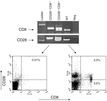
Screening of CD28–/–CD8–/–double-knockout mice. The ani-mals were screened by flow cytometry as well as by PCR (Figure 1). Whole blood was obtained from the retro-orbital plexus of one eye, and lymphocytes were isolated after hypotonic lysis of red blood cells. The cells were stained with rat anti-mouse CD8 FITC and hamster anti-mouse CD28 phycoerythrin (PE). Appropriate iso-type controls were used. All Ab’s were obtained from PharMingen (San Diego, California, USA). For the PCR screening, genomic DNA was extracted from tails according to QIAGEN tail DNA protocol (QIAGEN, Valencia, California, USA). CD28 PCR was performed in a 50-µl volume using 10×reaction buffer with 15 mM Mg++and 10 mM dNTP mix (Boehringer Mannheim Biochemicals, Indianapolis, Indiana, USA) in an auto-mated thermal cycler (Multiblock, Thermo Hybaid, Needham Heights, Massachusetts, USA). WT and knockout alleles were detected by multiplexing two primers specific for the WT allele with one primer for the knockout allele. This primer pairs with one of the WT primers to produce the knockout-specific band. Ten microliters of each PCR reaction was resolved on 1% agarose gels, and bands were visualized using ethidium bromide staining. Homozygous samples produced a sin-gle 740-bp band, and WT samples produced a sinsin-gle 600-bp band. Heterozygotes produced both bands. All PCR experiments included a no-template control and control reactions using DNA from known heterozygote and WT samples. CD8 PCR was performed as above, but using 10×reaction buffer, 25 mM Mg++(Perkin-Elmer Corp., Norwalk, Connecticut, USA), and primers specif-ic for the CD8 WT and knockout alleles. Homozygous samples produced a single 343-bp band, and WT pro-duced a single 265-bp band.
EAE-induction with MOG. MOG peptide 35-55 (MOG 35-55) (M-E-V-G-W-Y-R-S-P-F-S-R-V-V-H-L-Y-R-N-G-K) corresponding to the mouse sequence was synthesized by Quality Control Biochemicals Inc. (Hopkinton, Massachusetts, USA) and purified by HPLC. Peptide purity was greater than 99% after HPLC. Mice were immunized subcutaneously in the flanks with 150–200 µg of MOG peptide in 0.1 ml PBS and 0.1 ml CFA con-taining 0.4 mg Mycobacterium tuberculosis(H37Ra; Difco Laboratories, Detroit, Michigan, USA) and intraperi-toneally injected with 200 ng pertussis toxin (List Bio-logical Laboratories, Campbell, California, USA) on the
day of immunization and 48 hours later. EAE was scored as described previously (4): 0, no disease; 1, limp tail or isolated weakness of gait without limp tail; 2, par-tial leg paralysis; 3, total leg or parpar-tial hind-and front-leg paralysis; 4, total hind-leg hind-and partial front-leg paralysis; 5, moribund or dead animal.
In vivo CD8+T cell depletion. A mAb to mouse CD8 (clone 2.43, kind gift of H. Auchincloss, Jr., Brigham and Women’s Hospital) was administered at a dose of 100 µg intraperitoneally on days –6, –3, and –1 before immunization. For the late CD8+T cell depletion, the same dosages were given on days 14, 17, and 19 after immunization. Control animals received an equal dose of rat Ig (rat anti-mouse IgG-2b; Sigma-Aldrich, St. Louis, Missouri, USA). The effectiveness of the deple-tion was assessed by FACS analysis of peripheral T cells (both from blood and spleen) on days 0, 7, 14, and 30. The CD8+T cells were absent from days 0–14 and grad-ually reappeared afterward, but did not reach normal levels even on day 30 (8% CD8+T cells in Ab-depleted spleen as compared with 14% in naive spleen).
[image:3.576.305.526.385.593.2]Ex vivo CD8+T cell depletion using magnetic beads. A sin-gle cell suspension was prepared from CD28–/–mice splenocytes 14 days after immunization with MOG. The cells were first incubated with rat anti-mouse CD8 Ab (PharMingen) for 45 minutes at 4°C, washed twice in PBS, then incubated with Dynabeads (Dynal Biotech
Figure 1
ASA, Oslo, Norway) according to the manufacturer’s instructions for 45 minutes at 4°C. Bead-bound CD8+ T cells were then removed over a magnet by positive selection. The remaining cells were confirmed to be CD8 negative, with fewer than 5% CD8+T cells follow-ing flow cytometry usfollow-ing a FACScalibur and analyzed with Cellquest software (both Becton Dickinson Immunocytometry Systems, San Jose, California, USA).
CD8+T cell purification for in vitro suppression assays and
adoptive transfer experiments. To obtain 100% purified CD8+CD28–and CD8+CD28+T cells for in vitro and adoptive transfer studies, we first prepared a single-cell suspension from spleens of naive WT mice. The cells were then incubated with FITC-conjugated anti-CD8 Ab and PE-conjugated anti-CD28 Ab and sorted by a FACS sorting machine (Becton Dickinson Immunocy-tometry Systems). To obtain larger number of 100% purified CD8+CD28–T cells for adoptive transfer exper-iments, cell suspensions were prepared from spleens of CD28–/–mice. CD8+T cells were enriched (more than 85% CD8+) using CD8+ T cell–enrichment column (R&D Systems Inc., Minneapolis, Minnesota, USA). The cells were then incubated with FITC-conjugated anti-CD8 Ab and sorted by a FACS sorting machine (Becton Dickinson Immunocytometry Systems).
Enzyme-linked immunosorbent spot assay. Enzyme-linked immunosorbent spot assay (ELISPOT) plates (Cellular Technology Ltd., Cleveland, Ohio, USA) were coated with capture Ab’s against IL-4, IL-5, IL-10, or IFN-γ(PharMingen) in PBS and left overnight at 4°C. The assay was performed as described previously (4). Media used for ELISPOT assays consisted of complete media as described (4). Control wells contained spleno-cytes plus medium. One hundred microliters of the appropriate concentrations of antigen or mitogen were added to the other wells. The resulting spots were counted on a computer-assisted ELISPOT Image Ana-lyzer (Cellular Technology Ltd.). The results were then calculated as cytokine-producing cells per million splenocytes (unless otherwise specified).
Anti-MOG Ab assay. Serum samples were obtained from blood of immunized or naive mice and stored at –20°C. The assay was performed as previously pub-lished (4). Serum samples from immunized animals were compared with a naive serum standard.
In vitro regulatory assay and Transwell studies. To study the regulatory function of CD8+CD28–and CD8+CD28+ T cells in vitro, a novel suppression assay was estab-lished using the ELISPOT assay: splenocytes from naive or immunized CD8–/–mice were used as respon-der cells along with the appropriate mitogen or anti-gen. One hundred percent purified CD8+CD28–or CD8+CD28+ T cells were then added at different ratios to each well, either in direct contact with responder cells or separated from responder cells using eight-well strip inserts (Nalge Nunc Inter-national, Kamstrup, Denmark). The frequency of IFN-γ–producing responder cells was then deter-mined as described previously (4).
To analyze restriction elements of the regulatory CD8+CD28–T cells (MHC or nonclassical), Ab’s against MHC class I (H-2kb, 25 µg/ml) and Qa-1bmolecules (as high as 100 µg/ml) were added to the in vitro cultures of the suppression assays. To investigate the role of Fas-FasL–mediated killing of APCs by the CD8+CD28– cells, Ab’s against Fas (CD95) and FasL (CD95L) (as high as 100 µg/ml) were also added in vitro to the cul-tures of the suppression assay. All above Ab’s and appropriate isotype controls were obtained from PharMingen (San Diego, California, USA).
Functional mixed lymphocyte reaction. Splenocytes from naive CD8–/–mice were incubated with concanavalin A (Con A). Purified CD8+CD28–T cells or CD8+CD28+T cells were then added at the initiation of the 24-hour culture at a 1:2 ratio. The cells were then collected, and APCs were enriched by plastic adherence. Irradiated (30 Gy) APCs were then added as stimulators to BALB/c splenocytes as responder cells, and the frequency of IFN-γ–producing cells were compared in different groups using ELISPOT assay.
Analysis of APC function after interaction with CD8+CD28–
and CD8+CD28+T cells. Splenocytes from MOG-immu-nized CD8–/–mice were incubated with MOG and puri-fied CD8+CD28–T cells or CD8+CD28+T cells in a 2:1 ratio for 24 hours. CD11c+cells were then enriched using a DC isolation kit (Miltenyi Biotec, Auburn, Cal-ifornia, USA), irradiated (30 Gy), and added at a 1:1 ratio to 100% purified CD4+T cells isolated from MOG-immunized WT mice. The frequency of IFN-γ –produc-ing cells was then measured by ELISPOT assay.
Flow cytometry. Plastic adherent cells were collected, washed twice, and stained with CD11c-FITC (as a marker for DCs) and either CD80, CD86, or CD40 PE-conjugated mAb. Four-parameter analysis (forward scatter, side scatter, and two fluorescence channels) was used for data analysis. To rule out apoptosis of CD4+T cells as mechanism of action by regulatory T cells, cells from the experimental groups were stained with anti-mouse CD8 FITC and CD4 FITC, washed, and subse-quently stained with annexin V–PE and 7-AAD (using the apoptosis kit; PharMingen). For further charac-terization of the population of CD8+CD28– and CD8+CD28+T cells in naive WT mice, splenocytes are stained with CD8 FITC, CD28 APC, and a panel of PE-conjugated mAb’s including CD45RB, CD44, CD62L, CD25, CD122 (IL-2 receptor βchain), CD132 (com-mon βchain), CD210 (IL-10 receptor), and NK-1.1. We have further studied T cell receptor Vβexpression of the T cell repertoire reactive to MOG in WT, CD8–/–, CD28–/–, CD8–/–CD28–/–mice 12–14 days after immu-nization by flow cytometry. The following mAb’s were used: anti-Vβ2, anti-Vβ3, anti-Vβ4, anti-Vβ5.1,5.2, anti-Vβ6, anti-Vβ7, anti-Vβ8, anti-Vβ8.1,8.2, anti-Vβ8.3, anti-Vβ9, anti-Vβ10, anti-Vβ11, anti-Vβ12, anti-Vβ13, anti-Vβ14, and anti-Vβ17. All anti-mouse Ab’s above were obtained from PharMingen.
experi-mental group and examined histologically for inflam-matory infiltrate using H&E and demyelination using Luxol fast blue (LFB) staining. Mice were euthanized and perfused through the heart with 10 ml PBS, followed by 20 ml Bouin’s solution (Electron Microscopy Sciences, Fort Washington, Pennsylvania, USA). Whole brain and spinal cord sections were placed in Bouin’s solution for 72 hours and then paraffin embedded. Ten-micrometer sections were cut and placed on slides. Paraffin-embed-ded sections were deparaffinized by exposure to xylene and then decreasing graded concentrations of alcohol for 1 minute each. Sections were exposed to hematoxylin for 2 minutes, washed, and then exposed to eosin for 1 minute. Finally, sections were exposed to increasing grades of alcohol, xylene, and then mounted. For LFB staining paraffin-embedded sections were deparaf-finized as described above and then soaked in LFB solu-tion (Sigma-Aldrich) at 56°C overnight. Excess stain is rinsed off with 95% alcohol. Slides are differentiated in 0.05% lithium carbonate solution for 30 seconds and then placed in 70% ethyl alcohol. Sections are counter-stained in cresyl violet and then mounted.
Statistics. Incidence of disease and mortality between the two groups of animals were compared by using Fisher’s exact probability tests. The mean day of onset of disease and mean peak disease severity between any two groups of mice were analyzed by Mann-Whitney
Utests. The significance of differ-ences between means of spots was determined using the pairedttest.
Results
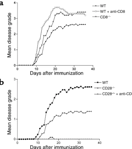
Role of CD8+T cells in EAE. We inves-tigated the role of CD8+ lympho-cytes in the MOG-induced chronic EAE model with particular empha-sis on CD28–/–mice that are natu-rally resistant to EAE (1–4). CD8+ lymphocytes constitute 9–14% of splenocytes in naive WT mice of which 60% are CD8+CD28+, while the remaining 40% are CD8+CD28– cells. Thus, overall, CD8+CD28+ cells make up 5.5–8.5% and CD8+CD28– cells 3.5–5.5% of all splenocytes (one representative example is shown in Figure 1). In WT mice, depletion of CD8+T cells (less than 1% CD8+ T cells in peripheral blood by flow cytometry) prior to disease induction worsens clinical disease significantly as compared with non–CD8-depleted controls (mean maximal grade 3.8 ± 0.9 versus 2.8 ± 1.4, P< 0.03). The clinical disease observed was similar to that observed in CD8–/–mice (Table 1, Figure 2a). The mortality of animals lacking CD8+T cells was higher than that of WT animals (Table 1), although it did not reach statistical significance.
CD28–/–mice develop EAE after CD8+T cell depletion. We and others have previously reported that unlike WT C57BL/6 mice, CD28–/–mice immunized with MOG 35-55 peptide are resistant to EAE (1, 3, 4). Adminis-tration of a depleting anti-CD8 mAb before immu-nization brings out clinical disease in CD28–/–animals (Table 1, Figure 2b), characterized immunohistologi-Table 1
Effects of absence of CD8 T lymphocytes on actively induced EAE in wild-type and CD28–/–mice
Strain Treatment Incidence Mortality Day of onset Mean maximum disease
WT Rat IgG 19/19 4/19 12 ± 1.7 2.8 ± 1.4 WT Anti-CD8 mAb 16/16 5/16 11.3 ± 2.3 3.8 ± 0.9A CD8–/– None 24/24 10/24 11.2 ± 2.2 3.8 ± 1.1A CD28–/– Rat IgG 1/15 0/15 14 0.03 ± 0.12 CD28–/– Anti-CD8 mAb 12/13B 0/13 14.8 ± 3C 1.7 ± 0.9A,B CD28–/– Late anti-CD8 mAb 8/11 0/11 24.75 ± 1.7 1.5 ± 0.38D CD28–/–CD8–/– None 8/15 8/8 11.6 ± 1.6 5B (atypical)
CD28–/–CD8–/– None 7/15B 0/7 20 ± 8.4C 1.2 ± 0.6A,B (typical)
CD28–/–CD8+/– None 8/8B 0/8 15.2 ± 2.8C 1.3 ± 0.9B,E
AP< 0.03 vs. WT; BP< 0.0001 vs CD28–/–; CP< 0.004 vs. WT; DP< 0.003 vs. CD28–/–; EP< 0.02 vs.
[image:5.576.51.362.80.222.2]CD28–/–CD8–/–.
Figure 2
[image:5.576.310.522.492.732.2]cally by intraparenchymal cellular infiltrates similar to those observed in WT mice (data not shown), whereas in nondepleted CD28–/– mice the infiltrates were restricted to the meninges and within blood vessels, similar to our previous report (4).
In CD8-depleted animals, 12 of 13 mice developed disease compared with only 1 in 15 in the non-deplet-ed CD28–/–animals. The disease onset in CD8-deplet-ed CD28–/– mice was delayed and significantly less severe, however, compared with WT mice (Table 1). To further investigate the role of CD8+T cells during the effector phase of EAE, we administered the depleting anti-CD8 mAb 2 weeks after immunization. The majority of these animals (8 of 11) also developed mild EAE (Table 1), with the onset being about 11 days after depletion. These data indicate a critical role for CD8+CD28–T cells in disease resistance in CD28–/– mice during the priming and effector phases of the immune response in vivo.
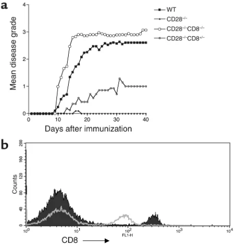
CD28–/– CD8–/– mice develop active EAE. To confirm the role of CD8+CD28–T cells in natural disease resistance in CD28–/–mice, we generated a new mouse colony that is deficient at both CD28 and CD8 loci by intercross-ing CD28–/–with CD8–/–mice (see Methods). All the homozygous double-knockout mice developed EAE when immunized with MOG peptide (Table 1, Figure 3a). Interestingly, these mice developed two different forms of clinical EAE: approximately 50% of animals developed a mild form of “typical EAE” with the classic ascending flaccid paralysis and a significantly later dis-ease onset as compared with WT controls. The remain-ing animals developed an “atypical” form of EAE, with the onset of symptoms not significantly altered com-pared with WT controls (Table 1). The clinical symp-toms of those with atypical disease were primarily asso-ciated with ataxia, spastic reflexes, loss of coordinated movements, spinning, and head tilt. The disease led to premature death in all animals within 24 hours of onset of clinical symptoms.
CD28-deficient mice heterozygous for the CD8 locus demonstrate decreased CD8 surface density compared with CD28–/–CD8+/+mice by flow cytometry (Figure 3b). We asked whether a decrease in CD8 expression would make CD28–/–animals susceptible to disease. Figure 3a shows that these mice develop mild typical EAE with a late onset of disease as compared with WT mice (Table 1). The disease course was halfway between that in CD28–/–and CD28–/–CD8–/–mice, indicating a dose-response effect of CD8 expression. It is interesting that decreased expression of surface CD8 in the heterozy-gous mice is linked to partial loss of regulatory function in vivo and suggests that surface CD8 is involved in the mechanism of CD8+CD28–T cell regulation.
To confirm the regulatory function of CD8+CD28–T cells in vivo, we developed an adoptive transfer system into CD8–/–animals that naturally develop EAE, which is more severe than WT animals. First, one million 100% purified CD8+CD28–T cells isolated from naive CD28–/– spleens were transferred into each CD8–/–recipient via
tail vein, and animals were then immediately immunized with MOG peptide. Adoptive transfer of CD8+CD28–T cells led to a significant decrease in disease severity as compared with untreated CD8–/–mice (P= 0.02, n= 10) (Figure 4a). Interestingly, the disease severity was com-parable to that in control WT animals (P= NS, n= 10). The onset of disease was significantly delayed as com-pared with CD8–/–mice (16.1 ± 4.25 versus 10.7 ± 1.6,
[image:6.576.302.532.353.596.2]P= 0.003). There was no significant difference in the dis-ease incidence and the mortality between the two groups (incidence in CD8–/–, 10 of 10 versus 9 of 10 in adoptive transfer group; mortality in CD8–/–animals, 7 of 10 ver-sus 3 of 10 in the transfer group, P= NS). Interestingly, adoptive transfer of 5 million purified CD8+CD28– cells did not result in further protection from disease (Figure 4b). Similar experiments with adoptive transfer of 0.5 ×10⁶purified CD8+CD28–T cells did not lead to significant suppression of EAE (data not shown), demonstrating the need for transfer of a minimum of one million cells. Next, the same experiment was repeat-ed using one million CD8+CD28–and CD8+CD28+T cells purified from naive WT mice. CD8+CD28+cells did not modulate the course of EAE as compared with con-trol animals (P= 0.14, n= 6) while CD8+CD28–T cells led
Figure 3
to a significant decrease in disease severity (P= 0.0019,
n= 6) (Figure 4b). These data confirm that CD8+CD28– T cells represent a distinct subset of CD8+T cells with regulatory function in vivo in EAE.
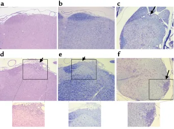
Finally, histological analysis of CD8–/–CD28–/–mice with typical EAE revealed the presence of inflammato-ry infiltrates and demyelination in the lumbar spinal cord starting at day 9 (Figure 5c), with no infiltrates or demyelination in the brainstem (Figure 5, a and b) or cervico-thoracic spinal cord (not shown). In contrast, CD28–/–CD8–/– mice with atypical EAE displayed
inflammatory infiltrates in the brainstem meninges and parenchyma, including the anterior pons depicted in Figure 5d as well as demyelination in the anterior pons (Figure 5e). In some mice from this group, inflammation and demyelination was seen in the later-al pons and vestibular nerve later-also. In addition, CD28–/–CD8–/– mice with atypical EAE had large inflammatory infiltrates in the cervical and thoracic spinal cords with demyelination (Figure 5f), but little to no infiltrate in the lumbar spinal cord.
Expansion of MOG-specific IFN-γ–producing CD4+T cells in
[image:7.576.57.286.55.304.2]the absence of CD8+T cells. We measured the frequency of MOG-specific Th1-producing (IFN-γ) and Th2-pro-ducing (IL-4, IL-5 and IL-10) cells on day 14 after immu-nization by ELISPOT in CD28–/–CD8–/–mice, CD28–/–, and CD28–/– CD8-depleted (by mAb therapy) mice. As Figure 4
Suppression of EAE by adoptive transfer of CD8+CD28–cells into CD8–/–mice. (a) 100% purified CD8+CD28–cells were generated from naive CD28–/–splenocytes and injected into CD8–/–recipients via the tail vein, as described in Methods. The recipient mice were then immu-nized with MOG peptide on the same day. The mean daily score for each group (n= 10) is shown on the yaxis. The course of CD8–/–mice (open squares), CD8–/–recipients of CD8+CD28–cells (open circles), and WT mice (filled squares) is shown. (b) One hundred percent puri-fied CD8+CD28+and CD8+CD28–T cells were isolated from spleens of naive WT mice. Approximately one million cells were then injected into each CD8–/–recipient as described above. The mean daily score for each group (n= 6) is shown on theyaxis. CD8+CD28–T cells (open circles) significantly suppress the EAE as compared with the control group (open squares), while CD8+CD28+T cells do not show any sig-nificant effect on disease course (filled circles). Adoptive transfer of approximately five million CD8+CD28–T cells did not lead to any fur-ther suppression of disease (filled squares).
Figure 5
H&E and LFB stains of sections of the brainstem and spinal cords from CD28–/–CD8–/– mice with typical EAE (a–c) and CD28–/–CD8–/–mice with atypical EAE (d–f). (a) H&E-stained section from the anterior pons showing no inflammatory infiltrates. (b) LFB staining in the pons showing no demyelination. (c) LFB staining showing small infiltrate and demyelination in the lateral lumbar spinal cord (arrow). (d) H&E-stained section from pons showing inflammatory infiltrate in the anterior pons (arrow). (e) LFB staining showing demyelination in the anterior pons (arrow). (f) LFB staining showing demyelination and large inflammatory infiltrates in the lateral thoracic spinal cord (arrow). Photomicrographs a–fare taken at
[image:7.576.192.534.483.736.2]shown in Figure 6a, the frequency of antigen-specific Th1 cells expanded significantly in both CD28–/–mice depleted of CD8+T cells as well as CD28–/–CD8–/–mice, as compared with CD28–/–control mice (P< 0.02 for all antigen concentrations). In contrast, the frequency of Th2-producing cells was not significantly altered in any of the studied groups (IL-4 spots of 56.5 ± 4.9 for CD28–/–versus 46.5 ± 4.9 for anti-CD8–treated group versus 53 ± 11.3 in CD28–/–CD8–/–the mouse). Similar results were found in the WT CD8-depleted and CD8–/– mice compared with WT controls (Figure 6b). These results indicate that CD8+CD28–T cells play a role in limiting expansion of MOG-specific Th1 cells after immunization, whereas Th2 clone size is unaltered.
Mice deficient in STAT4 lack IL-12 induced IFN-γ– production and Th1 differentiation and have been recently reported to be resistant to the induction of EAE (24). If CD8+T cells exert their regulatory function through limiting the expansion of Th1 clone size, one would hypothesize that removal of CD8+T cells would not overcome the disease resistance in STAT4–/–mice. As expected, depletion of CD8+T cells prior to disease induction in STAT4–/–mice did not worsen the clinical disease as compared with control non–CD8-depleted STAT4–/–mice (mean maximal grade of 0.6 ± 0.65 in depleted group versus 0.5 ± 0.7 in control group, n= 5 in each group). As shown in Figure 6c, CD8+T cell depletion in STAT4–/– mice did not lead to expansion of MOG-specific Th1 cells.
Enhancement of an anti–MOG 35-55 peptide Ab response in the absence of CD8+CD28–T cells. MOG-induced EAE is characterized by significant demyelination in the CNS, which is thought to be mediated by anti-MOG Ab’s (25, 26). We have shown previously that relative Ab titers from immunized CD28–/–mice sera were significantly lower than those from sera of immunized WT animals (4). These results are consistent with published data that CD28 signaling functions to augment T cell– dependent B cell growth and Ig secretion (27). To eval-uate the effect of CD8+CD28– T cells on humoral immune response, we compared the Ab titers of the dif-ferent groups of mice 14 days after immunization. We found that the relative Ab titers in sera from immu-nized CD28–/–mice were significantly lower than those from CD8-depleted CD28–/–animals (CD28–/–mean titer = 1/50; WT mean titer = 1/2,400; CD8-depleted CD28–/– mean titer = 1/3,200; and mean titer of CD28–/–CD8–/–mice = 1/3,200, P= 0.0001 for the latter two groups versus CD28–/–). Similarly, CD8-depleted WT mice demonstrate higher Ab titers in sera (WT mean titer = 1/2,400; CD8-depleted WT animals mean titer = 1/4,800; and mean titer of CD8–/– mouse = 1/5,600). These data establish that lack of CD8+CD28– T cells can cause expansion of CD4+Th1 cells that then provide help to B cells to promote Ab production.
Depletion of CD8+T cells ex vivo leads to expansion of Th1
CD4+T cells. Primed splenocytes obtained from MOG peptide–immunized CD28–/–mice were cultured with MOG 35-55 peptide for 24 hours in ELISPOT plates.
[image:8.576.298.532.294.533.2]CD8 depleted splenocytes were obtained after mag-netic bead separation and cultured similarly. Figure 6d shows increased frequency of IFN-γ–producing Th1 cells in CD8-depleted cultures consistent with the in vivo data described above. These data establish the inhibitory effect of CD8+CD28–T cells on Th1 clone size in vitro. The ex vivo CD8+T cell depletion did not result in any significant change in the fre-quency of Th2 cells (not shown), again consistent with the in vivo data described above. Furthermore, since TGF-βis a potential effector of immune regu-lation by CD8+ T cells (13, 28), we examined the supernatants of in vitro cultures by ELISA for TGF-β. There was no statistically significant difference in the TGF-βcontent from splenocyte cultures incubated with MOG before and after in vivo or ex vivo CD8+T cell depletion (data not shown). Thus, regulation by CD8+T cells appears to be independent of secretion of TGF-βor Th2 cytokines.
Figure 6
The natural resistance to EAE in CD28–/– is in part mediated by a negative regulatory signal provided by B7-1 through CTLA4 (4). There is also growing evi-dence that signals mediated by cell-surface molecules such as CTLA4 are also involved in the effector func-tion of some regulatory T cells (29–31). We investigat-ed whether CD8+CD28–T cells express CTLA4 in naive or immunized CD28–/–animals. We found that naive CD8+CD28–cells lack both intracellular CTLA-4 and surface CD25 expression (data not shown). CD8+T cells from immunized CD28–/–mice on day 14 lack CTLA4 expression, while CD4+ T cells show some CTLA-4 expression (3%). This low level of CTLA4 expression in CD4+T cells is consistent with published data indicating that CD28 costimulation is required for optimal CTLA-4 expression (32). Thus, regulatory CD8+CD28–T cells do not express CTLA4.
Expression of a panel of markers including memory markers (CD45RB, CD44, CD62L), cytokine receptors
(CD25, CD122, CD132, and CD210), and NK marker (NK1.1) was compared between the CD8+CD28– T cells with regulatory function and CD8+CD28+T cells that lack such function. The two subpopulations were only distinguished by the expression of CD122 (IL-2 receptor βchain): CD122 was expressed almost entire-ly by CD8+CD28+T cells. CD45RB and CD62L were both expressed by all CD8+CD28+and CD8+CD28–T cells, while none of the subpopulations expressed any NK1.1, CD25, or CD210. Expression of both CD132 and CD44 was comparable in the two populations (data not shown).
In vitro suppression induced by CD8+CD28–T cells requires
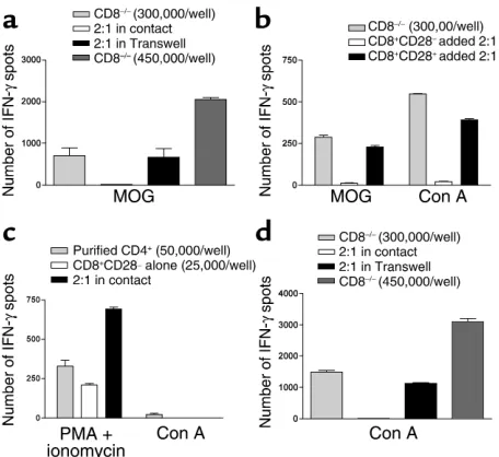
cell-cell contact and is APC dependent. To study the ability of CD8+CD28–T cells to suppress in vitro, a unique coculture system was set up using the ELISPOT assay. Using splenocytes from MOG-immunized CD8–/–mice as responder cells, we first demonstrated that 100% purified CD8+CD28–T cells derived from CD28–/–mice can suppress IFN-γproduction only if in direct contact with responder cells (Figure 7a). Titration of the same number of CD28+/+splenocytes as CD8+CD28–T cells into cultures did not lead to a decrease of IFN-γ, there-by excluding the possibility that an increase in total responder cell number was responsible for the sup-pressive effect (Figure 7a). Furthermore, CD8+CD28–T cells were able to induce suppression in a dose-depend-ent manner (responder cells–to–regulatory cells ratios of 2:1, 4:1, 16:1, with loss of suppression at a ratio of 32:1). This suppression was not a peculiarity of cells from CD28–/– animals, since CD8+CD28– but not CD8+CD28+cells from WT mice demonstrated similar suppressive activity in vitro (Figure 7b). These in vitro findings are consistent with our adoptive transfer experiments demonstrating that regulatory CD8+T cells do occur in WT mice and are confined to a sub-population of CD8+T cells lacking CD28 expression. Using the apoptosis kit, we have also determined that there was no increase in the number of apoptotic cells in either CD3+or CD3–populations whether they were incubated with CD8+CD28– or CD8+CD28+ T cells (data not shown). Furthermore, blockade of Fas-FasL interaction in the suppression assay cultures did not result in reversal of suppression by the CD8+CD28– cells in vitro (data not shown). These results exclude apoptosis of T cells or APCs as a mechanism of regula-tion by these cells.
CD8+CD28–T cells do not suppress IFN-γ produc-tion by purified CD4+T cells stimulated by PMA and ionomycin, while they suppress CD8–/– splenocytes stimulated with Con A by ELISPOT (Figure 7, b–d). Since T cell proliferation to Con A is dependent on the presence of APCs as accessory cells (33, 34) (confirmed in Figure 7c), these data, taken together with the Tran-swell culture system data, clearly demonstrate that CD8+CD28–T cells require cell-cell contact and APCs for their regulatory function.
To study whether CD8+CD28–T cell regulation is MHC class I restricted, we repeated the in vitro sup-Figure 7
[image:9.576.54.282.53.262.2]pression assays in the presence of anti-MHC class I–blocking Ab’s or appropriate isotype control Ab and measured the frequency of IFN-γ–producing cells by ELISPOT. We found that 288 ± 45 IFN-γspots were generated by responder cells incubated with MOG alone and 13 ± 5.7 spots by responder cells incubated with MOG in the presence of CD8+CD28–T cells at a ratio of 4:1, while 375.2 ± 100 spots were generated by responder cells after addition of MHC class I–block-ing Ab in the presence of CD8+CD28–T cells but not when isotype control (9.2 ± 4.5 spots) was added to the latter group (P = 0.005 between the last two groups). In contrast, addition of anti–Qa-1bto the in vitro suppression assays (11.75 ± 3.5 spots) did not result in reversal of suppression by regulatory CD8+CD28–T cells. These data indicate that the reg-ulatory functions of the CD8+CD28–T cells are MHC class I and not Qa-1brestricted.
CD8+CD28–T cells inhibit the upregulation of costimulatory
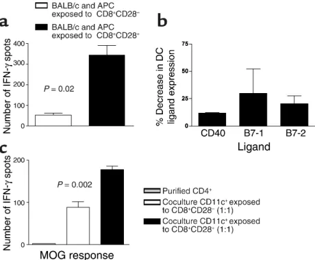
molecules on APCs leading to less efficient antigen presentation. To evaluate the effect of regulatory cells on APC func-tion, we first set up a functional MLR assay. CD8–/– splenocytes were cultured with purified CD8+CD28–T cells, purified CD8+CD28+T cells, or alone and stimu-lated with Con A for 24 hours. Plastic adherent cells were isolated from these cultures, irradiated, and used as stimulators in a MLR. APCs previously cultured with CD8+CD28–T cells have markedly decreased ability to stimulate BALB/c responder cells in allogeneic mixed-leukocyte reactions compared with APCs previously cul-tured with CD8+CD28+T cells (Figure 8a). Using flow cytometry, we then studied the expression of costimula-tory molecules by DCs (CD11c+) from the APC cultures described above. Figure 8b shows the mean decrease in the percentage of CD11c+cells that express costimula-tory molecules after coculture with CD8+CD28–T cells relative to CD11c+from cocultures with CD8+CD28+ cells. Thus, DCs precultured with CD8+CD28–cells have significantly downregulated CD80, CD86, and CD40 costimulatory molecules and are less-efficient APCs. To confirm these findings, we stimulated CD8–/– spleno-cytes from MOG-immunized mice in vitro with MOG peptide, in the presence of purified CD8+CD28– or CD8+CD28+T cells similar to the experiment above. CD11+cells isolated from these two culture conditions were then irradiated and used as APCs for 100% purified CD4+T cells from MOG-immunized WT mice (1:1 ratio) in ELISPOT plates. As seen in Figure 8c, CD11c+cells conditioned by interaction with CD8+CD28–T cells in vitro are significantly less efficient in presenting antigen to primed CD4+T cells.
Discussion
Previous reports from our group and others established that CD28–/–mice are generally resistant to EAE (1, 3, 4). Disease resistance, however, can be overcome by expo-sure to higher antigenic dose (4, 6) or by blocking CD80-CD152 interaction (4). In this report we show that regulatory CD8+CD28–T cells contribute to
[image:10.576.302.532.381.571.2]natu-ral resistance to EAE in the absence of CD28 costimu-lation. Ab depletion of CD8+T cells before immuniza-tion and on day 14 after immunizaimmuniza-tion brings out clin-ical disease in CD28–/–mice. These findings establish the regulatory functions of CD8+T cells both at the time of priming and at the time of the effector phase of the disease. Regulation by CD8+CD28–T cells was con-firmed by adoptive transfer of these cells into mice lack-ing CD8+T cells. In addition, using a novel regulatory assay by ELISPOT analysis, we clearly demonstrate the suppressive effect of CD8+CD28–T cells derived from CD28–/–mice in vitro. We have used our adoptive trans-fer model and in vitro regulatory assay to answer sever-al important questions. Do CD8+CD28–regulatory T cells occur in WT mice? Do CD8+CD28+cells have reg-ulatory functions? CD8+CD28–T cells from WT ani-mals clearly suppress IFN-γproduction in vitro and sup-press EAE in vivo similar to CD8+CD28– cells originating from CD28–/–mice. In contrast, CD8+CD28+ cells do not induce suppression in vitro or in vivo. CD8+CD28–T cells occurring in WT animals are unable to provide complete protection against EAE, but rather control the severity of disease. This is not surprising since it is likely that other regulatory cells such as CD4+CD25+(8) and/or mechanisms such as negative T cell regulatory pathways (CTLA4, PD1), as we have
Figure 8
recently published (4, 35), are also operational in regu-lating disease. Our data clearly show that regulatory function of CD8+T cells resides in the subpopulation lacking CD28 expression. CD8+CD28–regulatory T cells have only been described in humans (21, 23). Glatiramer acetate (GA), (Capoxone; Teva Pharmaceutical Indus-tries Ltd., St. Louis, Missouri, USA) is a random copoly-mer of glutamic acid, lysine, alanine, and tyrosine that is used therapeutically in patients with multiple sclero-sis (36). Recent work on the mechanism of the drug’s immunomodulatory effect demonstrated that GA leads to upregulation of GA-induced CD8+T cell responses with restoration to levels found in healthy individuals (37). As a corollary, treatment with GA may be in part mediated through the induction and restoration of this regulatory population of cells. Earlier work identified suppressor functions in CD8 cells that lacked CD28 expression (38, 39). Our findings that CD28–cells are enriched for suppressor T cells suggest that the lack of CD28 either predisposes toward suppressor phenotype and/or that CD28 is a marker of a distinct functional T cell subset that is subject to different influences (40, 41). Having established the existence of these two distinct subsets of CD8+T cells with different functions, we then compared the phenotype of these cells using a panel of activation markers, memory markers, cytokine recep-tors, and NK markers. With exception of CD122 all the markers were expressed in similar fashion in both CD8 subpopulations. CD8+CD122+cells are present exclu-sively in the CD8+CD28+subset of cells. CD8+CD122+T cells have been described in mice and were found to rap-idly produce a much greater amount of IFN-γafter CD3 stimulation in vitro compared with CD8+CD122– T cells (42). Moreover, they are able to stimulate CD4+T cells to produce IFN-γ, suggesting that these cells are an important cellular component in the Th1 immune response. Thus, the absence of such cells in the regula-tory subset of CD8 T cells is intriguing.
Our data also show that disease susceptibility is restored in our novel CD28–/–CD8–/–mice. It is interest-ing that half of these mice developed atypical EAE with ataxia, head tilting, and spinning. Histologically, there were remarkable differences in the distribution of the lesions that may account for the differences in the clini-cal symptoms observed: CD28–/–CD8–/–mice that devel-oped the typical form of EAE characterized by ascending flaccid weakness demonstrated small inflammatory infiltrates and demyelination in the lumbar spinal cord starting at day 9, with no infiltrates or demyelination in the brainstem or cervico-thoracic spinal cord. In con-trast, mice with atypical EAE showed significant infil-trates and demyelination in the brainstem. Previous reports of atypical EAE have attributed the various clin-ical presentations to the induction of T cells specific to nondominant peptides (43–45) or to the cytokine pro-file of the effector cells (46). We explored the possibility that the encephalitogenic T cell repertoire reactive to MOG may be different in the absence of CD8 T cells. Analysis of T cell receptor Vβexpression on CD4+T cells
in WT, CD8–/–, CD28–/–, and CD28–/–CD8–/–mice 12–14 days after immunization did not reveal any significant differences (data not shown). Given the low frequency of antigen-specific T cells in vivo, however, more sensitive assays such as immunoscope analysis of CD28–/–CD8–/– mice may help detect small differences in T cell reper-toire specific for MOG peptides and may provide insight into the mechanisms of atypical disease in these animals. Understanding the exact mechanisms leading to varying disease expression would have important clinical impli-cations, since the two patterns observed are in syngene-ic msyngene-ice immunized with the same peptide antigen.
Our in vitro experiments in CD28–/–CD8–/–mice clearly indicate that the suppression of EAE by CD8+ T cells may arise from their inhibitory effect on Th1 cell expansion. This is consistent with the findings that CD8+T cells preferentially downregulate CD4+ Th1 cells in vivo (16). We have observed a similar increased frequency of IFN-γ–producing CD4+T cells after double immunization that is also associated with clinical disease in CD28–/–mice (4). These results show that exceeding a threshold frequency of MOG-specific IFN-γ–secreting cells is permissive for disease induction. Accordingly, CD8+ T cell depletion in STAT4–/–mice does not overcome the resistance of the animals to the induction of EAE, as these mice are unable to expand their Th1 cells in vivo.
regulatory T cell in innate immunity. In our experi-ments, CD8+CD28–T cells did not appear to induce apoptosis in CD4+T cells, since the number of viable CD4 T cells incubated with CD8+CD28–was compara-ble with cells incubated with CD8+CD28+cells. More-over, with the exception of MHC class I blockade, addi-tion of Ab’s blocking Fas/FasL or anti–Qa-1bto in vitro suppression assays failed to reverse the suppression. The dose response of the in vivo regulation in relation to sur-face expression of CD8 is intriguing and suggests a role for surface CD8 molecules in the mechanism of regula-tion. The frequency of CD8+regulatory T cells has been reported to determine the susceptibility of a particular strain to EAE in a rat model (50).
Recently, several investigators reported that the sup-pressor function of CD4+CD25+T cells is mediated through CTLA-4 signaling both in vitro and in vivo (29–31). Unlike these regulatory CD4+T cells, however, CD8+CD28–T cells do not express CTLA-4. In human cell cultures, APCs exposed to CD8+CD28–regulatory T cells have impaired CD40-signaling pathway and do not upregulate B7 molecules (20, 22, 23, 51). In fact, recent work showed that CD8+CD28–alloantigen-specific T suppressor cells induce the upregulation of immuno-globulin-like transcript 3 (ILT3) and ILT4 on APCs, ren-dering these cells tolerogenic (52). PMA and ionomycin are known to induce accessory cell-independent T cell activation while Con A, similar to antigen, can only induce proliferation in the presence of such cells (33, 34). The failure of CD8+CD28–cells to elicit suppression in the former group demonstrated clearly the depend-ence of suppression on APCs in vitro. The impaired upregulation of costimulatory molecules on APCs may prevent the efficient stimulation of CD4+cells in the presence of CD8+CD28–T cells leading to a decrease in IFN-γ. Since IFN-γis known to upregulate costimulato-ry molecules on APCs (53), one could argue that lower expression of costimulatory molecules may be the con-sequence of decreased production of IFN-γ. The lack of a direct effect on CD4+T cells and the inability of regu-latory T cells to inhibit IFN-γproduction in the absence of APCs, however, make this explanation very unlikely. The exact mechanism of how CD8+CD28–T cells pre-vent upregulation of costimulatory molecule expression on APCs requires further investigation.
In summary, in this report we establish, we believe for the first time, the regulatory functions and mechanisms of action of CD8+CD28–T cells in an autoimmune dis-ease model in vivo. The mechanisms of disdis-ease regula-tion appear to be related to inhibiregula-tion of expansion of encephalitogenic CD4+Th1 cells. The inhibition was not cytokine mediated but instead required cell-to-cell inter-action between CD4+cells, CD8+CD28– T cells, and APCs. The regulatory CD8+CD28–T cells prevent the upregulation of costimulatory molecules on APCs, thus prohibiting efficient costimulation of CD4 T cells. We have also identified a novel disease model with different clinical and pathologic manifestations in the same genetic background using the same immunizing
anti-gen. Investigations of this disease model and of the prox-imal and molecular mechanisms of CD8+CD28– regula-tory cell functions have implications for understanding autoimmune disease phenotypes and development of novel therapeutic strategies to prevent and/or treat autoimmune diseases in humans.
Acknowledgments
This work was supported by research grants from the National Multiple Sclerosis Society (RG-2589 and RG-3504 to S.J. Khoury and FG-1287 to T. Chitnis) and the NIH (AI-40945, AI-43496, and AI-46130 to S.J. Khoury and PO1 AI-41521 to M.H. Sayegh). N. Najafian is a recipient of American Society of Trans-plantation Faculty Grant.
1. Oliveira-dos-Santos, A.J., et al. 1999. CD28 costimulation is crucial for the development of spontaneous autoimmune encephalomyelitis.
J. Immunol. 162:4490–4495.
2. Perrin, P.J., Lavi, E., Rumbley, C.A., Zekavat, S.A., and Phillips, S.M. 1999. Experimental autoimmune meningitis: a novel neurological disease in CD28-deficient mice. Clin. Immunol. 91:41–49.
3. Girvin, A.M., et al. 2000. A critical role for B7/CD28 costimulation in experimental autoimmune encephalomyelitis: a comparative study using costimulatory molecule-deficient mice and monoclonal antibody blockade. J. Immunol. 164:136–143.
4. Chitnis, T., et al. 2001. CD28-independent induction of experimental autoimmune encephalomyelitis. J. Clin. Invest. 107:575–583.
5. Karpus, W.J., et al. 1995. An important role for the chemokine macrophage inflammatory protein-1 alpha in the pathogenesis of the T cell-mediated autoimmune disease, experimental autoimmune encephalomyelitis. J. Immunol. 155:5003–5010.
6. Manickasingham, S.P., Anderton, S.M., Burkhart, C., and Wraith, D.C. 1998. Qualitative and quantitative effects of CD28/B7-mediated cos-timulation on naive T cells in vitro. J. Immunol.161:3827–3835. 7. Van Parijs, L., and Abbas, A.K. 1998. Homeostasis and self-tolerance in
the immune system: turning lymphocytes off. Science. 280:243–248. 8. Shevach, E.M. 2000. Regulatory T cells in autoimmunity*. Annu. Rev.
Immunol. 18:423–449.
9. Jiang, H., and Chess, L. 2000. The specific regulation of immune responses by CD8+ T cells restricted by the MHC class Ib molecule, Qa-1. Annu. Rev. Immunol. 18:185–216.
10. Jiang, H., Zhang, S.I., and Pernis, B. 1992. Role of CD8+ T cells in murine experimental allergic encephalomyelitis. Science. 256:1213–1215. 11. Koh, D.R., et al. 1992. Less mortality but more relapses in experimental
allergic encephalomyelitis in CD8–/–mice. Science. 256:1210–1213.
12. Gaur, A., Ruberti, G., Haspel, R., Mayer, J.P., and Fathman, C.G. 1993. Requirement for CD8+ cells in T cell receptor peptide-induced clonal unresponsiveness. Science. 259:91–94.
13. Miller, A., Lider, O., Roberts, A.B., Sporn, M.B., and Weiner, H.L. 1992. Suppressor T cells generated by oral tolerization to myelin basic protein suppress both in vitro and in vivo immune responses by the release of transforming growth factor beta after antigen-specific triggering. Proc. Natl. Acad. Sci. U. S. A. 89:421–425.
14. Chen, Y., Inobe, J., and Weiner, H.L. 1995. Induction of oral tolerance to myelin basic protein in CD8-depleted mice: both CD4+ and CD8+ cells mediate active suppression. J. Immunol. 155:910–916.
15. Jiang, H., et al. 1998. T cell vaccination induces T cell receptor Vbeta-spe-cific Qa-1-restricted regulatory CD8 (+) T cells. Proc. Natl. Acad. Sci. U. S. A. 95:4533–4537.
16. Jiang, H., Braunstein, N.S., Yu, B., Winchester, R., and Chess, L. 2001. CD8+ T cells control the TH phenotype of MBP-reactive CD4+ T cells in EAE mice. Proc. Natl. Acad. Sci. U. S. A. 98:6301–6306.
17. Lindsey, J.W., and Steinman, L. 1993. Competitive PCR quantification of CD4, CD8, ICAM-1, VCAM-1, and MHC class II mRNA in the central nervous system during development and resolution of experimental allergic encephalomyelitis. J. Neuroimmunol. 48:227–234.
18. Huseby, E.S., et al. 2001. A pathogenic role for myelin-specific CD8(+) T cells in a model for multiple sclerosis. J. Exp. Med. 194:669–676. 19. Ciubotariu, R., et al. 1998. Specific suppression of human CD4+ Th cell
responses to pig MHC antigens by CD8+CD28– regulatory T cells.
J. Immunol. 161:5193–5202.
20. Liu, Z., Tugulea, S., Cortesini, R., and Suciu-Foca, N. 1998. Specific sup-pression of T helper alloreactivity by allo-MHC class I-restricted CD8+CD28– T cells. Int. Immunol. 10:775–783.
sup-pressor T cells by peptide priming in vitro. Hum. Immunol. 59:690–699. 22. Li, J., et al. 1999. T suppressor lymphocytes inhibit NF-kappa
B-mediat-ed transcription of CD86 gene in APC. J. Immunol. 163:6386–6392. 23. Colovai, A.I., et al. 2000. Induction of xenoreactive CD4+ T-cell anergy
by suppressor CD8+CD28– T cells. Transplantation. 69:1304–1310. 24. Chitnis, T., et al. 2001. Effect of targeted disruption of STAT4and STAT6
on the induction of experimental autoimmune encephalomyelitis. J. Clin. Invest. 108:739–747. doi:10.1172/JCI200112563.
25. Linington, C., Engelhardt, B., Kapocs, G., and Lassman, H. 1992. Induc-tion of persistently demyelinated lesions in the rat following the repeat-ed adoptive transfer of encephalitogenic T cells and demyelinating anti-body. J. Neuroimmunol. 40:219–224.
26. Genain, C.P., and Hauser, S.L. 1996. Allergic encephalomyelitis in com-mon marmosets: pathogenesis of a multiple sclerosis-like lesion. Meth-ods. 10:420–434.
27. Klaus, S.J., et al. 1994. Costimulation through CD28 enhances T cell-dependent B cell activation via CD40-CD40L interaction. J. Immunol.
152:5643–5652.
28. Lider, O., Santos, L.M., Lee, C.S., Higgins, P.J., and Weiner, H.L. 1989. Suppression of experimental autoimmune encephalomyelitis by oral administration of myelin basic protein. II. Suppression of disease and in vitro immune responses is mediated by antigen-specific CD8+ T lym-phocytes. J. Immunol. 142:748–752.
29. Takahashi, T., et al. 2000. Immunologic self-tolerance maintained by CD25(+)CD4(+) regulatory T cells constitutively expressing cytotoxic T lymphocyte-associated antigen 4. J. Exp. Med. 192:303–310.
30. Salomon, B., et al. 2000. B7/CD28 costimulation is essential for the homeostasis of the CD4+CD25+ immunoregulatory T cells that control autoimmune diabetes. Immunity. 12:431–440.
31. Read, S., Malmstrom, V., and Powrie, F. 2000. Cytotoxic T lymphocyte-associated antigen 4 plays an essential role in the function of CD25(+)CD4(+) regulatory cells that control intestinal inflammation.
J. Exp. Med. 192:295–302.
32. Walunas, T.L., et al. 1994. CTLA-4 can function as a negative regulator of T cell activation. Immunity. 1:405–413.
33. Kern, D.E., Lachmann, L.B., and Greenberg, P.D. 1987. Lyt-2+ cells. Requirements for concanavalin A-induced proliferation and interleukin 2 production. J. Immunol.139:2880–2887.
34. Takeuchi, M., Kakiuchi, T., Taira, S., and Nariuchi, H. 1987. B cells as accessory cells in a Con A response of a T cell clone. Immunobiology.
176:1–13.
35. Salama, A.D., et al. 2003. Critical role of the programmed death-1 (PD-1) pathway in regulation of experimental autoimmune encephalomyelitis.
J. Exp. Med. 198:71–78.
36. Coyle, P.K., et al. 1995. Copolymer 1 reduces relapse rate and improves disability in relapsing-remitting multiple sclerosis: results of a phase III multicenter, double-blind placebo-controlled trial. The Copolymer 1 Multiple Sclerosis Study Group. Neurology. 45:1956–1961.
37. Karandikar, N.J., et al. 2002. Glatiramer acetate (Copaxone) therapy induces CD8+T cell responses in patients with multiple sclerosis. J. Clin.
Invest. 109:641–649. doi:10.1172/JCI200214380.
38. Damle, N.K., Mohagheghpour, N., Hansen, J.A., and Engleman, E.G. 1983. Alloantigen-specific cytotoxic and suppressor T lymphocytes are derived from phenotypically distinct precursors. J. Immunol.
131:2296–2300.
39. Li, S.G., et al. 1990. Human suppressor T cell clones lack CD28. Eur. J. Immunol. 20:1281–1288.
40. Monteiro, J., Batliwalla, F., Ostrer, H., and Gregersen, P.K. 1996. Short-ened telomeres in clonally expanded CD28–CD8+ T cells imply a replica-tive history that is distinct from their CD28+CD8+ counterparts.
J. Immunol. 156:3587–3590.
41. Nociari, M.M., Telford, W., and Russo, C. 1999. Postthymic development of CD28-CD8+ T cell subset: age-associated expansion and shift from memory to naive phenotype. J. Immunol. 162:3327–3335.
42. Takayama, E., et al. 2000. Mouse CD8+CD122+ T cells with intermedi-ate TCR increasing with age provide a source of early IFN-γproduction.
J. Immunol. 164:5652–5658.
43. Greer, J.M., et al. 1996. Immunogenic and encephalitogenic epitope clus-ters of myelin proteolipid protein. J. Immunol. 156:371–379. 44. Muller, D.M., Pender, M.P., and Greer, J.M. 2000. A neuropathological
analysis of experimental autoimmune encephalomyelitis with predom-inant brain stem and cerebellar involvement and differences between active and passive induction. Acta Neuropathol. 100:174–182. 45. Sobel, R.A., Waldner, H., Whitters, M.J., Collins, M., and Kuchroo, V.K.
2000. Genetic and epigenetic influence on EAE phenotypes induced with different encephalitogenic peptides. J. Neuroimmunol. 108:45–52. 46. Wensky, A., Garibaldi Marcondes, M.C., Lafaille, J.J., and Furtado, G.C.
2001. The role of IFN-gamma in the production of Th2 subpopulations: implications for variable Th2-mediated pathologies in autoimmunity.
J. Immunol. 167:3074–3081.
47. Mosmann, T.R., Li, L., and Sad, S. 1997. Functions of CD8 T-cell subsets secreting different cytokine patterns. Semin. Immunol. 9:87–92. 48. Cerwenka, A., Carter, L.L., Reome, J.B., Swain, S.L., and Dutton, R.W.
1998. In vivo persistence of CD8 polarized T cell subsets producing type 1 or type 2 cytokines. J. Immunol. 161:97–105.
49. Vukmanovic-Stejic, M., Vyas, B., Gorak-Stolinska, P., Noble, A., and Kemeny, D.M. 2000. Human Tc1 and Tc2/Tc0 CD8 T-cell clones display distinct cell surface and functional phenotypes. Blood. 95:231–240. 50. Sun, D., Whitaker, J.N., and Wilson, D.B. 1999. Regulatory T cells in
experimental allergic encephalomyelitis. III. Comparison of disease resistance in Lewis and Fischer 344 rats. Eur. J. Immunol. 29:1101–1106. 51. Liu, Z., Tugulea, S., Cortesini, R., Lederman, S., and Suciu-Foca, N. 1999. Inhibition of CD40 signaling pathway in antigen presenting cells by T suppressor cells. Hum. Immunol. 60:568–574.
52. Chang, C.C., et al. 2002. Tolerization of dendritic cells by TS cells: the crucial role of inhibitory receptors ILT3 and ILT4. Nat. Immunol.
3:237–243.

![1 (4 Chlorophenacyl) 4 methyl 5,6,7,8,9,10 hexahydrocycloocta[b]pyridin 2(1H) one](data:image/gif;base64,R0lGODlhAQABAIAAAP///wAAACH5BAEAAAAALAAAAAABAAEAAAICRAEAOw==)