The functional CD8 T cell response to HIV
becomes type-specific in progressive disease
Sang Kyung Lee, … , Judy Lieberman, Premlata Shankar
J Clin Invest.
2002;
110(9)
:1339-1347.
https://doi.org/10.1172/JCI16028
.
High levels of HIV-specific CD8 T cells are demonstrable throughout HIV disease using
laboratory assays that measure responses to consensus epitopes. In acute infection, the
dynamics of the antiviral CD8 T cell response correlate well with the decline in viremia.
However in chronic infection, although responses are detected against a broader spectrum
of epitopes, virus-specific CD8 T cells are apparently unable to control viral replication. To
investigate whether CD8 T cells responding to consensus epitopes may have lost their in
vivo relevance in the chronic phase because of viral evolution driven by immune pressure,
we compared the CD8 T cell response to CD4 T cell targets infected with either lab-adapted
HIV
IIIBor the patient’s own virus. The magnitude of the IFN-
g
response declined with
disease progression, especially to autologous virus. T cell receptor (TCR) clonotypes of
HIV
IIIBand autologous virus–responding cells were determined by sequencing TCR
b
chain variable (TCRBV) genes. In two of three asymptomatic donors, the dominant
clonotypes overlapped, whereas in five symptomatic patients, the TCR clonotypes
responding to HIV
IIIBvirus were completely different from those responding to autologous
virus. Moreover, in cytolytic assays, T cell lines derived from IFN-
g
+cells responding to
lab-adapted or autologous virus cross-recognized target cells infected with either virus in
asymptomatic subjects with shared TCR clonotypes but not in progressors with differing
clonotypes. […]
Article
Aging
Find the latest version:
Introduction
Studies of immune responses generated in HIV-infect-ed individuals suggest that CD8 T cells play an impor-tant role in host defense against the virus. This has been very effectively shown in acute HIV infection where the appearance of virus-specific CD8 T cells is temporally associated with a decline in plasma viremia (1–3). At the peak of the acute response, up to 5% of CD8 T cells specific for a single immunodominant epi-tope have been detected by MHC/peptide tetramer staining (4). Evidence for the vast mobilization of virus-specific CD8 T cells during primary infection is also provided by the substantial expansion of particular CD8 T cell BV subsets containing HIV-specific CTLs (5). Direct evidence for the role of CD8 T cells in viral control comes from the closely related simian immun-odeficiency virus model in Rhesus macaques, wherein elimination of CD8 T cells results in higher viral bur-den and rapid disease progression (6, 7).
Significant expansions of Vβsubsets containing CD8 T cells directed against the virus are also present dur-ing chronic infection (8–12). In fact, the CTL response, which is highly focused toward a few epitopes during primary HIV infection, broadens during the chronic phase (13). Paradoxically, despite the mobilization of this broad virus-specific CD8 T cell repertoire, most untreated chronically infected subjects are unable to control viral replication. The reasons for the inability of CD8 T cells to control viral replication are not com-pletely understood. HIV-specific CD8 T cells that are dominant in chronic infection may not be the ones associated with the dramatic clearance of virus in acute infection, because Vβfamilies that are significantly expanded during primary infection are rapidly down-sized and even eliminated (14). This is also borne out by recent studies showing substantial differences in the CTL epitopes targeted in primary versus chronic HIV infection (15). Persistence of virus may be facilitated by sequence mutations in key conserved epitopes target-ed by CD8+CTLs (16–24). The apparent breadth and intensity of CD8+T cells in chronic infection could in fact reflect attempts of the immune system to contain the virus by targeting new substituted epitopes to con-trol multiple epitope variants. In such a scenario, CD8 T cells generated before sequence mutations may per-sist as memory cells even when they are no longer rele-vant. In fact, use of assays that measure frequencies of HIV-specific cells by MHC/peptide tetramer staining or functional responses to relatively conserved consensus epitopes may result in misleading interpretations.
The functional CD8 T cell response to HIV becomes
type-specific in progressive disease
Sang Kyung Lee, Zhan Xu, Judy Lieberman, and Premlata Shankar
Center for Blood Research and Department of Pediatrics, Harvard Medical School, Boston, Massachusetts, USA
High levels of HIV-specific CD8 T cells are demonstrable throughout HIV disease using laboratory assays that measure responses to consensus epitopes. In acute infection, the dynamics of the antiviral CD8 T cell response correlate well with the decline in viremia. However in chronic infection, although responses are detected against a broader spectrum of epitopes, virus-specific CD8 T cells are appar-ently unable to control viral replication. To investigate whether CD8 T cells responding to consensus epitopes may have lost their in vivo relevance in the chronic phase because of viral evolution driven by immune pressure, we compared the CD8 T cell response to CD4 T cell targets infected with either lab-adapted HIVIIIBor the patient’s own virus. The magnitude of the IFN-γresponse declined with disease
progression, especially to autologous virus. T cell receptor (TCR) clonotypes of HIVIIIBand autologous
virus–responding cells were determined by sequencing TCR βchain variable (TCRBV) genes. In two of three asymptomatic donors, the dominant clonotypes overlapped, whereas in five symptomatic patients, the TCR clonotypes responding to HIVIIIB virus were completely different from those
responding to autologous virus. Moreover, in cytolytic assays, T cell lines derived from IFN-γ+cells
responding to lab-adapted or autologous virus cross-recognized target cells infected with either virus in asymptomatic subjects with shared TCR clonotypes but not in progressors with differing clono-types. Therefore, in advanced-stage patients, viral-specific CD8 T cells recognizing consensus epitopes persist from an earlier response but no longer effectively recognize autologous virus.
J. Clin. Invest.110:1339–1347 (2002). doi:10.1172/JCI200216028.
Received for publication May 29, 2002, and accepted in revised form September 3, 2002.
Address correspondence to: Premlata Shankar, Center for Blood Research and Department of Pediatrics, Harvard Medical School, 800 Huntington Avenue, Boston, Massachusetts 02115, USA. Phone: (617) 278-3476; Fax: (617) 278-3493;
E-mail: [email protected].
Conflict of interest: No conflict of interest has been declared.
Nonstandard abbreviations used: phytohemagglutinin (PHA); HIVIIIB(IIIB); T cell receptor (TCR); TCR βchain variable (TCRBV);
Most studies analyzing the CTL response to HIV-1 have relied on the use of recombinant vaccinia–infect-ed or synthetic peptide–pulsvaccinia–infect-ed targets. These conven-tional targets express antigens in excess, which may not be representative of antigen expression on physiologi-cally relevant CD4 T cell targets in vivo. This may be particularly important in HIV infection because MHC class I downmodulation by Nef may lower epitope den-sities on infected cells to levels below the threshold of recognition by low-avidity CD8 T cells (25, 26). More-over, most laboratory assays measure responses to viral gene products of laboratory strains of HIV, which may differ substantially from the patient’s viral strain.
To understand how representative the group-specific responses against lab-strain viruses are of the response to the patient’s own viruses, we used a novel assay to assess the response to HIV-infected primary CD4 T cell targets (27). We used these targets to quantitate the IFN-γresponse of circulating CD8 T cells against lab-strain and autologous HIV virus and to capture IFN-γ– producing cells to determine whether the dominant T cell clonotypes in cells responding to autologous virus overlap with those responding to lab-strain virus.
Methods
Subjects. The work was carried out on a cross section of HIV-infected subjects (Table 1). The study was approved by the Institutional Review Committee of the Center for Blood Research. Blood was drawn after obtaining informed consent, and PBMCs were isolated by Ficoll-Hypaque (Pharmacia Biotech Inc., Piscataway, New Jersey, USA) density-gradient centrifugation. Sam-ples were either freshly obtained or cryopreserved using a programmed cell freezer (model 9000; Gordinier Elec-tronics Inc., Roseville, Michigan, USA).
Generation of viral stocks. To generate autologous virus, CD4 T cells in PBMCs from each subject were positive-ly selected with immunomagnetic beads (Miltenyi
Biotec, Auburn, California, USA) and stimulated with phytohemagglutinin (PHA) (4 µg/ml) in RPMI 1640 supplemented with 15% heat-inactivated FCS and 60 IU/ml recombinant IL2 (Chiron Corp., Emeryville, Cal-ifornia, USA) to promote virus production. Super-natants were tested for p24 production using an HIV-1 p24 ELISA kit (NEN Life Science Products, Boston, Massachusetts, USA), and samples were used for infec-tion when the p24 level was over 100 ng/ml. In some cases, virus production was amplified by coculture with PHA blasts from HIV-seronegative subjects. HIVIIIB (IIIB) stock was generated in H9 cells from virus origi-nally obtained from Robert Gallo’s laboratory (Univer-sity of Maryland, Institute of Human Virology, Balti-more, Maryland, USA). HIVNL4-3 viral stock was generated from 293T cells transfected with the plasmid construct pNL4-3, obtained from the Research and Reference Reagent Program, Division of AIDS, Nation-al Institute of Allergy and Infectious Disease, NIH (Bethesda, Maryland, USA).
Generation of uniformly infected primary CD4 T cell targets for cell stimulation and cytotoxicity assays. CD4 T cells iso-lated from PBMCs by selection with immunomagnet-ic beads were stimulated with PHA (4 µg/ml). The CD4 PHA blasts were cultured in the presence of antiretro-viral drugs (500 nM Azido deoxythymidine [AZT] [Sigma Chemical Co., St. Louis, Missouri, USA], 30 nM Saquinavir [Hoffman La Roche, Basel, Switzerland], and 15 nM Ritonavir [Abbott Laboratory, Abbott Park, Illinois, USA]) for 1–2 weeks to guarantee that there was no autologous viral contamination. The cells were then infected with HIV strain IIIB, NL4-3, or autolo-gous virus at an moi of 0.01–0.1 or were used as unin-fected controls. After 3–5 days of culture, the infection status and MHC class I expression were verified by sur-face staining with HLA-ABC PE (DAKO Corp., Foster City, California, USA) and intracellular staining with p24 FITC-conjugated p24 antibody (Immunotech, Westbrook, Maine, USA). The infected cells were enriched by CD4 depletion as described (28).
Detection and capture of IFN-γ–producing HIV-specific CD8 T cells. CD4-depleted PBMCs (2 ×10⁶cells) were stimu-lated overnight (16–18 hours) with IIIB or autologous virus–infected CD4 T cells at an effector/target ratio ranging from 5:1 to 10:1. Control cultures were stimu-lated with uninfected CD4 blasts. The cultures were then treated with an IFN-γ catch reagent (Miltenyi Biotec) to generate an affinity matrix on the cell sur-face. After incubation at 37°C for 1 hour to allow cap-ture of secreted IFN-γonto the affinity matrix, the cells were labeled with IFN-γdetection antibody conjugated to PE. An aliquot of the cells was costained with anti-CD8 mAb and analyzed by flow cytometry. Finally, IFN-γ+cells were isolated using anti-PE microbeads (Miltenyi Biotec) per the manufacturer’s instructions.
[image:3.576.57.290.79.213.2]Identification of T cell receptorβchain variable and com-plementarity-determining region 3 sequences. RNA was puri-fied from IFN-γ–producing cells using a commercial RNA extraction kit (QIAGEN Inc., Valencia, California, Table 1
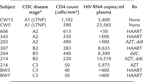
Clinical characteristics of study subjects
Subject CDC disease CD4 count HIV RNA copies/ml Rx stageA (cells/mm3) plasma
CW15 A1 (LTNP) 1,102 3,400 None
CW5 A1 (LTNP) 590 23,565 None
606 A2 615 <50 HAART
343 A2 550 <500 HAART
203 A2 490 <500 AZT, ddI
307 B2 283 8,633 HAART
204 B3 440 8,300 ddC
214 B2 220 14,510 AZT, ddC
216 C3 50 5,975 AZT
BW3 C3 40 <400 HAART
BW7 C3 50 <400 HAART
AStage A, asymptomatic; stage B, minor symptoms present (e.g., thrush);
stage C, major opportunistic infections present. Patients were classified by their lowest recorded CD4 counts. LTNPs were defined by infection for more than 5 years with CD4 T cell counts higher than 500 cells/mm3in the absence
USA), and cDNA was synthesized using the SMART RACE cDNA amplification kit (CLONTECH Labora-tories Inc., Palo Alto, California, USA), which provides an RT enzyme that adds a 5′oligo(dC) tail. An anchor oligonucleotide in the mix, which has a dG3′ tail, allows incorporation of the anchor sequence into the 5′end of the cDNA. The first-strand cDNA was used for anchored PCR with the upstream anchor primers provided by the manufacturer and a T cell receptor–specific (TCR-specific) Cβ outer Kd25 (5′
-GGCCAGGCACACCAGTGTGGCCTTTTGGGT-3′) and a Cβ inner primer, Kd24 (5′- TTCTGATGGCTCAAACA-CAGGAC-3′). Second strand cDNA was synthesized using a touchdown PCR for five cycles at 94°C for 5 seconds, 72°C for 2 minutes, followed by 5 cycles of PCR at 94°C for 5 seconds, 70°C for 10 seconds, and 72°C for 2 minutes. Finally, specific amplification of TCR βchain variable (TCRBV) genes was done with 25 cycles at 94°C for 5 seconds, 68°C for 10 seconds, and 72°C for 2 minutes. Subsequently, amplification reac-tions were diluted 50-fold with reaction buffer and a second round of PCR was performed with inner primers for another 20 cycles at 94°C for 5 seconds, 68°C for 10 seconds, and 72°C for 2 minutes. Ampli-fied PCR products were analyzed on 1.5% agarose gels and purified with the QIAquick PCR purification kit (QIAGEN Inc.). The purified PCR products were cloned into TA cloning vector pCR2.1 (Invitrogen Corp., Carlsbad, California, USA). For each sample, plasmid DNA was purified from multiple clones using a plasmid purification kit (QIAGEN Inc.). TCRBV genes were sequenced from the cloned DNA using an automated DNA sequencer (Applied Biosystems, Fos-ter City, California, USA).
Amplification of individual BV genes by RT-PCR. Total RNA from IFN-γ+cells was also used for amplification of individual Vβfamilies. cDNA was synthesized from total RNA using the TaqMan reverse transcription kit (Applied Biosystems). Aliquots of cDNA were amplified with 26 different 5′sense Vβ-specific primers and a common 3′ antisense Cβ primer using sequences described by Choi et al. (29). A sample with no cDNA served as negative control, and a sample with 5′and 3′ Cβoligonucleotides for amplification of the Cβregion served as positive control.
Generation of HIV-specific CTL lines. Bulk cell lines were generated by culturing the immunomagnetically select-ed IFN-γ+cells in the presence of irradiated allogeneic PBMC feeder cells and recombinant IL15 (25 ng/ml; R&D Systems Inc., Minneapolis, Minnesota, USA) for 10–14 days for use as effectors in cytotoxicity assays. Cul-ture was kept to the minimum to avoid altering the orig-inal clonal repertoire.
Chromium release assays. IIIB and autologous
virus–infected CD4 T cell targets, generated as described above, were labeled with 51Cr, washed, and distributed in round-bottom 96-well plates (5 ×10³cells in 100 µl). The bulk cultures from IIIB– and autologous virus– responding cells were used as effectors in triplicate wells
at effector/target ratios ranging from 1:1 to 5:1. The effector and target cells were incubated for 4 hours at 37°C. Supernatants (40 µl) from the cultures were har-vested and g counts measured on a Packard Microplate reader (Microplate scintillation counter; Packard Instrument Company, Downers Grove, Illinois, USA). Percent specific cytotoxicity was calculated from the average cpm as (average cpm minus spontaneous release/total release minus spontaneous release) ×100.
Results
Frequencies of IFN-γ–producing cells responding to primary CD4 T cell targets infected with lab-strain and autologous HIV decline with disease progression. HIV-infected CD4 T cells rapidly downmodulate CD4 expression, so homoge-neously infected CD4 T cells can be selected for use as targets by immunomagnetic depletion of CD4-express-ing uninfected cells from infected cultures (28, 30). Because infected cells are significantly enriched, the method eliminates variability in infectivity, making it possible to compare results from different samples and viral strains. We used this method to generate primary CD4 T cell targets infected with autologous or IIIB virus for use as stimulators to detect and capture func-tional IFN-γ–secreting, HIV-specific CD8 T cells. CD4 T cell blasts from each patient were treated with anti-retroviral drugs for 7 days to suppress endogenous virus. The cells were then infected with either IIIB or autologous virus, and after 3–5 days, were negatively enriched for infected cells by CD4 depletion. The level of enrichment of infected cells and the mean fluores-cence intensity of HIV p24 staining were comparable between CD4 stimulator cells infected with autologous virus and CD4 stimulator cells infected with IIIB virus. We also compared MHC class I expression between autologous and HIVIIIBvirus–infected targets, because the viral Nef protein is known to induce downmodula-tion of class I expression. Figure 1a depicts representa-tive data showing comparable p24 and MHC class I expression in CD4 T cells infected and enriched for autologous virus, IIIB, or another lab-adapted virus, NL4-3. As HIVIIIBhas been reported to contain multi-ple viral clones, some of which have dysfunctional nef
genes (26, 31), we also PCR-amplified and cloned the
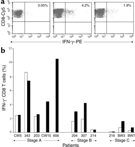
analysis. The frequency of cells producing an IFN-γ response to virus-infected targets was assessed in 11 seropositive subjects: five Centers for Disease Control (CDC) stage A subjects, including two long-term non-progressors (LTNPs); three CDC stage B subjects; and three CDC stage C subjects. The frequency of IFN-γ– producing CD8 T cells ranged from 0% to 10.6% (Fig-ure 2b). The frequency of IFN-γ+CD8 T cells respond-ing to autologous and IIIB virus–infected targets in subjects in whom response against both viruses could be evaluated suggests a decline in functional HIV-spe-cific IFN-γ–producing cells in more advanced donors (mean 4.4% ± 3.6% against autologous vs. 4.1% ± 2.8% against IIIB in 3 CDC stage A subjects, 1.3% ± 1.9% against autologous vs. 2.5% ± 1.9% against IIIB in three CDC stage B subjects, and 0.37% ± 0.3% against autol-ogous vs. 0.8% ± 0.75% in three CDC stage C subjects). In two additional CDC stage A subjects, the percent-ages of CD8 T cells producing an IFN-γresponse to HIVIIIBwere 10.6% and 4.3% of CD8 T cells, but no autologous virus could be generated in these patients despite several attempts. The decline in frequencies of
IFN-γ–secreting cells was statistically significant when the magnitude in the CDC stage A subjects was com-pared with that in the more advanced CDC stage B and C subject group (P< 0.02 for IIIB-specific response and
P< 0.04 for autologous virus–specific response). Sur-prisingly, twice as many IFN-γ+cells responded to lab-adapted IIIB virus than responded to autologous virus in CDC stage B and stage C subjects (median 1.15 vs. 0.55, respectively; P< 0.03).
Oligoclonal and nonoverlapping TCRBV usage in IFN-γ+
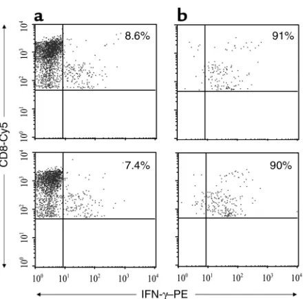
[image:5.576.60.286.56.279.2]CD8 T cells responding to targets infected with lab-strain and autologous virus. We also characterized the clonotypic composition of the responding cells by using anchored PCR to determine the TCRBV region and complemen-tarity-determining region 3 (CDR3) gene sequences in immunomagnetically selected IFN-γ+populations. IFN-γ–secreting cells were captured by immunomag-netic selection with αPE beads after external staining with PE-conjugated anti–IFN-γ (Figure 3). In most cases, more than 90% of the captured cells stained for IFN-γ. As the isolated cells were not fixed, they could be used for RNA isolation as well as for further culture. Anchored PCR was used for BV gene analysis because
Figure 2
Frequency of IFN-γ–producing cells responding to HIV-infected tar-gets declines in more advanced disease stages. (a) Representative flow cytometric analysis of surface IFN-γand CD8 staining on gated CD8 T cells in PBMCs from subject 307 after overnight stimulation with uninfected CD4 T cells (left) or HIVIIIB(middle) and autologous
[image:5.576.305.539.474.730.2](right) virus–infected CD4 T cell targets and capture of secreted IFN-γon the cell surface. The numbers shown are the percentages of CD8 T cells producing IFN-γ. (b) The percentage of CD8 T cells pro-ducing IFN-γin response to IIIB (black bars) and autologous (white bars) virus–infected targets are shown for subjects at various disease stages after subtracting background values for uninfected targets. In subjects 606 and CW15, the response was measured only against IIIB virus because no autologous virus could be generated.
Figure 1
Flow cytometric analysis of MHC class I and p24 expression on HIV-infected primary CD4 T cells used as stimulator cells. CD4 PHA blasts from representative subject 307 were used as uninfected con-trol (a) or were infected with HIVIIIB(b), NL4-3 (c), or autologous
this technique amplifies all TCRBV genes without qual-itative or quantqual-itative biases, unlike PCR amplification of individual TCRBV families. After PCR amplification, the product was analyzed by gel electrophoresis (Figure 4a). In each case, TCRBV genes were sequenced from 30–50 clones derived from the amplified PCR products (Table 2). To ensure the validity of the TCRBV gene sequences identified, in two subjects the analysis was performed in two independent experiments and yield-ed similar results (data not shown). For five samples, the authenticity of the analysis was also verified by direct PCR amplification of the reverse-transcribed RNA using a panel of upstream Vβprimers and a common down-stream Cβprimer. Although the specific BV+usage of the IFN-γ–producing cells could not be verified by anti-body staining immediately after stimulation because of TCR downmodulation that accompanies activation (data not shown), we could demonstrate the presence of
the specific BV+cells in the IFN-γ–selected population after a brief period of culture. Figure 4, b and c, shows the confirmatory studies for one subject for which all 50 clones sequenced from the captured cells responding to autologous virus had an identical TCRBV sequence belonging to the 7S1 family. In that case, the PCR analy-sis amplified only a single 7S1 band, and the antibody staining after culture showed that 90% of the cells stained for 7S1 antibody.
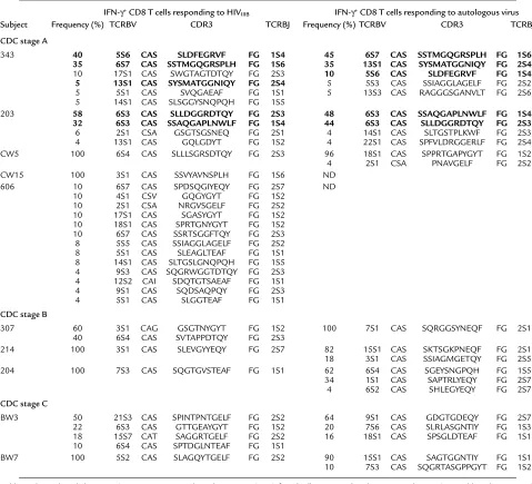
The TCR usage of CD8 T cells responding to lab-strain HIV was analyzed in ten subjects (Table 2). The response ranged from monoclonal to polyclonal, although in most subjects, one or two clonotypes dom-inated the response, with other minor clonotypes con-tributing less than 10% to the response. Because the specificity of the capture method is about 90% (see Fig-ure 1), it is difficult to tell whether clonotypes present at frequencies of below 10% are contaminating cells or are participating in the specific response. A polyclonal response with no clear dominant clonotype was seen in only one subject. This CDC stage A subject (no. 606), with 13 TCR clonotypes participating in the response, had the highest frequency of IFN-γ–producing cells (10.6%). All seven of the CD8 T cell clones generated from the IFN-γ+cells from this subject were HIV-specif-ic, suggesting that the TCR clonotypes identified were not because of contamination with nonspecific cells (data not shown). In the two LTNPs analyzed, the TCRBV gene usage was monoclonal.
[image:6.576.66.286.52.269.2]A similar clonotypic analysis was performed for CD8 T cells responding to autologous virus–infected targets Figure 3
Immunomagnetic selection of IFN-γ–secreting HIV-specific CD8 T cells. PBMCs from donor 343 were stimulated with autologous (upper pan-els) or IIIB (lower panpan-els) virus–infected primary CD4 T cells, stained for IFN-γ, and immunomagnetically isolated. Cells before (a) and after (b) selection were stained with anti–CD8-Cy5 antibody and analyzed for IFN-γ+cells within a CD8-gated population.
Figure 4
Representative analysis of TCRBV usage in CD8 T cells responding to HIV-infected targets. (a) Agarose gel analysis of anchored PCR ampli-fication of TCRBV genes from immunomagnetically captured IFN-γ+cells stimulated with autologous virus from representative subject 307.
[image:6.576.59.541.527.644.2]in the eight of the subjects for whom autologous virus could be generated (Table 2). The response to autolo-gous virus was also oligoclonal, with one or two clono-types dominating the response in most subjects. We compared the TCR clonotypes responding to lab-strain virus with those recognizing the patient’s own virus to determine whether group-specific epitopes in HIVIIIB contribute to the dominant response in vivo. In two of three CDC stage A subjects, the immunodominant TCR clonotypes against lab-adapted HIVIIIBvirus and autologous virus overlapped to a large extent, suggest-ing a focussuggest-ing of the response toward such conserved epitopes (Table 2). In both patients, clonotypes exclu-sive to autologous virus or lab-adapted virus constitut-ed less than 10% of the response. However, in all other subjects analyzed (CDC stage A, n = 1; stage B, n = 3;
and stage C, n = 2), the dominant TCR clonotypes used in response to autologous virus and lab-adapted IIIB virus were completely different.
Bulk cell lines generated from lab-strain HIV–specific IFN-γ+
[image:7.576.57.536.77.512.2]cells recognize autologous virus–infected targets and vice versa only in subjects with shared TCRBV usage. To test the in vivo relevance of the CD8 T cells responding to lab-strain virus, we evaluated the ability of cell lines derived from immunomagnetically selected HIVIIIB-specific IFN-γ+ cells to lyse autologous virus–infected targets (Figure 5). Tissue culture was kept to a minimum to avoid in vitro alterations of the native clonal composition. In CTL assays performed after 10–12 days of culture, HIVIIIB-specific T cell lines cross-recognized autologous virus–infected targets in CDC stage A subjects who shared TCRBV genes. However, bulk cell lines from Table 2
TCRBV and CDR3 amino acid sequences of CD8 T cells producing IFN-γin response to HIV-infected primary T cell targets
IFN-γ+CD8 T cells responding to HIV
IIIB IFN-γ+CD8 T cells responding to autologous virus
Subject Frequency (%) TCRBV CDR3 TCRBJ Frequency (%) TCRBV CDR3 TCRBJ
CDC stage A
343 40 5S6 CAS SLDFEGRVF FG 1S4 45 6S7 CAS SSTMGQGRSPLH FG 1S6
35 6S7 CAS SSTMGQGRSPLH FG 1S6 35 13S1 CAS SYSMATGGNIQY FG 2S4
10 17S1 CAS SWGTAGTDTQY FG 2S3 10 5S6 CAS SLDFEGRVF FG 1S4
5 13S1 CAS SYSMATGGNIQY FG 2S4 5 5S3 CAS SSIAGGLAGELF FG 2S2
5 5S1 CAS SVQGAEAF FG 1S1 5 13S3 CAS RAGGGSGANVLT FG 2S6 5 14S1 CAS SLSGGYSNQPQH FG 1S5
203 58 6S3 CAS SLLDGGRDTQY FG 2S3 48 6S3 CAS SSAQGAPLNWLF FG 1S4
32 6S3 CAS SSAQGAPLNWLF FG 1S4 44 6S3 CAS SLLDGGRDTQY FG 2S3
6 2S1 CSA GSGTSGSNEQ FG 2S1 4 14S1 CAS SLTGSTPLKWF FG 2S3 4 13S1 CAS GQLGDYT FG 1S2 4 22S1 CAS SPFVLDRGGERLF FG 2S4 CW5 100 6S4 CAS SLLLSGRSDTQY FG 2S3 96 18S1 CAS SPPRTGAPYGYT FG 1S2 4 2S1 CSA PNAVGELF FG 2S2
CW15 100 3S1 CAS SSVYAVNSPLH FG 1S6 ND
606 10 6S7 CAS SPDSQGIYEQY FG 2S7 ND
10 4S1 CSV GQGYGYT FG 1S2 10 2S1 CSA NRGVSGELF FG 2S2 10 17S1 CAS SGASYGYT FG 1S2 10 18S1 CAS SPRTGNYGYT FG 1S2 10 6S7 CAS SSRTSGGFTQY FG 2S3 8 5S5 CAS SSIAGGLAGELF FG 2S2 8 5S1 CAS SLEAGLTEAF FG 1S1 8 14S1 CAS SLTGSLGNQPQH FG 1S5 4 9S3 CAS SQGRWGGTDTQY FG 2S3 4 12S2 CAI SDQTGTSAEAF FG 1S1 4 9S1 CAS SQDSAQPQY FG 2S3 4 5S1 CAS SLGGTEAF FG 1S1
CDC stage B
307 60 3S1 CAG GSGTNYGYT FG 1S2 100 7S1 CAS SQRGGSYNEQF FG 2S1 40 6S4 CAS SVTAPPDTQY FG 2S3
214 100 3S1 CAS SLEVGYYEQY FG 2S7 82 15S1 CAS SKTSGKPNEQF FG 2S1 18 3S1 CAS SSIAGMGETQY FG 2S5 204 100 7S3 CAS SQGTGVSTEAF FG 1S1 62 6S4 CAS SGEYSNGPQH FG 1S5 34 1S1 CAS SAPTRLYEQY FG 2S7 4 6S2 CAS SHLEGYEQY FG 2S7
CDC stage C
BW3 50 21S3 CAS SPINTPNTGELF FG 2S2 64 9S1 CAS GDGTGDEQY FG 2S7 22 6S3 CAS GTTGEAYGYT FG 1S2 20 7S6 CAS SLRLASGNTIY FG 1S3 18 15S7 CAT SAGGRTGELF FG 2S2 16 18S1 CAS SPSGLDTEAF FG 1S1 10 6S4 CAS SPTDGLNTEAF FG 1S1
BW7 100 5S2 CAS SLAGQYTGELF FG 2S2 90 15S1 CAS SAGTGGNTIY FG 1S1 10 7S3 CAS SQGRTASGPPGYT FG 1S2
CDC stage B and C subjects in whom the TCR clono-types responding to IIIB and autologous virus were not shared did not cross-recognize autologous virus-infect-ed targets. When bulk lines generatvirus-infect-ed from IFN-γ+cells responding to autologous virus were tested against HIVIIIB-infected CD4 T cell targets, a similar pattern of cross-recognition was observed. Data from representa-tive subjects are shown in Figure 5.
To confirm that the results obtained with HIVIIIBdo not represent an aberrant response to an nef-deficient lab-strain virus, we also examined the lytic capability of the bulk cell line generated from IIIB-responsive IFN-γ+ cells against target cells infected with another lab-adapted viral isolate, NL4-3. This viral strain is known to have an intact nefgene (32). As shown in Figure 6, the bulk cell line from a CDC stage B subject (no. 307) exhibited comparable lysis of NL4-3 and IIIB virus–infected targets but failed to lyse autologous virus–infected targets. Similar results were obtained with a CTL clone generated from the IIIB-responsive cells from the same subject. These data indicate that the lack of recognition of autologous virus–infected targets was not because of low epitope densities induced by nef-mediated class I downmodulation.
The epitope specificity of this bulk line as well as the clone were mapped to a 10 amino acid region in Nef (LWIYHTQGYF, aa 112–121) using recombinant vac-cinia and overlapping 20mer peptide–pulsed B-lym-phoblastoid cell lines targets in cytotoxicity assays as described by Lieberman et al. (33) (data not shown). When the nefregion of the autologous virus was ampli-fied by PCR and sequenced, two amino acid changes were found within the 10mer epitope–containing region (LWVYNTQGYF) compared with the consensus
sequence in the IIIB and NL4-3 strains. This suggests that sequence mutation may be the underlying cause for nonrecognition of autologous virus–infected tar-gets by IIIB-responsive CTLs.
Discussion
This study examines the functional virus-specific CD8 T cell repertoire in the setting of a protracted infection with a virus with a propensity for mutation. We used primary CD4 T cell targets to compare the magnitude and clonotypic composition of CD8 T cells responding to lab-strain HIVIIIBwith those responding to autolo-gous viral isolates in HIV-infected individuals at differ-ent stages of disease. Our principal findings are that the magnitude of the functional response declines with dis-ease progression and that the TCR clonotypes of CD8 T cells responding to the two viruses overlap in the early stage of infection but completely diverge in later stages of the disease. Consequently, HIVIIIB-responding cells do not recognize autologous virus and vice versa in symp-tomatic patients. Moreover, the functional CD8 T cell response is oligoclonal in most patients. Taken togeth-er, these results suggest that conventional assays that focus on responses to consensus epitopes may not accu-rately reflect, but rather tend to overestimate, the response to autologous virus. Our results therefore underscore the need to develop approaches that more closely test the relevant in vivo responses.
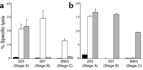
[image:8.576.58.293.56.172.2]The stimulator cells we used do not exactly mimic in vivo targets, because CD4 cells are cultured for a brief Figure 5
Autologous virus–infected targets are not lysed by IFN-γ+CD8 T cell
lines responding to HIVIIIBvirus and vice versa in samples from more
advanced donors. Short-term cultures from IFN-γ+cells responding
[image:8.576.307.538.425.583.2]to IIIB (a) or autologous virus (b) were tested against uninfected (black bars), IIIB (white bars), or autologous (gray bars) virus–infect-ed primary CD4 T cell targets. Representative data for subjects from each CDC stage are depicted at effector/target ratios ranging from 1:1 to 5:1. Data are presented as mean of experiments performed in triplicate. The cytotoxicity assays confirm the results of TCRBV sequencing. There was cross-recognition of IIIB and autologous virus by two of the stage A donor cell lines, but not by the cell lines from more advanced donors.
Figure 6
Bulk lines and clones derived from HIVIIIBvirus–responsive CD8 T cells
recognize IIIB and NL4-3 virus, but not autologous virus–infected CD4 T cell targets. A bulk cell line and clones were generated from CD8 T cells that produced IFN-γafter stimulation with HIVIIIB-infected CD4
period before use. Nevertheless, they more closely resemble the physiologically relevant targets than do the stimulator cells used in conventional methods. Unlike recombinant vaccinia–infected or synthetic pep-tide–pulsed targets, infected CD4 T cells, which are the natural targets of the virus, more closely reflect epitope densities achieved on infected targets in vivo (34). This is particularly important in light of reports that the nef protein downmodulates cell-surface MHC class I expression, which may further reduce epitope densities, making virus-infected cells poor targets for CTL lysis and possibly for stimulation of IFN-γsecretion (25, 26). The decline in frequency of IFN-γ–producing HIV-spe-cific cells in stage B and C disease is unexpected, consid-ering studies that show a high frequency of HIV-specif-ic CD8 T cells until late in the course of HIV disease (35–37). These studies have generally used MHC/peptide tetramer staining or IFN-γresponses after peptide stim-ulation to identify virus-specific CD8 T cells. One expla-nation may be that our use of virus-infected targets fil-ters out all of the responses that are not meaningful in the context of infected cells, including low-avidity or sub-dominant CD8 T cells that may produce IFN-γonly upon stimulation with an excess of peptides. This is also suggested by the predominantly oligoclonal and mono-clonal functional response that we observed against both lab-adapted and autologous virus in most of the sub-jects. Moreover, studies that use MHC/peptide tetramers identify HIV-specific CD8 T cells without regard to their functionality. Many investigators, ourselves included, have reported that a significant fraction of tetramer-pos-itive cells are unable to produce IFN-γwhen freshly test-ed ex vivo (27, 38–40). Functional deficiencies of HIV-specific cells in chronic infection can occur because of their generation or maintenance in a progressively CD4-deficient milieu (41–45). In fact, when we analyzed HIV-specific CD8 T cell frequencies with four different HLA-A2 and B8/peptide tetramers from five subjects in this study who expressed these HLA alleles (subjects CW5, 203, 204, 216, and 307), we found no correlation with the disease stage (ref. 27 and data not shown). Compa-rable frequencies of gag SLYNVATL/A2.1 tetramer-stain-ing CD8 T cells were seen in samples from an AIDS patient (no. 216) with no detectable IFN-γ+CD8 T cells and an LTNP (no. CW5) with over 2% IFN-γ+CD8 T cells (1.4% and 1.6%, respectively).
We were surprised to find that the overall frequency of IFN-γ–producing cells was lower in response to autologous virus than to HIVIIIBvirus stimulation. This finding suggests that CD8 T cells generated in response to consensus epitopes early in infection persist even after viral mutations make them irrelevant. The responses against any newly presented or substituted epitopes that come up because of viral mutations dur-ing chronic infection have to be generated in the settdur-ing of HIV-specific CD4 T helper cell deficiency and cytokine imbalance and may be functionally impaired. This may be particularly relevant because the HIV virus selectively targets HIV-specific CD4 T cells (46).
Although this needs to be investigated in a larger group of patients, these results suggest that in patients with more advanced disease, measurement of the IFN-γ response to lab-strain virus significantly overstates the functional response to autologous virus.
Although we have used only one lab-strain virus as representative of the response to relatively conserved epitopes, the most striking fact to emerge from the study is the discordance between the CD8 T cell clono-types responding to autologous virus and lab-strain virus, particularly in more advanced patients. The inability of the bulk cell lines from IIIB-responsive CD8 T cells to cross-recognize autologous virus–infected tar-gets in CTL assays corroborates the TCR sequencing data. Although HIVIIIBvirus has been reported to con-tain multiple viral clones, some of which have dys-functional nef genes (26, 31), our results cannot be explained on the basis of differences in class I expres-sion. Sequence analysis revealed that the HIVIIIBisolate we used for the study did not contain viral clones with a truncated nefgene, which was in keeping with our data showing comparable levels of class I expression on autologous and IIIB virus–infected targets (Figure 1). Moreover, in one representative subject, the dichotomy in recognition of autologous and lab-adapted virus could also be confirmed with NL4-3 virus, which has an intact nefgene (Figure 6).
One possible explanation for the persistence of virus-specific CD8 T cells that do not recognize the currently circulating autologous virus is that these are residual cells from a previously relevant response that are left behind as the CD8 T cell response diversifies in an effort to cope with viral mutations. In fact, our data showing lower frequencies of IFN-γ+ CD8 T cells responding to autologous virus compared with IIIB virus-infected tar-gets in CDC stage B and C subjects (P< 0.03) also hints at accumulation of cells that may have become irrelevant in the context of the evolving virus. It is well document-ed that memory T cells can persist indefinitely in mouse models in the absence of antigen (47). In fact, many HIV tetramer–positive cells exhibit memory-like phenotype, suggesting that they have not been recently activated by antigen (27, 48, 49). However, as different viral quasi-species exist in infected individuals, it is also possible that CD8 T cells reacting to group-specific epitopes may be keeping viral quasispecies that have not mutated these epitopes in control. This type of plasticity in the immune response may be a host strategy to abrogate the deleterious consequences of CTL escape (50, 51).
and intense host immune response, as the functional CD8 T cell response to the patient’s own viral isolate declines around the time disease symptoms develop.
Acknowledgments
We thank N. Manjunath, D. Zhang, and E. Song for discussions and comments. We also thank M. Leder-man, C. Lange, and J. Daily for patient samples. This work was supported by NIH grants 49792 and AI-45306 (to P. Shankar).
1. Borrow, P., Lewicki, H., Hahn, B.H., Shaw, G.M., and Oldstone, M.B. 1994. Virus-specific CD8+cytotoxic T-lymphocyte activity associated with control of viremia in primary human immunodeficiency virus type 1 infection. J. Virol.68:6103–6110.
2. Koup, R.A., et al. 1994. Temporal association of cellular immune responses with the initial control of viremia in primary human immun-odeficiency virus type 1 syndrome. J. Virol.68:4650–4655.
3. Pantaleo, G., et al. 1997. The qualitative nature of the primary immune response to HIV infection is a prognosticator of disease progression independent of the initial level of plasma viremia. Proc. Natl. Acad. Sci. USA.94:254–258.
4. Wilson, J.D., et al. 2000. Direct visualization of HIV-1-specific cytotoxic T lymphocytes during primary infection. AIDS.14:225–233. 5. Pantaleo, G., et al. 1994. Major expansion of CD8+T cells with a
pre-dominant V beta usage during the primary immune response to HIV.
Nature.370:463–467.
6. Schmitz, J.E., et al. 1999. Control of viremia in simian immunodeficien-cy virus infection by CD8+lymphocytes. Science.283:857–860. 7. Jin, X., et al. 1999. Dramatic rise in plasma viremia after CD8+T cell
depletion in simian immunodeficiency virus-infected macaques. J. Exp. Med.189:991–998.
8. Than, S., et al. 1999. Clonal dominance patterns of CD8 T cells in rela-tion to disease progression in HIV-infected children. J. Immunol.
162:3680–3686.
9. Gorochov, G., et al. 2001. Down-regulation of CD8+T-cell expansions in patients with human immunodeficiency virus infection receiving high-ly active combination therapy. Blood.97:1787–1795.
10. Kolowos, W., et al. 1999. Biased TCR repertoire in HIV-1-infected patients due to clonal expansion of HIV-1-reverse transcriptase-specific CTL clones. J. Immunol.162:7525–7533.
11. Weiss, L., et al. 1998. Persistent expansion, in a human immunodefi-ciency virus-infected person, of V beta-restricted CD4+CD8+T lympho-cytes that express cytotoxicity-associated molecules and are committed to produce interferon-gamma and tumor necrosis factor-alpha. J. Infect. Dis.178:1158–1162.
12. Mion, M., et al. 1997. TCR expression and clonality analysis in periph-eral blood and lymph nodes of HIV-infected patients. Hum. Immunol.
57:93–103.
13. Dalod, M., et al. 1999. Weak anti-HIV CD8+T-cell effector activity in HIV primary infection. J. Clin. Invest.104:1431–1439.
14. Pantaleo, G., et al. 1997. Evidence for rapid disappearance of initially expanded HIV-specific CD8+T cell clones during primary HIV infection.
Proc. Natl. Acad. Sci. USA.94:9848–9853.
15. Goulder, P.J., et al. 2001. Substantial differences in specificity of HIV-specific cytotoxic T cells in acute and chronic HIV infection.J. Exp. Med.
193:181–194.
16. Phillips, R.E., et al. 1991. Human immunodeficiency virus genetic vari-ation that can escape cytotoxic T cell recognition. Nature.354:453–459. 17. McMichael, A. 1998. T cell responses and viral escape. Cell.93:673–676. 18. Koup, R.A. 1994. Virus escape from CTL recognition. J. Exp. Med.
180:779–782.
19. Couillin, I., et al. 1994. Impaired cytotoxic T lymphocyte recognition due to genetic variations in the main immunogenic region of the human immunodeficiency virus 1 NEF protein. J. Exp. Med.180:1129–1134. 20. Soudeyns, H., et al. 1999. Selective pressure exerted by
immunodomi-nant HIV-1-specific cytotoxic T lymphocyte responses during primary infection drives genetic variation restricted to the cognate epitope. Eur. J. Immunol.29:3629–3635.
21. Borrow, P., et al. 1997. Antiviral pressure exerted by HIV-1-specific cyto-toxic T lymphocytes (CTLs) during primary infection demonstrated by rapid selection of CTL escape virus. Nat. Med.3:205–211.
22. Price, D.A., et al. 1997. Positive selection of HIV-1 cytotoxic T lympho-cyte escape variants during primary infection.Proc. Natl. Acad. Sci. USA.
94:1890–1895.
23. Goulder, P.J., et al. 1997. Late escape from an immunodominant cyto-toxic T-lymphocyte response associated with progression to AIDS. Nat. Med.3:212–217.
24. Koenig, S., et al. 1995. Transfer of HIV-1-specific cytotoxic T lympho-cytes to an AIDS patient leads to selection for mutant HIV variants and subsequent disease progression. Nat. Med.1:330–336.
25. Collins, K.L., Chen, B.K., Kalams, S.A., Walker, B.D., and Baltimore, D. 1998. HIV-1 Nef protein protects infected primary cells against killing by cytotoxic T lymphocytes. Nature.391:397–401.
26. Yang, O.O., et al. 2002. Nef-mediated resistance of human immunode-ficiency virus type 1 to antiviral cytotoxic T lymphocytes. J. Virol.
76:1626–1631.
27. Shankar, P., et al. 2000. Impaired function of HIV-specific CD8(+)T cells in chronic HIV infection. Blood.96:3094–3101.
28. Shankar, P., Xu, Z., and Lieberman, J. 1999. Viral-specific cytotoxic T lymphocytes lyse HIV-infected primary T lymphocytes by the granule exocytosis pathway. Blood.94:3084–3093.
29. Choi, Y.W., et al. 1989. Interaction of Staphylococcus aureus toxin “superantigens” with human T cells. Proc. Natl. Acad. Sci. USA.
86:8941–8945.
30. Ferrari, G., et al. 1997. Clade B-based HIV-1 vaccines elicit cross-clade cytotoxic T lymphocyte reactivities in uninfected volunteers. Proc. Natl. Acad. Sci. USA.94:1396–1401.
31. Kuiken, C., et al. 1999. Human retroviruses and AIDS 1999: a compila-tion and analysis of nucleic acid and amino acid sequences. Theoretical Biology and Biophysics Group, Los Alamos National Laboratory. Los Alamos, New Mexico, USA.
32. Olivetta, E., et al. 2000. cis expression of the F12 human immunodefi-ciency virus (HIV) Nef allele transforms the highly productive NL4-3 HIV type 1 to a replication-defective strain: involvement of both Env gp41 and CD4 intracytoplasmic tails. J. Virol.74:483–492.
33. Lieberman, J., et al. 1992. Cytotoxic T lymphocytes from HIV-1 seropos-itive individuals recognize immunodominant epitopes in Gp160 and reverse transcriptase. J. Immunol.148:2738–2747.
34. Tsomides, T.J., et al. 1994. Naturally processed viral peptides recognized by cytotoxic T lymphocytes on cells chronically infected by human immunodeficiency virus type 1. J. Exp. Med.180:1283–1293. 35. Betts, M.R., et al. 2001. Analysis of total human immunodeficiency virus
(HIV)-specific CD4(+) and CD8(+) T-cell responses: relationship to viral load in untreated HIV infection. J. Virol.75:11983–11991.
36. Gea-Banacloche, J.C., et al. 2000. Maintenance of large numbers of virus-specific CD8+T cells in HIV-infected progressors and long-term non-progressors. J. Immunol.165:1082–1092.
37. Migueles, S.A., and Connors, M. 2001. Frequency and function of HIV-specific CD8(+) T cells. Immunol. Lett.79:141–150.
38. Goepfert, P.A., et al. 2000. A significant number of human immunode-ficiency virus epitope-specific cytotoxic T lymphocytes detected by tetramer binding do not produce gamma interferon. J. Virol.
74:10249–10255.
39. Kostense, S., et al. 2002. Persistent numbers of tetramer(+) CD8(+) T cells, but loss of interferon-gamma(+) HIV-specific T cells during progression to AIDS. Blood.99:2505–2511.
40. Kostense, S., et al. 2002. Functional restoration of human immunodefi-ciency virus and Epstein-Barr virus-specific CD8(+) T cells during high-ly active antiretroviral therapy is associated with an increase in CD4(+) T cells. Eur. J. Immunol.32:1080–1089.
41. Kalams, S.A., and Walker, B.D. 1998. The critical need for CD4 help in maintaining effective cytotoxic T lymphocyte responses. J. Exp. Med.
188:2199–2204.
42. Picker, L.J., and Maino, V.C. 2000. The CD4(+) T cell response to HIV-1.
Curr. Opin. Immunol.12:381–386.
43. Rosenberg, E.S., et al. 1997. Vigorous HIV-1-specific CD4+T cell respons-es associated with control of viremia. Science.278:1447–1450. 44. Spiegel, H.M., et al. 2000. Human immunodeficiency virus type 1- and
cytomegalovirus-specific cytotoxic T lymphocytes can persist at high fre-quency for prolonged periods in the absence of circulating peripheral CD4(+) T cells. J. Virol.74:1018–1022.
45. Trimble, L.A., and Lieberman, J. 1998. Circulating CD8 T lymphocytes in human immunodeficiency virus-infected individuals have impaired function and downmodulate CD3 zeta, the signaling chain of the T-cell receptor complex. Blood.91:585–594.
46. Douek, D.C., et al. 2002. HIV preferentially infects HIV-specific CD4+T cells. Nature.417:95–98.
47. Ahmed, R., and Gray, D. 1996. Immunological memory and protective immunity: understanding their relation. Science.272:54–60.
48. Champagne, P., et al. 2001. Skewed maturation of memory HIV-specif-ic CD8 T lymphocytes. Nature.410:106–111.
49. Altman, J.D., et al. 1996. Phenotypic analysis of antigen-specific T lym-phocytes. Science. 274:94–96.