www.impactjournals.com/oncotarget/ Oncotarget, Vol. 7, No. 33
INTRODUCTION
Several genetic mutations are usually necessary for the onset of cancer [1]. In case of childhood acute lymphoblastic leukemia (ALL) only few mutations are required [2]. In most cases, products deriving from
chromosomal translocations are the oncogenic initiating or driving lesions and have been characterized at the molecular and functional level during the last decades. In addition to the widely known gene fusions, genomic technologies - SNP arrays or whole genome sequencing - also allowed the identification of complementing genetic
COBL is a novel hotspot for IKZF1 deletions in childhood acute
lymphoblastic leukemia
Bruno Almeida Lopes1, Claus Meyer2, Thayana Conceição Barbosa1, Udo zur Stadt3, Martin Horstmann3,4,5, Nicola C. Venn6, Susan Heatley7,8, Deborah L. White7,8, Rosemary Sutton6, Maria S. Pombo-de-Oliveira1, Rolf Marschalek2, Mariana Emerenciano1
1Pediatric Hematology-Oncology Program, Research Center, Instituto Nacional de Câncer, Rio de Janeiro, RJ, Brazil
2Diagnostic Center of Acute Leukemia/Institute of Pharmaceutical Biology/ZAFES, Goethe-University of Frankfurt, Biocenter, Germany
3Center for Diagnostics, University Medical Center Hamburg Eppendorf, Hamburg, Germany 4Research Institute Children’s Cancer Center, Hamburg, Germany
5Department of Pediatric Hematology and Oncology, University Medical Center Hamburg-Eppendorf, Hamburg, Germany 6Children's Cancer Institute, Lowy Cancer Research Centre UNSW, Sydney, New South Wales, Australia
7South Australian Health and Medical Research Institute (SAHMRI), Adelaide, South Australia, Australia 8Discipline of Medicine, University of Adelaide, Adelaide, South Australia, Australia
Correspondence to: Mariana Emerenciano, email: memerenciano@inca.gov.br
Keywords:acute lymphoblastic leukemia, COBL, IKZF1, RAG, relapse
Received: May 03, 2016 Accepted: June 30, 2016 Published: July 13, 2016
ABSTRACT
IKZF1 deletion (ΔIKZF1) is an important predictor of relapse in childhood B-cell precursor acute lymphoblastic leukemia. Because of its clinical importance, we previously mapped breakpoints of intragenic deletions and developed a multiplex
PCR assay to detect recurrent intragenic ΔIKZF1. Since the multiplex PCR was not able to detect complete deletions (IKZF1 Δ1-8), which account for ~30% of all ΔIKZF1, we aimed at investigating the genomic scenery of IKZF1 Δ1-8. Six samples of cases
with IKZF1 Δ1-8 were analyzed by microarray assay, which identified monosomy 7,
isochromosome 7q, and large interstitial deletions presenting breakpoints within COBL
gene. Then, we established a multiplex ligation-probe amplification (MLPA) assay and
screened copy number alterations within chromosome 7 in 43 diagnostic samples with IKZF1 Δ1-8. Our results revealed that monosomy and large interstitial deletions
within chromosome 7 are the main causes of IKZF1∆1-8. Detailed analysis using long distance inverse PCR showed that six patients (16%) had large interstitial deletions
starting within intronic regions of COBL at diagnosis, whichis ~611 Kb downstream
of IKZF1,suggesting that COBL is a hotspot for ΔIKZF1. We also investigated a series
of 25 intragenic deletions (∆2–8, ∆3–8 or ∆4–8) and 24 relapsed samples, and found
one IKZF1-COBL tail-to-tail fusion, thus supporting that COBL is a novel hotspot for
ΔIKZF1. Finally, using RIC score methodology, we show that breakpoint sequences of
IKZF1 ∆1-8 are not analog to RAG-recognition sites, suggesting a different mechanism of error promotion than that suggested for intragenic ΔIKZF1.
alterations that are important contributors of hematological malignancies. One of the identified alterations concerns the IKZF1 gene, located at 7p12, encoding the transcription factor Ikaros, that is essentially involved in the development of the B-cell lineage [3].
The biology of IKZF1 is complex because this gene consists of 8 exons, and encodes 11 different splice variants [4]. Five of these isoforms are translated into proteins that are acting as transcriptional activators (1, 2, 2a, 3, 3a), while six isoforms result in dominant-negative versions of Ikaros (4, 4a, 5, 6, 7, 8). The Ikaros protein composes a regulatory complex involving hematopoietic transcription factors (E2A, EBF1-3, and PAX5) with the distinct function of driving lymphoid development [5], which is impaired by “dominant-negative” isoforms.
In 2007, the first study demonstrating the importance of IKZF1 deletions (ΔIKZF1) in leukemia was published [2]. Genome wide SNP array analyses of 242 patients revealed that several genes involved in B-lineage development are mutated in B-cell precursor ALL (BCP-ALL) patients. These initial findings were confirmed by a contemporary study that investigated 40 leukemia patients [6], which identified again recurrent submicroscopic deletions in several genes linked to B-lineage development (PAX5, EBF1, TCF3, IKZF1 and others). Thereafter, clinical studies revealed that leukemia patients with ΔIKZF1 should be classified as high-risk, as the presence of a deletion was shown to be an independent prognostic factor for event-free survival in pediatric BCP-ALL [7]. Of note, a subsequent international study showed that all types of ΔIKZF1 are associated with unfavorable prognosis [8]. The clinical effects of ΔIKZF1 and the results from basic research have been thoroughly reviewed [9].
Due to the clinical importance of ΔIKZF1, multiplex ligation-probe amplification (MLPA) assays have been developed to detect such alterations in BCP-ALL. As ΔIKZF1 are predictive of relapse and given the lack of sensitivity of MLPA assays, scientific efforts focused on novel diagnostic tools for minimal residual disease investigation. In 2011, a real-time quantitative PCR assay was developed to detect the most common microdeletion in ALL IKZF1Δ3-6 (now known as IKZF1Δ4-7), which results in the Ik6 transcript [10]. This was followed in 2013 by two studies that developed multiplex PCR assays to detect recurrent intragenic deletions of IKZF1, such as Δ2-3, Δ2-7, Δ2-8, Δ4-7, and Δ4-8 [11, 12]. Although these methodologies present a higher sensitivity of at least 10-2, they are not able to detect complete deletions (IKZF1 Δ1-8), which represent ~30% of all deletions. Therefore, the aim of this study was to unravel the genomic landscape of IKZF1 Δ1-8.
RESULTS
Clinical and genetic characteristics of BCP-ALL with IKZF1 complete deletions
Patients with IKZF1 Δ1-8 were mainly male (60.5%), aged 1-9 years old (72.1%) at diagnosis, and only 30.2% had WBC ≥ 50 × 109/L. The predominant immunophenotype was c-ALL (65.1%).
Copy number alterations (CNAs) in genes frequently altered in BCP-ALL were compared between patients with or without IKZF1 deletions in order to discover possible concomitant alterations associated with IKZF1 Δ1-8. The results showed that IKZF1 ∆1-8 deletions extended to its surrounding genes, such as ZPBP (deleted in 85.7% of cases), FIGNL1 (90.5%) and DDC (90.5%). On the other hand, deletions of EBF1 and BTG1 were respectively absent or rarely (4.1%) identified in patients with IKZF1 ∆1-8 (Supplementary Table S1).
Genome-wide CNAs in samples with complete deletion of IKZF1 at diagnosis
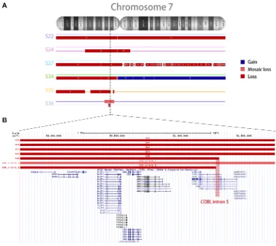
Six random DNA samples of BCP-ALL with IKZF1 ∆1-8 were selected for the microarray assay. The details of each sample are presented in Supplementary Table S2. Microarray analysis revealed that samples with IKZF1 ∆1-8 presented large deletions within chromosome 7 (n = 5) and partial loss of 9p arm (n = 3), as shown in Supplementary Figure S1. Three sorts of alterations on chromosome 7 were found for samples with IKZF1 ∆1-8: monosomy 7 (n = 2), large interstitial deletions (n = 3), and isochromosome 7q (n = 1) (Figure 1A). Interestingly, two patients (S35 and S36) with large interstitial deletions presented breakpoints within COBL intron 5 (Figure 1B) downstream of IKZF1. On the other hand, there was no indication of a genetic hotspot upstream of IKZF1, as50% of cases presented loss of chromosome 7 short arm, and the remaining patients had remarkably variable breakpoints at the 7p telomeric side.
Screening CNAs on chromosome 7 with the customized MLPA
Because the microarray data showed that a diverse spectrum of alterations within chromosome 7 promotes IKZF1 ∆1-8, we designed two customized MLPAs to identify such CNAs in the whole series of patients included in this study. The MLPA probes were distributed throughout chromosome 7, and most of them were placed within COBL. The main characteristics and localization of the MLPA probes are described in Supplementary Table S4, and probe sequences are available upon request.
results were concordant with microarray experiments. As expected, patient S22 (with monosomy 7 by microarray) presented monoallelic deletion for all of the probes tested on chromosome 7, while patients S35 and S36 (with COBL intron 5 rearrangements) presented deletion of a series of probes within COBL, thus correctly indicating the breakpoint was localizing within intron 5. Although our MLPA did not detect all of the expected CNAs (e.g. patient S24 bears an interstitial deletion within chromosome 7 and patient S34 has isochromosome 7q), the analysis of whole probe set contributed to the correct interpretation of results. Thus, we confirmed that our custom MLPAs were able to detect distinct CNAs within chromosome 7.
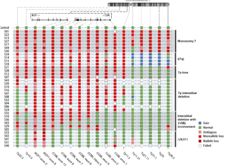
Subsequently, the custom MLPAs were used to investigate CNAs for chromosome 7 in 43 BCP-ALL pediatric patients with IKZF1 ∆1-8. The MLPA results could not be interpreted for seven samples, which were excluded from further analysis. Five sorts of alterations were identified on chromosome 7: monosomy 7 (n = 7), isochromosome 7q (n = 4), 7p loss (n = 4), large interstitial deletions within the
7p arm (n = 10), IKZF1 ∆1-8 with breakpoints within COBL (n = 6), and IKZF1 complete deletion without involvement of surrounding regions (n = 5) (Figure 2).
Breakpoint sequences of IKZF1 ∆1-8
[image:3.612.104.507.302.661.2]We designed four long-distance inverse PCRs (LDI-PCRs) as well as two multiplexed long-distance PCRs (MP-PCRs) assays in order to find the sequence of the breakpoints predicted by both microarray and MLPA results. The sequences of breakpoints for COBL rearrangements are summarized in Supplementary Figure S3. Patient S10 presented a large interstitial deletion on the short arm of chromosome 7, comprising the whole IKZF1 until COBL intron 5 and a subsequent ~1.1 Mb inversion. Patients S35 and S36 presented interstitial deletions of 18.8 and 1.7 Mb within 7p arm, respectively. The deletions fused COBL intron 5 to non-coding regions of the short arm of chromosome 7: 7p14.3-COBL (S35) and 7p12-COBL (S36).
Figure 1: Copy number alterations in the chromosome 7 of six samples (S22, S24, S27, S34, S35, and S36) with complete deletion of IKZF1. (A) The figure shows chromosome 7 CNAs, where red and blue lines indicate deletions and amplifications, respectively. (B) UCSC Genome Browser closer view of the region containing IKZF1 and its surrounding genes, showing complete deletions of IKZF1
Characteristics of patients with breakpoints in
COBL
COBL rearrangements were found in six patients with IKZF1 ∆1-8 using our customized MLPA and LDI-PCR. We also investigated IKZF1-COBL fusions in 25 newly diagnosed ALL samples with IKZF1 3′-end deletions (i.e. retention of IKZF1 exon 1 and a heterozygous deletion from exons 2, 3 or 4 through to exon 8). None of these 25 samples had IKZF1-COBL fusions. In addition, we detected a single patient with IKZF1-COBL fusion when RNA sequencing was performed on 24 relapsed BCP-ALL samples. Our initial MLPA analysis did not find this deletion, but closer inspection showed ratios of 0.84–0.93 for exons 4–8, consistent with 15–30% of cells in the sample having both IKZF1-COBL and CDNK2A microdeletions. This patient had an mRNA fusion between IKZF1 exon 3 and a cryptic exon located in intron 5 of the COBL gene, resulting in an arbitrary fusion protein (Supplementary Figure S4). We successfully sequenced the breakpoints of this patient and three others
(S10, S35, S36, and S48) (Supplementary Figure S3). Due to PCR size limitations, the breakpoint region of the remaining three samples with COBL rearrangements by MLPA (S03, S05, and S29) could not be confirmed by Sanger sequencing. Patient and laboratory characteristics of cases with COBL-rearrangements as well as their clinical data are described in Table 1.
Identification of possible mechanisms underlying
the deletions occurrence
[image:4.612.79.542.319.660.2]After sequencing the breakpoints, we explored which mechanism would be involved in the generation of the interstitial deletions and gene fusions found in this study. First, we investigated RAG1/2 recombination signal sequences (RSSs) nearby the breakpoints. However, this analysis showed that most of the breakpoint sequences had RIC scores for 12RSSs and 23RSSs below the critical threshold (-38.81 and -58.45, respectively), and were not associated with functional RSSs. Only the patient S48 with IKZF1-COBL fusion presented significant RSS
sequences at the proximity of the breakpoint site. Then, we compared the RIC scores of complete deletions and intragenic deletions of IKZF1 from our previous report [12]. As summarized in the Figure 3A–3C, RSSs and RIC scores were significantly different between these groups. Intragenic deletions, including IKZF1-COBL fusions, presented RAG analog sequences, while complete deletions did not present functional RSSs, supporting the hypothesis that another mechanism may be associated with complete deletions of IKZF1. Moreover, data analyses from publicly available tracks from the ENCODE consortium showed that the active chromatin regions associated with the presence H3K4me1 and DNase hypersensitive sites were located at the promoter region, as well as intron 5 and intron 7 of COBL gene (Figure 3D).
DISCUSSION
Nearly 30% of all ∆IKZF1 in pediatric BCP-ALL comprise complete deletions of IKZF1, termed IKZF1 ∆1-8. Here, we developed novel methods to characterize
[image:5.612.59.563.83.395.2]these patient cases in more detail. Our results showed that most of the IKZF1 ∆1-8 derive from large interstitial deletions within chromosome 7, spanning genes either upstream (ZPBP and C7orf72, ~210 Kb upstream of IKZF1) or downstream (FIGNL1 and DDC, 39 and 53 Kb downstream, respectively) of IKZF1. Interestingly, EBF1 and BTG1 deletions were found in patients with intragenic IKZF1 deletions, but they were rarely deleted in the cohort of IKZF1 ∆1-8 patients, corroborating with a previous report showing that BTG1 and EBF1 deletions co-occur in ALL [13]. Our data suggest that IKZF1 ∆1-8 promotes the development of a leukemogenic process that is independent of alterations in EBF1 and BTG1. Furthermore, a recent study found that mutual BTG1 and IKZF1 deletions cooperatively increased the incidence of relapse in pediatric BCP-ALL cases. However, the patients were not stratified based on IKZF1 deletion subgroups [14]. Therefore, BTG1 deletions may play a synergistic role with intragenic IKZF1 deletions, but the same might not be true for IKZF1 ∆1-8.
Table 1: Demographic, laboratory and clinical characteristics of
IKZF1
deleted cases with
COBL
involvement
Characteristics Patient identification
S03 S05 S10 S29 S35 S36 S48
Age (yrs) 1† 5 5 16 1§ 5 15
Sex M F M M F F M
Laboratory
WBC (×109/L) 5.0 5.7 16.4 55.0 459.6 7.5 NA
Immunophenotype c-ALL c-ALL Pre-B c-ALL c-ALL c-ALL Pre-B
ALL subtype NA NA NA High hyperdiploid
ETV6-RUNX1 ETV6-RUNX1 B-other
CDKN2A/B status Deleted wt NA Deleted Deleted wt Deleted
PAX5 status Deleted Deleted NA Deleted Deleted wt wt
Clinical data
Clinical trial COALL97 COALL92 COALL97 GBTLI-93 None¶ GBTLI-93 UKALLR3
CNS disease No No No No NA No No
MRD (D33) Negative Negative Negative NA - NA 2E-2
CR (D33) Yes Yes Yes NA - NA Yes
NCI risk group LR LR LR HR HR LR HR
Relapse NA Yes Yes Yes No No Yes
Time to relapse (yrs) NA 6.5 9.0 2 - - 3.3
Outcome NA Alive Alive Dead Dead Alive Dead
Follow-up (mo) NA 96 47 13 0.5 69 8
c-ALL, common acute lymphoblastic leukemia; CNS, central nervous system; CR, complete morphological remission; F, female; HR, high risk; LR, low risk; M, male; MRD, minimal residual disease; mo, months; NA, not available; NCI, national cancer institute; SR, standard risk; wt, wild-type; yrs, years.
†13 months-old at diagnosis. §20 months-old at diagnosis.
Contrary to intragenic deletions, IKZF1 ∆1-8 is characterized by larger chromosomal deletions on chromosome 7. After a first screening round with microarrays in order to identify CNAs within chromosome 7, we developed a novel MLPA assay and performed detailed analyses to better characterize such alterations. In this study, children with BCP-ALL presented IKZF1 ∆1-8 due to monosomy 7 (7/36, 19%) or large interstitial deletions that occurred on chromosome 7 (16/36, 44%). Alterations such as isochromosome 7q, 7p loss, and IKZF1 ∆1-8 without involvement of surrounding regions were also found, but at much lower frequency. Earlier studies have also associated IKZF1 ∆1-8 to monosomy 7 and interstitial deletions on chromosome 7 [11, 15, 16].
Our results also revealed seven patients with BCP-ALL bearing large interstitial deletions that all started within intronic regions of the COBL, whichis localized
[image:6.612.70.548.268.541.2]~611 Kb downstream of IKZF1. The frequency of COBL rearrangements varied for complete (16.7%) and intragenic (0%) deletions of IKZF1 at diagnosis, and were found in 4.2% of cases at relapse. COBL is an actin nucleator and contains three copies of the WH2 (WASP homology 2) actin-binding domain, thus promoting actin polymerization [17]. It has important roles for neuronal morphogenesis and regulation of microvillar length. Its alterations have been associated to neuronal disorders (autism spectrum disorders) [18], and autoimmune diseases (eg. type 1 diabetes) [19]. In 2013, Meyer et al. characterized ∆IKZF1 in pediatric BCP-ALL patients, and described for the first time an IKZF1-COBL tail-to-tail fusion as a consequence of an ~800 kb interstitial deletion between IKZF1 intron 1 and COBL intron 5 [12]. In the present study, we also found an IKZF1-COBL fusion in a relapsed sample. In 2015, Baughn et al. found one patient
with BCP-ALL and normal karyotype that presented a ~917 Kb interstitial deletion within chromosome 7, leading to IKZF1 ∆1-8. Again, the breakpoint was located within COBL intron 6 [20]. In 2011, Flach et al. described one patient with an evolution from myelodysplastic syndrome to acute myeloid leukemia after accumulation of 7p12.1– 12.2 deletion ranging from IKZF1 to COBL [21]. In 2016, Duployez et al. reported a patient with myeloproliferative neoplasm who progressed to blast crisis upon acquisition of biallelic IKZF1 deletions, as well as EBF1 and CDKN2A/B deletions. In this case, IKZF1 deletion involved its surrounding genes, from VWC2 until COBL [22]. Furthermore, Gonzalez-Gonzalez et al. used a SNP-array to identify a ~941 Kb amplification between IKZF1 and COBL intron 2 in a patient with metastatic colorectal cancer [23]. These data show that COBL rearrangements are recurrently found in IKZF1 ∆1-8, and are also found in cases with intragenic deletions of IKZF1 ( IKZF1-COBL fusions), suggesting a relationship between genes located at 7p12.1 (IKZF1, DDC, GRB10, and COBL) and cancer. Interestingly, breakpoints within COBL were also found in autism spectrum disorders [18]. Although our data supported by these aforementioned studies suggest that COBL is a downstream hotspot for ∆IKZF1, the breakpoint sites varied considerably at the telomeric side of deletions. This finding has been concordant in both SNP array and MLPA screenings. For that reason, it was not possible to investigate any breakpoint hotspot upstream of IKZF1 and, consequently, it is not feasible to include detection of IKZF1 ∆1-8 in the multiplex PCR panels previously published.
Several studies have suggested that RAG-recognition errors might promote intragenic ∆IKZF1, based on the identification of RSS-analogue sequences in the vicinity of the identified breakpoints [24, 25]. However, the association between RAG recombination and IKZF1 ∆1-8 is still unclear. Using the RIC score methodology, we have demonstrated that breakpoint sequences of IKZF1 ∆1-8 investigated were not similar to RSSs. Also, IKZF1 ∆1-8 with COBL rearrangements did not present additional nucleotides at the breakpoint sites, suggesting that complete deletions of IKZF1 are not attributed to neither aberrant RAG activity nor terminal deoxynucleotidyl transferase (TdT) involvement. On the other hand, RSSs analogous sequences and additional nucleotides were found for patients with intragenic deletions of IKZF1, including patients with IKZF1-COBL. Therefore, analyses of breakpoint sequences reveal that intragenic deletions are possibly mediated by RAG-recombination events, while the remaining large interstitial deletions and monosomy 7 leading to IKZF1 ∆1-8 are the result of other mechanism that caused chromosome instability. Such mechanisms could involve genomic hotspots due to the changes in the architecture of chromosomes, or, other mechanisms that are associated with DNA double-strand breakage. It is noteworthy that the breakpoints found in our study
positioned within accessible chromatin regions, therefore, it is plausible that such area is more susceptible to double-strand DNA breaks.
In conclusion, we demonstrate that monosomy 7 and large interstitial deletions within chromosome 7 are the main causes of complete deletions of IKZF1. COBL rearrangements were recurrently found in these patients, showing that COBL represents a genetic hotspot for ∆IKZF1. Both cases with IKZF1-COBL had breakpoints within one base pair in COBL intron 5, so screening of new patient sets with 3′-IKZF1 deletions may reveal similar patients with rare RAG initiated deletions. Further investigation of COBL rearrangements are needed to better characterize its role in BCP-ALL, and to answer the question whether the deletion of COBL or other genes localizing between IKZF1 and COBL could be important for leukemogenesis and prognosis. For this purpose, we developed a customized MLPA assay for the evaluation of CNAs within COBL.
MATERIALS AND METHODS
Subjects
First, forty-three diagnostic samples of children with IKZF1 ∆1-8 were selected, being 24 patients enrolled in a Brazilian previously published study [26] and 19 patients registered in the German CoALL [12]. Briefly, IKZF1 Δ1-8 were analyzed by MLPA (SALSA MLPA P335-A3-B2 probe mix and/or SALSA MLPA P202-B1, MRC Holland, Amsterdam, The Netherlands), according to the manufacturer’s recommendations. Based on the data obtained in this first screening, we also investigated two additional series of patients: (i) with lack of IKZF1 exon 8, namely ∆2–8, ∆3–8 or ∆4–8 (n = 25), being 13 Brazilian and 12 Australian samples (ANZCHOG ALL8 or AIEOP-BFM ALL2009 trials), which were identified by MLPA analysis of 399 and 568 new diagnosis samples, respectively, and (ii) 24 relapse samples investigated by both MLPA and sequencing analysis. In accordance with the Declaration of Helsinki, clinical data collection (e.g. gender, age at diagnosis, white blood cell (WBC) count at diagnosis, and ALL subtype) and laboratory procedures have been evaluated and approved by the Ethics Committees of Instituto Nacional de Câncer-INCA (#33243214.7.0000.5274) and the Sydney Children’s Hospital Network LNR.13.SCHN.367.
Microarray assay
and labeling, the DNA was hybridized to the microarray for 16 hours, washed on the GeneChip Fluidics Station 450, stained with Affymetrix GeneChip Stain Reagents, and scanned on the GeneChip Scanner 3000 7G (Affymetrix. Inc., Santa Clara, CA, USA). Data were analyzed using Chromosome Analysis Suite software version 3.0 (Affymetrix. Inc., Santa Clara, CA, USA) based on the GRCh37/hg19 build of the Human Genome Assembly.
Multiplexed long-distance PCR
The breakpoints of interstitial deletions or gene fusion indicated by microarray were confirmed by MP-PCRs in order to analyze the breakpoints at the nucleotide level. The reaction was performed with a set of ten primers flanking a region of ~20 Kb surrounding each breakpoint. Amplification was performed with PCR Extender System (5Prime, Germany) and the primers listed in Supplementary Table S3.
Customized multiplex ligation-dependent probe amplification
Two in-house customized MLPA assays were designed to investigate CNAs within chromosome 7, with a special focus on COBL. The design of the probes was based on the manual “Designing synthetic MLPA probes”, version 14 (MRC-Holland); probe details are described in Supplementary Table S4. The validation was performed by a comparison of CNA data between microarray and customized MLPA. In brief, 100 ng of genomic DNA were denatured and hybridized overnight with the customized probes. Then, the probes were ligated and amplified with SALSA MLPA EK1 reagents (MRC Holland, The Netherlands). The fragments were separated by ABI 3,500 Genetic Analyzer (Applied Biosystems, EUA), and analyzed with GeneMarker v1.85 (SoftGenetics), where the relative copy numbers are normalized according to the peaks observed in controls.
Long distance inverse PCR
The LDI-PCR was used to analyze breakpoints within COBL intron 5. The technique was previously described for KMT2A rearrangements detection, and basically consists of seven steps: (1) DNA digestion with restriction enzymes, (2) religation of the ends to form circular DNA, (3) amplification of the circular DNA of interest (4) Agarose gel electrophoresis to separate derivative bands from wild type bands (5) gel extraction of derivative bands (6) sequencing of derivative bands (7) via BLAST alignment of the identified sequence with the human genome [27]. The primer sequences are listed in Supplementary Table S3.
Identification of mechanisms leading to IKZF1
deletions
RAG1/2 RSSs were investigated along the breakpoint sequences. The “RSS database” searches RSS sequences, consisting of a heptamer (5′-CACAGTG-3′) and a nonamer (5′-ACAAAAACC-3′) separated by either 12 or 23 nucleotides (12RSS and 23RSS), and classifies the sequences based on the “RIC score”, which estimates the similarity between the sequence of interest and the RSS consensus sequence [28]. 12RSSs and 23RSSs greater than -38.81 and -58.45, respectively, were attributed as possibly functional. In addition, DNase-seq and ChIP-seq data were retrieved from ENCODE and visualized with Integrative Genomics Viewer (IGV) version 2.3.77 to assess chromatin structure of COBL.
Statistical analysis
This study compared clinical-demographic characteristics and CNAs between samples according to IKZF1 status (IKZF1 Δ1-8 vs. wild-type or intragenic deletions) with Fisher’s exact test using SPSS Statistics 18 (IBM, EUA). For the analysis of RAG consensus sequences at breakpoints, we used an unpaired t test to compare RIC scores among IKZF1 deletion subgroups. GraphPad Prism 5 (GraphPad Software, Inc., California, USA) software) was used for this analysis. P-values < 0.05 were interpreted as statistically significant.
ACKNOWLEDGMENTS
CONFLICTS OF INTEREST
The authors declare no conflicts of interest.
FUNDING
This investigation was supported by the Brazilian National Counsel of Technological and Scientific Development (CNPq#447385/2014-3), by the Fundação Carlos Chagas Filho de Amparo à Pesquisa do Estado do Rio de Janeiro (FAPERJ#E-26/110.533/2014), and Instituto Nacional de Câncer (INCA). ME has been supported by CNPq (PQ-2014#304142/2014-0) and FAPERJ (JCNE#2015-2017) research scholarships. RM is being supported by the DFG grant Ma 1876/11-1. RS and DW acknowledge funding support from the National Health and Medical Research Council in Australia.
A
uthor’s contributions
BAL and ME wrote the manuscript. BAL, CM, TCB, US and NV performed and analyzed laboratory data. MH, SH, DLW and RS contributed with clinical and demographical data. MSPO and RM contributed to the writings and critical analysis of the data. CM and ME contributed to the conception of the study, writings and critical analysis of the data. All authors contributed with revision of the final version of the manuscript.
REFERENCES
1. Lawrence MS, Stojanov P, Polak P, Kryukov GV, Cibulskis K, Sivachenko A, Carter SL, Stewart C, Mermel CH, Roberts SA, Kiezun A, Hammerman PS, McKenna A, et al. Mutational heterogeneity in cancer and the search for new cancer-associated genes. Nature. 2013; 499:214–218.
2. Mullighan CG, Goorha S, Radtke I, Miller CB, Coustan-Smith E, Dalton JD, Girtman K, Mathew S, Ma J, Pounds SB, Su X, Pui CH, Relling MV, et al. Genome-wide analysis of genetic alterations in acute lymphoblastic leukaemia. Nature. 2007; 446:758–764.
3. Georgopoulos K, Moore DD, Derfler B. Ikaros, an early lymphoid-specific transcription factor and a putative mediator for T cell commitment. Science. 1992; 258:808–812. 4. Rebollo A, Schmitt C. Ikaros, Aiolos, Helios: transcription
regulators and lymphoid malignancies. Immunol Cell Biol. 2003; 81:171–175.
5. Ramirez J, Lukin K, Hagman J. From hematopoietic progenitors to B cells: mechanisms of lineage restriction and commitment. Curr Opin Immunol. 2010; 22:177–184. 6. Kuiper RP, Schoenmakers EF, van Reijmersdal SV,
Hehir-Kwa JY, van Kessel AG, van Leeuwen FN, Hoogerbrugge PM. High-resolution genomic profiling of childhood ALL reveals novel recurrent genetic lesions
affecting pathways involved in lymphocyte differentiation and cell cycle progression. Leukemia. 2007; 21:1258–1266. 7. van der Veer A, Waanders E, Pieters R, Willemse ME,
Van Reijmersdal SV, Russell LJ, Harrison CJ, Evans WE, van der Velden VH, Hoogerbrugge PM, Van Leeuwen F, Escherich G, Horstmann MA, et al. Independent prognostic value of BCR-ABL1-like signature and IKZF1 deletion, but not high CRLF2 expression, in children with B-cell precursor ALL. Blood. 2013; 122:2622–2629.
8. Boer JM, van der Veer A, Rizopoulos D, Fiocco M, Sonneveld E, de Groot-Kruseman HA, Kuiper RP, Hoogerbrugge P, Horstmann M, Zaliova M, Palmi C, Trka J, Fronkova E, et al. Prognostic value of rare IKZF1 deletion in childhood B-cell precursor acute lymphoblastic leukemia: an international collaborative study. Leukemia. 2015; 30:32–38.
9. Jia M, Wang ZJ, Li JY, Yang SL, Zhao HZ, Cheng YP, Luo ZB, Tang YM. The impact of IKZF1 deletion on the prognosis of acute lymphoblastic leukemia: an updated meta-analysis. Cancer Biomark. 2014; 14:493–503. 10. Venn NC, van der Velden VH, de Bie M, Waanders E,
Giles JE, Law T, Kuiper RP, de Haas V, Mullighan CG, Haber M, Marshall GM, Md N, van Dongen JJ, et al. Highly sensitive MRD tests for ALL based on the IKZF1 Delta3-6 microdeletion. Leukemia. 2012; 26:1414–1416.
11. Caye A, Beldjord K, Mass-Malo K, Drunat S, Soulier J, Gandemer V, Baruchel A, Bertrand Y, Cave H, Clappier E. Breakpoint-specific multiplex polymerase chain reaction allows the detection of IKZF1 intragenic deletions and minimal residual disease monitoring in B-cell precursor acute lymphoblastic leukemia. Haematologica. 2013; 98:597–601.
12. Meyer C, Zur Stadt U, Escherich G, Hofmann J, Binato R, Barbosa TC, Emerenciano M, Pombo-de-Oliveira MS, Horstmann M, Marschalek R. Refinement of IKZF1 recombination hotspots in pediatric BCP-ALL patients. Am J Blood Res. 2013; 3:165–173.
13. Waanders E, Scheijen B, van der Meer LT, van Reijmersdal SV, van Emst L, Kroeze Y, Sonneveld E, Hoogerbrugge PM, van Kessel AG, van Leeuwen FN, Kuiper RP. The origin and nature of tightly clustered BTG1 deletions in precursor B-cell acute lymphoblastic leukemia support a model of multiclonal evolution. PLoS Genet. 2012; 8:e1002533.
14. Scheijen B, Tijchon E, van Ingen Schenau D, Marke R, van Emst L, Van Der Meer LT, Kuiper RP, Boer JM, Pieters R, Hoogerbrugge PM, Den Boer ML, van Leeuwen FN. Tumor Suppressors BTG1 and IKZF1 Cooperate during Mouse Leukemia Development and Impact Relapse Rate in Childhood Acute Lymphoblastic Leukemia. Blood. 2015; 126:905–905.
Philadelphia chromosome positive acute lymphoblastic leukemia are nonrandom and may be associated with outcome. Leukemia. 2004; 18:693–702.
16. Martinelli G, Iacobucci I, Storlazzi CT, Vignetti M, Paoloni F, Cilloni D, Soverini S, Vitale A, Chiaretti S, Cimino G, Papayannidis C, Paolini S, Elia L, et al. IKZF1 (Ikaros) deletions in BCR-ABL1-positive acute lymphoblastic leukemia are associated with short disease-free survival and high rate of cumulative incidence of relapse: a GIMEMA AL WP report. J Clin Oncol. 2009; 27:5202–5207.
17. Winckler B, Schafer DA. Cordon-bleu: a new taste in actin nucleation. Cell. 2007; 131:236–238.
18. Griswold AJ, Ma D, Cukier HN, Nations LD, Schmidt MA, Chung RH, Jaworski JM, Salyakina D, Konidari I, Whitehead PL, Wright HH, Abramson RK, Williams SM, et al. Evaluation of copy number variations reveals novel candidate genes in autism spectrum disorder-associated pathways. Hum Mol Genet. 2012; 21:3513–3523.
19. Winkler C, Krumsiek J, Lempainen J, Achenbach P, Grallert H, Giannopoulou E, Bunk M, Theis FJ, Bonifacio E, Ziegler AG. A strategy for combining minor genetic susceptibility genes to improve prediction of disease in type 1 diabetes. Genes Immun. 2012; 13:549–555. 20. Baughn LB, Biegel JA, South ST, Smolarek TA, Volkert S,
Carroll AJ, Heerema NA, Rabin KR, Zweidler-McKay PA, Loh M, Hirsch B. Integration of cytogenomic data for furthering the characterization of pediatric B-cell acute lymphoblastic leukemia: a multi-institution, multi-platform microarray study. Cancer Genet. 2015; 208:1–18.
21. Flach J, Dicker F, Schnittger S, Schindela S, Kohlmann A, Haferlach T, Kern W, Haferlach C. An accumulation of cytogenetic and molecular genetic events characterizes the progression from MDS to secondary AML: an analysis of 38 paired samples analyzed by cytogenetics, molecular mutation analysis and SNP microarray profiling. Leukemia. 2011; 25:713–718.
22. Duployez N, Nibourel O, Ducourneau B, Grardel N, Boyer T, Bories C, Darre S, Coiteux V, Berthon C, Preudhomme C, Roche-Lestienne C. Acquisition of genomic events leading to lymphoblastic transformation in a rare case of myeloproliferative neoplasm with BCR-JAK2
fusion transcript. Eur J Haematol. 2016; 97:399–402. doi: 10.1111/ejh.12752.
23. Gonzalez-Gonzalez M, Fontanillo C, Abad MM, Gutierrez ML, Mota I, Bengoechea O, Santos-Briz A, Blanco O, Fonseca E, Ciudad J, Fuentes M, De Las Rivas J, Alcazar JA, et al. Identification of a characteristic copy number alteration profile by high-resolution single nucleotide polymorphism arrays associated with metastatic sporadic colorectal cancer. Cancer. 2014; 120:1948–1959. 24. Mullighan CG, Miller CB, Radtke I, Phillips LA, Dalton J,
Ma J, White D, Hughes TP, Le Beau MM, Pui CH, Relling MV, Shurtleff SA, Downing JR. BCR-ABL1 lymphoblastic leukaemia is characterized by the deletion of Ikaros. Nature. 2008; 453:110–114.
25. Iacobucci I, Storlazzi CT, Cilloni D, Lonetti A, Ottaviani E, Soverini S, Astolfi A, Chiaretti S, Vitale A, Messa F, Impera L, Baldazzi C, D’Addabbo P, et al. Identification and molecular characterization of recurrent genomic deletions on 7p12 in the IKZF1 gene in a large cohort of BCR-ABL1-positive acute lymphoblastic leukemia patients: on behalf of Gruppo Italiano Malattie Ematologiche dell’Adulto Acute Leukemia Working Party (GIMEMA AL WP). Blood. 2009; 114:2159–2167.
26. Barbosa TC, Terra-Granado E, Quezado Magalhaes IM, Neves GR, Gadelha A, Guedes Filho GE, Souza MS, Melaragno R, Emerenciano M, Pombo-de-Oliveira MS. Frequency of copy number abnormalities in common genes associated with B-cell precursor acute lymphoblastic leukemia cytogenetic subtypes in Brazilian children. Cancer Genet. 2015; 208:492–501.
27. Meyer C, Schneider B, Reichel M, Angermueller S, Strehl S, Schnittger S, Schoch C, Jansen MW, van Dongen JJ, Pieters R, Haas OA, Dingermann T, Klingebiel T, et al. Diagnostic tool for the identification of MLL rearrangements including unknown partner genes. Proc Natl Acad Sci U S A. 2005; 102:449–454.