Comment Article HERR et al.
INTRODUCTION
Radical cystectomy, with a pelvic lymph-node dissection, is widely regarded as the ‘reference standard’ treatment for invasive bladder cancer [1]. Survival after cystectomy is predicted by the pathological stage of the primary bladder tumour and nodes, as well as the quality of cystectomy [2]. Chemotherapy may improve the subsequent survival [3], but even with combined therapy the quality of surgery influences the outcome. For example, a subset analysis of a randomized,
cooperative group trial of neoadjuvant chemotherapy plus cystectomy showed that the surgical factors of margin status, extent of node dissection, number of nodes resected and individual surgeon’s experience (by training and volume) were independent predictors of overall survival [4]. Neoadjuvant chemotherapy was more likely to improve the survival of patients with muscle-invasive bladder cancer if they received a high-quality operation by an experienced surgeon. Despite mounting evidence that the quality of surgery matters, there are no universally accepted standards for radical cystectomy and pelvic node dissection, because the quality of surgery varies widely among individual surgeons and patients
What constitutes an adequate radical cystectomy and pelvic node dissection for bladder cancer? What benchmarks can be used to define the quality of surgery in an individual patient? We attempted to derive a set of standards for radical cystectomy and node dissection by compiling the inclusive cystectomy experience of 16 surgeons operating on 1091 cases over 3 years (2000–2002) from four centres experienced in treating bladder cancer [5]. Participating surgeons performed cystectomy for cure or palliation in ‘all comers’, regardless of age or comorbidity, and declined to operate in <1% of patients for health reasons. Our collaborative surgical results reflect, as much as possible, what cystectomy can achieve in
unselected patients presenting with diverse clinical situations.
We evaluated surgical and pathological features defined by radical cystectomy and pelvic node dissection among patients of varying ages, health states, clinical stages of bladder cancer, and previous treatments (pelvic surgery, chemotherapy and radiation therapy) for bladder or other pelvic malignant or benign disease. A quarter of the patients were aged >75 years and 20% were octogenarians; 16% had been treated previously. Half had advanced pelvic disease (pT3–4) and 20% had positive lymph nodes. Surgical endpoints included soft-tissue margin status, extent of pelvic node dissection, number of nodes examined and individual surgeon volume.
Of the 16 surgeons, seven operated on <50 cases, five on 50–100 and four completed >100. There was no significant difference between surgeons or institutions in the surgical quality or type of patient operated, except the surgeons with the highest volume tended to operate on more elderly, sicker, and pre-treated patients than surgeons with lower volumes. Surgeons used a standard or extended bilateral node dissection in 80% of patients and 20% had a limited (9%) or no node dissection (11%). A limited node dissection was used in 35% of patients aged >75 years and in half receiving previous extensive pelvic treatment. A standard node dissection was sometimes impractical if the operation had to be completed quickly in infirm patients, or was impossible because of previous pelvic surgery, chemotherapy or radiation.
STANDARDIZATION OF RADICAL CYSTECTOMY: TIME TO COUNT
AND BE COUNTED
HARRY W. HERR, JOSEPH A. SMITH* and
JAMES E. MONTIE†
–
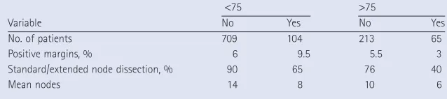
Memorial Sloan-Kettering Cancer Center, New York, NY, *Vanderbilt University Medical Center, Nashville, TN, and †University of Michigan Medical Center, Ann Arbor, MI, USATable 1 shows the surgical outcomes of margin status, extent of node dissection and average node counts, accounting for patient variability. Although the overall positive margin rate was 6.5%, margins were positive in 12% of patients with locally advanced disease. Older, and pre-treated, patients had less extensive node dissections and lower node counts than younger, healthier patients who had not received previous pelvic therapy. The mean (median) number of nodes examined for all patients was 12.5 (11), but varied widely among individual patients having anatomically similar node dissections. This variability could be from anatomical differences between individuals or could reflect differences in the method of pathological review. Using such benchmarks, we think that experienced surgeons who regularly perform cystectomy (at least 10 per year) should achieve negative surgical margins in >90% of cases and remove a mean of 10–14 nodes, recognizing that such standards will not be met in some of the most difficult cases.
C O M M E N T S
Second, are surgical standards important? We think they are for the outcome in individual patients, and for the design and evaluation of multimodal studies in bladder cancer. Who, where and how well surgery is performed could influence follow-up care, including adjuvant therapies. Recognized and accepted standards might also serve to elevate the overall quality of cystectomy in the future. This alone may prove to be as or more important than anticipated improvements in chemotherapy.
The practical issues involved in creating uniform standards for cystectomy can be formidable but are clearly surmountable. It is time for surgeons to count, and be counted.
REFERENCES
1 Stein JP, Skinner DG. Results with
radical cystectomy for treating bladder cancer: a ‘reference standard’ for high-grade, invasive bladder cancer. BJU Int 2003; 92: 12–5
2 Herr HW, Bochner BH, Dalbagni G et al.
Impact of the number of lymph nodes retrieved on outcome in patients with muscle invasive bladder cancer. J Urol 2003; 167: 1295–7
3 Grossman HB, Natale RB, Tangen CM
et al. Neoadjuvant chemotherapy plus
cystectomy compared with cystectomy alone for locally advanced bladder cancer.
N Engl J Med 2003; 349: 859–62
4 Herr HW, Faulkner JR, Grossman HB
et al. Surgical factors impact bladder
cancer outcomes: a cooperative group report. J Clin Oncol 2004; in press
5 Bladder Cancer Collaborative Group.
Standardization of radical cystectomy and pelvic lymph node dissection for bladder cancer: a collaborative group report.
J Urol 2004; 171: 1823–6
Correspondence: Harry W. Herr, Urology,
Memorial Sloan-Kettering Cancer Center, New York, USA.
e-mail: [email protected]
September 2004 944 Comment Article OSBORN et al.
SHOULD THE THERAPEUTIC APPROACH TO PROSTATE CANCER
WITH SEMINAL VESICLE INVASION BE REVIEWED: IMPROVING
FUNCTIONAL RESULTS WITHOUT DIMINISHING ONCOLOGICAL
OUTCOME?
JONATHAN R. OSBORN, ALISTAIR R. RAMSDEN*,
[image:2.595.69.388.122.193.2]GERALD W. CHODAK and RAJENDRA A. PERSAD†
–
Midwest Urology Research Foundation, Chicago, IL, USA, *Royal United Hospital, Bath, and †Bristol Royal Infirmary, Bristol, UKTABLE 1 Standards of radical cystectomy and pelvic node dissection, using data from the Bladder Cancer
Collaborative Group for 1091 patients aged <75 or >75 years with or with no previous treatment
Variable
<75 >75
No Yes No Yes
No. of patients 709 104 213 65
Positive margins, % 6 9.5 5.5 3
Standard/extended node dissection, % 90 65 76 40
Mean nodes 14 8 10 6
It is generally accepted that the presence of seminal vesicle invasion (SVI) in a resected specimen is an unfavourable prognostic factor in prostate cancer [1–3]. SVI has been reported in 5–13.6% of recent radical prostatectomy specimens [4–6]. Various studies have assessed the prediction of recurrence and prognosis in patients with
prostate cancer and SVI treated by radical prostatectomy. In these studies, factors influencing prognosis have been identified by retrospective analysis, and include preoperative PSA level, age, Gleason score (biopsy and resected specimen) and percentage of cancer in biopsy specimens [7–11].
In refining these criteria, Ramsden and Chodak [12] described a significant improvement in prognosis for men with SVI if the histology showed negative margins, unilateral SVI or perineural invasion. They concluded that this may be applied to patients with SVI to direct adjuvant therapy and guide postoperative counselling.
At the other end of the prognostic spectrum, what should the surgical approach be with regard to the SVs? It has been suggested that predicting SVI before surgery would enable surgeons to develop a SV-sparing prostatectomy, in the belief that this may reduce morbidity. It has been argued that preserving the SVs may result in less dissection close to the neurovascular bundle, with a theoretically lower risk of damaging it. It still has to be determined whether extirpation of the SVs changes the outcome. Some argue that SVI is the harbinger of metastatic disease, and therefore that any radical local treatment is destined to fail if SVI is present.
There are several counter-arguments; after the prostate has been removed the SVs serve no known purpose, and that the higher risk to the neurovascular bundle might be from dissection around the prostatic apex, rather than during SV dissection. In addition, there is a risk of leaving undetected carcinoma from isolated metastasis to the SV tip, even if the cut margins of the amputated SV are free from tumour. Type 3 SVI (isolated metastasis) is found in 13% of specimens containing SVI [13].
The current approach by some British surgeons is partial amputation of the SVs, as opposed to complete excision. This permits some pathological assessment of SVI, allowing an evaluation of the prognosis. Traditionally, unlike lymph node sampling, it was thought that resecting the SVs was of therapeutic rather than diagnostic value. It is likely that tumour metastasis to the tip of the SV would be missed by this technique. However, it is unlikely that SV dissection would be therapeutic if SVI is present.
C O M M E N T S
confidently predict patients at low risk of SVI before surgery with a guarantee of oncological safety, the current practice of extirpation of the seminal vesicles at radical prostatectomy should continue.
REFERENCES
1 Montie JE. Current prognostic factors for
prostate carcinoma. Cancer 1996; 78: 341–4
2 Ravery V, Boccon-Gibod LA,
Meulemans A et al. Predictive value of
pathological features for progression after radical prostatectomy. Eur Urol 1994; 26: 197–201
3 Mukamel E, de Kernion JB, Hannah J
et al. The incidence and significance of
seminal vesicle invasion. Cancer 1987; 59: 1535–8
4 Eastham JA, Kattan MW, Riedel E et al.
Variations among individual surgeons in the rate of positive surgical margins in radical prostatectomy specimens. J Urol 2003; 170: 2292–5
5 Zlotta AR, Djavan B, Petein M et al.
Prostate specific antigen density of the transition zone for predicting pathological stage of localized prostate cancer in patients with serum prostate specific antigen less than 10 ng/ml. J Urol 1998; 160: 283–6
6 Quinn DI, Henshall SM, Brenner PC et al.
Prognostic significance of preoperative factors in localized prostate carcinoma treated with radical prostatectomy: importance of percentage of biopsies that contain tumor and the presence of biopsy perineural invasion. Cancer 2003; 97: 1884–93
7 Bostwick DG, Qian J, Bergstralh E et al.
Prediction of capsular invasion and seminal vesicle invasion in prostate cancer. J Urol 1996; 155: 1361–7
8 Salomon L, Anastasiadis AG, Johnson
CW et al. Seminal vesicle involvement
after radical prostatectomy: predicting risk factors for progression. Urology 2003; 62: 304–9
9 Sofer M, Savoie M, Kim SS, Civantos
F, Soloway MS. Biochemical and
pathological predictors of the recurrence of prostatic adenocarcinoma with seminal vesicle invasion. J Urol 2003; 169: 153–6
10 Bloom KD, Richie JP, Schultz D,
Renshaw A, Saegaert T, D’amico AV. Invasion of seminal vesicles by adenocarcinoma of the prostate: PSA
outcome determined by preoperative and postoperative factors. Urology 2004; 63: 333–6
11 Koh H, Kattan MW, Scardino PT et al.
A nomogram to predict seminal vesicle invasion by the extent and location of cancer in systematic biopsy results. J Urol 2003; 170: 1203–8
12 Ramsden AR, Chodak GW. An analysis of
risk factors for biochemical progression in patients with seminal vesicle invasion. Validation of Kattans nomogram in a pathological sub group. BJU Int 2004; 94: 961–4
13 Ohori M, Scardino PT, Lapin SL,
Seale-Hawkins C, Link J, Wheeler TM. The
mechanisms and prognostic significance of seminal vesicle involvement by prostate cancer. Am J Surg Pathol 1993; 17: 1252–61
14 Terris MK, McNeal JE, Freiha FS,
Stamey TA. Efficacy of transrectal
ultrasound-guided seminal vesicle biopsies in the detection of seminal vesicle invasion by prostate cancer. J Urol 1993; 149: 1035–9
15 Allepuz Losa CA, Sanz Velez JI, Gil Sanz
MJ, Mas LP, Rioja Sanz LA. Seminal
vesicle biopsy in prostate cancer staging.
J Urol 1995; 154: 1407–11
16 Ohori M, Shinohara K, Wheeler TM et al.
Ultrasonic detection of non-palpable seminal vesicle invasion: a
clinicopathological study. Br J Urol 1993; 72: 799–808
17 Ikonen S, Karkkainen P, Kivisaari L et al.
Endorectal magnetic resonance imaging of prostatic cancer: comparison between fat-suppressed T2-weighted fast spin echo and three-dimensional dual-echo, steady-state sequences. Eur Radiol 2001; 11: 236–41
18 Rorvik J, Halvorsen OJ, Albrektsen G,
Ersland L, Daehlin L, Haukaas S. MRI
with an endorectal coil for staging of clinically localised prostate cancer prior to radical prostatectomy. Eur Radiol 1999; 9: 29–34
Correspondence: Alistair R. Ramsden, Urology,
Royal United Hospital, Bath, UK. e-mail: [email protected] September 2004
944 Comment Article DONOGHUE AND CREW
ADJUVANT TOPICAL TREATMENT OF UPPER URINARY TRACT
UROTHELIAL TUMOURS
–
JOHN P. O’DONOGHUE and JEREMY P. CREW
–
Department of Urology, The Churchill Hospital, Oxford, UK
INTRODUCTION
TCC of the ureter and renal pelvis is relatively uncommon and accounts for <5% of all cases of urothelial neoplasia. The standard treatment has been nephroureterectomy with excision of a cuff of bladder. The idea of organ-sparing surgery was first reported in 1945 for ureteric tumours [1]. With developments in endoscopic and percutaneous techniques, organ-sparing surgery is feasible in patients with inadequate renal reserve, at high risk of bilateral disease, or with significant comorbidity. Organ-sparing surgery is also suited to patients with low-grade disease and a normal contralateral kidney, because these lesions have a low risk of invasiveness and metastasis. Huben et al. [2] reported a median survival of 66.8 months for patients with low-grade vs 14.1 months for those with high-grade disease. In 83% of these patients, low- and high-grade tumours
matched low- and high-stage disease. Percutaneous treatment is possible when the ureteroscopic approach is difficult. Inaccessible infundibulo-calyceal tumours or large pelvic tumours are more readily treated by the percutaneous approach. This route may be necessary in patients where retrograde access is difficult because there are anatomical anomalies.
C O M M E N T S
adriamycin and interferon-a have also been tried. The agent can be instilled directly through a nephrostomy tube after the position has been checked by imaging, to prevent obstruction or extravasation. The agent is instilled via gravity and linked to a manometer so that the intrarenal pressure does not exceed 25 cmH2O [4]. Alternatively, it
may be given retrogradely via a ureteric catheter, by bladder instillation with the patient in the Trendelenberg position after inserting a ureteric stent, or via a urethral catheter in a patient with VUR [5].
Unfortunately, the results of intravesical treatment are varied. Herr [6] reported on a patient with a single kidney and a pT2G3 papillary tumour and multifocal carcinoma
in situ at the PUJ. After pelvectomy,
ureterectomy and autotransplantation the patient received six weekly courses of intravesical BCG. Thirteen months after surgery the patient was free of recurrence and had negative cytology. A report by Orihuela and Smith [7] found an 80% recurrence rate in patients who did not receive adjuvant BCG, vs 17% among those who did. However, a follow-up study showed no survival advantage with adjuvant immunotherapy [8]. Studer et al. [9] used Pasteur BCG in 10 renal units (eight patients) with cytological evidence of carcinoma in situ. In all but one patient the cytology became negative. A study by Martinez-Pineiro et al. [10] of 42 upper tracts treated solely by endourological means gave a recurrence rate of 24%. After intravesical adjuvant BCG or mitomycin C the recurrence rate was 12.5% and 14%, respectively (mean follow-up 30.6 months). Shoenberg et al. [11] reported on 10 patients with solitary kidneys and upper tract TCC; the patients received an intravesical BCG instillation as an adjunct to percutaneous resection, with no reported morbidity. Six of the patients had no recurrence, one had recurrence at 19 months, and one developed metastases at 15 months. One died from disease progression. Further studies using topical BCG after organ-sparing surgery are listed in Table 1 [12–16].
Mitomycin C is a cross-linking agent and in part inhibits DNA synthesis. Topical mitomycin C is well tolerated in the upper urinary tract and there are no reports of impairment in renal function after instillation. Smith et al. [17] reported on two patients with bladder cancer and VUR who developed lower ureteric recurrences. They received
intravesical treatment for 18 months, and after 2 years they were reported to be disease-free. Eastham and Huffman [4] reported on the use of mitomycin C as topical therapy after endoscopic resection of superficial TCC of the renal pelvis or ureter. Seven patients were treated over a 4-year period and were either not fit for more aggressive treatment or had a solitary kidney. Five patients had no evidence of disease whilst one had a marked decrease in tumour burden. Table 1 also shows other studies using mitomycin C [4,18].
After radical cystectomy, 2.4–8.5% of patients develop upper tract TCC [19]. Factors associated with a greater risk of recurrence after surgery include high grade and stage, multifocality, carcinoma in situ, and TCC in the prostatic urethra and distal ureter [20]. Recurrence in the upper tract after cystectomy is usually aggressive and has a poor prognosis. The treatment of choice is nephroureterectomy but in patients with bilateral disease or a solitary kidney, and low-grade/low-stage disease, conservative treatment has been used; whilst the results appear encouraging, the follow-up is short and patients few [21].
Upper urinary tract tumours are rare and the most appropriate treatment for low-grade tumours is not entirely clear. The results appear encouraging but no individual study has shown a statistical improvement in survival and recurrence rates. What is known about intravesical chemotherapeutic and immunological treatments after conservative surgery is from retrospective studies with small cohorts of patients and with heterogeneous characteristics. The reasons
for inconclusive results include; (i) too few patients to show clinical significance; (ii) inadequate contact time between the urothelium and the agent being instilled; and (iii) possible differences between upper tract tumours and bladder tumours. Large prospective multicentre studies are needed to clarify the situation. Meanwhile, ablative surgery remains the standard for managing upper tract TCC but in circumstances where this may be problematic, conservative resection with adjuvant topical
chemotherapy/immunotherapy is an option.
REFERENCES
1 West SA. Conservative surgery in certain
benign tumours of the ureter. J Urol 1945;
53: 97
2 Huben RP, Mounzer AM, Murphy GP.
Tumour grade and stage as prognostic variables in upper tract urothelial tumours. Cancer 1998; 62: 2016–20
3 Wallace DMA, Wallace DM, Whitfield
HN, Hendry WG, Wickham JEA. The late
results of conservative surgery for upper tract urothelial carcinomas. Br J Urol 1981; 53: 537–41
4 Eastham JA, Huffman JL. Technique of
mitomycin C instillation in the treatment of upper urinary tract urothelial tumours.
J Urol 1993; 150: 324–5
5 Jabbour ME, Smith AD. Primary
percutaneous approach to upper urinary tract transitional cell carcinoma. Urol Clin North Am 2000; 27: 739–50
6 Herr HW. Durable response of a
[image:4.595.240.560.109.246.2]carcinoma in situ of the renal pelvis to topical Bacillus Calmette Guerin. J Urol 1985; 134: 531–2
TABLE 1 Studies using adjuvant topical BCG or adjuvant mitomycin C after organ-sparing surgery
Ref N patients Regimen Tumour stage/grade Recurrence rate (%) Follow-up, months
BCG
[12] 17 Retrograde Ta/T1/CIS/G2–G4 28.5 11–64
[13] 14 Percutaneous NG 12.5 NG
[14] 13 Percutaneous Ta/G1–G2 13 6–36
[15] 8 Percutaneous NG 12.5 9–59
[16] 9 Retrograde NG 22 4–41
Mitomycin C
[4] 7 Percutaneous NG 28.5 1–12
[18] 20 Retrograde G1–G3 54 30
C O M M E N T S
7 Orihuela E, Smith AD. Percutaneous
treatment of transitional cell carcinoma of the upper urinary tract. Urol Clin North Am 1988; 15: 425–31
8 Jarrett TW, Sweetser PM, Weiss GH,
Smith AD. Percutaneous management of transitional cell carcinoma of the renal collecting system: 9-year experience. J Urol 1995; 154: 1629
9 Studer UE, Casanova G, Kraft R, Zingg
EJ. Percutaneous Bacillus Calmette-Guerin perfusion of the upper urinary tract for carcinoma in situ. J Urol 1989; 142: 975–7
10 Martinez-Pineiro JA, Matres MJG,
Martinez-Pineiro L. Endourological treatment of upper tract urothelial carcinomas; analysis of a series of 59 tumours. J Urol 1996; 156: 377–85
11 Shoenberg MP, Van Arsdalen KN, Wein
A J. The management of transitional cell carcinoma in solitary renal units. J Urol 1991; 146: 700–3
12 Sharpe JR, Duffy G, Chin JL. Intrarenal
bacillus Calmette Guerin therapy for upper urinary tract carcinoma in situ. J Urol 1993; 149: 457–60
13 Bellman GC, Sweetser P, Smith AD.
Complications of intracavitary bacillus Calmette Guerin after percutaneous resection of upper urinary tract transitional cell carcinoma. J Urol 1994; 151: 13–5
14 Patel A, Fuchs GJ. New techniques for
the administration of topical adjuvant therapy after endoscopic ablation of upper urinary tract transitional cell carcinoma. J Urol 1998; 159: 71–5
15 Vasavada SP, Streem SB, Novick AC.
Definitive tumour resection and percutaneous bacillus Calmette Guerin for management of renal pelvic transitional cell carcinoma in solitary kidneys. Urology 1995; 45: 381–6
16 Nonomura N, Ono Y, Nozawa M et al.
Bacillus Calmette Guerin perfusion therapy for the treatment of transitional cell carcinoma in situ of the upper urinary tract. Eur Urol 2000; 38: 701–5
17 Smith AD, Orihuela E, Crowley AR.
Percutaneous management of renal pelvic tumours: a treatment option in selected cases. J Urol 1987; 137: 852–6
18 Keeley FX Jr, Bagley DH. Adjuvant
mitomycin C following endoscopic treatment of upper tract transitional cell carcinoma. J Urol 1997; 158: 2074–7
19 Balaji KC, McGuire M, Grotas J,
Grimaldi G, Russo P. Upper tract recurrences following radical cystectomy. an analysis of prognostic factors, recurrence pattern and stage at presentation. J Urol 1999; 162: 1603–6
20 Malkowicz SB, Skinner DG.
Development of upper tract carcinoma after cystectomy for bladder carcinoma. Urology 1990; 36: 20–2
21 Braslis KG, Soloway MS. Management
of ureteral and renal pelvic recurrence after cystectomy. Urol Clin North Am 1994; 21: 653–9
Correspondence: John P. O’Donoghue,
Department of Urology, Churchill Hospital, Headington, Oxford, OX3 7L J, UK. e-mail: [email protected]
September 2004 944 Comment Article HAMM ET AL.
RED-CELL SALVAGE IN UROLOGICAL SURGERY
REBECCA S. HAMM,
MARK DAUGHERTY and MALCOLM C. CRUNDWELL – Department of Urology, Royal
Devon and Exeter NHS Trust, Exeter, Devon, UKSignificant blood loss during major urological surgery is common and in the UK is usually replaced by transfusion of donated homologous blood. This is not always entirely satisfactory because of the increasingly limited availability and the well-documented risks associated with homologous blood transfusion. Most recently concerns have been raised about possible contamination of donated blood with variant Creuzfeldt-Jakob disease [1] which has caused the Blood
Transfusion Service to discontinue donations from those who have received a blood transfusion since 1 January 1980. This is likely to have a significant affect on the stocks of donated blood and means that alternatives to homologous blood transfusion should be actively pursued.
Autologous blood transfusion is the collection of blood from an individual for the purpose of re-infusion into the same individual at a later
time. Autologous blood transfusion can be in the form of preoperative autologous blood donation (PABD), intraoperative cell salvage (IOCS) or postoperative cell salvage and acute normovolaemic haemodilution (ANH).
The first PABD was described in 1921 by F.C. Grant in a patient undergoing surgery for a cerebellar tumour. PABD became standard medical practice in the 1920s and 30s and was frequently used by Cushing during cranial surgery. It was not until World War II that transfusion practice changed and homologous products became readily available.
Cell salvage with direct reinfusion was described in haemothorax cases in 1931 by Brown and Debenheim. In 1943 Arnold Griswold developed the first cell salvage auto-transfusion device, collecting blood in a bottle by suction, straining it through a cheese cloth and then re-infusing it. Modern cell salvage techniques still use this basic principle.
In ANH, 1–3 units of whole blood are drawn from the patient after the onset of anaesthesia and the volume is replaced with colloid or crystalloid volume expanders. Any blood lost during surgery is therefore more dilute and the withdrawn blood can be re-infused at the end of the procedure.
In modern cell salvage techniques (IOCS) salvaged blood is aspirated from the surgical field, anticoagulated in the suction device and then collected in a sterile collection container. When an adequate amount of blood has been collected it is pumped into a spinning centrifuge bowl. Red cells, being the heaviest components of blood, collect at the lowest point in the bowl and supernatant containing the other components of blood spill over into a waste bag. Sterile saline solution is pumped through the spinning centrifuge bowl and displaces the lighter remaining contaminants. Once this process is complete the red blood cells are re-suspended and can be transfused, usually immediately, or can be stored under specific conditions for up to 6 h.
C O M M E N T S
field, be taken into the cell salvage machine, and because they are heavy like red cells, returned to the patient. The use of leukocyte depletion filters can reduce the risk of such occurrence but concerns remain as to whether use of IOCS may compromise surgical cure. Malignant cells have been found in the circulation during surgery even when IOCS is not used [2], but the significance of these circulating cells is not clear. If re-infused cells were clinically significant it would be expected that patients having had IOCS might present with early widespread metastases, or at least unexpected lung metastases. This has not proven to be the case in bladder, renal or prostate cancer. The four reports in Table 1 [3–6] illustrate experience of nearly 250 cases of IOCS, with no cases of early unexpected metastasis.
Since 1996 it has been the policy in our unit to have IOCS available at all major open urological surgery. We have experience of >150 cases, including 74 cystectomies, 30 radical nephrectomies and 37 radical prostatectomies, and have transfused over 600 units of salvaged blood. There have been no cases of early lung or diffuse metastases suggestive of spread of malignant disease by IOCS.
The theoretical risk of metastatic spread secondary to IOCS has not become apparent, although the very real risk of spread of viral infection and other complications of homologous transfusion remain.
REFERENCES
1 Llewelyn CA, Hewitt PE, Knight RSG
et al. Possible transmission of variant Creutzfeldt-Jakob disease by blood transfusion. Lancet 2004; 363: 417–21
2 Oefelein MG, Kaul K, Herz B et al.
Molecular detection of prostate epithelial cells from the surgical field and peripheral circulation during radical prostatectomy. J Urol 1996; 155: 238–42
3 Klimberg I, Sirois R, Wajsman Z, Baker
J. Intraoperative autotransfusion in urologic cancer. Arch Surg 1986; 121: 1326–9
4 Hart O, Klimberg I, Wajsman Z.
Intraoperative autotransfusion in
radical cystectomy for carcinoma of the bladder. Surg Gynae Obs 1989; 168: 302– 6
5 Davis M, Sofer M, Gomez-Marin O,
Bruck D, Soloway M. The use of cell salvage during radical retropubic prostatectomy: does it influence cancer recurrence. BJU Int 2003; 91: 474–6
6 Gray C, Amling C, Polston G, Powell C,
Kane C. Intraoperative cell salvage in radical retropubic prostatectomy. Urology 2001; 58: 740–5
Correspondence: Rebecca S. Hamm,
[image:6.595.239.557.97.255.2]Department of Urology, Royal Devon and Exeter NHS Trust, Exeter, Devon, UK. e-mail: [email protected]
TABLE 1 Reports of IOCS in urology
Ref Date N patients Operation Outcome Comment
[3] 1986 24 RC Two died from local recurrence, LN met IOCS is safe
10 RP One pelvic recurrence
13 RN Two lung metastases
[4] 1989 49 RC Six patients died by follow-up mean
23.8 months
Overlap of patients with above series
[5] 2003 87 RP No difference in outcome for IOCS over
pre-donation or no transfusion
IOCS is safe
[6] 2001 62 RP No difference in progression-free survival
with 101 patients who pre-donated blood
No increased risk of early biochemical progression