Molecular Microbiology (2005) 58(2), 349–357 doi:10.1111/j.1365-2958.2005.04842.x First published online 9 September 2005 Blackwell Science, LtdOxford, UKMMIMolecular Microbiology0950-382XBlackwell Publishing Ltd, 2005? 2005582349357Review ArticleStructure and function of chaperone SecBJ. Zhou and Z. Xu
Accepted 5 August, 2005. *For correspondence. E-mail zhaohui@ umich.edu; Tel. (+1) 734 615 2077; Fax (+1) 734 763 6492.
MicroReview
The structural view of bacterial translocation-specific
chaperone SecB: implications for function
Jiahai Zhou and Zhaohui Xu*
Department of Biological Chemistry, Medical School and Life Sciences Institute, University of Michigan, 210 Washtenaw Avenue, Ann Arbor, MI 48109-2216, USA.
Summary
SecB is a molecular chaperone that functions in bacterial post-translational protein translocation pathway. It maintains newly synthesized precursor polypeptide chains in a translocation-competent state and guides them to the translocon via its high-affinity binding to the ligand as well as to the membrane-embedded ATPase SecA. Recent advances in eluci-dating the structures of SecB have enabled the exam-ination of protein function in the structural context. Structures of SecB from both Haemophilus influenzae
and Escherichia coli support the early two-subsite polypeptide-binding model. In addition, the detailed molecular interaction between SecB and SecA was revealed by a structure of SecB in complex with the C-terminal zinc-containing domain of SecA. These observations explain the dual role of SecB plays in the translocation pathway, as a molecular chaperone and a specific targeting factor. A model of SecB–SecA complex suggests that the binding of SecA to SecB changes the conformation of the polypeptide binding sites in the chaperone, enabling transfer of precursor polypeptides from SecB to SecA. Recent studies also show the presence of a second zinc-independent SecB binding site in SecA and the new interaction might contribute to the function of SecB.
Introduction
The role of molecular chaperones in protein folding has been well established over the last decade. In general, molecular chaperones have a particular affinity for non-native conformations that exist in protein folding interme-diates. Interaction between folding intermediates and
chaperone stabilizes folding intermediates and prevents them from aggregation. Molecular chaperones also play important roles in protein translocation because the majority of proteins are translocated across the mem-brane as unfolded polypeptides. Therefore, it is important for them to be kept in an unfolded state to remain compe-tent for translocation.
SecB is a molecular chaperone specialized in the post-translational protein translocation pathway of some proteobacteria. It binds to newly synthesized precursor polypeptides (preproteins) and stabilizes them in an unfolded and non-aggregated state after they exit from the ribosome translation tunnel (Lecker et al., 1989; 1990; Liu et al., 1989; Breukink et al., 1992). It also delivers prepro-teins to the membrane-embedded translocon via its spe-cific interaction with SecA, an ATPase that provides part of the actual energy for translocation (Hartl et al., 1990; de Cock and Tommassen, 1992; Hoffschulte et al., 1994; Fekkes et al., 1998). The role of SecB in preprotein export was first revealed by Kumamoto and Beckwith (1983;
1985) when they found that secB mutations resulted in
translocation defects for a subset of secretory proteins in Escherichia coli. Extensive work has been carried out on SecB as its first identification (Weiss et al., 1988; Kuma-moto, 1989). Several excellent reviews on the function of SecB have appeared in the past few years (Randall and Hardy, 1995; 2002; Fekkes and Driessen, 1999; Kim and Kendall, 2000; Driessen, 2001; Driessen et al., 2001). In this review, we will focus on advances in our understand-ing of the molecular mechanism of SecB function based on recent structural analyses (Xu et al., 2000; Dekker et al., 2003; Zhou and Xu, 2003).
SecB structure
-350 J. Zhou and Z. Xu
© 2005 The Authors
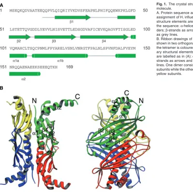
helices. The dimer is a flat molecule formed by a 180°
rotation of one monomer with respect to the other and pairing them together via strand β1 and helix α1. As a result of the pairing, there is a surface-exposed antiparal-lel β-sheet on one face of the SecB dimer. The dimer is mainly stabilized by main chain hydrogen bonds between the two antiparallel β1 strands from the two monomers. Two SecB dimers associate to form a tetramer by sand-wiching four long α1 helices between the eight-stranded antiparallel β-sheets. The tetramer is very stable, with an estimated tetramer–dimer equilibrium constant at pH 7.6 well below 20 nM (Muren et al., 1999). The dimer–dimer interface is stabilized by polar interactions involving side-chains from the four α1 helices.
The amino acid sequence identity between E. coli SecB and H. influenzae SecB is 59% and, as expected, the two structures are very similar. Differences between them are found mainly in the loop regions. There is a two-residue difference in the connecting loop between strands β3 and
β4: residues 76–79 in H. influenzae SecB and residues 69–70 in E. coli SecB (Xu et al., 2000; Dekker et al., 2003). This flexible β-hairpin region is close to the nega-tively charged SecA-binding surface of SecB (see below) and may affect interaction with SecA.
The extreme C-terminus of SecB subunits is not visible in the electron density maps of either of the crystal struc-tures. Proton nuclear magnetic resonance (NMR) spec-troscopy demonstrated that this region is highly mobile (Volkert et al., 1999). Deletion of the C-terminal tail pro-duces a stable truncated SecB protein that retains its ability to bind to non-native polypeptide in vitro but causes a defect in protein export when overproduced in vivo. The defect can be alleviated by overproduction of SecA, sug-gesting that the C-terminal tail of SecB may interact with SecA. Deletion of the C-terminal tail also leads to a two-fold decrease in affinity for non-native polypeptide (Dia-mond and Randall, 1997). As peptide binding to SecB protects this tail region from proteolysis (Randall, 1992),
A
[image:2.595.59.458.50.447.2]B
Fig. 1. The crystal structure of the SecB molecule.
A. Protein sequence and secondary structure assignment of H. influenzae SecB. Secondary structure elements are indicated underneath the sequence: α-helices are drawn as cylin-ders; β-strands as arrows; and other elements as grey lines.
Structure and function of chaperone SecB 351
it is likely that the tail contributes to peptide binding. A structure of SecB–peptide complex will provide the nec-essary molecular details about how the C-terminal tail of SecB packs against the rest of the structure and regulates the preprotein translocation process.
Polypeptide binding
In the Sec-dependent pathway, protein translocation can-not occur if newly synthesized polypeptide chains are either folded or aggregated (Randall and Hardy, 1986; Weiss et al., 1988; de Cock et al., 1992). To keep newly synthesized proteins in the translocation-competent state, SecB recognizes the non-native conformation within the mature regions of preproteins and binds with high affinity (the dissociation constant is around 5 nM to 50 nM) (Ran-dall and Hardy, 1995; Ran(Ran-dall et al., 1997; 1998; Topping and Randall, 1997). Although SecB does not directly bind to the leader/signal sequence of preproteins (Randall et al., 1990), the presence of this sequence is neverthe-less crucial for export. First, the leader sequence can significantly slow down the rate of spontaneous folding of preproteins and, thereby, increase the probability of bind-ing by SecB (Hardy and Randall, 1991). Second, leader sequences bind to a specific domain of SecA and are thought to be important for the transfer of preproteins from SecB to SecA (Lill et al., 1990; Fekkes et al., 1998; Baud et al., 2002).
Randall and colleagues carried out extensive studies to define the SecB-binding frame within the preprotein. By analysing proteolytic digestion fragments of complexes between SecB and its natural ligands maltose-binding protein, galactose-binding protein or oligopeptide-binding
protein, the SecB-binding frame was found to be located in the mature region of the three preproteins and to span a stretch of approximately 150–170 residues (Topping and Randall, 1994; Khisty et al., 1995; Randall and Hardy, 1995; Smith et al., 1997). Based on their early studies of a SecB-binding peptide library (Randall, 1992), a model was proposed for the interaction of SecB with its ligand (Randall and Hardy, 1995). The model suggests that there are two types of peptide-binding motifs or structures that can be bound by SecB: flexible stretches of polypeptide of approximately 15 residues in length and exposed hydrophobic regions within the non-native polypeptide. The initial interaction occurs at the extended and flexible binding site. Saturation of these binding sites induces a conformational change in SecB that leads to exposure of hydrophobic sites for ligand binding.
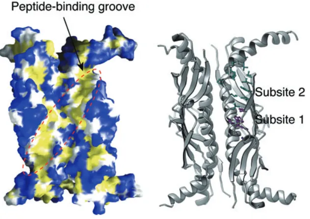
Consistent with Randall’s hypothesis (Randall and Hardy, 1995), it was proposed that there are two peptide binding subsites in SecB based on the crystal structure of H. influenzae protein (Fig. 2) (Xu et al., 2000). A similar situation was observed more recently in the crystal struc-ture of E. coli SecB (Dekker et al., 2003). Subsite 1 cor-responds to the deep section of the peptide-binding channel of SecB and may recognize the hydrophobic and aromatic region within the non-native polypeptide because most of the residues lining subsite 1 are aromatic and conserved. Conformational variation among the dif-ferent subunits of SecB suggests that this region is struc-turally flexible and provides a possible explanation for the necessary plasticity for peptide binding. Subsite 1 is large enough to accommodate a hypothesized nine-residue ‘SecB-binding motif’ (Knoblauch et al., 1999). Subsite 2 is much shallower and more open than subsite 1. It is
[image:3.595.59.375.477.701.2]352 J. Zhou and Z. Xu
© 2005 The Authors fore able to accommodate a more extended region within
the non-native polypeptide. In the crystal structure of H. influenzae SecB, the N-terminal region of a neighbouring molecule in the crystal lattice inserts itself into subsite 2 (J. Zhou and Z. Xu, unpubl. results). It interacts with sub-site 2 by adopting an extended conformation and forming regular main chain hydrogen bonds with strand β2 of the β-sheet. The pattern of interaction likely holds true for the non-native polypeptide ligand.
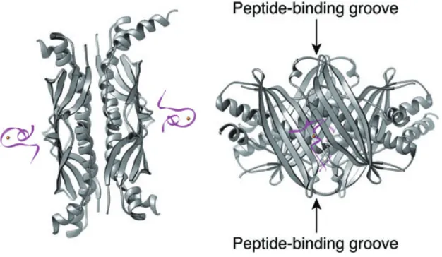
The molecular symmetry within SecB gives rise to four identical binding sites. While affinity for individual sites might be low, simultaneous occupancy of multiple binding sites will ensure high-affinity binding of polypeptide to SecB. As mentioned above, the SecB-binding frame of its natural ligands spans a stretch of approximately 150–170 residues (Topping and Randall, 1994; Khisty et al., 1995; Smith et al., 1997). While the same SecB molecule does not necessarily occupy the entire stretch of residues, one can speculate that the long unstructured polypeptide seg-ments might wrap around the chaperone to occupy the binding sites on both sides of SecB. It is sufficiently long to loop out from one side through either the path across the top of chaperone or the path across the side of the chaperone. In both cases, the ligand makes close contact with the extreme C-terminal part of SecB. This could explain why polypeptide binding protects these protease-sensitive C-terminal tails (Randall, 1992) and why deletion of the tail part of SecB decreases its affinity for polypep-tide ligands (Diamond and Randall, 1997).
SecA recognition
SecB directs the bound preprotein into the translocation pathway via its specific interaction with membrane-bound SecA (Fekkes et al., 1998). SecB binds to SecA with low affinity in solution (dissociation constant is about 1.6 μM) (den Blaauwen et al., 1997). The binding affinity increases
significantly when SecA is bound to the membrane-embedded translocon SecYEG complex (the dissociation constant is 10–30 nM) (Fekkes et al., 1997). The binding is even tighter (dissociation constant around 10 nM) if SecB is loaded with a polypeptide ligand (Hartl et al., 1990; Fekkes et al., 1997). The SecB binding site on SecA is localized primarily at the extreme C-terminus of SecA, although additional sites have also been suggested (Woodbury et al., 2000) (see below for more discussion). Removal of the C-terminal 22 residues of SecA causes a deficiency in SecB-mediated preprotein translocation (Fekkes et al., 1997).
The crystal structure of H. influenzae SecB in complex with the last 27 C-terminal residues of H. influenzae SecA (SecAc) provides details about the molecular interaction between the two proteins (Fig. 3) (Zhou and Xu, 2003). SecB uses the solvent-exposed surface of the eight-stranded β-sheet formed by two of the four subunits to interact with one SecAc peptide. In the crystal structure, the SecAc peptide is mainly stabilized by a bound zinc atom. The zinc atom is co-ordinated by three highly con-served cysteines and a histidine. Substitution of these residues in SecA by serine abolishes the ability of SecB to promote preprotein translocation (Rajapandi and Oliver, 1994). The well-structured SecA C-terminal region is nec-essary for SecB interaction, as the interaction is disrupted by treatment of SecA with a zinc chelator and restored by the addition of ZnCl2 (Fekkes et al., 1999). Two recent
solution NMR structures of E. coli SecAc peptide suggest that structural changes in SecAc upon binding to SecB
are minimal (Dempsey et al., 2004; Matousek and
[image:4.595.59.372.519.701.2]Alex-andrescu, 2004). The crystal structure also shows four residues in H. influenzae SecAc, Arg878, Asn879, Lys889 and Lys879, contribute significantly to the binding of SecB. Replacement of any of these residues by alanine in SecA abolishes its binding to SecB (Zhou and Xu, 2003).
Structure and function of chaperone SecB 353
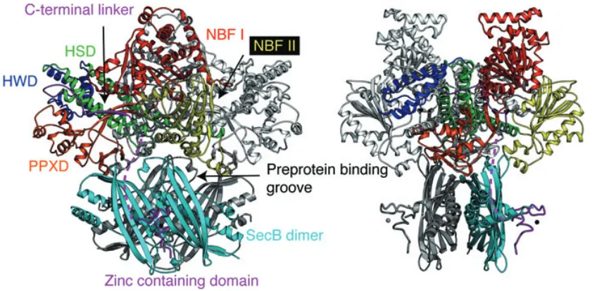
To visualize how SecB might interact with SecA, the crystal structure of Bacillus subtilis SecA (Hunt et al., 2002) was manually docked onto the structure of H. influenzae SecB–SecAc complex (Fig. 4) (Zhou and Xu, 2003). Although B. subtilis does not possess SecB, high sequence identity (46%) between SecA molecules from B. subtilis and H. influenzae suggests that the two proteins are likely to share a very similar overall structure. In addition, a hybrid ‘B. subtilis SecA’ with its C-terminal 27 residues substituted with the corresponding sequence from H. influenzae SecA binds to H. influenzae SecB (J. Zhou and Z. Xu, unpubl. results). Three major constraints were used in the docking procedure. First, the C-terminal tails of SecA (not seen in any of the three published
structures: Hunt et al., 2002; Sharma et al., 2003;
Osborne et al., 2004) project into a space beneath the core of the SecA dimer based on the positions of the observed C-terminal ends in the SecA structure. Second, a symmetric complex is assumed to form between SecB and SecA. This requires that the twofold axis of SecA dimer be aligned with the twofold axis relating the two SecA-binding surfaces within a SecB tetramer. Third, the distance between the observed C-terminal ends in the SecA structure and the start of the SecAc segments needs to fit about 15 amino acids missing between these two points of connection. If these 15 amino acids all adopt extended backbone conformations, they could extend over a span of about 50 Å. In this model, the distance between the two points of connection (dashed line in
Fig. 4) is 37 Å. Although the docking procedure is only approximate, the resulting model allows us to speculate about the interaction between SecB and SecA on a struc-tural term.
The crystal structure of SecA (Fig. 4) (Hunt et al., 2002; Sharma et al., 2003; Osborne et al., 2004) shows that it contains two nucleotide-binding motifs (NBF I and NBF II) with a preprotein cross-linking domain (PPXD)
(Kimura et al., 1991) inserted in between at the
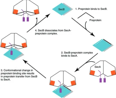
[image:5.595.88.512.420.627.2]N-terminal two-thirds of the sequence. The C-N-terminal one-third of the sequence consists of the α-helical scaffold (HSD) and the helical wings domains (HWD) as well as the extreme C-terminal zinc-containing domain. As shown in Figs 4 and 5, SecB–preprotein complex binds to SecA from the bottom direction via the zinc-binding domains of SecA (step 2 in Fig. 5). One of the SecB polypeptide-binding grooves located on the top of the molecule is sandwiched between SecB and SecA. This positions SecB-bound preprotein directly beneath the SecA molecule and near its preprotein cross-linking domain, which makes direct preprotein transfer feasible. One could imagine that the simultaneous binding of the C-terminal tails of the SecA dimer to SecB changes the relative orientation of the two eight-stranded β-sheets in SecB, and therefore the conformation of the polypeptide-binding groove. This could lead to a decrease in the affin-ity of SecB for the bound preprotein, resulting in the release of preprotein from SecB and subsequent transfer to SecA (step 3 in Fig. 5).
© 2005 The Authors The proposed SecA–SecB interaction model assumes
that SecA symmetrically interacts with SecB as a dimer and the interaction is mediated via the C-terminal zinc-containing domain. This is supported by the SecAc–SecB complex structure as well as binding studies using various SecA and SecB mutants (Fekkes et al., 1997; 1999; Zhou and Xu, 2003). In particular, both C-terminal tails of SecA dimer appear to be necessary to form a tight SecA–SecB complex as heterodimers of wild-type SecA and a C-terminal truncated form of SecA are defective in SecB binding (Fekkes et al., 1997). Recent studies using chi-meric SecA proteins also suggest that the C-terminal tail of SecA is necessary and sufficient to mediate the specific interaction between SecB and SecA. B. subtilis lacks a SecB proteins and its SecA does not bind to either E. coli or H. influenzae SecB. By genetically switching the C-terminal tail of B. subtilis SecA to the corresponding part from either E. coli SecA or H. influenzae SecA, the new hybrid ‘B. subtilis SecA’ can now bind to the cognate SecB, depending on which tail the chimeric protein con-tains (J. Zhou and Z. Xu, unpubl. data).
In addition to the above-mentioned interaction, Randall and colleagues discovered a second site of SecA–SecB interaction. By analysing complexes formed between E. coli SecA and variants of E. coli SecB, they found that the translocation defective variants of SecB still form a com-plex with SecA, even though they cannot interact with the C-terminal tail of SecA (Woodbury et al., 2000). This hypothesis was further supported by their titration calorim-etry and sedimentation velocity centrifugation data
(Ran-dall et al., 2004). The second binding site was now
[image:6.595.111.484.56.373.2]Structure and function of chaperone SecB 355
must be weakened to ensure the release of SecB from the membrane. This would facilitate the C-terminal helix of SecB to interact with the dimer interface of SecA. It is possible that the interaction then weakens the SecA dimer and leads to conformational changes in SecA, which might also contribute to the transfer of preprotein from SecB to SecA (Jilaveanu et al., 2005; Or et al., 2005; Randall et al., 2005). To fully address the functional role of the C-terminal independent SecA–SecB interaction will require the determination of its structure.
Other functions of SecB
Besides its function in the Sec-dependent protein translo-cation pathway, SecB is also the chaperone for the secre-tion of the HasA haemophore through the type I protein translocation pathway in Serratia marcescens (Delepel-aire and Wandersman, 1998; Sapriel et al., 2003). HasA is a protein secreted by the Gram-negative bacteria under iron starvation conditions to assist the utilization of exter-nal haem as an iron source. Efficient secretion of HasA requires the protein in an unfolded state, which is main-tained by the binding of SecB. In vitro studies have shown that SecB specifically binds to unfolded HasA and slows down its folding rate significantly (Wolff et al., 2003). Therefore, it is likely that SecB uses the hydrophobic polypeptide-binding groove to recognize unfolded HasA. Elimination of SecB in S. marcescens affects the secretion of both HasA and proteins translocated through the Sec-dependent pathway (Sapriel et al., 2003). Although the chaperoning mechanism of SecB in the two pathways appears to be similar, the downstream interaction partners are different. The SecB residues implicated in SecA bind-ing are not important for HasA secretion. Further bio-chemical and structural characterization of the type I pathway-specific protein–protein interactions in SecB will help elucidate the mechanism.
SecB may also act as a general chaperone, as it can bind to denatured luciferase and facilitates the subse-quent refolding of the protein by the DnaK/DnaJ system
(Knoblauch et al., 1999). Recent studies showed that
overproduction of SecB can suppress the temperature-sensitive and the aggregation-prone phenotypes caused by elimination of both DnaK and trigger factor in E. coli (Ullers et al. 2004). It should be noted that this reflects a rather aberrant condition in which the cognate folding machinery is highly compromised, while a rescue is observed only upon overproduction of SecB. Whether this reflects a genuine in vivo function of SecB remains to be seen. The structural basis for this activity of SecB is unknown but likely involves the polypeptide binding site used in protein translocation pathway.
SecB has also been shown to interact directly with bacterial ribosome-bound chaperone trigger factor (Ha
et al., 2004). The dissociation constant between E. coli SecB and E. coli trigger factor is ∼6 μM, as determined by surface plasmon resonance. However, complex forma-tion between the two proteins was not observed in a gel filtration chromatography experiment using purified mate-rials. Isothermal titration calorimetry suggested that the dissociation constant is much lower (in the mM range) (J. Zhou and Z. Xu, unpubl. results). If they do interact, which part of SecB structure is involved in interaction? Little is known for the physiological role this interaction might play in vivo. Could it be possible that SecB receives the newly synthesized preprotein from trigger factor rather than through random collision in the cytosol? Clearly, further studies are necessary to clarify these issues.
Conclusions
The recent crystal structures of SecB and its complex with the C-terminal tail of SecA have shed new light on the molecular mechanism by which the small bacterial chaperone SecB functions in protein translocation. SecB employs hydrophobic, solvent-exposed surfaces to stabi-lize the non-native conformation that exists in preproteins. The predominant interactions between SecB and prepro-teins involve non-specific main chain hydrogen bonds and hydrophobic interactions. As SecB does not seem to rec-ognize any particular sequence motifs within preproteins, this structural feature of SecB ensures that different pre-protein ligands can be recognized. In contrast, the inter-action between SecB and SecA is highly specific. Not surprisingly, the protein–protein interface within the SecB– SecA complex comprises highly conserved residues. Interactions between the two proteins are mediated by specific side-chain hydrogen bonds. Replacement of these residues by alanines has a drastic effect on the stability of the molecular complex.
© 2005 The Authors Institutes of Health Grant (GM60997). Z.X. is a University of
Michigan Biological Sciences Scholar and a Pew Scholar in Biomedical Sciences.
References
Baud, C., Karamanou, S., Sianidis, G., Vrontou, E., Politou, A.S., and Economou, A. (2002) Allosteric communication between signal peptides and the SecA protein DEAD motor ATPase domain. J Biol Chem277: 13724–13731. den Blaauwen, T., Terpetschnig, E., Lakowicz, J.R., and
Driessen, A.J. (1997) Interaction of SecB with soluble SecA. FEBS Lett416: 35–38.
Breukink, E., Kusters, R., and De Kruijff, B. (1992) In-vitro
studies on the folding characteristics of the Escherichia coli
precursor protein prePhoE. Evidence that SecB prevents the precursor from aggregating by forming a functional complex. Eur J Biochem208: 419–425.
de Cock, H., and Tommassen, J. (1992) SecB-binding does not maintain the translocation-competent state of prePhoE.
Mol Microbiol6: 599–604.
de Cock, H., Overeem, W., and Tommassen, J. (1992) Bio-genesis of outer membrane protein PhoE of Escherichia coli. Evidence for multiple SecB-binding sites in the mature portion of the PhoE protein. J Mol Biol224: 369–379. Dekker, C., de Kruijff, B., and Gros, P. (2003) Crystal
struc-ture of SecB from Escherichia coli. J Struct Biol144: 313– 319.
Delepelaire, P., and Wandersman, C. (1998) The SecB chap-erone is involved in the secretion of the Serratia marce-scens HasA protein through an ABC transporter. EMBO J
17: 936–944.
Dempsey, B.R., Wrona, M., Moulin, J.M., Gloor, G.B., Jalile-hvand, F., Lajoie, G., et al. (2004) Solution NMR structure and X-ray absorption analysis of the C-terminal zinc-binding domain of the SecA ATPase. Biochemistry 43: 9361–9371.
Diamond, D.L., and Randall, L.L. (1997) Kinetic partitioning. Poising SecB to favor association with a rapidly folding ligand. J Biol Chem272: 28994–28998.
Driessen, A.J. (2001) SecB, a molecular chaperone with two faces. Trends Microbiol9: 193–196.
Driessen, A.J., Manting, E.H., and van der Does, C. (2001) The structural basis of protein targeting and translocation in bacteria. Nat Struct Biol8: 492–498.
Fekkes, P., and Driessen, A.J. (1999) Protein targeting to the bacterial cytoplasmic membrane. Microbiol Mol Biol Rev
63: 161–173.
Fekkes, P., van der Does, C., and Driessen, A.J. (1997) The molecular chaperone SecB is released from the carboxy-terminus of SecA during initiation of precursor protein translocation. EMBO J16: 6105–6113.
Fekkes, P., de Wit, J.G., van der Wolk, J.P., Kimsey, H.H., Kumamoto, C.A., and Driessen, A.J. (1998) Preprotein transfer to the Escherichia coli translocase requires the co-operative binding of SecB and the signal sequence to SecA. Mol Microbiol29: 1179–1190.
Fekkes, P., de Wit, J.G., Boorsma, A., Friesen, R.H., and
tional identification of the SecB homologue in Methanococ-cus jannaschii and direct interaction of SecB with trigger factor. Biochem Biophys Res Commun315: 1039–1044. Hardy, S.J., and Randall, L.L. (1991) A kinetic partitioning
model of selective binding of nonnative proteins by the bacterial chaperone SecB. Science251: 439–443. Hartl, F.U., Lecker, S., Schiebel, E., Hendrick, J.P., and
Wick-ner, W. (1990) The binding cascade of SecB to SecA to SecY/E mediates preprotein targeting to the E. coli plasma membrane. Cell63: 269–279.
Hoffschulte, H.K., Drees, B., and Muller, M. (1994) Identifi-cation of a soluble SecA/SecB complex by means of a subfractionated cell-free export system. J Biol Chem269: 12833–12839.
Hunt, J.F., Weinkauf, S., Henry, L., Fak, J.J., McNicholas, P., Oliver, D.B., and Deisenhofer, J. (2002) Nucleotide control of interdomain interactions in the conformational reaction cycle of SecA. Science297: 2018–2026.
Jilaveanu, L.B., Zito, C.R., and Oliver, D. (2005) Dimeric SecA is essential for protein translocation. Proc Natl Acad Sci USA102: 7511–7516.
Khisty, V.J., Munske, G.R., and Randall, L.L. (1995) Mapping of the binding frame for the chaperone SecB within a nat-ural ligand, galactose-binding protein. J Biol Chem 270: 25920–25927.
Kim, J., and Kendall, D.A. (2000) Sec-dependent protein export and the involvement of the molecular chaperone SecB. Cell Stress Chaperones5: 267–275.
Kimura, E., Akita, M., Matsuyama, S., and Mizushima, S. (1991) Determination of a region in SecA that interacts with presecretory proteins in Escherichia coli. J Biol Chem266: 6600–6606.
Knoblauch, N.T., Rudiger, S., Schonfeld, H.J., Driessen, A.J., Schneider-Mergener, J., and Bukau, B. (1999) Substrate specificity of the SecB chaperone. J Biol Chem 274: 34219–34225.
Kumamoto, C.A. (1989) Escherichia coli SecB protein asso-ciates with exported protein precursors in vivo. Proc Natl Acad Sci USA86: 5320–5324.
Kumamoto, C.A., and Beckwith, J. (1983) Mutations in a new gene, secB, cause defective protein localization in Escher-ichia coli. J Bacteriol154: 253–260.
Kumamoto, C.A., and Beckwith, J. (1985) Evidence for spec-ificity at an early step in protein export in Escherichia coli.
J Bacteriol163: 267–274.
Lecker, S., Lill, R., Ziegelhoffer, T., Georgopoulos, C., Bassford, P.J., Jr, Kumamoto, C.A., and Wickner, W. (1989) Three pure chaperone proteins of Escherichia coli
– SecB, trigger factor and GroEL – form soluble complexes with precursor proteins in vitro. EMBO J8: 2703–2709. Lecker, S.H., Driessen, A.J., and Wickner, W. (1990)
ProOmpA contains secondary and tertiary structure prior to translocation and is shielded from aggregation by asso-ciation with SecB protein. EMBO J9: 2309–2314. Lill, R., Dowhan, W., and Wickner, W. (1990) The ATPase
activity of SecA is regulated by acidic phospholipids, SecY, and the leader and mature domains of precursor proteins.
Structure and function of chaperone SecB 357
Liu, G., Topping, T.B., and Randall, L.L. (1989) Physiological role during export for the retardation of folding by the leader peptide of maltose-binding protein. Proc Natl Acad Sci USA86: 9213–9217.
Matousek, W.M., and Alexandrescu, A.T. (2004) NMR struc-ture of the C-terminal domain of SecA in the free state.
Biochim Biophys Acta1702: 163–171.
Muren, E.M., Suciu, D., Topping, T.B., Kumamoto, C.A., and Randall, L.L. (1999) Mutational alterations in the homotet-rameric chaperone SecB that implicate the structure as dimer of dimers. J Biol Chem274: 19397–19402. Or, E., Boyd, D., Gon, S., Beckwith, J., and Rapoport, T.
(2005) The bacterial ATPase SecA functions as a mono-mer in protein translocation. J Biol Chem 280: 9097– 9105.
Osborne, A.R., Clemons, W.M., Jr, and Rapoport, T.A. (2004) A large conformational change of the translocation ATPase SecA. Proc Natl Acad Sci USA 101: 10937– 10942.
Rajapandi, T., and Oliver, D. (1994) Carboxy-terminal region of Escherichia coli SecA ATPase is important to promote its protein translocation activity in vivo. Biochem Biophys Res Commun200: 1477–1483.
Randall, L.L. (1992) Peptide binding by chaperone SecB: implications for recognition of nonnative structure. Science
257: 241–245.
Randall, L.L., and Hardy, S.J. (1986) Correlation of compe-tence for export with lack of tertiary structure of the mature species: a study in vivo of maltose-binding protein in
E. coli. Cell46: 921–928.
Randall, L.L., and Hardy, S.J. (1995) High selectivity with low specificity: how SecB has solved the paradox of chaperone binding. Trends Biochem Sci20: 65–69.
Randall, L.L., and Hardy, S.J. (2002) SecB, one small chap-erone in the complex milieu of the cell. Cell Mol Life Sci
59: 1617–1623.
Randall, L.L., Topping, T.B., and Hardy, S.J. (1990) No spe-cific recognition of leader peptide by SecB, a chaperone involved in protein export. Science248: 860–863. Randall, L.L., Topping, T.B., Hardy, S.J., Pavlov, M.Y.,
Fre-istroffer, D.V., and Ehrenberg, M. (1997) Binding of SecB to ribosome-bound polypeptides has the same character-istics as binding to full-length, denatured proteins. Proc Natl Acad Sci USA94: 802–807.
Randall, L.L., Hardy, S.J., Topping, T.B., Smith, V.F., Bruce, J.E., and Smith, R.D. (1998) The interaction between the chaperone SecB and its ligands: evidence for multiple sub-sites for binding. Protein Sci7: 2384–2390.
Randall, L.L., Crane, J.M., Liu, G., and Hardy, S.J. (2004) Sites of interaction between SecA and the chaperone SecB, two proteins involved in export. Protein Sci 13: 1124–1133.
Randall, L.L., Crane, J.M., Lilly, A.A., Liu, G., Mao, C., Patel, C.N., and Hardy, S.J. (2005) Asymmetric binding between
SecA and SecB two symmetric proteins: implications for function in export. J Mol Biol348: 479–489.
Sapriel, G., Wandersman, C., and Delepelaire, P. (2003) The SecB chaperone is bifunctional in Serratia marcescens: SecB is involved in the Sec pathway and required for HasA secretion by the ABC transporter. J Bacteriol185: 80–88. Sharma, V., Arockiasamy, A., Ronning, D.R., Savva, C.G., Holzenburg, A., Braunstein, M., et al. (2003) Crystal struc-ture of Mycobacterium tuberculosis SecA, a preprotein translocating ATPase. Proc Natl Acad Sci USA100: 2243– 2248.
Smith, V.F., Hardy, S.J., and Randall, L.L. (1997) Determi-nation of the binding frame of the chaperone SecB within the physiological ligand oligopeptide-binding protein. Pro-tein Sci6: 1746–1755.
Topping, T.B., and Randall, L.L. (1994) Determination of the binding frame within a physiological ligand for the chaper-one SecB. Protein Sci3: 730–736.
Topping, T.B., and Randall, L.L. (1997) Chaperone SecB from Escherichia coli mediates kinetic partitioning via a dynamic equilibrium with its ligands. J Biol Chem 272: 19314–19318.
Topping, T.B., Woodbury, R.L., Diamond, D.L., Hardy, S.J., and Randall, L.L. (2001) Direct demonstration that homotetrameric chaperone SecB undergoes a dynamic dimer–tetramer equilibrium. J Biol Chem276: 7437–7441. Ullers, R.S., Luirink, J., Harms, N., Schwager, F., Georgop-oulos, C., and Genevaux, P. (2004) SecB is a bona fide generalized chaperone in Escherichia coli. Proc Natl Acad Sci USA101: 7583–7588.
Volkert, T.L., Baleja, J.D., and Kumamoto, C.A. (1999) A highly mobile C-terminal tail of the Escherichia coli protein export chaperone SecB. Biochem Biophys Res Commun
264: 949–954.
Weiss, J.B., Ray, P.H., and Bassford, P.J., Jr (1988) Purified secB protein of Escherichia coli retards folding and motes membrane translocation of the maltose-binding pro-tein in vitro. Proc Natl Acad Sci USA85: 8978–8982. Wolff, N., Sapriel, G., Bodenreider, C., Chaffotte, A., and
Delepelaire, P. (2003) Antifolding activity of the SecB chap-erone is essential for secretion of HasA, a quickly folding ABC pathway substrate. J Biol Chem278: 38247–38253. Woodbury, R.L., Topping, T.B., Diamond, D.L., Suciu, D., Kumamoto, C.A., Hardy, S.J., and Randall, L.L. (2000) Complexes between protein export chaperone SecB and SecA. Evidence for separate sites on SecA providing bind-ing energy and regulatory interactions. J Biol Chem 275: 24191–24198.
Xu, Z., Knafels, J.D., and Yoshino, K. (2000) Crystal structure of the bacterial protein export chaperone secB. Nat Struct Biol7: 1172–1177.