Inonotus Obliquus Extracts decreased expression of MMP1 mRNA via JNK-AP-1 axis Young Joo Kim, Hwa Jun Cha*
Osan university, Department of Beauty & Cosmetics
*Corresponding author: Hwa Jun Cha, Department of Beauty & Cosmetics, Osan university, 45 Cheonghak-ro, Osan, Gyeonggi-do, Republic of Korea. E-mail: [email protected]
Abstracts
In present study, the effect of Inonotus Obliquus extracts used in traditional medicine was investigated on the expression of matrix metalloproteinases-1 (MMP-1) in the normal human dermal fibroblasts. As shown our results, extracts of Inonotus Obliquus decreased MMP1 expression in oxidative stress-exposed normal human dermal fibroblasts. Additionally,
Inonotus Obliquus extracts decreased AP-1 transcriptional activity and phospho-JNK in oxidative stress exposed normal human dermal fibroblasts. The results suggest that Inonotus Obliquus extracts decreased MMP1 expression using decreasing AP-1 transcriptional activity and phospho-JNK. Therefore, Inonotus Obliquus extracts has potential to reduce formation of wrinkle and to use as a cosmetic ingredient.
Introduction
Skin aging is caused by external and internal factors [1, 2]. A common feature of aging process
is the creation of wrinkles [3]. Wrinkling is mostly caused by destruction of the extracellular
matrix (ECM) in the dermis. ECMs in the dermis are mainly composed of collagen type I, III,
VII, VII and elastin, proteoglycans and fibronectin, especially collagen type I accounting for
more than 70% [4, 5]. Therefore, the assembly and disassembly of collagen type I plays an
important role in the structure of the dermis. Matrix metalloproteases (MMP1) which is one of
disassemble enzymes destructs collagen type I [6, 7, 8]. MMP1 is highly expressed in natural
and photorealistic aging, which induce destruction and alteration of the dermal structure [4, 5].
Therefore, in the cosmetics field, skin aging is suppressed by maintaining the structure of the
ECM through controlling the expression of MMP1 or Collagen type I [9, 10, 11]. In
photo-ageing, expression of MMP1 is increased by ultraviolet radiation or reactive oxygen species
(ROS), which is major factor of photo-ageing, which induced phosphorylation of c-Jun
N-terminal kinase (JNK), which is one of the MAP kinase families, and then p-JNK activates
c-JUN, the main factor of AP-1 complex [12]. Thus, the activation of AP-1 complex, a
transcription factor, increases the expression of MMP1, since MMP1 is one of the target genes
of AP-1. The ageing of the skin can be protected through phytochemicals which regulate
collagen synthesis and degradation related genes, such as MMP1 [13]. Therefore, in this study,
we investigate that Inonotus Obliquus extracts regulates expression of MMP1, and suggest the potential as a functional anti-wrinkle ingredient of cosmetics.
Materials and Methods Cell culture
Normal human dermal fibroblasts (nHDFs) were purchased and used by Lonza Inc. (USA).
by adding 10% total bovine serum (FBS; Gibco, USA) and 10% P/S solution (Gibco, USA;
penicillin (100 unit/mL), and streptomycin (100g/mL)). The incubation conditions were
incubated at 37°C, 95% humidity, and 5% CO2 incubator, and cells between 3 and 15
generations were used in the experiment.
Preparation of Inonotus Obliquus extracts
The dried plant material was ground into powder with a mill. The finely pulverized samples
were weighed (5 g), and 500 ml of water or 70% ethanol solution in water was added,
respectively. The mixtures were extracted with sonication, respectively. The extract was later
filtered using filter papers (Whatman No. 3; Whatman, England), and centrifuged for 10 min
at 4℃, 5,000 rpm. The filtered extracts concentrated under vacuum using a rotary evaporator
(Rotavapor R-100; Buchi, Switzerland). The frozen extract was evaporated to a freeze-dried
powder for 72 h using a laboratory freeze dryer (ILSHIN, Korea). The powder of Inonotus Obliquus extracts was dissolved into DMSO, and then Inonotus Obliquus extracts was stored at −20℃ before use.
Cell viability assay
The cell viability was measured using WST-1 assay. In 96 well plate, 3×10³ nHDFs were incubated in a DMEM containing 10% FBS for 24 h. And then each experimental indicated
condition was treated and then incubated again for 24 h. After incubation, EZ-CYTOX
(Daeillab, Korea) based on WST-1 assay was added with 10% and the cells incubated for 30
min in 37°C. After the incubation, the absorbance was measured at a wavelength of 450 nm
with the plate reader (Bio-Rad, USA).
Trizol (Invitrogen, USA) was used to extract total RNA. Total RNA (2 μg) was
reverse-transcribed using a reverse transcription master premix (Enzynomics, Korea). qRT-PCR was
performed using
HOT FIRE Pol EvaGreen PCR Mix Plus (Solis BioDyne, Estonia).
The primer of MMP1 used MMP1 forward primer, 5'-CTTCTGGAGGCAAGGAC-3; MMP1
reverse primer and 5'-TGCCCCCCATTCATTCATTCA-3'; The primer of β-actin used β-actin
forward primer, CGACAGGATGCAGAAGGAG-3’ and β-actin reverse primer,
5’-ACATCTGCTGGAAGGTGGA-3’. Normalization was relatively quantified using β-actin,
and the melting curve was measured to determine whether the non-specific band was present.
Western blot
The cells washed twice with 1X PBS, followed by lysis in radio-immuno-precipitation assay
(RIPA) lysis buffer (Thermo Scientific, IL, USA) and centrifugation at 14,000 rpm for 10 min
at 25℃. And then a total cellular protein content is measured using the Pierce® BCA method
(Thermo Scientific). Whole cell lysates (20 µg protein each) were separated by 10%
SDS-PAGE, transferred to a polyvinylidene difluoride membrane (Roche), and probed with primary
antibodies specific to JNK and phosphor-JNK (Santa Cruz Biotechnology, TX, USA). After
washing, membranes were incubated with horseradish peroxidase-conjugated secondary
antibody (Santa Cruz). Immunodetection was performed using a chemiluminescence method
(SuperSignal; Pierce Biotechnology, IL, USA) and then normalized with β-actin.
Luciferase assay
The genes of Reporter Plasmid (pGL-TRE, 1 μg) and normalization Plasmid (pCMV-β-gal, 0.2
μg) were tranfected to nHDFs using Hilymix (Dojindo, Japan) and cultivated for 24 h. And
then each experimental indicated condition was treated. After treatment of Inonotus Obliquus
(Promega, USA) at 4°C for 10 min. And the supernatant was separated and used for luciferase
assay. The Luciferase assay was performed using the luciferase agent (Promega) and the
luminous intensity was measured using the Luminometer (Veritas, USA). Normalization
corrected the Luciferase assay by measuring the activity of o-Nitrophenyl
β-D-galactopyranoside (ONPG) assay.
Statistical analysis
The Student t -test method was used to determine the statistical significance of the experimental
results. It was also determined that the value of p<.05 was significant.
Results
Cytotoxicity Measurement of Inonotus Obliquus Extracts in nHDFs
To identify cytotoxicity of Inonotus Obliquus extracts in nHDFs, cell viability was measured after treating 0, 25, 50, and 100 μg/ml Inonotus Obliquus extracts 24 h. As shown in Figure 1, cells were found to be non-toxic under 50 μg/ml Inonotus Obliquus extracts. Therefore, the efficacy was measured from further experiments using 50 μg/ Inonotus Obliquus extracts.
MMP1 mRNA expression suppression of Inonotus Obliquus extracts in nHDFs
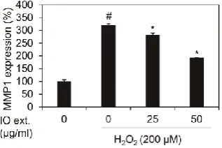
We showed whether Inonotus Obliquus extracts decreased MMP1 mRNA expression in nHDFs with 200 μM H2O2. Each condition cells were harvested and RNA isolated. And cDNA
was synthesized using isolated RNA, and the expression of MMP1 was verified as qRT-PCR.
The results of the experiment revealed that the expression of MMP1 mRNA was increased by
3.20 times (320.25%) by 200 μM H2O2 against the control group as shown in Figure 2. In
1.92 times (192.34%) in 200 μM H2O2 exposed nHDFs. Therefore, Inonotus Obliquus extracts
have been confirmed to reduce MMP1 mRNA caused by oxidative stress.
AP-1 transcriptional activity suppression of Inonotus Obliquus extracts in nHDFs
The increase of MMP1 mRNA expression due to oxidative stress was induced by
phosphorylation at c-Jun, a major component protein of AP-1 complex. Therefore, we
investigated the transcription activity of AP-1 by Inonotus Obliquus extracts, which was measured by TPA Response Element (TRE), the DNA binding site of AP-1 (Rise et al., 1989;
Park et al., 2003). As shown in Figure 3, we demonstrated that 200 μM H2O2 increase
8.26-fold (826.36%) in transcription activity of the AP-1 complex. However, treating Inonotus Obliquus extracts decreased the transcription activity of AP-1 complex by 3.07 times (307.46%) by 50 μg/ml in 200 μM H2O2 exposed nHDFs. Therefore, Inonotus Obliquus extracts is verified
to reduce the expression of MMP1 by regulating the transcription activity of the AP-1 complex
(Figure 3).
JNK phosphorylation suppression of Inonotus Obliquus extracts in nHDFs
AP-1 activity is regulated by phospho-JNK in oxidative stress. Thus, we showed whether
Inonotus Obliquus extracts decreases phospho-JNK in 200 μM H2O2 exposed nHDFs (Leppä
et al. ,1998; Karin, 1995). As shown Figure 4, JNK phosphorylation was elevated by oxidative
stress, 200 μM H2O2, and co-treatment of 200 μM H2O2 and Inonotus Obliquus extracts showed
that phospho-JNK decreased compared to 200 μM H2O2 single treatment (Figure 4). Inonotus Obliquus extracts regulated the transcription activity of the AP-1 complex by inhibiting JNK, thereby regulating the increased MMP1 expression caused by oxidative stress. Overall,
Discussion
In this study, we confirm the anti-aging mechanism of the Inonotus Obliquus extracts. As shown in the results of this paper, the Inonotus Obliquus extracts was found to be non-cellular toxic at concentrations below 50 μg/ml (Figure 1). MMP1 overexpression caused by oxidative
stress was decreased by 50 μg/ml Inonotus Obliquus extracts (Figure 2). We also found that this regulation of MMP1 was due to reduced transcription activity of AP-1 through decrease
of JNK activity (Figure 3, 4). In aged skin, MMP1 decomposed extracellular matrix by
degradation of collagen type 1. Especially, MMP1 is elevated by photo or natural
ageing-mediated oxidative stress [1, 2]. The oxidative stress induced JNK phosphorylation, and then
phospho-JNK elevates transcriptional activity of AP-1, which is finally upregulated MMP1
expression [14, 15]. Thus, our results suggest that Inonotus Obliquus extracts decreases MMP1 mRNA expression using downregulating AP-1 transcriptional activity and JNK
phosphorylation [16, 17]. Moreover, through the results of this study, we identified that the
Inonotus Obliquus extracts is a cosmetic material that can control the decomposition of the collagen by reducing the expression of MMP1, which breaks down the collagen of the skin,
and thus presents a possibility as a new functional cosmetic ingredient for reducing wrinkles.
Conclusion
Treatment with Inonotus Obliquus extracts attenuated MMP1 expression in nHDFs, as determined by qRT-PCR, without causing cytotoxicity. Furthermore, Inonotus Obliquus
extracts down-regulated AP-1 transcriptional activity and phosphorylation of JNK. These
data collectively indicate that Inonotus Obliquus extracts are potent inhibitors of skin aging.
This research was supported by Osan University in 2019 (Kim, YJ) and Basic Science Research
Program through the National Research Foundation of Korea (NRF) funded by the Ministry of
Education (2017R1D1A1B03028380).
Conflict of interest
The authors state no conflict of interest.
Acknowledgments
This research was supported by Osan University in 2019 (Kim, YJ) and Basic Science Research
Program through the National Research Foundation of Korea (NRF) funded by the Ministry of
Education (2017R1D1A1B03028380).
References
1. Chung, J.H., Youn, S.H., Kwon, O.S., Cho, K.H., Youn, J.I., Eun, H.C. Regulations of
collagen synthesis by ascorbic acid, transforming growth factor-beta and interferongamma in
human dermal fibroblasts cultured in three-dimensional collagen gel are photoaging- and
agingindependent. J Dermatol Sci. 1997, 15, 188-200.
2. Rittié, L., Fisher, G.J. UV-light-induced signal cascades and skin aging. Ageing Res Rev.
2002, 1, 705-720.
3. Fisher, G.J., Kang, S., Varani, J., Bata-Csorgo, Z., Wan, Y., Datta, S., Voorhees, J.J.
Mechanisms of Photoaging and Chronological Skin Aging. Arch Dermatol. 2002, 138,
1462-1470.
(Ed.), Extracellular Matrix. Amsterdam, Netherlands, Harwood academic publishers, 1996,
pp22-67.
5. Uitto, J. Connective tissue biochemistry of the aging dermis. Age-associated alterations in
collagen and elastin. Dermatol Clin. 1986, 4, 433-446.
6. Chung, H.Y., Kim, H.J., Shim, K.H., Kim, K.W. Dietary modulation of prostanoid synthesis
in the aging process: role of cyclooxygenase-2. Mech Aging Dev. 1999, 111, 97-106.
7. Coulomb, B., Dubertet, L., Merrill, C., Touraine, R., Bell, E. The collagen lattice: a model
for studying the physiology, biosynthetic function and pharmacology of the skin. Br J Dermatol.
1984, Suppl 27, 83-87.
8. Fisher, G.J., Voorhees, J.J. Molecular mechanisms of photoaging and its prevention by
retinoic acid: ultraviolet irradiation induces MAP kinase signal transduction cascades that
induce AP-1-regulated matrix metalloproteinases that degrade human skin in vivo. J Investig
Dermatol Symp Proc. 1998, 3, 61-68.
9. Varani, J., Spearman, D., Perone, P., Fligiel, S.E.G., Datta, S., Wang, Z.Q., Voorhees, J.J.
Inhibition of type I procollagen synthesis by damaged collagen in photoaged skin and by
collagenase-degraded collagen in vitro. Am J Pathol. 2001, 158, 931-942.
10. Di, L.C., Pei, Y., Travers, J.B. Augmentation of ultraviolet B radiation-induced tumor
necrosis factor production by the epidermal platelet-activating factor receptor. J Biol Chem.
1999, 274, 26917-26921.
11. Angel, P., Szabowski, A., Schorpp-Kistner, M. Function and regulation of AP-1 subunits
in skin physiology and pathology. Oncogene. 2001, 20, 2413-2423.
12. Fisher. G.J., Wang, Z.Q., Datta, S.C., Varani, J., Kang, S., Voorhees, J.J. Pathophysiology
of premature skin aging induced by ultraviolet light. N Engl J Med. 1997, 337, 1419-1428.
13. Dasgupta, J., Kar, S., Liu, R., Joseph, J., Kalyanaraman, B., Remington, S.J., Chen, C.,
metalloproteinase-1 through c-Jun-N-terminal kinase. J Cell Physiol. 2010, 225, 52-62.
14. Leppä, S., Saffrich, R., Ansorge, W., Bohmann, D. Differential regulation of c-Jun by ERK
and JNK during PC12 cell differentiation. EMBO J. 1998, 17, 4404-4413.
15. Karin, M. The regulation of AP-1 activity by mitogenactivated protein kinases. J Biol Chem.
1995, 270, 16483-16486.
16. Risse, G., Jooss, K., Neuberg, M., Brüller, H.J., Müller, R. Asymmetrical recognition of
the palindromic AP1 binding site (TRE) by Fos protein complexes.E MBO J 1989, 8,
3825-3832.
17. Park, K.K., Jung, E., Chon, S.K., Seo, M., Kim, H.W., Park, T. Finding of TRE (TPA
responsive element) in the sequence of human taurine transporter promoter. Adv Exp Med Biol.
Figure legends
Figure 1. Cytotoxicity of extracts of Inonotus Obliquus in human dermal fibroblasts
Effects of Inonotus Obliquus extracts on the cell viability were expressed as a percentage of control at the indicated concentrations. Values are mean±standard deviation (S.D.) from
Figure 2. Effects of extracts of Inonotus Obliquus on MMP1 mRNA expression in human dermal fibroblasts.
Effects of Inonotus Obliquus extracts on MMP1 mRNA expression against H2O2 treatment.
MMP1 mRNA expression were normalized to β-actin. The results are expressed as the
mean±S.D. from triplicate experiments. # p<.05 compared with non-treated cells. * p<.05
Figure 3. Effects of extracts of Inonotus Obliquus on promoter activity of TRE in human dermal fibroblasts.
TRE activity was estimated by using TRE-luciferase reporter assay. TRE activity were
normalized to β-galactosidase. The graphs are expressed as the mean±S.D. from triplicate
experiments. # p<.05 compared with non-treated cells. * p<.05 compared with 200 μM H2O2
Figure 4. Effects of extracts of Inonotus Obliquus on JNK phosphorylation in human dermal fibroblasts.
JNK and p-JNK protein expression were assessed by using western blot assay. β-Actin served
as an internal control, and different concentrations of Inonotus Obliquus extracts (0, 25, and 50 µg/mL) were treated in human dermal fibroblasts. JNK band density was measured by Image
J software and normalized by band density of p-JNK. The graphs are expressed as the
mean±S.D. from triplicate experiments. # p<.05 compared with non-treated cells. * p<.05