Review
1
Structure and function of multimeric G-quadruplexes
2
Sofia Kolesnikova 1,2 and Edward A. Curtis 1,*
3
1 The Institute of Organic Chemistry and Biochemistry of the Czech Academy of Sciences, Prague 166 10,
4
Czech Republic
5
2 Department of Biochemistry and Microbiology, University of Chemistry and Technology, Prague 166 28,
6
Czech Republic
7
* Correspondence: [email protected]
8
Received: date; Accepted: date; Published: date
9
Abstract: G-quadruplexes are noncanonical nucleic acid structures formed from stacked guanine
10
tetrads. They are frequently used as building blocks and functional elements in fields such as
11
synthetic biology and also thought to play widespread biological roles. G-quadruplexes are often
12
studied as monomers but can also form a variety of higher-order structures. This increases the
13
structural and functional diversity of G-quadruplexes, and recent evidence suggests that it could
14
also be biologically important. In this review we describe the types of multimeric topologies
15
adopted by G-quadruplexes and highlight what is known about their sequence requirements. We
16
also summarize the limited information available about potential biological roles of multimeric
17
G-quadruplexes and suggest new approaches that could facilitate future studies of these structures.
18
Keywords: G-quadruplex; dimer; tetramer; multimer; oligomer; telomere; promoter; R-loop;
19
DNA:RNA hybrid
20
21
1. Introduction
22
The B-form double helix is the most well known nucleic acid structure but it is not the only one.
23
Other examples include double helices with geometries that differ from that of classical B-form
24
DNA, such as Z-DNA [1], triple helices [2], and even four-stranded structures in which canonical
25
A-T and C-G base pairs are absent [3-4]. Among these noncanonical folds, the G-quadruplex (GQ)
26
has received the most attention. This is a four-stranded structure typically stabilized by stacked
27
guanine tetrads connected by short loops [3, 5-6]. Because of their high stability, structural
28
versatility, and functional diversity, GQs have been widely used as building blocks and functional
29
elements in fields such as synthetic biology [7-8]. Sequences with the potential to form GQs are also
30
abundant in the genomes of higher eukaryotes [9-10], and recent studies using GQ-specific
31
antibodies indicate that these structures can form in the context of cells [11-12]. GQs are thought to
32
play a variety of biological roles. These including regulation of transcription, translation, DNA
33
replication, and RNA localization [13-15].
34
Most biological studies to date have focused on monomeric GQs. However, GQs can also
35
adopt a variety of multimeric forms. These include relatively small structures such as dimers.
36
They also include larger ones like G-wires, which can contain hundreds of GQ monomers.
37
Although the ability of GQs to multimerize has long been recognized, the possibility that such
38
high-order structures form in the context of cells has received less attention. From our perspective,
39
however, two observations suggest that this possibility should be considered. First, the cellular
40
concentrations of GQs are surprisingly high, especially in higher eukaryotes. For example, current
41
estimates suggest that human cells contain at least 716,000 DNA GQs [16] in a volume (for a HeLa
42
cell nucleus) of 0.22 pL [17]. This corresponds to a cellular GQ concentration of 6 M. Not all of
43
these GQs will be present at the same time in the cell cycle, and not all will be capable of forming
44
dimers. On the other hand, this concentration is orders of magnitude higher than that required for
45
efficient GQ multimerization. For example, in a recent study we identified GQs with dissociation
46
constants of dimer formation as low as 35 nM [18]. The hypothesis that GQs form multimeric
47
structures in cells is also compelling when the myriad evolutionary and functional advantages of
48
this mechanism are considered [19-22]. These include the ability to regulate biochemical function
49
based on concentration, to detect ligands with enhanced sensitivity by cooperative binding, and to
50
modulate activity in a rapid and reversible manner by exchanging dimerization partners (Figure 1).
51
Inspired by these considerations, in this review we first describe the types of multimeric topologies
52
adopted by GQs and review what is known about their sequence requirements. We then
53
summarize the limited information currently available about the potential biological roles of
54
multimeric GQs in cells and suggest new approaches that could facilitate future studies of these
55
structures, especially in the context of cells.
56
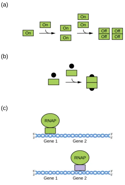
Figure 1. Regulation of biochemical function by multimerization. (a) Concentration-based control of
57
biochemical function. In this scheme, monomers are biochemically active and dimers are not. At
58
concentrations below the dissociation constant for dimer formation, most of the population is
59
monomeric and in the active state, while at concentrations above the dissociation constant most of
60
the population is dimeric and in the inactive state. (b) Enhanced sensitivity to ligand concentration
61
by cooperative binding. In this scheme, ligand binding is independent when binding sites are
62
monomeric but cooperative when they are linked by multimerization. This leads to all-or-none
63
binding and enhanced sensitivity to ligand concentration. (c) Modulation of biochemical activity by
64
the exchange of dimerization partners. In this scheme, the gene transcribed by RNA polymerase is
65
determined by the DNA-binding specificity of its dimerization partner.
66
2. Definition of multimeric G-quadruplexes and modes of multimerization
67
A multimer is an aggregate of molecules consisting of multiple monomers. Nucleic acids
68
typically multimerize (hybridize) through duplex formation via the Watson-Crick base pairing of
69
two complementary strands. A double helix provides DNA with structural stability, determined
70
by hydrogen bonding and base-stacking interactions, and facilitates replication [23-24], while
71
(a)
(c) (b)
3’ 5’
RNAP
Gene 1
5’ 3’
Gene 2
3’ 5’
RNAP
Gene 1
5’ 3’
Gene 2 On
On On
Off Off
On On
On
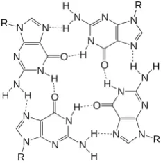
Figure 2. Chemical structure of a GQ tetrad.
72
double-stranded RNA facilitates genetic interference [25]. Duplex formation is the most common
73
mechanism of multimerization, but not the only one. Multimerization can also occur using a distinct
74
hydrogen-bonding pattern called Hoogsteen base pairing. This enables formation of a G-tetrad, the
75
building block of a unique type of nucleic acid structure called a G-quadruplex (GQ). To form a
76
G-tetrad, four guanines associate via eight hydrogen bonds from both the Watson-Crick and
77
Hoogsteen faces of the base (Figure 2). G-tetrads stack on top of one another giving rise to a GQ
78
(Figure 3). To form a GQ, a sequence typically needs to contain segments of at least two guanines
79
separated by mixed-sequence loop nucleotides. The most widely used models allow loops of 1 to 7
80
nucleotides [9-10] but in some cases loops can be longer [26-28]. In addition, it is becoming
81
increasingly clear that GQs can accommodate bulges [29], and noncanonical tetrads have also been
82
observed in high-resolution structures [30-36]. Even more complicated topologies are seen in the
83
Spinach, Mango, and Class V GTP aptamers, in which the four clusters of guanines that form the
84
tetrads in the GQ are far apart in the primary sequence [37-39]. Guanines in the G-tetrad can in
85
principle come from one, two, three, or four guanine-rich (G-rich) strands. GQs formed from only
86
one strand are typically defined as intramolecular (unimolecular) (Figure 3A-B), although, as
87
discussed below, multiple GQs on a single strand can also interact to form higher-order structures.
88
GQs that contain more than one strand are termed intermolecular (multimolecular or multimeric)
89
and can be classified according to the number of strands as bimolecular (dimeric) (Figure 3C),
90
trimolecular (trimeric) (Figure 3D), or tetramolecular (tetrameric) (Figure 3E). Multimeric GQs
91
formed from identical strands are called homomultimeric, whereas those composed from
92
non-identical strands are called heteromultimeric.
93
While the number of molecules in a GQ is the standard way to classify multimers, another
94
approach is to consider the structure of the multimerization interface. GQs utilize two primary
95
modes of multimerization. In the first mode, interfaces are formed by tetrads from two different
96
Figure 3. Formation of GQs from different numbers of strands. (a) Intramolecular (unimolecular) GQ
97
with antiparallel strands. (b) Intramolecular (unimolecular) GQ with parallel strands. (c) Bimolecular
98
(dimeric) GQ. (d) Trimolecular (trimeric) GQ. (e) Tetramolecular (tetrameric) GQ. Note that each of
99
these structures can in principle contain all parallel strands, all antiparallel strands, or a mix of
100
parallel and antiparallel strands.
101
N
N N N
O
N R
H
H H N N
N N
O
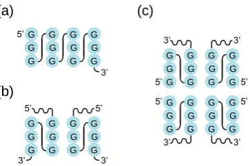
N R
H H
H N
N N
N
O N
R
H H
H
N N
N N O
N
R
H H
GQs stacked on top of one another. When intramolecular (unimolecular) GQs multimerize
102
using this mechanism, individual nucleic acid strands first assemble into monomeric GQs such that
103
all four guanines in each G-tetrad come from the same nucleic acid strand. Monomeric GQ
104
subunits then stack on top of one another via π-π stacking interactions of terminal interfaces to form
105
higher-order GQ structures (Figure 4A). Intermolecular (multimeric) GQs can also multimerize in
106
this way if all of the guanines in one of the tetrads at the interface come from one GQ and all of the
107
guanines in the other come from a different GQ (Figure 4A). GQ subunits can stack in three
108
different orientations: 5’ to 3’ (head-to-tail), 5’ to 5’ (head-to-head), and 3’ to 3’ (tail-to-tail).
109
Examples of these orientations can be found in [40] (5’ to 3’), [41] (5’ to 5’), and [42] (3’ to 3’).
110
Experimental and molecular dynamics data suggest that 5′ to 5′ stacking is the most common
111
orientation for GQ structures due to a favorable stacking geometry [41, 43]. The propensity of
112
subunits to stack is also influenced by the topology of intermolecular GQ subunits. Stacking is
113
favorable for parallel GQs (i.e. GQs with all strands oriented in the same direction and with
114
propeller loops on the sides of the tetrads). In antiparallel GQ structures, which have strands in
115
opposite orientations, lateral and diagonal loops are positioned above and below the GQ axis which
116
can impede stacking interactions of terminal G-tetrads [44]. Stacking interactions can also facilitate
117
formation of higher-order structures from tandem GQ subunits folded on a single strand. Such a
118
structure was proposed as one of the models of the human telomere overhang [45-46]. In addition
119
to playing important roles in multimerization, stacking interactions are the most important mode of
120
ligand binding to GQs. End-stacking GQ ligands either bind to terminal GQs tetrads or intercalate
121
between tandem GQs to stabilize the multimeric structure [47-52].
122
In the second mode of multimerization, guanines from two or more nucleic acid strands
123
hydrogen bond to form tetrads, so that interfaces occur within rather than between tetrads. These
124
intermolecular G-tetrads then stack to form GQs of various lengths (Figure 4B), sometimes using
125
slipped strands [53]. Various G-rich oligonucleotides multimerize via this mode, and a large body
126
of literature has investigated their formation [5-6]. As early as 1988, Sen and Gilbert demonstrated
127
that oligonucleotides containing motifs of four, five or six contiguous guanines fold into tetrameric
128
structures (Sen and Gilbert, 1988) [54]. One year later, two different groups proposed the formation
129
of dimeric GQs from two telomere-derived G-rich sequences [55-56]. Sen and Gilbert [57] observed
130
that short oligonucleotides with three consecutive guanines at the 3’ end assembled
131
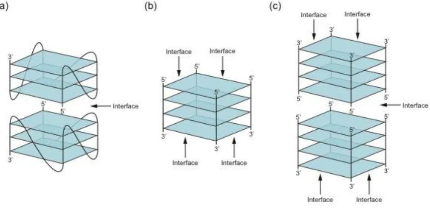
Figure 4. Types of interfaces in multimeric GQs. (a) First mode of multimerization. Interfaces are
132
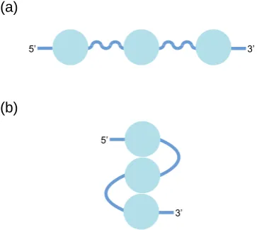
formed between tetrads which stack on top of one another in a 5' to 5' , 3' to 3', or 5' to 3' arrangement.
133
(b) Second mode of multimerization. Interfaces are formed within tetrads made up of guanines from
134
multiple DNA strands. (c) Structure combining these two modes of multimerization.
into four, eight and twelve-stranded GQ structures. Formation of these structures starts with the
136
self-assembly of slipped tetramers in which strands are not perfectly aligned but contain two
137
overhanging guanines at the 3’end. Tetrameric subunits associate with one another via hydrogen
138
bonding between these extra guanines.
139
Higher-order GQs which combine these two modes of multimerization have also been
140
described [18, 42, 58-61]. An illustrative example is a GQ structure assembled from eight
141
d(TGGGGT) strands (Figure 4C). The structural subunit is a tetramolecular GQ consisting of four
142
perfectly aligned strands, each in an identical 5′-3′ orientation (second mode of multimerization).
143
Two structural subunits then stack at their 5’ interfaces forming an octamer (first mode of
144
multimerization) [58-59].
145
Higher-order GQ structures are often initially characterized using low resolution techniques
146
such as circular dichroism, dimethylsulfate footprinting, native PAGE, mass spectrometry, and
147
analytical ultracentrifugation. These methods can be used to establish that the sequence forms a
148
GQ, identify the guanines in tetrads, and determine the number of strands in the structure.
149
Higher-order GQs can be visualized in greater detail using NMR and X-ray crystallography.
150
Examples of high-resolution structures which utilize the first mode of multimerization include 5'-5'
151
stacked dimers with canonical [41, 62-63] or extended [64] tetrads at the interface. Structures which
152
use the second mode of multimerization include interlocked dimers which occur in the promoters
153
and introns of oncogenes [65-67]. A structure which combines these two modes of multimerization
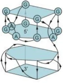
154
is that of a parallel-stranded tetrameric GQ formed from d(TGGGGT) strands [58-59]. Additional
155
examples are discussed in [5, 50, 68] and elsewhere.
156
3. Sequence requirements of multimeric G-quadruplexes
157
The propensity of a GQ sequence to fold into a particular multimeric structure depends on a
158
number of factors including the type and concentration of cation in the buffer, the presence of
159
molecular crowding agents, the nucleic acid concentration, the sequence length and the sequence
160
composition [5, 68-70]. Under conditions approximating the intracellular environment, the main
161
factor controlling higher-order structure formation is the sequence. In the context of GQ
162
Figure 5. Sequence requirements of multimeric GQs. (a) Example of a canonical GQ. (b) Variant
163
containing two rather than four G-runs. Such a sequence can form a multimeric GQ but not a
164
monomeric one. (c) Variant containing G-runs of two rather than three nucleotides. In some cases
165
such sequences form multimeric rather than monomeric structures. (d) Variant containing mutations
166
in tetrads. Such mutations can induce formation of both dimeric and tetrameric structures. (e)
167
Variant containing overhanging nucleotides. Such variants typically cannot stack via the first mode
168
of multimerization, but the ability to interact via the second mode of multimerization is unaffected.
169
(f) Variant containing an extended central loop. Longer loops favor formation of antiparallel rather
170
than parallel GQs, and such loops can interfere with the ability of GQs to stack via the first mode of
171
multimerization.
172
(a)
(e) (b)
(c)
(d)
(f)
G G G T GG G A A G G G T G GG
5’ 3’
G G G T G G G3’ 5’
G G
G G G G G G
T
T A A
5’ 3’
G G G T GGG A A G G G T G GG
5’ A A3’
G G G T GG G A A G G G T G GG
5’ A A A A A 3’
G A G T G AG A A G G G T G GG
formation, a large number of sequences, ranging from short strands with only one G-run to up to
173
20000 nucleotide long sequences with roughly 3300 G-runs, have been investigated (58-59, 71). The
174
key sequence features affecting multimerization are discussed below and summarized in Figure 5.
175
The minimum requirement for a sequence to fold into an intramolecular GQ is typically four
176
runs of at least two guanine bases separated by loops ranging from one to seven nucleotides (Figure
177
5A). Sequences falling into this category often either fold into monomeric GQs that do not interact
178
with one another or form multimeric structures by the first mode of multimerization (stacking of
179
monomeric subunits). Structure prediction is nevertheless complicated by reports of sequences
180
containing four stretches of guanines which form intertwined dimers using the second mode of
181
multimerization [65-67, 72]. Sequences with fewer than four G-runs can typically only adopt GQ
182
structures by interacting to form multimeric structures using the second mode of multimerization
183
(Figure 5B). Sequences with two or three G-runs separated by short loops usually combine both
184
types of multimerization and readily assemble into structures with eight, twelve, or even more
185
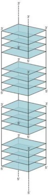
strands [57]. At the extreme end of this continuum lie the G-wires [73], long linear ladder-like
186
structures formed from numerous slipped GQ tetrameric subunits (Figure 6). G-wires are longer
187
than any other higher-order GQs with maximum lengths depending on the method of preparation
188
as well as the sequence. For example, a DNA sample of d[G4T2G4] formed linear G-wires ranging
189
from 7 to 100 nm in length. A 100 nm G-wire was calculated to contain 75 GQ blocks composed of
190
140 full and 20 half strands [74]. Positioning of the GQ subunits within the structure can be
191
controlled by adding GC bases to the terminal ends, which enables the formation of G:C:G:C tetrads
192
linking the subunits [75].
193
Figure 6. Structure of a G-wire.
In addition to the number of G-runs in the sequence, multimerization also depends on the
195
length of the G-run (Figure 5C). An instructive example is a study of structural transitions caused
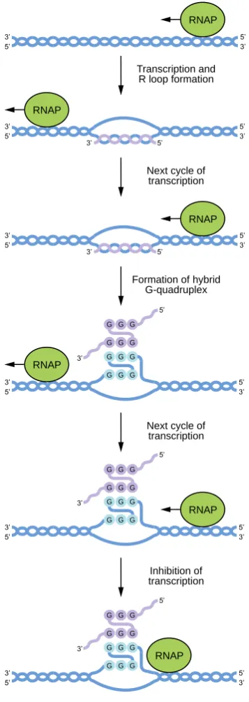
196
by truncations of guanine tracts in the telomere-derived DNA strand d(G4T4G4) [76]. The original
197
sequence and its 5’ truncated analog d(G3T4G4) formed stable dimeric structures that did not
198
undergo conversion into higher-order structures. In contrast, the 3’ truncated sequences d(G3T4G3)
199
and d(G4T4G3) formed a mixture of dimeric, trimeric and tetrameric GQs in a
200
concentration-dependent manner. This structural polymorphism can be explained by varying
201
stabilities of the dimeric structures formed by the reference and the various truncated sequences.
202
The reference d(G4T4G4)2 dimer contains sixteen guanines which form four G-tetrads, and is
203
therefore more stable than the three-tetrad dimers formed by truncated sequences. A bimolecular
204
structure formed from two d(G3T4G4) strands is special in that, in contrast to the antiparallel dimers
205
formed by d(G3T4G3) and d(G4T4G3), it contains three parallel strands. This specific strand
206
orientation stabilizes the d(G3T4G4)2 dimer and thereby prevents it from rearranging into
207
higher-order structures.
208
A third feature of G-runs that can affect multimerization is the presence of bulges (interruptions
209
of G-runs by non-G nucleotides) (Figure 5D). Despite bulges being tolerated by various GQs, they
210
can greatly influence structural stability. The extent to which GQ stability is reduced depends on a
211
number of factors such as the location of the bulges within the GQ sequence, the context of the
212
sequence and the overall GQ topology [29]. The introduction of bulges does not necessarily result
213
in structural changes, and in some cases they do not prevent monomeric GQs from forming. On the
214
other hand, GQs with reduced stability are prone to structural reorganizations, including
215
higher-order structure formation. Bulged nucleotides as drivers of multimerization were
216
systematically investigated in a recent study that analyzed dimerization and tetramerization as a
217
function of guanine substitutions in G-runs [18]. The reference sequence used in this study,
218
d(G3TG3AAG3TG3A), folds into a parallel three-tetrad GQ with nucleotides 2, 6, 11 and 15 forming
219
the central tetrad (Figure 7A). Despite containing an exposed 5' tetrad, only monomers were
220
observed when this sequence was analyzed on native gels. Dimerization and tetramerization was
221
induced by introducing substitutions of guanines for other nucleotides at certain positions in the
222
central tetrad. Bulged G-runs prevented these sequences from forming stable intramolecular GQs
223
by themselves and increased their propensity to form multimeric structures. Sequences containing
224
substitutions at position 2, 6 or both were proposed to form
225
Figure 7. Mutations in tetrads can induce GQ multimerization. (a) Secondary structure of a GQ we
226
are studying in our group with the sequence GGGTGGGAAGGGTGGGA. We previously generated
227
a library containing all possible mutations in the central tetrad in this structure (at positions 2, 6, 11
228
and 15) and tested these variants for a series of biochemical activities associated with GQs [18, 82-83
229
127]. (b) Proposed secondary structure of dimers formed by variants containing mutations at
230
positions 2, 6, or both in the central tetrad of the reference GQ. The 5' part of this structure, which
231
contains the mutated nucleotides, is represented by a wavy black line. (c) Proposed secondary
232
structure of tetramers formed by variants containing mutations at positions 11, 15, or both in the
233
central tetrad of the reference GQ. The 3' part of this structure, which contains the mutated
234
nucleotides, is represented by a wavy black line.
intertwined dimers with a core of three canonical G-tetrads at the 3’ end (Figure 7B). Sequences
236
with substitutions at positions 11, 15 or both were instead proposed to form intertwined dimers
237
containing a core of three canonical G-tetrads at the 5’ end (Figure 7C). Such sequences also formed
238
tetramers which, in accord with the most favorable mode of stacking, were proposed to consist of
239
two dimers stacked in a 5’ to 5’ orientation (Figure 7C). Intertwined dimers formed from sequences
240
with other possible combinations of substitutions (2 and 11, 2 and 15, 6 and 11, 6 and 15) would only
241
contain two stacked G-tetrads [18]. This structural arrangement cannot effectively stabilize the
242
structures, which probably explains why these sequences did not also multimerize.
243
GQ sequences can be designed to begin and/or end with G-runs or they can contain
244
overhanging non-G nucleotides at the 5’ and/or 3’ ends (Figure 5E). Flanking nucleotides often
245
inhibit stacking interactions by sterically hindering formation of interfaces by terminal tetrads [57,
246
77]. In some cases even the introduction of a 5' phosphate group is sufficient to inhibit 5'-5'stacking
247
[73]. This knowledge can be used when probing multimerization mechanisms: for multimers
248
stacked in a 5’ to 5’ orientation, addition of flanking nucleotides at the 5’ end should interfere with
249
the stacking interaction, while addition of flanking nucleotides to the 3' end should not [18].
250
Nevertheless, some studies report the stacking of GQ subunits which contain overhanging
251
nucleotides. In these structures, flanking nucleotides either radiate out from the GQ axis or become
252
a part of various unusual structures such as thymine triads [78], guanine:uridine octads [79] or
253
guanine:cytidine octads [60].
254
The role of loop nucleotides in multimeric GQ assembly has received relatively little attention
255
from researchers, and most studies investigating multimeric GQs have not analyzed the effects of
256
loop length and sequence composition. Despite this, examples in which loop nucleotides affect GQ
257
multimerization have been described. Loop length can indirectly influence multimerization by
258
affecting GQ topology (Figure 5F). Sequences bearing at least one single-nucleotide loop tend to
259
adopt a parallel-stranded orientation, whereas longer loops increase the likelihood that sequences
260
will fold into mixed-strand or antiparallel GQs [80]. As discussed above, this can affect the ability
261
of GQs to interact by the first mode of multimerization: the propeller loops of parallel strand GQs do
262
not generally affect their ability to form multimers through stacking on their terminal tetrads,
263
whereas the lateral and diagonal loops of antiparallel GQs sometimes do. Loop sequence can also
264
play an important role in GQ structure formation. One mechanism by which this can impact the
265
global structure of the GQ is by formation of non-canonical extended G-tetrads. For example,
266
d(GGA)4 strands assemble into dimers by stacking on a guanine:adenine heptad interface formed by
267
two monomeric subunits. Three loop adenines provide hydrogen bonds donors and acceptors in the
268
heptad plane and hence play an indispensable role in dimerization [64]. Moreover, several studies
269
have shown that mutating nucleotides not thought to directly interact with tetrads can lead to major
270
changes in the GQ structure. For example, changing the loops in a human telomere-derived
271
sequence from TTA to AAA abolishes GQ formation [81]. Recent findings in our lab also show that
272
mutations in loops can affect the propensity of sequences to form multimeric GQs [82-83].
273
In summary, these studies indicate that the main sequence features affecting multimerization
274
are the number and length of the G-runs, the sequence composition of the G-runs, the presence of
275
flanking nucleotides at the 5' and 3' ends of the GQ, and the length and sequence of loop nucleotides
276
(Figure 5). Despite much being understood about the sequence requirements of multimeric GQs, it
277
is generally not possible to predict higher-order GQ topology from sequence. Instead, experimental
278
data from low-resolution methods such as circular dichroism, dimethylsulfate footprinting, native
279
PAGE, mass spectrometry, and analytical ultracentrifugation is used in combination with
280
high-resolution techniques such as NMR and X-ray crystallography.
281
4. Potential biological roles of multimeric GQs at the tips of telomeres
282
The first discussions of biologically relevant higher-order GQ structures emerged as the length
283
and sequence composition of telomeres was being characterized [84]. In the majority of eukaryotes,
284
the nucleotide sequence of telomeres consists of a G-rich strand running 5’ to 3’ toward the end of
285
the chromosome and a complementary C-rich strand. For most of their length, telomeres are
double-stranded, except for a single-stranded G-rich 3’overhang. While the telomeric sequence is
287
similar in diverse species, the length of the overhang is not. For example, in Tetrahymena the
288
overhang typically consists of tandem repeats of the sequence d(TTGGGG) and is 14 to 21
289
nucleotides long [85]. On the other hand, in humans the sequence of the overhang is d(TTAGGG)
290
and it ranges in length from 125 to 275 nucleotides [86-87].
291
While the composition and length of the telomeric overhang has been characterized in many
292
species, its secondary structure is still a matter of debate. The first studies of short telomere-derived
293
synthetic oligonucleotides reported that these sequences folded into several topologically different
294
guanine-rich structures [54-56, 88]. Subsequent studies showed that these correspond to GQ
295
structures, although several different topologies have been reported [6, 89-90]. Examples include
296
parallel-stranded tetrameric and antiparallel dimeric GQs assembled from short telomeric sequences
297
bearing one and two G-runs, respectively as well as intramolecular monomeric GQs formed by
298
sequences containing four G-runs.
299
Because the human telomeric overhang contains multiple repeats of segments with four G-runs,
300
it can potentially fold into a higher-order structure containing a series of monomeric GQs [91]. A
301
single-stranded DNA with several monomeric GQs can adopt at least two different structural
302
arrangements (Figure 8). The first one is termed “beads-on-a-string”, and assumes that monomeric
303
GQ units do not interact with one another [92]. A second model proposes that GQ subunits
304
associate with one another through stacking interactions between the terminal tetrads of consecutive
305
GQs to form high-order structures [45-46]. Despite evidence for both models, a structure in which
306
GQs stack on one another appears to be most likely [91]. Structures consistent with this model have
307
been observed by AFM and EM [71, 93-94], and methods such as FRET have provided additional
308
evidence for interactions between neighboring GQ subunits [94]. Molecular dynamics simulations
309
also support the idea that telomeric GQ monomers can stack to form higher order structures [45-46].
310
Based on comparisons between calculated and experimentally measured sedimentation coefficients
311
it has been proposed that a structure consisting of hybrid rather than all-parallel GQs is most likely
312
[95]. Despite these advances, a high-resolution structure of the telomeric overhang has not yet been
313
reported.
314
In vivo evidence for the presence of GQs at telomeres came from studies reporting the binding
315
of GQ-specific antibodies to the tips of telomeres [11-12, 96]. These experiments also showed that
316
telomerase co-localizes with GQ antibodies at chromosomal ends. Telomerase catalyzes telomere
317
elongation, and is directly involved in GQ unfolding [97]. Although these studies indicate that
318
telomeric DNA adopts a GQ structure in vivo, they do not distinguish between monomeric and
319
multimeric GQs since the antibody has similar affinities for both types of structures. While
320
Figure 8. Possible structures of telomeric GQs. (a) Beads-on-a-string model in which telomeric GQs
321
do not interact. (b) Model in which telomeric GQs stack on one another to form higher-order
322
structures.
323
5’
3’
5’ 3’
(a)
antibodies specific for multimeric GQs have not yet been developed, several small molecules specific
324
for multimeric GQs have recently been reported [51, 98-100]. One of these compounds, IZNP-1
325
(triaryl-substituted imidazole derivative), binds and stabilizes multimeric GQs by intercalating into
326
the cavity between two stacked GQ monomers. IZNP-1 treated cells contain a greater number of
327
BG4 foci (human GQ structure-specific antibody foci) at telomeres than untreated cells. They also
328
exhibit signs of DNA damage and telomere shortening, which can lead to cell cycle arrest, apoptosis
329
and senescence. Taken together, these findings suggest that multimeric GQs can form at telomeres
330
under certain conditions and contribute to telomere dysfunction [51].
331
5. Potential biological roles of multimeric G-quadruplexes in promoters
332
Visualization of IZNP-1 induced BG4 foci confirmed that telomeric ends have the ability to
333
form multimeric GQs. Surprisingly, however, the percentage of fluorescent foci at telomeres was
334
relatively low (38%) and the majority of antibodies localized to other genomic regions. One
335
implication of this observation is the possibility that multimeric GQs exist outside telomeric DNA.
336
If so, multimeric GQs might have roles beyond chromosomal capping.
337
A role for monomeric GQ structures in transcriptional regulation was proposed following the
338
discovery that putative GQ sequences are highly enriched in nuclease hypersensitive regions within
339
promoters [13]. In addition, more than 40% of human genes contain at least one GQ motif in their
340
promoter. While formation of monomeric intramolecular GQs has been investigated for a number
341
of these genes including c-MYC, KRAS, and BCL2 [101-104], only a few examples of genes likely to
342
be regulated by multimeric GQs in their promoters have been identified so far. The most
343
well-characterized example is an unusual tetrad:heptad:heptad:tetrad (T:H:H:T) dimer generated
344
from a d(GGA) repeatsequence. An oligonucleotide containing four such repeats folds into an
345
intramolecular structure in which a guanine tetrad and a guanine-adenine heptad are stacked on top
346
of one another. The NMR structure indicates that two monomeric subunits stack 5’ to 5’ on the
347
heptad plane, resulting in a dimeric T:H:H:T structure [64] (Figure 9). A similar structure was
348
adopted by an oligonucleotide containing eight copies of the repeat [105]. The d(GGA) repeat
349
sequence occurs in twelve nearly perfect tandem repeats in the c-MYB promoter. In theory, this
350
sequence can give rise to three independent T:H building subunits (with four tandem repeats per
351
building block), which could then stack in different combinations to form dimers. A study
352
investigating this possibility proposed that a T:H:H:T motif formed by stacking interactions between
353
the first and the third building blocks functions as a negative regulator of c-MYB promoter activity.
354
The authors also identified a transcription factor, MAZ, which binds to the T:H:H:T structure and
355
could play a role in the regulation of c-MYB expression [106-107]. The c-MYB promoter is not the
356
only genomic region rich in GGA repeats, and similar motifs have been identified in the regulatory
357
regions of other genes such as NCAM [108], SPARC [109], KRAS [110],
358
Figure 9. A dimeric GQ formed by GGA repeats. Structure of a dimeric GQ formed by the sequence
359
(GGA)8. See [64] and [105] for more information about this structure.
and CCNB1IP1 [111] (Islam et al., 2019). Although it is tempting to assume that the transcription of
361
genes with multiple d(GGA) repeats in the promoter is regulated by T:H:H:T structures, the results
362
presented in [106] and [107] do not completely rule out the possibility that inhibition of transcription
363
is mediated by monomeric forms of these GQs. Testing the effects of antibodies or small molecules
364
specific for the T:H:H:T structure on the extent of transcriptional inhibition could provide further
365
insight into the structural organization of the d(GGA) repeat region in this promoter.
366
Another multimeric GQ structure that could potentially regulate gene expression occurs in the
367
promoter of hTERT, the gene encoding the reverse transcriptase of telomerase. The core promoter
368
of hTERT contains twelve consecutive G-runs and can therefore form multiple GQs. One proposed
369
structural model for this region consists of two stacked GQs separated by a 26 nucleotide loop which
370
contains another four G-runs and adopts a hairpin structure [112]. An alternative model suggests
371
that all of the G-runs in the sequence participate in GQ folding and that the final structure consists of
372
three parallel GQs tightly stacked on top of one another [40, 113]. Although the available evidence
373
does not indicate which model is correct, it does support the idea that a G-rich structure in this
374
promoter regulates hTERT expression [112-115].
375
The hypothesis that multimeric GQs in promoters regulate gene expression is also supported by
376
a study investigating the regulatory sequences of muscle-specific genes [116]. The promoter
377
regions of these genes contain a high frequency of G-runs that form both homodimeric and
378
heterodimeric structures in vitro. One example is the ITGA7 promoter region, which contains two
379
G-rich sequences, each capable of forming an intramolecular monomeric GQ, separated from one
380
another by 85 nucleotides. These two G-rich sequences formed a heterodimeric structure when
381
analyzed on native gels. This structure is hypothesized to play a role in the regulation of mouse
382
myogenic gene expression. Consistent with this hypothesis, the myogenic determination protein
383
MyoD binds the heterodimeric structure but not a monomeric GQ formed by one of the
384
heterodimer-forming sequences. MyoD exhibited a similar binding preference for a homodimeric
385
GQ formed by a G-rich region in the promoter of the sMtCK gene. Although this G-rich sequence
386
occurs only once in the sMtCK promoter region and therefore cannot assemble into a homodimer in
387
vivo, several G-rich clusters are present in adjacent genomic regions. It would therefore be of
388
interest to determine whether a heterodimeric GQ structure can form in this region and is bound by
389
MyoD [116].
390
Taken together, these data indicate that multimeric GQ structures can form in the promoter
391
regions of several genes and are consistent with the idea that such structures can regulate gene
392
expression. Many additional promoters contain multiple G-runs. For example, genes containing
393
more than eight G-tracts include TRIM13 [67] and c-MYC [102-103]. Although these are candidates
394
for genes regulated by multimeric GQs, our current understanding of GQ folding precludes reliable
395
prediction of higher-order GQ structure from sequence. As discussed above, the development of
396
antibodies or small molecules that selectively stabilize or destabilize specific multimeric GQ
397
structures should greatly facilitate analysis of their potential biological roles in promoters.
398
6. Potential biological roles of RNA-DNA hybrid G-quadruplexes
399
In addition to the multimeric DNA GQs discussed so far, multimeric GQs can also form from
400
combinations of DNA and RNA strands [117]. These structures are called DNA:RNA hybrid GQs.
401
They can be readily assembled in vitro, and also form during transcription when guanines in the
402
nontemplate DNA strands of genes interact with guanines in RNA molecules encoded by these
403
genes using the second mode of multimerization (Figure 10). DNA:RNA hybrid GQs inhibit
404
transcription, presumably by impeding the progress of RNA polymerase along the DNA template.
405
From the perspective of gene regulation, this is a compelling mechanism because it can act as a
406
negative feedback loop to turn transcription off when mRNA levels are sufficiently high (Figure 1A).
407
The first hint that DNA:RNA hybrid GQs act as regulatory elements came from analysis of a region
408
in the human mitochondrial genome called conserved sequence block II (CSB II) [118]. A G-rich
409
element in CSB II induces transcriptional termination and promotes formation of an unusually stable
410
structure in the template DNA. The stability of this structure suggested that it was not a
conventional R-loop, in which an RNA transcript and the DNA template encoding it are held
412
together by Watson-Crick base pairing. Instead, it was proposed that this structure was a GQ
413
containing both DNA and RNA strands. Consistent with this hypothesis, a structure was detected
414
by native PAGE that required for its formation the presence of both a DNA oligonucleotide
415
corresponding to the G-rich strand of the template and an RNA oligonucleotide corresponding to
416
the G-rich portion of the RNA transcript. Formation of this structure was inhibited when guanine
417
bases in the DNA strand were replaced by 7-deazaguanine or adenosine, both of which normally
418
prevent GQ formation. The circular dichroism spectrum of this structure was consistent with that
419
of a GQ, and it was resistant to ribonucleases A and H, both of which degrade RNA strands in
420
canonical DNA:RNA duplexes. Additional characterization using DMS fingerprinting, which can
421
distinguish guanines in tetrads from those in single or double-stranded DNA due to their reduced
422
sensitivity to DMS modification, and Zn-TTAPc-mediated photocleavage, which preferentially
423
cleaves GQs, provided additional evidence that the structure is a DNA:RNA hybrid GQ [119].
424
Similar methods were used to show that a DNA:RNA hybrid GQ also forms in the human NRAS
425
gene [120]. As was the case for the CSB II motif, the structure in the NRAS gene impeded T7 RNA
426
polymerase during in vitro transcription reactions. It also inhibited expression of luciferase from
427
plasmids transfected into cultured human cells. Only two stretches of guanines in the non-template
428
strand of the gene are needed to form the DNA:RNA hybrid GQ, with the other two provided by the
429
RNA transcript encoded by the template strand. DNA:RNA hybrid GQs containing two rather
430
than three tetrads can also form, although they are less stable than those containing three tetrads
431
[121]. Bioinformatic studies indicate that sequences with the potential to form DNA:RNA hybrid
432
GQs occur in 97% of human genes [122]. These motifs are enriched in and near promoters,
433
especially downstream of the transcription start site on the nontemplate strand, and this pattern of
434
enrichment is evolutionarily conserved in mammals [122]. Taken together, these studies support
435
the idea that DNA:RNA hybrid GQs represent an important regulatory element in higher
436
eukaryotes.
437
Additional studies have explored the mechanism of formation of DNA:RNA hybrid GQs
438
during transcription. Zhang and colleagues developed a method to distinguish R-loops from
439
DNA:RNA hybrid GQs based on differences in the sensitivities of these structures to ribonucleases
440
such as RNase A [123]. This was used to show that conventional R-loops form prior to DNA:RNA
441
hybrid GQs during in vitro transcription of a template containing the CSB II motif [123]. Formation
442
of DNA:RNA hybrid GQs was also inhibited by the presence of a C-rich oligonucleotide
443
complementary to the G-rich region of both the nontemplate strand and the nascent RNA transcript.
444
Based on these experiments it was proposed that a canonical R-loop is formed in the first round of
445
transcription of the CSB II motif. After the RNA strand in the R-loop is displaced by RNA
446
polymerase in the next round of transcription, it forms a DNA:RNA hybrid GQ with the
447
nontemplate strand and prevents further transcription (Figure 11). Real-time FRET studies indicate
448
that R-loops also stabilize DNA:RNA hybrid GQs [124], probably because they prevent the
449
nontemplate strand from forming a duplex with the template strand. The mechanical strength of
450
DNA:RNA hybrid GQs has also been investigated using optical tweezers [125]. These experiments
451
used a template containing the sequence (GGGGA)4, which occurs downstream of the transcription
452
start site on the nontemplate strand in several hundred human genes [125]. Transcription reactions
453
were performed in the absence of CTP, which caused the polymerase to stall 15 nucleotides
454
downstream of the GQ at the position of the first G in the template. Stalled complexes were
455
tethered between beads and stretched to determine their mechanical stabilities. This revealed that
456
DNA:RNA hybrid GQs are more mechanically stable and form more readily in the nontemplate
457
strand than DNA GQs.
458
Recent results from our laboratory raise the possibility that formation of DNA:RNA hybrid GQs
459
can be regulated by biologically important small molecules [83]. These experiments used a mutant
460
GQ previously shown by our group to bind GTP [126-127] and to form multimers [18]. When
461
folded in the absence of GTP, this sequence forms multimeric GQs as well as monomers that are
462
probably unfolded. When folded in the presence of physiological concentrations of GTP, however,
the monomeric form of the GQ is stabilized and formation of multimers is supressed [83]. NMR
464
studies indicate that the GTP ligand is incorporated into a tetrad, and together with other data
465
support a model in which GTP is incorporated into a cavity in the central tetrad of the monomer
466
created by a G to A mutation. Hundreds of examples of sequences with the potential to form
467
GTP-dependent structures were identified in the human genome, some of which were evolutionarily
468
conserved in primates. Ongoing experiments in our group are investigating possible links between
469
GTP-dependent formation of DNA:RNA hybrid GQs and transcriptional regulation.
470
Figure 10. Model for the regulation of transcription by DNA:RNA hybrid GQs formed between the
471
noncoding strands of G-rich genes and RNA molecules transcribed from these genes. The newly
472
synthesized RNA transcript is shown in purple. See [123] for more information about this model.
473
7. Conclusions
474
3’ 5’
5’ 3’
3’ 5’
5’ 3’
3’ 5’
3’ 5’
5’ 3’
3’ 5’
3’ 5’
5’ 3’ G G G
G G G G G G
G G G
3’ 5’
5’ 3’ G G G
G G G G G G
G G G
3’ 5’
5’ 3’ G G G
G G G G G G
G G G
RNAP Transcription and
R loop formation
Inhibition of transcription Next cycle of transcription Formation of hybrid
G-quadruplex Next cycle of transcription
RNAP
RNAP
RNAP
RNAP
RNAP
5’
3’
5’
5’ 3’
GQs can form a variety of multimeric structures ranging in size from dimers to G-wires. Most
475
contain interfaces formed by either stacked tetrads in adjacent GQ monomers (first mode of
476
multimerization) or guanines from two or more nucleic acid strands that hydrogen bond to form a
477
G-tetrad (second mode of multimerization). Some progress has been made in understanding the
478
sequence requirements of multimeric GQs. For example, overhanging nucleotides often inhibit
479
multimerization by interfering with the stacking of tetrads, while mutations in tetrads can promote
480
multimerization by destabilizing the monomeric form of the structure. Despite this, the existence of
481
unusual folds such as intertwined dimers formed from sequences containing four stretches of
482
guanines highlight our inability to predict the structures of multimeric GQs from primary sequence.
483
The propensity of GQs to multimerize in vitro, the high concentrations of GQs in eukaryotic cells,
484
and the advantages of multimerization as a regulatory mechanism raise the possibility that GQ
485
multimerization could be biologically important. Although this hypothesis is only starting to be
486
explored, several lines of evidence suggest that higher-order GQ structures form in both telomeres
487
and promoters. Furthermore, recent studies demonstrate that DNA:RNA hybrid GQs formed
488
between the nontemplate strands of genes and the RNA transcripts they encode inhibit
489
transcription, and suggest that this could be an important regulatory mechanism in higher
490
eukaryotes. We anticipate that future progress in defining the biological roles of multimeric GQs
491
will require the development of tools analogous to those used to characterize monomeric GQs.
492
Examples include bioinformatic models that can identify sequences with the potential to form
493
multimeric GQs in sequenced genomes, antibodies and small molecules specific for different types
494
of multimeric GQs, techniques to visualize multimeric GQs in cells, and high-throughput methods
495
to map multimeric GQs in genomic DNA (including those containing both DNA and RNA strands).
496
The development and application of such tools has the potential to provide a wealth of new
497
infomation about multimeric GQs, especially with respect to their potential biological roles.
498
Author Contributions: Conceptualization, E.A.C.; Writing – Original Draft Preparation, S.K. and E.A.C;
499
Writing – Review & Editing, S.K. and E.A.C; Visualization, S.K. and E.A.C; Supervision, E.A.C.; Project
500
Administration, E.A.C.; Funding Acquisition, E.A.C.
501
Funding: This work was supported by an IOCB start-up grant awarded to E.A.C. and "Chemical biology for
502
drugging undruggable targets (ChemBioDrug)" No. CZ.02.1.01/0.0/0.0/16_019/0000729 from the European
503
Regional Development Fund (OP RDE).
504
Acknowledgments: We thank Václav Veverka and colleagues at the IOCB for helpful discussions.
505
Conflicts of Interest: The authors declare no conflicts of interest.
506
References
507
1. Rich, A. DNA comes in many forms. Gene 1993, 135, 99-109.
508
2. Hu, Y.; Cecconello, A.; Idili, A.; Ricci, F.; Willner ,I. Triplex DNA nanostructures: from basic properties to
509
applications. Angew. Chem. Int. Ed. Engl. 2017, 56, 15210-15233.
510
3. Davis, J.T. G-quartets 40 years later: from 5'-GMP to molecular biology and supramolecular chemistry.
511
Angew. Chem. Int. Ed. Engl. 2004, 43, 668-698.
512
4. Day, H.A.; Pavlou, P.; Waller, Z.A. I-Motif DNA: structure, stability and targeting with ligands. Bioorg.
513
Med. Chem. 2014, 22, 4407-4418.
514
5. Burge, S.; Parkinson, G.N.; Hazel, P.; Todd, A.K.; Neidle, S. Quadruplex DNA: sequence, topology and
515
structure. Nucleic Acids Res. 2006, 34, 5402-5415.
516
6. Phan, A.T. Human telomeric G-quadruplex: structures of DNA and RNA sequences. FEBS J. 2010, 277,
517
1107-1117.
518
7. Yatsunyk, L.A.; Mendoza, O.; Mergny, J.L. "Nano-oddities": unusual nucleic acid assemblies for
519
DNA-based nanostruc-tures and nanodevices. Acc. Chem. Res. 2014, 47, 1836-1844.
520
8. Mergny, J.L.; Sen, D. DNA quadruple helices in nano-technology. Chem. Rev. 2019, 119, 6290-6325.
521
9. Huppert, J.L.; Balasubramanian, S. Prevalence of quadruplexes in the human genome. Nucleic Acids Res.
522
2005, 33, 2908-2916.
10. Todd, A.K.; Johnston, M.; Neidle, S. Highly prevalent putative quadruplex sequence motifs in human
524
DNA. Nucleic Acids Res. 2005, 33, 2901-2907.
525
11. Lam, E.Y.N.; Beraldi, D.; Tannahill, D.; Balasubramanian, S. G-quadruplex structures are stable and
526
detectable in human genomic DNA. Nat. Commun. 2013, 4, 1796.
527
12. Biffi, G.; Tannahill, D.; McCafferty, J.; Balasubramanian, S. Quantitative visualization of DNA
528
G-quadruplex structures in human cells. Nat. Chem. 2013, 5, 182-186.
529
13. Huppert, J.L.; Balasubramanian, S. G-quadruplexes in promoters throughout the human genome. Nucleic
530
Acids Res. 2007, 35, 406-413.
531
14. Kendrick, S.; Hurley, L.H. The role of G-quadruplex/i-motif secondary structures as cis-acting regulatory
532
elements. Pure Appl. Chem. 2010, 82, 1609-1621.
533
15. Rhodes, D.; Lipps, H.J. G-quadruplexes and their regulatory roles in biology. Nucleic Acids Res. 2015, 43,
534
8627-8637.
535
16. Chambers, V.S.; Marsico, G.; Boutell, J.M.; Di Antonio, M.; Smith, G.P.; Balasubramanian, S.
536
High-throughput sequencing of DNA G-quadruplex structures in the human genome. Nat. Biotechnol.
537
2015, 33, 877-881.
538
17. Fujioka, A.; Terai, K.; Itoh, R.E.; Aoki, K.; Nakamura, T.; Kuroda, S.; Nishida, E.; Matsuda, M. Dynamics of
539
the Ras/ERK MAPK cascade as monitored by fluorescent probes. J. Biol. Chem. 2006, 281, 8917-8926.
540
18. Kolesnikova, S.; Hubálek, M.; Bednárová, L.; Cvačka, J.; Curtis, E.A. Multimerization rules for
541
G-quadruplexes. Nucleic Acids Res. 2017, 45, 8684-8696.
542
19. Traut, T.W. Dissociation of enzyme oligomers: a mechanism for allosteric regulation. Crit. Rev. Biochem.
543
Mol. Biol. 1994, 29, 125-163.
544
20. Goodsell, D.S.; Olson, A.J. Structural symmetry and protein function. Annu. Rev. Biophys. Biomol. Struct.
545
2000, 29, 105-153.
546
21. Amoutzias, G.D.; Robertson, D.L.; Van de Peer, Y.; Oliver, S.G. Choose your partners: dimerization in
547
eukaryotic transcription factors. Trends Biochem. Sci. 2008, 33, 220-229.
548
22. Matthews, J.M.; Sunde, M. Dimers, oligomers, everywhere. Adv. Exp. Med. Biol. 2012, 747, 1-18.
549
23. Watson, J.D.; Crick, F.H.C. Molecular structure of nucleic acids: a structure for deoxyribose nucleic acid.
550
Nature 1953, 171, 737-738.
551
24. Yakovchuk, P.; Protozanova, E.; Frank-Kamenetskii, M.D. Base-stacking and base-pairing contributions
552
into thermal stability of the DNA double helix. Nucleic Acids Res. 2006, 34, 564-574.
553
25. Fire, A.; Xu, S.; Montgomery, M.K.; Kostas, S.A.; Driver, S.E.; Mello, C.C. Potent and specific genetic
554
interference by double-stranded RNA in Caenorhabditis elegans. Nature 1998, 391, 806-811.
555
26. Smirnov, I.; Shafer, R.H. Effect of loop sequence and size on DNA aptamer stability. Biochemistry 2000, 39,
556
1462-1468.
557
27. Bourdoncle, A.; Estevez Torres, A.; Gosse, C.; Lacroix, L.; Vekhoff, P.; Le Saux, T.; Jullien, L.; Mergny, J.L.
558
Quadruplex-based molecular beacons as tunable DNA probes. J. Am. Chem. Soc. 2006, 128, 11094-11105.
559
28. Guedin, A.; Gros, J.; Alberti, P.; Mergny, J.L. How long is too long? Effects of loop size on G-quadruplex
560
stability. Nucleic Acids Res. 2010, 38, 7858-7868.
561
29. Mukundan, V.T.; Phan,A.T. Bulges in G-quadruplexes: broadening the definition of
562
G-quadruplex-forming sequences. J. Am. Chem. Soc. 2013, 135, 5017-5028.
563
30. Cheong, C.; Moore, P.B. Solution structure of an unusually stable RNA tetraplex containing G- and
564
U-quartet structures. Biochemistry 1992, 31, 8406-8414.
565
31. Patel, P.K.; Hosur, R.V. NMR observation of T-tetrads in a parallel stranded DNA quadruplex formed by
566
Saccharomyces cerevisiae telomere repeats. Nucleic Acids Res. 1999, 27, 2457-2464.
567
32. Patel, P.K.; Bhavesh, N.S.; Hosur, R.V. NMR observation of a novel C-tetrad in the structure of the SV40
568
repeat sequence GGGCGG. Biochem. Biophys. Res. Commun. 2000, 270, 967-971.
569
33. Zhang, N.; Gorin, A.; Majumdar, A.; Kettani, A.; Chernichenko, N.; Skripkin, E.; Patel, D.J. Dimeric DNA
570
quadruplex containing major groove-aligned A-T-A-T and G-C-G-C tetrads stabilized by inter-subunit
571
Watson-Crick A-T and G-C pairs. J. Mol. Biol. 2001, 312, 1073-1088.
572
34. Pan, B.; Xiong, Y.; Shi, K.; Deng, J.; Sundaralingam, M. Crystal structure of an RNA purine-rich tetraplex
573
containing adenine tetrads: implications for specific binding in RNA tetraplexes. Structure 2003, 11,
574
815-823.
575
35. Kimura, T.; Xu, Y.; Komiyama, M. Human telomeric RNA r(UAGGGU) sequence forms parallel tetraplex
576
structure with U-quartet. Nucleic Acids Symp. Ser. (Oxf.) 2009, 53, 239-240.