doi:10.1136/gut.51.3.356
2002;51;356-361
Gut
G Farthing
M Bajaj-Elliott, P Fedeli, G V Smith, P Domizio, L Maher, R S Ali, A G Quinn and M J
(ß-defensins 1 and 2) expression during gastritis
Modulation of host antimicrobial peptide
http://gut.bmj.com/cgi/content/full/51/3/356
Updated information and services can be found at:
These include:
References
http://gut.bmj.com/cgi/content/full/51/3/356#otherarticles
6 online articles that cite this article can be accessed at:
http://gut.bmj.com/cgi/content/full/51/3/356#BIBL
This article cites 30 articles, 13 of which can be accessed free at:
service
Email alerting
top right corner of the article
Receive free email alerts when new articles cite this article - sign up in the box at the
Notes
http://journals.bmj.com/cgi/reprintform
To order reprints of this article go to:
go to:
Gut
STOMACH
Modulation of host antimicrobial peptide (β-defensins 1
and 2) expression during gastritis
M Bajaj-Elliott, P Fedeli, G V Smith, P Domizio, L Maher, R S Ali, A G Quinn,
M J G Farthing
. . . .
Gut 2002;51:356–361
Background:β-Defensins are a newly identified family of antimicrobial peptides that are expressed by epithelia on mucosal surfaces where their production is augmented by infection or inflammation. Helicobacter pylori colonises the gastric epithelium causing persistent gastric inflammation leading to antral and corpus gastritis, and peptic ulcer disease.
Aims:To evaluate the role ofβ-defensins in the innate immune response of the gastric epithelium to
infection and inflammation, we have assessed mRNA expression and regulation of humanβ-defensins
1 and 2 (hBD1, hBD2) byH pylori and proinflammatory stimuli. We have also compared gene and
peptide expression of these bactericidal agents inH pylori induced gastritis with that in normal gastric
mucosa.
Methods:Modulation of expression of hBD1 and hBD2 by various stimuli was studied in three (AGS, MKN7, MKN45) gastric epithelial cell lines by quantitative competitive reverse transcription-polymerase chain reaction (RT-PCR). Defensin mRNA expression was measured by semiquantitative RT-PCR in gastritis tissue and compared with controls. Peptide localisation was assessed by immunohis-tochemistry.
Results:CytotoxicH pylori and interleukin 1β(IL-1β) markedly upregulated expression of hBD2 in a dose and time dependent manner in both AGS and MKN7 cell lines. A modest increase in hBD1
expression was also noted during infection. Interestingly, induction of hBD1 gene expression by IL-1β
was only observed in MKN7 cells. The magnitude of this response was delayed and reduced compared with hBD2 expression. In gastric biopsies, hBD2 was undetectable in normal gastric antrum
but a marked increase was observed in H pylori positive gastritis compared with control tissue
(p<0.001). Constitutive expression of hBD1 was observed in normal gastric mucosa and there was a significant increase in gastritis (p<0.05). Immunohistochemistry revealed a parallel increase in hBD1 and hBD2 peptide expression in gastritis tissue with positive staining confined to the surface epithelium of the gastric glands.
Conclusions:Modulation ofβ-defensin expression by pathogenic and/or inflammatory stimuli and their cellular localisation places these antimicrobial peptides in the front line of innate host defence in the human stomach.
I
n various pathophysiological conditions such as inflamma-tion and infecinflamma-tion, the epithelium, together with intraepi-thelial lymphocytes and lamina propria immune cells, coor-dinate local innate and adaptive immune responses at both sensory and effector levels.1 2Helicobacter pyloriis a gram nega-tive bacterial pathogen that infects the stomach, exhibiting tropism for human gastric epithelium.3
The bacterium attaches, colonises the gastric mucosa, and elicits both cellular and humoral immune responses.4–6 H pylori
associated gas-troduodenal diseases are characterised by inflammation of the mucosa accompanied by massive recruitment of activated immune cells (particularly neutrophils) to the lamina propria. There is a general consensus that the resulting host tissue damage observed is due to the combined effects of both bacte-rial virulence factors and host inflammatory mediators.7–9
In recent years two members of a new family of antimicrobial peptides,β-defensins, have been identified and shown to be expressed by epithelia of various mucosal surfaces where their production is augmented by infection or inflammation.10–14
Humanβ-defensin 1 (hBD1) is constitu-tively expressed in epithelia and preliminary biochemical analyses suggest the peptide may exists in various isoforms; this is primarily due to differential processing of the amino terminus of its precursor molecule.13
At present the anti-microbial potential of these individual isoforms of hBD1 remains unclear. Several studies have also shown that unlike
hBD1, the second member of this familyβ-defensin 2 (hBD2) is only expressed during inflammatory and infectious episodes.15 16Previously, Wadaet alshowed increased expres-sion of hBD2 byH pyloriin the MKN45 gastric epithelial cell line and this effect was found to be dependent on the presence of thecagpathogenicity island in the bacterial genome.17
More recently, O’Neilet alhave also shown upregulation of hBD2 by
H pyloriin the AGS gastric epithelial cell line.18
To further our understanding of the mechanism(s) involved in the host defence toH pyloriand the potential role of these peptides during infection and inflammation, we have further studied regulation of hBD1 and hBD2. Our in vitro studies show for the first time that mRNA expression of hBD1 is not only constitutive but can be further modulated during infection. We have also confirmed marked upregulation of hBD2 byH pyloriand cytokine stimuli. To test the hypothesis that hBD1 and hBD2 are involved in the mucosal response to
Helicobacter, we further assessed expression of these genes inH
pylori induced gastritis tissue. We observed a significant
. . . .
Abbreviations:hBD, humanβ-defensin; IL, interleukin; IFN-γ, interferon
γ; RT-PCR, reverse transcription-polymerase chain reaction; TNF, tumour necrosis factor; GAPDH, glyceraldehyde-3-phosphate dehydrogenase; MOI, multiplicity of infection.
See end of article for authors’ affiliations
. . . .
Correspondence to: M Bajaj-Elliott, Department of Adult and Paediatric Gastroenterology, St Bartholomew’s and the Royal London School of Medicine and Dentistry, Turner St, London E1 2AD, UK;
[email protected] Accepted for publication 18 December 2001
increase in hBD1 and hBD2 mRNA and peptide expression in gastritis compared with control tissue. Our present study suggests that surface expression of hBD1 and hBD2 may form a critical component of the host innate defence to potential noxious stimuli in the human stomach.
MATERIALS AND METHODS Cells and reagents
The human gastric cell lines AGS, MKN7, and MKN45 were maintained in RPMI 1640 culture medium supplemented with 10% heat inactivated fetal calf serum (FCS; Sigma, Poole, UK), 2 mML-glutamine, 100 units/ml penicillin, 100 µg/ml
streptomycin, and 1% non-essential amino acids (Life Technologies, Paisley, UK). Cells (1×10⁵
) were seeded into 25 cm2area flasks and maintained at 37°C in 5% CO
2.
H pyloriculture and cytokine stimulation protocols
H pyloristrain NCTC 11637 (CagA/VacA positive) was grown on blood agar base 2 plates supplemented with 5% (v/v) horse blood (Oxoid, Basingstoke, UK) for three days under microaerobic conditions (Campy-Pak Systems; BBL, UK). Prior to use, the bacteria were resuspended in phosphate buff-ered saline and quantified by measurement of optical density at 450 nm. For bacterial/epithelial coculture experiments, the confluent epithelial cell layer was maintained in RPMI 1640 media as before, without antibiotics.
For cytokine stimulation experiments, the confluent cell layer was maintained in serum free (0.5% fetal calf serum) media for 16 hours before addition of various cytokines. Recombinant interleukin (IL)-1β, tumour necrosis factor α (TNF-α), interferonγ(IFN-γ), and IL-17 were obtained from R&D Systems (Abingdon, UK).
Patients and tissues
This study was approved by the Research Ethics Committee of the East London and City Health Authority. For reverse transcription-polymerase chain reaction (RT-PCR) analysis, mucosal antral biopsies were obtained from patients with his-tologically proved active gastritis (n=10,H pyloripositive) and 12 control patients endoscoped for symptoms of non-ulcer dypepsia or reflux in whom biopsies showed no significant histological abnormality. Biopsies were immediately snap fro-zen and stored at −70°C. Patients who had received anti-inflammatory, immune modulating, or antisecretory drugs in the previous month were excluded.H pyloriinfection was determined by biopsy urease test (CLOtest) and by histo-logical examination of tissue samples using cresyl-fast violet staining as well as routine haematoxylin and eosin staining.
RNA standard for quantitative RT-PCR
To allow quantification of hBD1 and hBD2 mRNA expression by RT-PCR, we constructed a plasmid that encodes a standard RNA molecule. Sequence specific primers used in the study were as follows: hBD1 sense primer (5′109
TTGTCTGAGAT-GGCCTCAGGTGGTAAC136
3′); hBD1 antisense primer (5′ 362 ATACTTCAAAAGCAATTTTCCTTTAT337
3′); hBD2 sense primer (5′11
CCAGCCATCAGCCATGAGGGTCTT34
3′); and hBD2 anti-sense primer (5′287
CATGTCGCACGTCTCTGATGAGGGAGC261 3′).
The above primer sequences were cloned into plasmid pMBEK as described in detail previously.19
To generate stand-ard RNA, the plasmid was linearised with Hind III and transcribed in vitro using T7 RNA polymerase under conditions recommended by the supplier (Promega, South-ampton, UK). Using the same primer set, RT-PCR for hBD1 yielded a standard PCR product of 434 bp whereas the target size was 253 bp. Similarly, the size difference between the standard hBD2 PCR product (486bp) versus the target tissue PCR product (276 bp) allowed quantification (number of mRNA transcripts/µg total cellular RNA) of each of the β-defensins.
Semiquantitative RT-PCR was carried out on the gastric biopsy samples. Expression of hBD1 and hBD2 was normal-ised to expression of the housekeeping gene glyceraldehyde-3-phosphate dehydrogenase (GAPDH). GAPDH primers used in this study were: sense primer 5′630
CTACTGGCGCTGGCAAG-GCTGT651
3′and antisense primer 5′989
GCCATGAGGTCCAC-CACCCTGCTG966
3′.
RNA isolation and PCR amplification
All reagents for RNA extraction and RT-PCR analysis were obtained from Life Technologies. Total RNA isolation from cells (AGS, MKN7, and MKN45) and mucosal gastric biopsies were performed using a monophasic solution of phenol and guanidine thiocyanate (TRIzol), as recommended by the sup-pliers. Serial 10-fold dilutions of the standard (1 ng to 100 fg) were cotranscribed with total cellular RNA (∼5µg) to generate cDNA using Moloney murine leukaemia virus reverse transcriptase. PCR was routinely performed using 0.5 units of
Taqpolymerase, 20 pmol of each oligonucleotide primer, 1.5 mM MgCl2, and 200µM deoxynucleotide triphosphates in a final reaction volume of 50µl. The amplification cycle was as follows: 94°C denaturation for 90 seconds; 58°C annealing for 90 seconds; and 72°C extension for 90 seconds; for 37 cycles. Following amplification, the PCR products were analysed on 2% agarose gels and band intensities were quantified by den-sitometry (Kodak Digital Science Electrophoresis Documenta-tion and Analysis System 120; Life Technologies). The ratios of band intensities of the PCR products from the standard RNA and target RNA were plotted against the starting number of standard RNA molecules on a double logarithmic scale. In this way, the point at which the starting number of standard RNA transcripts is equal to the starting amount of cellular target RNA can be determined. This technique allows us to quantify the number of mRNA transcripts in a sample to a sensitivity of 103
molecules/µg total RNA.
Semiquantitative RT-PCR was performed on biopsy speci-mens. cDNA was prepared from the samples as described above and the product was divided into three aliquots. PCR for hBD1, hBD2, and the housekeeping gene GAPDH was termi-nated after 37 cycles of amplification where we obtained opti-mum product yield in the linear phase of the reaction. The resulting products were analysed as above.
Immunohistochemistry
Immunohistochemistry was performed on archival paraffin blocked full thickness samples of resection tissue and biopsies from patients with varying grades of gastritis. Dewaxed and rehydrated 4µm sections were initially incubated for 15 min-utes in a 3% H2O2/methanol mix to inhibit endogenous peroxi-dase activity. For both hBD1 and hBD2 staining, antigen retrieval was performed by microwaving sections in 0.1 M cit-rate buffer at pH 6.0 for 30 minutes before further incubation with neat horse serum for 15 minutes in order to reduce non-specific binding. Sections were incubated with primary polyclonal antibodies for hBD1 and hBD2 (raised in rabbit) at 1:800 dilution overnight at 4°C.20
Both positive (skin tissue) and negative (without primary antibody) controls were also included. After appropriate washes, the sections were incu-bated with biotinylated antirabbit IgG antibody, followed by avidin-peroxidase complex for 30 minutes at room tempera-ture. Visualisation was performed using the 3, 3′ -diaminobenzidine method. All reagents used in the above procedure were purchased from Vector Laboratories Ltd (Peterborough, UK).
Statistical analysis
Comparison of hBD1 and hBD2 mRNA expression among the various groups of patients was performed using the Mann-Whitney U test.
RESULTS
A dose dependent increase in hBD1 mRNA expression with increasing number of infecting organisms was observed in both AGS (fig 1A) and MKN7 (fig 1B) cell lines over a 24 hour incubation period. Constitutive expression of hBD1 was sixfold lower in MKN7 cells compared with the AGS cell line. Irrespective of this basal difference, the increase in hBD1 mRNA expression levels byH pyloriwas found to be similar in both cell lines. The maximum level of expression was observed at a multiplicity of infection (MOI) of 100. A representative quantitative RT-PCR gel showing increased hBD1 mRNA expression with increasing MOI is shown in fig 2. No hBD2 was detected in control cells but on infection with the bacte-rium there was a significant increase in hBD2 mRNA expres-sion, even at low MOI (10 bacteria/cell). Similar high levels of hBD2 expression were achieved in each cell line. On maximal stimulation there was no significant difference between expression levels of hBD1 and hBD2. Interestingly, hBD1 and hBD2 expression was found to be constitutive and high (>107 transcripts/µg total RNA) in MKN45 cells (data not shown), suggesting dysregulation of defensin gene expression in this cell line.
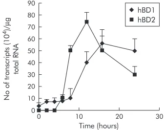
Due to the low constitutive expression of hBD1 in MKN7 cells, this cell line was chosen for further analyses. The kinet-ics of induction of hBD1 and hBD2 were studied at an MOI of 100, as this ratio of infectivity was found to be most potent, as shown above. Time course experiments showed that expres-sion of hBD2 began 3–4 hours after infection (fig 3), then markedly increased to attain maximal expression at eight hours, followed by declining levels at 12 and 24 hours. In con-trast, induction of hBD1 mRNA expression was much slower than that for hBD2 with the first increase observed at eight hours and maximum expression occurring at 12 hours post-infection.
IL-1βincreases expression of hBD1 and hBD2 in MKN7 cells
Several previous studies have shown that proinflammatory cytokines such as IL-1βand TNF-αcan increaseβ-defensin expression.15 21
We have also examined the effect of a panel of proinflammatory cytokines (IL-1β, TNF-α, IFN-γ, IL-17) on hBD1 and hBD2 mRNA expression in MKN7 cells. Among the cytokines tested, IL-1β(5 ng/ml) was found to be the only stimulant for hBD1 and hBD2 in this cell line. As observed during infection withH pylori, IL-1βalso caused an increase in hBD1 and hBD2 mRNA expression in a time dependent man-ner, with a similar earlier maximal increase in hBD2 expression (12 hoursv16 hours for hBD1; fig 4). However, the magnitude of induction of both peptides was much less and Figure 1 Dose dependent increase in humanβ-defensin 1 and 2
(hBD1, hBD2) mRNA expression in AGS (A) and MKN7 (B) cell lines withHelicobacter pylori NCTC 11637 strain. Cells were infected for 24 hours at 37°C. Results are mean (SEM) of four independent experiments. A significant increase (p<0.001) in hBD1 and hBD2 mRNA expression was observed on infection at a multiplicity of infection of 100.
C 10 100
No of bacteria/epithelial cell
1000 200 A 150 100 50 0 No of mRNA transcripts (10 4)/ µ g total RNA
C 10 100
No of bacteria/epithelial cell1000
150 B 0 No of mRNA transcripts (10 4)/ µ g total RNA 100 50 0 hBD1 hBD2 hBD1 hBD2
Figure 2 A representative quantitative reverse
transcription-polymerase chain reaction gel illustrating induction of humanβ-defensin 1 (hBD1) mRNA expression (that is, target band) with increasing multiplicity of infection in MKN7 cells.
Standard (434 bp)
Target (253 bp)
103
102
10 0
No of bacteria/epithelial cell
Figure 3 Time dependent increase in humanβ-defensin 1 and 2 (hBD1, hBD2) mRNA expression byHelicobacter pylori. MKN7 cells were coinfected at a multiplicity of infection (MOI) of 100 for 24 hours. hBD2 upregulation was initially noted at three hours with a maximum increase observed at eight hours. hBD1 mRNA expression was much slower with a maximum increase observed at 12 hours. Results are mean (SEM).
140 120 100 80 60 40 20 0
0 10 20
Time (hours) 30 No of transcripts (10 4)/ µ g total RNA hBD1 hBD2
Figure 4 Interleukin 1β(IL-1β) is a stimulant of humanβ-defensin 1 and 2 (hBD1, hBD2) mRNA expression. MKN7 cells were incubated with IL-1β(5 ng/ml) for 24 hours. As seen in fig 2, the kinetics of the increase in hBD2 expression were more rapid than for hBD1. The magnitude of response was much lower than that observed with Helicobacter pylori.
0 90
0 10 20
Time (hours) 30
delayed in IL-1βtreated cells when compared with H pylori infected cells.
In vivo expression of hBD1 and hBD2
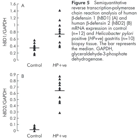
To resolve the potential effect ofH pyloricolonisation on hBD1 and hBD2 mRNA expression in the gastric mucosa, semiquan-titative RT-PCR analyses was performed on biopsies from patients with gastritis and compared with control tissue (fig 5A, B). Control biopsies showed constitutive expression of hBD1 whereas hBD2 mRNA was undetectable. Expression of hBD2 was markedly upregulated inH pyloripositive gastritis (p<0.001). Upregulation of hBD1 also achieved statistical sig-nificance (p<0.05) in gastritis compared with control tissue. To further confirm increased expression of hBD1 and hBD2 in the gastric epithelium of H pylori positive gastritis, immunohistochemistry was performed on archival paraffin sections of gastric biopsy specimens. Increased positive stain-ing for hBD1 and hBD2 peptide expression was observed in gastritis (fig 6C, D) compared with control tissue (fig 6A, B). Staining was confined to the surface epithelium and the gas-tric pits of the gasgas-tric gland, with no staining detected in the lamina propria or in inflammatory cells of the gastric mucosa.
DISCUSSION
Helicobacter pylori plays a major role in the pathogenesis of chronic antral gastritis and peptic ulcer disease.22
There is accumulating evidence that both bacterial factors and subse-quent host defence responses contribute to the ongoing inflammatory processes that determine the clinical manifesta-tions of the disease.
During infection, detection of chemokines (C-C and C-X-C) and early recruitment of neutrophils in the gastric mucosa are indicative of the active participation of innate immune mechanisms.23–25
In the present study, we have expanded the repertoire of the innate immune responses by the host to
Helicobacterinfection. Studies to date have shown constitutive expression of hBD1 by various mucosal surface epithelia, sug-gesting a “surveillance-like” role for this peptide in maintain-ing tissue homeostasis in the absence of infection.10
In the present study, we show that the constitutive expression of hBD1 can be further modulated during infection and inflam-mation. Daleet alhave also recently noted similar modulation of hBD1 expression in inflamed periodontal tissue.26
These two studies to date therefore imply a greater role for hBD1 in innate host defence than was previously thought. At present,
regulation of hBD1 gene and peptide expression during infec-tious and inflammatory episodes remains unclear. Using the sensitive technique of competitive RT-PCR, we observed induction of hBD1 mRNA expression in AGS and MKN7 cells 24 hours after infection withH pylori. We observed great vari-ation in basal expression of hBD1 in different gastric epithelial cell lines. For example, initial experiments conducted in MKN45 cells showed a spontaneous high level expression of Figure 5 Semiquantitative
reverse transcription-polymerase chain reaction analysis of human
β-defensin 1 (hBD1) (A) and humanβ-defensin 2 (hBD2) (B) mRNA expression in control (n=12) andHelicobacter pylori positive (HP+ve) gastritis (n=10) biopsy tissue. The bar represents the median. GAPDH,
glyceraldehyde-3-phosphate dehydrogenase.
1.6 1.4 1.2 1 0.8 0.6 0.4 0.2 0
hBD1/GAPDH
Control HP+ve A
0
hBD2/GAPDH
Control HP+ve B
0.1 0.2 0.3 0.4 0.5 0.6 0.7 0.8 0.9
Figure 6 Detection ofβ-defensins by immunohistochemistry. (A) (humanβ-defensin 1 (hBD1)) and (B) (humanβ-defensin 2 (hBD2)) show control gastric mucosa with very faint staining for both peptides. (C) and (D) (Helicobacter pylori positive gastritis) show positive staining for hBD1 and hBD2 peptides, respectively, in the surface epithelium and in gastric pits.
hBD1 and hBD2 in unstimulated cells which was not modulated by the bacterium. O’Neil et al have recently reported no change in constitutive expression of hBD1 in AGS cells byH pylori(MOI of 10).18
In contrast, our coculture infec-tion experiments were conducted at a greater multiplicity of infection (MOI 100), and the initial increase in hBD1 expres-sion was first detected only at eight hours. The combined effect of variation in basal hBD1 levels in different cell lines, bacterial strains, and variable numbers of infecting organisms may partly explain the results obtained in different laborato-ries.
Unlike hBD1, hBD2 peptide is not expressed by epithelia in the absence of infection. As expected, no hBD2 was detected in control cell lines and in control gastric biopsy tissue. We have confirmed upregulation of hBD2 mRNA by H pylori, as reported previously.17 18
In time course studies, we observed marked upregulation of hBD2 expression as early as three hours post-infection in MKN7 cells. The kinetics of hBD1 induction in comparison were much slower as the first increase was noted only at eight hours post-infection. At present, the identity of both bacterial virulence factors and host signalling events involved in the induction of defensin expression is limited. Although a crucial role for the bacterial “cag pathogenicity island” in induction of hBD2 mRNA expression has been observed,17
its potential role in the regu-lation of hBD1 expression has not been studied to date. Wada
et alhave recently shown unequivocally the involvement of the transcription factor nuclear factorκB in induction of hBD2.27 In contrast, no nuclear factorκB binding sites have been iden-tified in the promoter sequence of hBD1.28
The molecular mechanism(s) that may play a part in the activation of these peptides requires further investigation.
Several studies have shown that proinflammatory cytokines such as IL-1βand TNF-αcan directly increase expression of mammalian defensins.15 18 21
In the present study, we explored the effects of various cytokines on expression of hBD1 and hBD2 in gastric epithelial cell lines and found IL-1βto be a potent stimulant of hBD2 in all three gastric cell lines tested. In contrast, the effect of IL-1βon hBD1 expression was only observed in MKN7 cells with no induction in AGS and MKN45 cells. It is probable that the high basal levels of hBD1 observed in AGS and MKN45 cell lines may contribute to masking of cytokine response. Interestingly, two previous studies have reported no effect of IL-1βon hBD1 expression.18 29
As differ-ent cancer cell lines were used in the various studies, the data taken together suggest that the degree of cellular differentia-tion may play a role inβ-defensin gene regulation. Recent studies by El-Omaret alhave suggested that the presence of IL-1 gene cluster polymorphism in susceptible individuals is associated with an increased risk of H pylori induced hypochlorhydria and gastric cancer.30
At present, the role of β-defensins during inflammatory episodes is unclear but spe-cific induction by IL-1βmay have major consequences in the innate host-defence response to bacterial colonisation and pathogenesis of gastritis and related complications.
To confirm our observations of increased hBD1 and hBD2 expression byH pyloriin tissue culture cell lines, we analysed mRNA and peptide expression of both peptides in gastric biopsy specimens. Using semiquantitative RT-PCR, we found increased expression of hBD1 and hBD2 inH pylorimediated gastritis. As no hBD2 mRNA was detectable in control tissue, the increase observed in gastritis was found to be much greater than that observed for hBD1. Immunohistochemical analyses revealed a parallel increase in both hBD1 and hBD2 peptide expression during bacterial induced inflammation. This pilot study on analyses of hBD1 and hBD2 expression from human gastric biopsies suggests a potential role for these peptides in host innate defence to infection and inflammation.
H pyloriinduced chronic inflammation is dependent on the persistence of the organism in the gastric antrum and/or fun-dus. Several factors such as the low immunological activity of
H pylori lipopolysaccharide,31
bacterial expression of Lewis antigen,32 interference with antigen processing and presentation,33
and increased expression of IL-1034 have all been implicated in the increased colonisation and ineffective clearance ofH pylorifrom the gastric mucosa. The potential role and contribution of antimicrobial peptides at the surface of the gastric epithelium and their interactions with factors that favour colonisation of the bacterium are still unclear and warrant further investigation.
ACKNOWLEDGEMENT
Supported by the Special Trustees of St Bartholomew’s and the Royal London School of Medicine and Dentistry (MBE).
. . . . Authors’ affiliations
M Bajaj-Elliott, P Fedeli, G V Smith, L Maher,Department of Adult and Paediatric Gastroenterology, St Bartholomew’s and the Royal London School of Medicine and Dentistry, Turner St, London, UK
P Domizio,Department of Histopathology, St Bartholomew’s and the Royal London School of Medicine and Dentistry, Turner St, London, UK
R S Ali, A G Quinn,Centre for Cutaneous Research, St Bartholomew’s and the Royal London School of Medicine and Dentistry, Turner St, London, UK
M J G Farthing,Faculty of Medicine, University of Glasgow, South Park Terrace, Glasgow G12 8LG, UK
REFERENCES
1Kagnoff MF, Eckmann L. Epithelial cells as sensors for microbial
infection.J Clin Invest 1997;100:6–10.
2McKay DM, Baird AW. Cytokine regulation of epithelial permeability
and ion-transport.Gut 1999;44:283–9.
3Cover TL, Blaser MJ.Helicobacter pylori and gastroduodenal disease.
Ann Rev Med 1992;43:135–45.
4Bamford KB, Fan X, Crowe SE,et al. Lymphocytes in the human gastric
mucosa duringHelicobacter pylori have a T helper cell 1 phenotype.
Gastroenterology 1998;114:482–92.
5Fox JG, Beck P, Dangler CA,et al. Concurrent enteric helminth infection
modulates inflammation and gastric immune responses and reduces
Helicobacter-induced gastric atropy. Nat Med 2000;6:536–42.
6Zhang QB, Etolhi G, Dawodu JB,et al. Relationship between mucosal
levels ofHelicobacter-pylori specific IgA, interleukin-8 and gastric
inflammation.Clin Sci 1999;96:409–14.
7Censini S, Lange C, Xiang Z,et al. cag, a pathogenicity island of
Helicobacter pylori, encodes type I-specific and disease-associated
virulence factors.Proc Natl Acad Sci USA 1996;93:14648–53.
8Satin B, Del Giudice G, Della Bianca V,et al. The neutrophil-activating
protein (HP-NAP) ofHelicobacter pylori is a protective antigen and a
major virulence factor.J Exp Med 2000;191:1467–76.
9Allen LA. Modulating phagocyte activation: the pros and cons of
Helicobacter pylori virulence factors. J Exp Med 2000;191:1451–4.
10Zhao C, Wang I, Lehrer RI. Widespread expression of beta-defensin
hBD-1 in human secretory glands and epithelial cells.FEBS Lett
1996;396:319–22.
11Goldman MJ, Anderson GM, Stolzenberg ED,et al. Human
β-Defensin-1 is a salt-sensitive antibiotic in lung that is inactivated in cystic
fibrosis.Cell 1997;88:553–60.
12Harder J, Bartels J, Christophers E,et al. A peptide antibiotic from
human skin.Nature 1997;387:861.
13Valore EV, Park CH, Quayle AJ,et al. Humanβ-defensin 1: An
antimicrobial peptide of urogenital tissues.J Clin Invest
1998;101:1633–42.
14Diamond G, Bevins CL.β-Defensins: Endogenous antibiotics of the
innate host defence response.Clin Immunol Immunopathol
1998;83:221–5.
15O’Neil DA, Martin Porter E, Elewaut D,et al. Expression and regulation
of the human beta-defensins hBD-1 and hBD-2 in intestinal epithelium.J
Immunol 1999;163:6718–28.
16Hiratsuka T, Nakazato M, Date Y,et al. Identification of human
beta-defensin-2 in respiratory tract and plasma and its increase in
bacterial pneumonia.Biochem Biphys Res Commun 1998;249:943–7.
17Wada A, Mori N, Oishi K,et al. Induction of human beta-defensin 2
mRNA expression byHelicobacter pylori in human gastric cell line
MKN45 cells oncag pathogenicity island. Biochem Bipohys Res
Commun 1999;263:770–4.
18O’Neil DA, Cole SP, Martin-Porter E,et al. Regulation of human
β-defensins by gastric epithelial cells in response to infection with
Helicobacter pylori or stimulation with interleukin-1. Infect Immun
2000;68:5412–15.
19Bajaj-Elliott M, Breese EJ, Poulsom R,et al. Keratinocyte growth factor in
inflammatory bowel disease: increased mRNA transcripts in ulcerative colitis compared with Crohn’s disease in biopsies and isolated mucosal
20Ali RS, Falconer A, Ikram M,et al. Expression of the peptide antibiotics
humanβ-defensin 1 and humanβ-defensin 2 in normal human skin.J
Invest Dermatol 2001;117:106–11.
21Russell JP, Diamond G, Tarver AP,et al. Co-ordinate induction of two
antibiotic genes in tracheal epithelial cells exposed to the inflammatory
mediators lipopolysacchride and tumor necrosis factor-alpha.Infect
Immun 1996;64:1565–8
22Kuipers EJ.Helicobacter pylori and the risk and management of
associated diseases: gastritis, ulcer disease, atrophic gastritis and gastric
cancer.Aliment Pharmacol Ther 1997;11:71–88.
23Crabtree JE. Gastric mucosal inflammatory responses toHelicobacter
pylori. Aliment Pharmacol Ther 1996;10:29–37.
24Sharma SA, Tummuru KR, Blaser MJ,et al. Activation of IL-8 gene
expression byHelicobacter pylori is regulated by transcription factor
nuclearκB in gastric epithelial cells.J Immunol 1998;160:2401–7.
25Mori N, Wada A, Hirayama T,et al. Activation of intercellular adhesion
molecule 1 expression byHelicobacter pylori is regulated by NF-κB in
gastric epithelial cancer cells.Infect Immun 2000;68:1806–14.
26Dale BA, Kimball JR, Krisanaprakornkit S,et al. Localised antimicrobial
peptide expression in human gingiva.J Periodont Res 2001;36:285–94.
27Wada A, Ogushi K, Kimura T,et al. Helicobacter pylori-mediated
transcriptional regulation of the humanβ-defensin 2 gene requires NFκB.
Cell Microbiol 2001;3:115–23.
28Liu L, Zhao C, Heng HHQ,et al. The human beta-defensin-1 and
alpha-defensins are encoded by adjacent genes:Genomics
1997;43:316–20.
29Mathews M, Jia HP, Guthmiller JM,et al. Production ofβ-defensin
antimicrobial peptides by the oral mucosa and the salivary glands.Infect
Immun 1999;67:2740–5.
30El-Omar EM, Carrington M, Chow WH,et al. Interleukin-1
polymorphisms associated with increased risk of gastric cancer.Nature
2000;404:398–402.
31Moran AP. Cell surface characteristics ofHelicobacter pylori. FEMS
Immunol Med Microbiol 1995;10:271–80.
32Heneghan MA, McCarthy CF, Moran AP. Relationship of blood group
determinants onHelicobacter pylori lipopolysaccharide with host
lewis phenotype and inflammatory response. Infect Immun
2000;68:937–41.
33Molinari M, Salio M, Galli C,et al. Selective inhibition of Ii-dependent
antigen presentation byHelicobacter pylori toxin VacA. J Exp Med
1998;187:135–40.
34Bauditz J, Ortner M, Beirbaum M,et al. Production of IL-12 in gastritis
relates to infection withHelicobacter pylori. Clin Exp Immunol
1999;117:316–23.
www.gutjnl.com
Sign up to receive the table of contents by email every month. You can select from three alerts: Table of Contents (full), TOC Awareness (notice only); Gut related announcements.