A Novel Approach for Glaucoma Detection Using Cup Disk and
Rim Disk Ratio
Dr.Arvind Kundu ; G.Priyanka ; K.Priyanka 1Co-Guide ; 2Guide ; 3 M.Tech Student
Dept Of Ece, Scient Institute Of Technology,Ibrahimpatnam,Hyderabad.
ABSTRACT
Glaucoma is a continual ailment
which if no longer detected in early phases
can lead to permanent blindness. The
clinical techniques used by
ophthalmologists like HRT and OCT is
highly-priced and time-consuming. As a
result, there is a have got to advance
automatic pc aided process which is able
to observe the stage of glaucoma
efficiently and in much less time.
Optic disk Ratio and optic cup
Ratio are high facets which help in
diagnosing glaucoma. As a consequence
suitable segmentation of optic disk and
optic cup plays a predominant role in
detecting the disease. In this paper, an
adaptive threshold cantered approach
which is unbiased of snapshot fine and
invariant to noise is used to section optic
disk, optic cup, Neuroretinal rim and cup
to disk ratio is calculated to monitor
glaucoma. An additional ocular
parameter, rim to disk ratio is regarded
which in mixture with CDR gives extra
reliability in deciding on glaucoma.
Operations are utilizing to the
method the whole photo to makes the
procedure extra amazing. In the end, an
SVM classifier has been used to categorize
the graphics as glaucomatous or not
glaucomatous.
The experimental outcome bought
are in comparison with those of
ophthalmologist and are located to have
extra efficient and high accuracy of 90%.
Also, in addition, the proposed system is
faster having low computational cost.
As compared to different tricky
instruments, digital fondues camera is
more cost effective and is more commonly
used in general eye examination.
INTRODUCTION
Changes in the structural look of the optic
nerve head (ONH) and Retinal Nerve Fiber
Layer (RNFL) had been reported to
precede the development of visual rea loss
in glaucoma.1–3 Detection of ONH and
RNFL damage is, accordingly, vital for
early prognosis of glaucoma. Latest
concentration has additionally been
thickness measurements for glaucoma
prognosis.
Retinal ganglion cells are also
misplaced in the posterior pole in
glaucoma, the place these cells could
constitute 80% to 85% of the retinal
thickness in the macular neighborhood.
Optical coherence tomography (OCT) is
an optical imaging system that presents
high resolution and reproducible graphics
of the RNFL that discriminate
glaucomatous from healthful topics.6 –
eleven even though OCT has been used,
for probably the most phase, to evaluate
RNFL thickness, latest upgrades in the
program even have made feasible the
evaluation of ONH topography and
macular thickness for glaucoma prognosis
and comply with-up.
A prior investigation validated that
OCT ONH measurements correlate well
with topographic measurements obtained
by way of nonlocal scanning another
imaging procedure that evaluates the
ONH.12 other reports have also shown
that OCT macular thickness measurements
are drastically thinner in glaucomatous in
comparison with healthy eyes.5,thirteen–
15 although the capacity of OCT ONH and
macular thickness measurements to
differentiate glaucomatous from healthful
topics has been pronounced to be slash
than RNFL thickness parameters, no be
taught has but provided a comparison of
those three methods within the equal
population.
EXISTING METHOD
Optic disk and optic cup are top
elements which help in diagnosing
glaucoma. Accordingly, proper
segmentation of optic disk and optic cup
plays a primary position in detecting the
sickness. In this paper an adaptive
threshold established a system which is
unbiased of picture first-class and invariant
to noise is used to phase optic disk, optic
cup, Neuroretinal rim, and cup to disk ratio
is calculated to monitor glaucoma. Another
ocular parameter, rim to disk ratio (RDR)
is viewed which in combination with CDR
gives more reliability in picking out
PROPOSED APPROACH
In current method rather of
processing usually a retinal picture, by
way of taking the region around optic disk,
they stated whether or not the eye is
glaucomatous or now not. That is the
drawback of discovering the Glaucoma the
place it exists external the Optic Disk.
Via since this problem and we
enforce new method for detecting
glaucoma to procedure the entire photo
after which decided where the eye defects.
RGB fundus image is used as an enter. The
most important regions of detecting
Glaucoma are optic Cup, Optic disk, and
Rim subject.
By utilizing these three parameters
and Processing the entire photograph then
we get the Output, the place the attention
is effected with Glaucoma and Which
stage the attention is suffering from
Glaucoma. Additionally, we use
Morphological Operations to get the
Output extra Accuracy and more effective.
This system helps in rapid processing and
significant computerized screening of
glaucoma.Eachsubjectunderwenta
opthalmogic examination including review
of medical history, best-corrected visual
acuity, slit-lamp bio microscopy,
intraocular pressure (IOP) measurement
using Goldman application tonometry,
gonioscopy, dilated fundoscopic
examination using a 78-diopter lens,
stereoscopic optic disk photography, and
automated perimeter using 24-2 Swedish
Interactive Threshold Algorithm. To be
included, subjects had to have best
corrected visual acuity of 20/40 or better,
spherical refraction within 5.0 diopters and
cylinder correction within 3.0 diopters, and
open angles on gonioscopy.
Eyes with the coexisting retinal
disease, uveitis, or no glaucomatous optic
neuropathy were excluded from this
investigation. One eye of each patient was
randomly selected for inclusion in the
study. Normal control eyes had intraocular
pressures of 21 mm Hg or less with no
history of increased IOP and a normal
visual field result. The normal visual field
was defined as a mean deviation and
pattern standard deviation within 95%
confidence limits and a Glaucoma
Hemifield Test (GHT) within normal
limits.
Normal control eyes also had a
healthy appearance of the optic disk and
RNFL (Retinal Nerve Fiber Layer,
cupping, optic disk hemorrhage, or RNFL
defects), as evaluated by clinical
glaucomatous if they had repeatable (two
consecutive) abnormal visual field test
results, defined as a PSD outside of the
95% normal confidence limits or a
Glaucoma Hemi field Test result outside
normal limits, regardless of the appearance
of the optic disk. Average MD of the
glaucomatous eyes on the visual field test
nearest the imaging date was 4.96 dB.
According to the
Hodapp-Parrish-Anderson16 grading scale of severity of
visual field defects, 61 patients (69%)
were classified as having early visual field
defects, 15 patients (17%) had moderate
defects, and 12 patients (14%) had severe
visual field defects.
Although the appearance of the
optic disk on stereo photographs was not
used as an inclusion criterion, the results of
stereo photograph assessment were used
for comparison with Stratus OCT ONH
measurements. Simultaneous stereoscopic
optic disk photographs (TRC-SS; Topcon
Instrument Corp of America, Paramus,
New Jersey, USA) were evaluated by two
experienced graders, and each was masked
to the subject’s identity and to the other
test results.
The graders visually estimated the
horizontal and vertical cup/disk ratios
based on the contour of the cup. The mean
value of the two graders was used as a
final grading. Subjects underwent ocular
imaging with dilated pupils using the
commercially available optical coherence
tomograph, Stratus OCT (Carl Zeiss
Meditec, Dublin, California, USA). All
patients had optic nerve head, RNFL
thickness, and macular thickness scans
obtained during the same visit.
OCT employs the principles of the
low-coherence interferometer and is
analogous to ultrasound B-mode imaging
but uses light instead of sound to acquire
high-resolution images of ocular
structures. In brief, a low coherence
near-infrared (840 nm) light beam is directed
onto a partially reflective mirror (beam
splitter) that creates two light beams, a
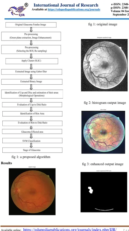
fig 1: a proposed algorithm
Results
fig 1: original image
fig 2: histogram output image
fig 4: our proposed output image for perfect segmentation disease image
CONCLUSION
The evaluation of stratus oct
program-provided parameters confirmed
that par papillary RNFL measures and
ONH topographic parameters had the
perfect vigour to discriminate
glaucomatous from healthy eyes. Areas
beneath the ROC curves and sensitivities
at reasonable and excessive specificities
were identical for the high-quality
parameters from each of these two
approaches to evaluation.
We additionally observed that a
blend of selected RNFL and ONH
parameters in a linear discriminate operate
resulted in additional improvement of the
diagnostic accuracy of OCT. The ROC
curve areas for the Stratus OCT RNFL
measurements have been similar to those
obtained with the prior models of this
technological know-how.
The areas beneath the ROC curves
for the prior OCT models were reported to
range from zero.79 to zero.94, depending
on the parameter and characteristics of the
population. In reviews evaluating the
diagnostic potential of a couple of OCT
parameters, the RNFL thickness in the
inferior region regularly had the first-class
performance to discriminate healthful eyes
from eyes with early to reasonable
glaucoma with sensitivities between sixty
seven% and seventy nine% for
specificities 90%.9,11,26 In our be taught,
the parameter inferior thickness also had
the highest field under the ROC curve,
with sensitivity of 65% for specificity at
95%. The parameter ordinary thickness
also had a equivalent efficiency.
REFERENCES
[1] Inoue N., Yanashima K., Magatani
K .Kurihara T. “Development of a simple
diagnostic method for the glaucoma using
ocular fundus pictures” proceedings of
2005 IEEE, Engineering in medicine and
biology 27th annual conference, shanghai,
China, pp 3355-3358, January 2006.
[2] Aquino A., Gegundez-Arias M.E,
Marin D. “Detecting the optic disk
boundary in digital fundus images using
morphological, edge detection and feature
extraction techniques” IEEE transactions
on medical imaging, Vol.29, pp1860-1869,
November 2010.
[3] Li.H, Chutatape O. “A model
based approach for automated feature
extraction in fundus images”, proceedings
of 9th IEEE international conference on
computer vision (ICCV’03),Vol.1,pp
[4] Yang X., Hamaguchi S., Sun Y.,
Xiao S. “Detect of optic disk centre based
on Gaussian vessel detector and tangent
information transform” IEEE 2011, 4th
international conference on biomedical
engineering and informatics
(BMEI),Vol.1,pp 250-254, October 2011.
[5] Nayak J., Acharya R., Bhatt P.S.,
Nakul Shetty,Lim T.C. “Automated
diagnosis of Glaucoma using fundus
images”, Springer science + Business
media, LLC 2008.
[6] Fengshou Yin, Jiang Liu, Damon
Wing Kee Wong, Ngam Meng Tan, Carol
Cheung, Manibhaskaran, Tien Yin Wong
“Automated segmentation of optic disk
and optic cup in fundus images for
glaucoma diagnosis”, 25th international
Symposium on computer based medical
system pp.1-6, June 2012.
[7] Dua S., Acharya U.R.,Chowriappa
P.,Sree S.V., “Wavelet based energy
features for glaucomatous image
classification” IEEE transaction on
information technology in biomedicine,
Vol. 16, no.1,pp 80-87, January 2012.
Dr. Arvind Kundu
He did B. Tech from H.P. University (SHIMLA) in Electronics & Communication. He did M. Tech from M.D. University (ROHTAK) in Electronics & Communication Engineering. He did Ph. D from Ranchi University and area of research is ADHOC Networks, EMBEDDED System, Cryptography, Message authentication Protocol, Image Processing, Routing protocol etc. He is working as HOD ECE Department at SCIENT INSTITUTE OF TECHNOLOGY, IBRAHIMPATNAM.
Ms. G.PRIYANKA
She received M.E from OSMANIA UNIVERSITY in Embedded Systems and VLSI Design. She did B.Tech from JNTU HYDERABAD in Electronics and Communication Engineering. She is working as an Assistant Professor in SCIENT Institute of technology. Her areas of interest in Digital concepts and developing the various IEEE based projects using Verilog, System Verilog, and Verification. Developing on algorithms of Digital oriented concepts and concepts on modulation using MATLAB and QCA relevant concepts. She is specialized in developing Front End and Back end based projects and also