Hypoxia promotes thyroid differentiation
of native murine induced pluripotent stem cells
YIPENG YANG
1,#, YUNSHU LU
1,#, TONG CHEN
1, SHENGLAI ZHANG
1, BINGFENG CHU
1, YURONG GONG
1,
WEIXIN ZHAO
2, JIAN ZHU*
,1and YINGBIN LIU*
,11General Surgery Department, Xin Hua Hospital of Shanghai Jiao Tong University School of Medicine, Shanghai, China and 2Wake Forest Institute for Regenerative Medicine, Wake Forest School of Medicine, Winston Salem, NC, USA
ABSTRACT Hypothyroidism is a very common hormonal deficiency and the stem cell technology which developed in the recent years may offer a therapeutic strategy for treating this disorder. Hy-poxia has been demonstrated to play an important role in embryonic formation and development and to modulate stem cell differentiation. However, the influence of oxygen tension on thyroid differentiation has not been studied. In this study, we used murine induced pluripotent stem (iPS) cells for thyroid cell differentiation under normoxic and hypoxic conditions and compared differ-entiation efficiency in morphology, function, gene and protein expression under both conditions. We found that hypoxia promoted adhesion and outgrowth of embryoid bodies (EBs) derived from murine iPS cells. Expression of endodermal markers (Foxa2 and Gata4) and thyroid transcription factors (Pax8 and Nkx2.1) was increased by hypoxia at both gene and protein levels during early-mid differentiation stages (p<0.05). And so were the thyroid specific markers NIS and TSHR at the end of the experiment (p<0.05). In addition, functional iodide uptake by differentiated cells was also increased after hypoxia. Thyroid differentiation from iPS cells is enhanced under hypoxia and this may involve hypoxia inducible factors (HIFs) and their downstream gene FGF2. Our data offer a foundation for understanding thyroid development and provide a potentially more efficient way to use cell therapy for treating thyroid deficiency.
KEY WORDS:
differentiation, thyroid, hypoxia, promotion, iPS cell
Introduction
Thyroid gland produces two essential hormones which impact almost every organ and cell of the body and get involved in variet-ies of biological processes (Sewell et al., 2014). Hypothyroidism, an irreversible change (Ma et al., 2015), is the most common hormonal deficiency and can be precipitated by various diseases (Hollowell et al., 2002; McLachlan et al., 2004). Recently, stem cell technologies have advanced and provide a new strategy to treat thyroid deficiency by tissue engineering and regenerative medi-cine. Successful differentiation from murine embryonic stem cells (mESCs) to thyrocyte-like cells was first reported by Lin’s group (Lin et al., 2003). Arufe and colleagues described directed deriva-tion of thyroid follicular cells by ESCs and proved that thyrotrophin, insulin, and insulin-like growth factor-1 (IGF-1) were essential for this differentiation (Arufe et al., 2006; Arufe et al., 2009). For in
www.intjdevbiol.com
*Address correspondence to: Yingbin Liu. General Surgery Department, Xin Hua Hospital of Shanghai Jiao Tong University School of Medicine, 1665 Kongjiang Road, Shanghai 200092, China. E-mail: liuybphd@126.com. Tel: +86-13918803900 - or Jian Zhu. General Surgery Department, Xin Hua Hospital of Shanghai Jiao Tong University School of Medicine, 1665 Kongjiang Road, Shanghai 200092, China. E-mail: 69zj@sina.com. Tel: +86-18918601360.
#Note: These authors contributed equally to this work.
Accepted: 17 June 2016. Edited by: Sally Dunwoodie.
ISSN: Online 1696-3547, Print 0214-6282
© 2016 UPV/EHU Press Printed in Spain
Abbreviations used in this paper: DE, definitive endoderm; FGF2, fibroblast growth
factor 2; HIF, hypoxia inducible factor; iPS cell, induced pluripotent stem cell; NIS, sodium iodide symporter; Tg, thyroglobulin; TPO, thyroperoxidase; TSHR, thyrotrophin receptor.
(Ma et al., 2015).
The relatively hypoxic uterus is necessary for early formation and development of mammalian and human embryos (Dunwoodie, 2009; Fischer et al., 1993), and the influence of hypoxia on prolif-eration, differentiation and function of cells, especially in stem cells is of interest (Millman et al., 2009; Mohyeldin et al., 2010; Wion et al., 2009). Research indicates that culturing stem cells in a low oxygen environment better maintained pluripotent differentiation ability and reduced chromosomal abnormalities (Ezashi et al., 2005; Zachar et al., 2010). Hypoxia can enhance differentiation from stem cells of various cell types such as endothelial (Prado-Lopez et al., 2010; Shin et al., 2011) and neural cells (Mondragon-Teran et al., 2009) as well as retinal, lung, and heart progenitor cells (Bae et al., 2012; Garreta et al., 2014; van Oorschot et al., 2011). Therefore, we hypothesized that oxygen tension may influence differentiation into thyroid cells. In this study, we performed the thyroid induction with unmodified iPS cells which are safer in clinical application than virally transfected iPS cells with Nkx2.1/Pax8 by a new protocol. We mimicked physiological hypoxic environments and compared differentiation efficiency in morphology, function, gene and protein expression to normoxic condition. We found that the low oxygen tension could enhance the thyroid derivation from murine iPS cells (miPSC).
Results
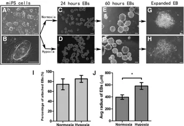
Embryoid body (EB) formation and adhesion under normoxia and hypoxia
Fig. 1A, B showed murine iPS cell clones on irradiated MEF feeder cells. In our study, the suspension cultured miPS cells were able to form EBs in the EB induction medium under both hypoxic and normoxic conditions after 12-24 hours. EB size, density and numbers formed after 24 and 60 h were similar under both conditions (Fig. 1C–F). EB adhesion onto gelatin coated culture dishes and outgrowth after reattachment are necessary for further differentiation
of iPS cells in our protocol. Thus, we compared EB adhesion ratios and cell morphology under both conditions and Fig. 1I showed that the hypoxia bar was higher but this was not statistically different (Fig. 1I.). Morphologically, EBs had better outgrowth and spread farther after reattachment under hypoxia during the same culture period (Fig. 1G, H, J). Therefore, hypoxia promoted adhesion and outgrowth of EBs but low oxygen does not influence the formation of EBs.
Hypoxia promotes thyroid endoderm induction
After EB formation, we used high concentration of Activin A (50ng mL-1) and gradually reduced FBS to induce the definitive endoderm (DE), the next step of thyroid differentiation from iPS cells. An anterior foregut endoderm was induced with a high con-centration of NOGGIN (BMP signaling inhibitor) and SB-431542 (TGF-b signaling inhibitor). In this study, We also supplemented differentiation medium with hTSH, mBMP4, mFGF2 and FGF10 for the induction of thyroid endoderm.
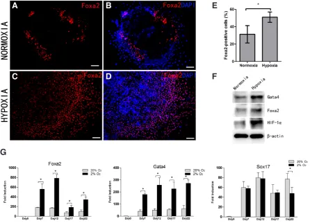
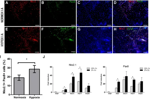
To measure gene expression, we isolated total RNA from cultured cells at specific times and quantitative real-time PCR confirmed that cells cultured under both conditions expressed Foxa2, Gata4 and Sox17—all endodermal markers (Mora-Castilla et al., 2014) from day 7 of differentiation (Fig. 2G). The levels of Gata4 and Foxa2 were much higher in hypoxia (more than 3 folds) on day 7 and 12 (Fig. 2G). But Sox17 expression under both culture conditions was similar on these two time points. mRNA expression of thyroid transcription factor 1 (TTF1, Nkx2.1) and Pax8 which were consider to be the crucial transcription factors to promote the expression of thyroid-specific genes (Mu et al., 2012) were also measured and these two factors were confirmed during the differentiation under both culture conditions and expression both was significantly up-regulated with hypoxia in the endodermal stage (Fig. 3J).
With immuno-fluorescent staining, we confirmed that cells were positive for Foxa2 (located in the periphery of expanded EBs) on day 10 under both conditions (Fig. 2A–D). Foxa2-positive cells
Fig. 1. Formation and adhesion of em-bryoid bodies (EB) under normoxic and hypoxic conditions. (A,B) Mouse iPS cell clones on irradiated MEF feeder cells under low and high magnification. (C,E)
under hypoxic conditions were greater than under normoxic cul-ture (Fig. 2E, p<0.05). Cells that co-expressed Nkx2.1 and Pax8 potentially correspond to thyrocytes, so double-staining for these two markers was done on day 15. We found the better outgrowth of the cell clusters than day 10 of differentiation and there were abundant Nkx2.1-positive and Pax8-positive cells in both culture conditions (Fig. 3). The percentage of Nkx2.1+ Pax8+ cells in hy-poxia was significantly higher than in normoxia (Fig. 3I, p<0.05). In addition, thyrotrophin receptor (TSHR) was expressed at the relatively early stage of differentiation in a similar area as Foxa2 and immuno-fluorescent staining confirmed stronger expression of TSHR protein under hypoxia (Fig. 4). Besides that, western blot data on day 10 confirmed increased Foxa2 and Gata4 protein ex-pression in response to low oxygen tension (Fig. 2F). In summary, enhancement of thyroid endodermal markers expression occurs with hypoxia during this stage.
HIFs are important transcription factors that mediate cell respons-es to hypoxia (Ng et al., 2010). HIFs contain several subtyprespons-es which can affect hundreds of downstream genes in differentiation and play a crucial role in embryonic development. Thus, we measured HIF-1a and HIF-2a expression under both conditions and quantitative real-time PCR data (Fig. 5) showed that hypoxia significantly increased expression of both HIF-1a and HIF-2a during thyroid differentiation
(p<0.05) and these increases were impressive during early stages of differentiation (Fig. 5E). Fig. 5E also confirmed down-regulation of HIF-1a and HIF-2a during the later differentiation period. The result of western blot indicated stronger expression of HIF-1a protein was induced by hypoxia (Fig. 2F) and immuno-fluorescent staining for HIF-1a on day 10 of differentiation indicated that HIF-1a was expressed widely and strongly among cells with hypoxia but not as much with normal oxygen (Fig. 5A-D). As one of the important downstream genes of HIFs (Simon et al., 2008), fibroblast growth factor 2 (FGF2) is considered to be necessary for thyroid lineage specification from mouse pluripotent stem cells (Kurmann et al., 2015). In order to avoid the interference of exogenous factors, we measured mRNA expression of FGF2 under both conditions with HIFs and the result indicated that hypoxia also significantly increased the level of FGF2 at all the time points (Fig. 5E).
Hypoxia promotes the induction of thyroid-like cells from the anterior foregut endoderm cells
After the thyroid endoderm stage, IGF1, KI and high concentra-tions of hTSH which could push the endodermal cells to thyroid cells were used for further differentiation. After expression of Nkx2.1 and Pax8 at the onset of thyroid development, thyroglobulin (Tg, a precursor of thyroxine), thyroperoxidase (TPO, needed for thyroid
Fig. 2. Hypoxia enhances the expression of the endodermal markers.(A-D)Representative images of immuno-fluorescent staining for Foxa2 on day 10 of the differentiation process under hypoxia and normoxia. Scale bars = 100 mm. (E) Percent Foxa2+ cells under hypoxia or normoxia. Significant differences exist between the two culture conditions. *(p<0.05). (F) Western blot analysis on day 12 of differentiation. b-actin was an internal control.
(G) Expression levels of the endodermal markers Foxa2, Gata4 and Sox17 relative to undifferentiated miPS cells (day0) under hypoxic (black bar) and normoxic conditions (gray bar) by quantitative real-time PCR. Data are from three independent experiments and p<0.05 (*) indicates significant differ-ences between groups.
B
C
D
A
E
F
hormone synthesis), sodium/iodide symporter (NIS, transport I- for synthesis of thyroid hormone) and thyroid stimulating hormone receptor (TSHR) were regarded as the thyroid-specific markers for mature and functional thyroid follicular cells (Lin et al., 2003; Arufe et al., 2006). In our study, TPO, NIS and TSHR were
ex-pressed by the induction protocol and their expression as well as that of Nkx2.1 and Pax8, were significantly higher under hypoxia (p<0.05, Fig. 6I). But the expression of Tg was not detected at any time point in this study.
Co-expression of NIS and TSHR is an exclusive characteristic of functional thyrocytes, so we measured NIS and TSHR protein at the end of differentiation and the polygonal glandular epithelium like cells which were positive for NIS and TSHR grew in spreading clusters under both conditions on day 22 (Fig. 6A-H). These cells could be regarded as the thyroid like cells and appeared in bigger pools with hypoxia. The percentage of NIS+TSHR+ cells under hypoxic condition was significantly higher than under normoxic condition (Fig. 6J, p<0.05). Western blot showed higher expres-sion of NIS and TSHR after differentiation under hypoxia (Fig. 6K). For the function analysis, the radioactive iodide uptake of the
Fig. 4. Representative images of immuno-fluorescent staining for TSHR on day 10 of the differentiation process. (A) TSHR-positive cells (green) in normoxic culture (B) merged picture with DAPI. (C) TSHR-positive cells in hypoxic culture and (D) merged picture with DAPI. TSHR was expressed at the early stage of differentiation in the periphery of expanded EBs and showed stronger expression under hypoxia. Scale bars, 100 mm.
B
C
D
A
G
B
C
D
E
F
H
A
I
J
Fig. 5. Hypoxia increases the expression of HIFs and its downstream gene FGF2 during the thy-roid differentiation.(A-D)
Representative images of immuno-fluorescent stain-ing for HIF-1a on day 10 of the differentiation process under normoxia (A,B) and hypoxia (C,D). Scale bars, 100 mm. (E) Expression levels of HIF-1a, HIF-2a and FGF2 relative to undif-ferentiated miPS (day0) cells under hypoxic (black bar) and normoxic conditions (gray bar) by quantitative real-time PCR. Data are from three independent experiments and p<0.05 (*) indicates significant differences be-tween groups.
B
C
D
A
E
Fig. 6. Hypoxia promotes the induction of thyroid-like cells. (A-H)Representative images of double immuno-fluorescent stain-ing TSHR and NIS on day 22 of the differentiation process under hypoxia and normoxia. The results showed the polygonal glandular epithelium like cells which were positive for NIS and TSHR grew in spreading clusters. Scale bars, 100 mm. (I) Expression levels of the thyroid specific markers NIS, TSHR and TPO relative to undif-ferentiated miPS cells (day0) under hypoxic (black bar) and normoxic conditions (gray bar) by quantitative real-time PCR. (J) Percent TSHR+ NIS+ cells in hypoxic and normoxic culture conditions. Significant dif-ferences exist between the two culture conditions. *(p<0.05). (K)
Western blot analysis on day 22 of differentiation. b-actin was an internal control. (L) Radioactive iodide uptake after 22 days of dif-ferentiation and undifferentiated miPS cells were the control. Data are from three independent experi-ments performed in triplicate and
p<0.05 (*) indicates significant differences between groups.
G
B
C
D
E
F
H
A
I
differentiated cells was measured at the end of the experiment. We observed that I- uptake ability of the cells could be induced by our differentiation protocol under both oxygen conditions (Fig. 6L). Since the basal level of iodide uptake with the addition of NIS blocker sodium perchlorate was stable, the iodide uptake of the cells differentiated under hypoxia was significantly higher than under hypoxia (Fig. 6L, p<0.05).
Discussion
Thyroid deficiency is common and about 12% of the population in US may suffer the thyroid related conditions during lifetime (Sewell et al., 2014; Agrawal et al., 2015). There have been lots of studies about stem cell technologies which can be the key tools for tissue engineering and regenerative medicine to treat this condition in the recent years. We optimized a differentiation protocol with non-Nkx2.1/Pax8 modified iPS cells which were considered safer in clinical application. Hypoxia is considered to be associated with some pathological states, such as ischemia, inflammation and some cancers (Semenza, 2001). For example in the thyroid related field, Warner S. Simonides’ study dem-onstrated that hypoxia may reduce metabolic rate in the tissue during hypoxic-ischemic injury through HIF-1 and DIO3 mediated thyroid hormone signaling regulation (Simonides et al., 2008). However, there was no report about the effect of oxygen tension on thyroid differentiation and thyroid hormone release. We mim-icked 2% oxygen tension at EB induction and found that EB size and number did not differ under hypoxic or normoxic conditions (Fig. 1). Although this result was unexpected to some extent, our data for EB adhesion and outgrowth are similar to other reports (Bae et al., 2012; Garita-Hernandez et al., 2013; Lee et al., 2012) and this suggests a promoting effect of hypoxia. Gelatin is com-monly used to coat the culture plates and dishes for cell growth and differentiation due to its beneficial physical and biochemical properties. Our protocol also use gelatin coated culture plates for EB attachment and further differentiation. However, the cells can only form 2D monolayer on the gelatin coated plates. Maybe matrigel, collagen or certain type of hydrogel can be used in further study for the formation of 3D structure.
Ectoderm, mesoderm, and endoderm are within EBs derived from iPS cells (Poh et al., 2014) and thyroid cells arise from a specific domain of the anterior foregut endoderm which devel-ops from the definitive endoderm (DE) during embryogenesis (Grapin-Botton et al., 2000). Therefore, we transformed EBs into DE with high concentrations of Activin A and supplemented differentiation medium with BMP and TGF-b signaling inhibitors to achieve anteriorization of derived DE. The originally formed EBs were trypsinized into single cells in our modified protocol so that the cells could be sufficiently exposed to Activin A and other factors. We found that the formation of EBs was difficult for murine iPS cells without serum, but serum is considered a poor inducer of endoderm (Kubo et al., 2004). Therefore, we used the gradually reduced FBS in endodermal induction. In this stage, we proved that low oxygen increased expression of the endoderm-specific markers Gata4 and Foxa2 and this result indicated that hypoxia may take effect from the early differentia-tion of thyroid cells. We saw no similar changes for Sox17 and why this occurred is not clear. With immuno-fluorescent staining, we noted that cells expressing Foxa2 (important transcription
factor associated with development of endoderm-derived tissues) was identified in the periphery of expanded EBs due to more exposure to differentiation medium. This also suggested the importance of EB attachment and outgrowth during differentia-tion. Meanwhile, early expression of TSHR at a similar area to Foxa2 illustrated an important role of TSH in early-mid stages and enhancement by hypoxia. Nkx2.1 and Pax8 are key regu-lators of thyroid development (Altmann et al., 2005; Di Palma et al., 2003) and Nkx2.1 or Pax8 knock-out mice may manifest thyroid agenesis and congenital hypothyroidism occur in the patients with mutations of these two genes (Krude et al., 2002; Macchia et al., 1998). We found more Nkx2.1+Pax8+ cells which are thought to have the potential to become thyroid cells under hypoxic culture. Such superiority due to hypoxia may directly result in better thyroid differentiation.
Conclusion
We have performed the thyroid differentiation from murine iPS cells under both hypoxic and normoxic conditions by using a new method. We offer the first evidence of the influence of thyroid in-duction by oxygen tension. Morphology, gene, protein and function analysis suggest that hypoxia promoted adhesion and outgrowth of EBs derived from murine iPC cells, and enhanced endodermal induction and thyroid differentiation. HIFs mediated regulation of FGF2 may get involved in this promotion and our data offer a foundation for understanding thyroid development and a potentially efficient approach for cell therapy.
Materials and Methods
Maintenance of mouse iP cells in culture
A mouse induced pluripotent stem cell line (mouse primary iPS cells-WP5) was purchased from Stemgent (08-0007; Cambridge, MA) and it was generated from unmodified mouse embryonic fibroblasts (Meissner
et al., 2007). The mouse iPS cells were maintained on feeder cells
(irra-diated mouse embryonic fibroblast, MEF) in miPSC cell culture medium composed of knockout DMEM (Gibco, Grand Island, NY), 15% (v/v) embryonic stem cell-qualified fetal bovine serum (FBS, Gibco), 1% (v/v) non-essential amino acids (NEAA, Gibco), 1% (v/v) L-glutamine (Gibco), 300 mM b-mercaptoethanol (Sigma-Aldrich St.Louis, MO) and 500 U ml-1
Stemfactor recombinant mouse LIF (Stemgent). Cells were incubated at 37 °C under 5% CO2 and 20% O2.
Thyroid differentiation from miPS cells
Hypoxic culture conditions were created using a Thermo Scientific incuba-tor (HERAcell 150i, Waltham,MA). Cells were incubated in normoxic (20% O2) and hypoxic condition (2% O2) at the initiation of differentiation and these
oxygen tension conditions were maintained throughout the experiment. Definitive endoderm Formation. Before embryoid body (EB) induction, cells were dissociated with 0.05% trypsin for 3 mins and incubated on 0.1% gelatin coated-culture dishes for 45 min at 37 °C to separate MEF feeder cells. Then, miPS cells were transferred into 60-mm culture-grade petri dishes coated with 2% agar (1x10⁵ cells mL-1) for self-aggregation.
EB induction medium was composed of Iscove′s Modified Dulbecco′s Medium (IMDM, Gibco), 5% (v/v) PFHM-II Protein-Free Hybridoma Me-dium (Gibco),15% stem cell-qualified FBS (Gibco), 1% NEAA (Gibco), 1% L-glutamine (Gibco), 100 mM b-mercaptoethanol (Sigma-Aldrich) and 0.5 mM ascorbic acid (Sigma-Aldrich). After 60 h in suspension culture, EBs were harvested with a 40-mm cell strainer and trypsinized (1 min, 37 °C) into single cells which were subsequently plated into definitive endoderm induction medium consisting of 75% (v/v) IMDM (Gibco), 25% (v/v) Ham’s Modified F12 medium (Cellgro, Manassas, VA), 10% stem cell-qualified FBS, 5% (v/v) knockout serum replacement medium (KSR, Gibco), 1% NEAA, 1% L-glutamine, 100 mM b-mercaptoethanol, 0.5 mM ascorbic acid, 5 mg mL-1 transferrin (Sigma-Aldrich), 5 mg mL-1 insulin, 50 ng mL-1 recombinant
Activin A (R&D Systems, Minneapolis, MN) and 100 mU mL-1 recombinant
human thyroid-stimulating hormone (hTSH, Fitzgerald Industries, Acton, MA). After 12 h, culture medium was changed to definitive endoderm in-duction medium supplemented with 3% (v/v) FBS and 10% KSR. After 36 h, EBs were transferred to serum-free endoderm induction medium with 15% KSR and incubated for another 24 h.
Thyroid endoderm formation. After 120 h (5 days) of differentiation, the EBs were harvested again and re-plated in 0.1% gelatin coated 24- and 6-well culture plates (5-8 EBs/cM2) for adhesion culture in anterior
defini-tive endoderm differentiation medium composed of 75% IMDM (Gibco), 25% Ham’s Modified F12 medium (Cellgro), 1% stem cell-qualified FBS (Gibco), 15% KSR, 1% NEAA, 1% L-glutamine, 100mM b-mercaptoethano, 0.5 mM ascorbic acid, 5mg mL-1 transferrin, 5mg mL-1 insulin, 100mU mL-1
hTSH, 50ng mL-1 mNoggin (R&D Systems) and 10mM SB431542
(Sigma-Aldrich). For the EB adhesion test, we counted the number of the attached EBs on the culture plates after two days adhesion culture and calculated the percentage in the initially seeded EBs. After total 7 days differentiation, the medium was switched to thyroid differentiation medium (75% IMDM, 25% Ham’s Modified F12 medium, 15% KSR, 1% NEAA, 1% L-glutamine, 100mM b-mercaptoethanol, 0.5 mM ascorbic acid, 10mg mL-1 transferrin,
7.5mg mL-1 insulin, 1mU mL-1 hTSH) supplemented with 10ng mL-1 mBMP4
(R&D Systems), 500ng mL-1 mFGF2 (R&D Systems) and 100ng mL-1 FGF10
(R&D Systems). The cells were grown for another 5 days. In addition, to compare the outgrowth of EBs under both oxygen conditions, the average radius of expanded EBs was measured (the distance from the center of the cells to the periphery in multiple directions) 4 days after reattachment. Thyroid differentiation. On day 12 of differentiation, culture medium was changed to thyroid differentiation medium supplemented with 50 ng mL-1 recombinant mouse IGF1 (R&D Systems). Cells were incubated for
10 days and on differentiation day 16, KI (100mM, Sigma-Aldrich) was
added into the medium.
Immuno-fluorescence staining
Cells cultured in 24-well plates were stained on the scheduled time points. After fixing samples in 4% (v/v) paraformaldehyde for 15 min on ice, some samples were permeabilized with 1% (v/v) triton X-100 for nuclear staining. After three washings in PBS, samples were incubated in serum-free protein blocking (Dako) solution at room temperature for 1 h and then with primary antibodies (Dako) overnight at 4 °C (300 mL/well).
Then, cells were washed and incubated in secondary antibodies for 1.5 h at room temperature in the dark. Nuclei were counterstained with DAPI (1:500, Invitrogen) and samples were mounted with mounting medium (Vectashield, Burlingame, CA). Samples were incubated in secondary antibody solution only for negative controls. Images were taken under a fluorescent microscope (Leica DMI3000B) and analyzed using ImageJ 1.46r. Four replicates were performed and five random fields of each as-say were taken for analysis
The antibodies we used: polyclonal rabbit anti-sodium iodide symporter (NIS) antibody (sc-134515, Santa Cruz Biotechnology, CA, 1:50), monoclonal mouse anti-TSH receptor antibody (sc-53542, Santa Cruz Biotechnology, CA, 1:50), monoclonal rabbit anti-Foxa2 antibody (ab108422, Abcam, 1:350), monoclonal mouse anti-Pax8 antibody (MA1-117, Invitrogen, 1:100), monoclonal rabbit anti-Nkx2.1 antibody (ab76013, Abcam, 1:300), monoclonal mouse anti-HIF-1a antibody (sc-13515, Santa Cruz, 1:50),
goat anti-rabbit IgG (A-11012, Alexa Fluor® 594-conjugated; Invitrogen), goat anti-mouse IgG (A-11001, Alexa Fluor® 488-conjugated; Invitrogen), donkey anti-mouse IgG (A-21203, Alexa Fluor® 594-conjugated; Invitrogen).
Western blot analysis
After washing, cell protein from 6-well culture plates was isolated with RIPA lysis buffer containing 1% proteinase/phosphatase inhibitor. Samples were centrifuged to remove insoluble material and protein was quantified with a BCA protein quantification kit. Samples were mixed with 2x load-ing buffer with b-mercaptoethanol and boiled for denaturation (95 °C for
5 min). Then, ~20 mg protein from each sample was separated via 10%
SDS-PAGE and transferred to nitrocellulose membranes. Nonspecific protein-binding sites were blocked with 5% skim milk and samples were incubated in primary antibody overnight at 4 °C. Next, samples were washed with 0.1% Tween-20 and incubated with secondary antibody for 1 h. Finally, chemiluminescent reagent (GE) was applied and immunoreactive bands were visualized with a luminescent image analyzer (ImageQuant400, GE).
The antibodies we used: polyclonal rabbit anti-Gata4 antibody (ab84593, Abcam, 1:1000), polyclonal goat anti-Foxa2 antibody (ab108422, Abcam, 1:500), monoclonal mouse anti-HIF-1a antibody (ab16066, Abcam, 1:1000),
RNA extraction
Total RNA was extracted from samples cultured in 6-well plates with PerfectPure RNA fibrous tissue kit (5 Prime Inc., Gaithersburg, MD) accord-ing to the manufacturer’s instructions. Nucleic acid concentrations were measured with a Nanodrop 2000 spectrophotometer (Thermo-scientific, Waltham, MA) and isolated RNA was stored at -80 °C for later analysis.
RNA was reverse transcribed to cDNA using a high-capacity cDNA reverse transcription kit (Applied Biosystems, Foster City, CA).
Quantitative real-time PCR analysis
cDNA (25 ng) from each sample was used for quantitative real-time PCR analysis with an ABI Prism 7300 PCR thermocycler (Applied Biosystems, Foster City, CA) with a SYBR Premix Ex Taq (Takara, Minamikusatsu, Japan). The housekeeping gene b-actin was used for normalization by
calculating relative values of expression fold-changes using the 2−ΔΔCt
method. Each sample was measured in triplicate and data are from 3 inde-pendent experiments. The primer sequences were shown as follows: TSHR (forward), 5’-GTCTGCCCAATATTTCCAGGATCTA-3’; TSHR (reverse), 5’-GCTCTGTCAAGGCATCAGGGT-3’; NIS (forward), 5’-AGCTGCCAA-CACTTCCAGAG-3’; NIS (reverse), 5’-GATGAGAGCACCACAAAGCA-3’; TPO (forward), 5’-ACAGTCACAGTTCTCCACGGATG-3’; TPO (reverse), 5’-ATCTCTATTGTTGCACGCCCC-3’; Tg (forward), 5’-GTCCAATGC-CAAAATGATGGTC-3’; Tg (reverse), 5’-GAGAGCATCGGTGCTGTTAAT-3’; Pax8 (forward), 5’-CAGCCTGCTGAGTTCTCCAT-3’; Pax8 (reverse), 5’-CT-GTCTCAGGCCAAGTCCTC-3’; Gata4 (forward), 5’-GAGGGTGAGCCTG-TATGTAATGC-3’; Gata4 (reverse), 5’-TGGCATTGCTGGAGTTACCG-3’; Nkx2.1 (forward), 5’- GGCGCCATGTCTTGTTCT-3’; Nkx2.1 (reverse), 5’-GGGCTCAAGCGCATCTCA-3’; Foxa2 (forward), 5’-CAGAACTCCATC-CGCCACTC-3’; Foxa2 (reverse), 5’-CTTCAGTGCCAGTTGCTTCTCA-3’; Sox17 (forward), 5’-GCCATCAGAACACCAAAACCA-3’; Sox17 (reverse), 5’-GCCATCAGAACACCAAAACCA-3’; HIF-1a (forward),
5’-ACCTTCATC-GGAAACTCCAAAG-3’; HIF-1a (reverse),
5’-CTGTTAGGCTGGGAAAAGT-TAGG-3’; MOP2 (forward), 5’-CTGAGGAAGGAGAAATCCCGT-3’; MOP2 (reverse), 5’-TGTGTCCGAAGGAAGCTGATG-3’; FGF2 (forward), 5’-TG-GTGACCACAAGCTGAATG-3’; FGF2 (reverse), 5’-TCCCTTGATAGACA-CAACTCCTC-3’; b-actin (forward), 5’-AGAGGGAAATCGTGCGTGAC-3’; b-actin (reverse), 5’-TGCCACAGGATTCCATACCC-3’.
Radioactive iodide uptake
After washing in PBS solution, the cells were incubated in IMDM me-dium containing 1mU mL-1 hTSH and 20mM sodium iodide supplemented
with 10mCi/ml carrier-free Na125I for 45 min at 37 °C under 5% CO 2 and
20% O2, with or without 40mM sodium perchlorate. Then the radioactive
medium was removed and the cells were washed again with PBS. To measure the radioactivity, 95% cold ethanol was added and the amount of
125I- was quantitated by a g-counter. DNA was extracted with Puregene kit
(Gentra Systems) and measured with a Nanodrop 2000 spectrophotometer (Thermo-scientific) in parallel experiments. The values of iodide uptake were expressed as picomoles per microgram of DNA.
Data analyses
Data are means ± SD and SPSS (v19.0) software was used for statistical
analysis. Group comparisons were performed using the Student’s t-test and statistical differences were considered to be significant when p ≤ 0.05.
Acknowledgment
This work was supported by Science and Technology Foundation of Shanghai Jiao Tong University Medical School (Grant No. 13xj22003) and Shanghai Health Bureau Key projects of scientific research fund(Grant No. 20124015). The authors are grateful to Dr. Hualin Yi, Dr. Wei Gong and Dr. Ping Dong for all the help in this research.
References
AGRAWAL P, PHILIP R, SARAN S, GUTCH M, RAZI M S, AGROIYA P, GUPTA K (2015). Congenital hypothyroidism. Indian J Endocrinol Metab 19: 221-227. ALTMANN A, SCHULZ R B, GLENSCH G, ESKERSKI H, ZITZMANN S, EISENHUT
M, HABERKORN U (2005). Effects of Pax8 and TTF-1 thyroid transcription factor gene transfer in hepatoma cells: imaging of functional protein-protein interaction and iodide uptake. J Nucl Med 46: 831-839.
ANTONICA F, KASPRZYK D F, OPITZ R, IACOVINO M, LIAO X H, DUMITRESCU A M, REFETOFF S, PEREMANS K, MANTO M, KYBA M et al.,2012). Generation of functional thyroid from embryonic stem cells. Nature 491: 66-71.
ARUFE M C, LU M, KUBO A, KELLER G, DAVIES T F, LIN R Y (2006). Directed differentiation of mouse embryonic stem cells into thyroid follicular cells. Endo-crinology 147: 3007-3015.
ARUFE M C, LU M, LIN R Y (2009). Differentiation of murine embryonic stem cells to thyrocytes requires insulin and insulin-like growth factor-1. Biochem Biophys Res Commun 381: 264-270.
BAE D, MONDRAGON-TERAN P, HERNANDEZ D, RUBAN L, MASON C, BHAT-TACHARYA S S, VERAITCH F S (2012). Hypoxia enhances the generation of retinal progenitor cells from human induced pluripotent and embryonic stem cells. Stem Cells Dev 21: 1344-1355.
CAZARIN J M, ANDRADE B M, CARVALHO D P (2014). AMP-activated protein kinase activation leads to lysome-mediated NA(+)/I(-)-symporter protein degradation in rat thyroid cells. Horm Metab Res 46: 313-317.
Di PALMA T, NITSCH R, MASCIA A, NITSCH L, Di LAURO R, ZANNINI M (2003). The paired domain-containing factor Pax8 and the homeodomain-containing factor TTF-1 directly interact and synergistically activate transcription. J Biol Chem 278: 3395-3402.
DUNWOODIE S L (2009). The role of hypoxia in development of the Mammalian embryo. Dev Cell 17: 755-773.
EZASHI T, DAS P, ROBERTS R M (2005). Low O2 tensions and the prevention of differentiation of hES cells. Proc Natl Acad Sci USA 102: 4783-4788.
FISCHER B, BAVISTER B D (1993). Oxygen tension in the oviduct and uterus of rhesus monkeys, hamsters and rabbits. J Reprod Fertil 99: 673-679.
GARITA-HERNANDEZ M, DIAZ-CORRALES F, LUKOVIC D, GONZALEZ-GUEDE I, DIEZ-LLORET A, VALDES-SANCHEZ M L, MASSALINI S, ERCEG S, BHAT-TACHARYA S S (2013). Hypoxia increases the yield of photoreceptors differentiating from mouse embryonic stem cells and improves the modeling of retinogenesis in vitro. Stem Cells 31: 966-978.
GARRETA E, MELO E, NAVAJAS D, FARRE R (2014). Low oxygen tension enhances the generation of lung progenitor cells from mouse embryonic and induced plu-ripotent stem cells. Physiol Rep 2: e12075.
GRAPIN-BOTTON A, MELTON D A (2000). Endoderm development: from patterning to organogenesis. Trends Genet 16: 124-130.
GROENMAN F, RUTTER M, CANIGGIA I, TIBBOEL D, POST M (2007). Hypoxia-inducible factors in the first trimester human lung. J Histochem Cytochem 55: 355-363.
HOLLOWELL J G, STAEHLING N W, FLANDERS W D, HANNON W H, GUNTER E W, SPENCER C A, BRAVERMAN L E (2002). Serum TSH, T(4), and thyroid antibodies in the United States population (1988 to 1994): National Health and Nutrition Examination Survey (NHANES III). J Clin Endocrinol Metab 87: 489-499. KRUDE H, SCHUTZ B, BIEBERMANN H, von MOERS A, SCHNABEL D, NEITZEL
H, TONNIES H, WEISE D, LAFFERTY A, SCHWARZ S et al.,2002). Choreo-athetosis, hypothyroidism, and pulmonary alterations due to human NKX2-1 haploinsufficiency. J Clin Invest 109: 475-480.
KUBO A, SHINOZAKI K, SHANNON J M, KOUSKOFF V, KENNEDY M, WOO S, FEHLING H J, KELLER G (2004). Development of definitive endoderm from embryonic stem cells in culture. Development 131: 1651-1662.
KURMANN A A, SERRA M, HAWKINS F, RANKIN S A, MORI M, ASTAPOVA I, UL-LAS S, LIN S, BILODEAU M, ROSSANT J et al.,2015). Regeneration of Thyroid Function by Transplantation of Differentiated Pluripotent Stem Cells. Cell Stem Cell 17: 527-542.
vascular-lineage differentiation through HIF1-mediated inverse regulation of Oct4 and VEGF. EMBO Mol Med 4: 924-938.
LIN R Y, KUBO A, KELLER G M, DAVIES T F (2003). Committing embryonic stem cells to differentiate into thyrocyte-like cells in vitro. Endocrinology 144: 2644-2649. MA R, MORSHED S A, LATIF R, DAVIES T F (2015). Thyroid cell differentiation
from murine induced pluripotent stem cells. Front Endocrinol (Lausanne) 6: 56. MACCHIA P E, LAPI P, KRUDE H, PIRRO M T, MISSERO C, CHIOVATO L, SOUABNI
A, BASERGA M, TASSI V, PINCHERA A et al.,1998). PAX8 mutations associated with congenital hypothyroidism caused by thyroid dysgenesis. Nat Genet 19: 83-86. MCLACHLAN S M, RAPOPORT B (2004). Why measure thyroglobulin autoantibodies
rather than thyroid peroxidase autoantibodies? Thyroid 14: 510-520.
MEISSNER A, WERNIG M, JAENISCH R (2007). Direct reprogramming of genetically unmodified fibroblasts into pluripotent stem cells. Nat Biotechnol 25: 1177-1181. MILLMAN J R, TAN J H, COLTON C K (2009). The effects of low oxygen on self-renewal and differentiation of embryonic stem cells. Curr Opin Organ Transplant 14: 694-700.
MOHYELDIN A, GARZON-MUVDI T, QUINONES-HINOJOSA A (2010). Oxygen in stem cell biology: a critical component of the stem cell niche. Cell Stem Cell 7: 150-161. MONDRAGON-TERAN P, LYE G J, VERAITCH F S (2009). Lowering oxygen tension
enhances the differentiation of mouse embryonic stem cells into neuronal cells. Biotechnol Prog 25: 1480-1488.
MORA-CASTILLA S, TEJEDO J R, TAPIA-LIMONCHI R, DIAZ I, HITOS A B, CA-HUANA G M, HMADCHA A, MARTIN F, SORIA B, BEDOYA F J (2014). Transient Downregulation of Nanog and Oct4 Induced by DETA/NO Exposure in Mouse Embryonic Stem Cells Leads to Mesodermal/Endodermal Lineage Differentiation. Stem Cells Int 2014: 379678.
MU D, HUANG R, LI S, MA X, LOU C, KUANG A (2012). Combining transfer of TTF-1 and Pax-8 gene: a potential strategy to promote radioiodine therapy of thyroid carcinoma. Cancer Gene Ther 19: 402-411.
NG K M, LEE Y K, CHAN Y C, LAI W H, FUNG M L, LI R A, SIU C W, TSE H F (2010). Exogenous expression of HIF-1 alpha promotes cardiac differentiation of embryonic stem cells. J Mol Cell Cardiol 48: 1129-1137.
POH Y C, CHEN J, HONG Y, YI H, ZHANG S, CHEN J, WU D C, WANG L, JIA Q, SINGH R et al.,2014). Generation of organized germ layers from a single mouse embryonic stem cell. Nat Commun 5: 4000.
PRADO-LOPEZ S, CONESA A, ARMINAN A, MARTINEZ-LOSA M, ESCOBEDO-LUCEA C, GANDIA C, TARAZONA S, MELGUIZO D, BLESA D, MONTANER D et al.,2010). Hypoxia promotes efficient differentiation of human embryonic stem cells to functional endothelium. Stem Cells 28: 407-418.
SAINI Y, HARKEMA J R, LAPRES J J (2008). HIF1alpha is essential for normal intrauterine differentiation of alveolar epithelium and surfactant production in the newborn lung of mice. J Biol Chem 283: 33650-33657.
SEMENZA G L (2001). Hypoxia-inducible factor 1: oxygen homeostasis and disease pathophysiology. Trends Mol Med 7: 345-350.
SEWELL W, LIN R Y (2014). Generation of thyroid follicular cells from pluripotent stem cells: potential for regenerative medicine. Front Endocrinol (Lausanne) 5: 96. SHIN J M, KIM J, KIM H E, LEE M J, LEE K I, YOO E G, JEON Y J, KIM D W, CHAE J I, CHUNG H M (2011). Enhancement of differentiation efficiency of hESCs into vascular lineage cells in hypoxia via a paracrine mechanism. Stem Cell Res 7: 173-185.
SIMON M C, KEITH B (2008). The role of oxygen availability in embryonic develop-ment and stem cell function. Nat Rev Mol Cell Biol 9: 285-296.
SIMONIDES W S, MULCAHEY M A, REDOUT E M, MULLER A, ZUIDWIJK M J, VISSER T J, WASSEN F W, CRESCENZI A, DA-SILVA W S, HARNEY J et al.,2008). Hypoxia-inducible factor induces local thyroid hormone inactivation during hypoxic-ischemic disease in rats. J Clin Invest 118: 975-983.
TAKAHASHI K, TANABE K, OHNUKI M, NARITA M, ICHISAKA T, TOMODA K, YA-MANAKA S (2007). Induction of pluripotent stem cells from adult human fibroblasts by defined factors. Cell 131: 861-872.
van OORSCHOT A A, SMITS A M, PARDALI E, DOEVENDANS P A, GOUMANS M J (2011). Low oxygen tension positively influences cardiomyocyte progenitor cell function. J Cell Mol Med 15: 2723-2734.
WION D, CHRISTEN T, BARBIER E L, COLES J A (2009). PO(2) matters in stem cell culture. Cell Stem Cell 5: 242-243.
YAMANAKA S (2008). Induction of pluripotent stem cells from mouse fibroblasts by four transcription factors. Cell Prolif 41 Suppl 1: 51-56.
Int. J. Dev. Biol. (2011) 55: 365-375 http://dx.doi.org/10.1387/ijdb.103214ec
Sox17-dependent gene expression and early heart and gut development in Sox17-deficient mouse embryos
Sabine Pfister, Vanessa J. Jones, Melinda Power, Germaine L. Truisi, Poh-Lynn Khoo, Kirsten A. Steiner, Masami Kanai-Azuma, Yoshiakira Kanai, Patrick P. L. Tam and David A. F. Loebel
Int. J. Dev. Biol. (2011) 55: 45-58
5 yr ISI Impact Factor (2013) = 2.879
http://dx.doi.org/10.1387/ijdb.103158sp
Estrogen regulation of placental angiogenesis and fetal ovarian development during primate pregnancy
Eugene D. Albrecht and Gerald J. Pepe Int. J. Dev. Biol. (2010) 54: 397-407 http://dx.doi.org/10.1387/ijdb.082758ea
A twist of insight - the role of Twist-family bHLH factors in development
Ralston M. Barnes and Anthony B. Firulli Int. J. Dev. Biol. (2009) 53: 909-924 http://dx.doi.org/10.1387/ijdb.082747rb
Genetic disruption of the growth hormone receptor does not influence motoneuron survival in the developing mouse
Sean A Parsons, Glen B Banks, Jenny A Rowland, Karen T Coschigano, John J Kopchick, Michael J Waters and Peter G Noakes