Amphioxus regeneration:
evolutionary and biomedical implications
ILDIKÓ M.L. SOMORJAI*
,1,21Biomedical Sciences Research Complex, North Haugh and
2Scottish Oceans Institute, East Sands, University of St Andrews, St Andrews, Scotland, UK
ABSTRACT Regeneration is a variable trait in chordates, with some species capable of impressive abilities, and others of only wound healing with scarring. Regenerative capacity has been reported in the literature for 5 species from two cephalochordate genera, Branchiostoma and Asymmetron. Its cellular and molecular bases have been studied in some detail in only two species: tail regenera-tion in the European amphioxus B. lanceolatum; and oral cirrus regeneraregenera-tion in the Asian species B. japonicum. Gene expression analyses of germline formation and posterior elongation in cephalo-chordate embryos provide some insight into regulation of progenitor and stem cell function. When combined with functional studies of gene function, including overexpression and knockdown, these will open the door to amphioxus as a good model not only for understanding the evolution of regeneration, but also for biomedical purposes.
KEY WORDS:
amphioxus, cephalochordate, regeneration, chordate, stem cell
Preamble
When I first began to search for literature describing cepha-lochordate regeneration a decade ago, I came across Asexual propagation and regeneration (1960), by Vorontsova and Lios-ner. Chapter XVII was devoted to reparative regeneration in the Echinodermata and the Lower Chordata. On page 283, under the heading “Acrania”, they wrote:
“There are only a few studies devoted to regeneration in amphi-oxus. Biberhoffer (sic) (1906) discovered regenerative phenomena in the most anterior parts of amphioxus containing portions of the notochord (Fig. 149). Regeneration of the posterior part of the body is doubtful (Probst, 1930)”.
The latter is accompanied by a line drawing, reproduced from Biberhofer (1906) showing at best limited regeneration of the anterior end of an amphioxus. This is a faithful account of the poverty of research into amphioxus regeneration until the end of the 20th century.
Following a brief introduction to the deuterostome regeneration context, I review here the literature on cephalochordate regenera-tion, as well as recent advances that have informed us on how amphioxus regeneration fits into the wider picture of chordate regeneration and stem cell biology.
www.intjdevbiol.com
*Address correspondence to: Ildikó M.L. Somorjai. Biomedical Sciences Research Complex, North Haugh, University of St Andrews, St Andrews, KY169ST, Scotland, UK. e-mail: [email protected] - Tel: +44 (0)133 446 3628. Fax: +44 (0)133 446 2595 - web: http://www.st-andrews.ac.uk/profile/imls
http://orcid.org/0000-0001-5243-6664
Accepted: 27 August, 2017; Accepted: 22 September, 2017.
ISSN: Online 1696-3547, Print 0214-6282
© 2017 UPV/EHU Press Printed in Spain
Abbreviations used in this paper: BMP, bone morphogenetic protein, dpa, days post-amputation, FGF, fibroblast growth factor, PGC, primordial germ cell, PSC, posterior stem cell, RTK, receptor tyrosine kinase, Wnt, Wingless/Int.
The chordate regeneration context
Most organisms are subject to injuries at some stage of their lives that require a physiological and cellular response in order to ensure survival. This can range from daily wear-and-tear associ-ated with normal ageing and cellular turnover, to traumatic events such as amputation. While wound healing and tissue replacement are common, the regrowth of lost body parts has a real energetic cost. The ability to regenerate as a trait is therefore extremely variable in the animal kingdom both in terms of frequency as well as penetrance, depending both on the ontogenetic stage of the organism as well as on the damaged tissue (Bely and Nyberg 2010; Seifert and Voss 2013).
regenerate well as larvae (reviewed in Carnevali 2006). The hemi-chordate Ptychodera flava is also emerging as a good system in which to study axial regenerative processes (Luttrell et al., 2016). A similar diversity in regenerative ability is apparent in the chordate lineages (Fig. 1). At one extreme, regeneration of the whole body from small pieces or circulating cells can be achieved, as seen in some colonial ascidians (Voskoboynik et al., 2008). This may be attributable to their use of asexual budding as their primary reproductive strategy (Kürn et al., 2011). Solitary ascidians also regenerate, but comparatively less well (Jeffery 2015). At the other end of the spectrum, many birds and mammals regenerate poorly as adults. For instance, laboratory mice can regenerate their digit tips, but only providing the amputation occurs within the nail bed (Lehoczky et al., 2011) in a process that relies on resident stem cells (Rinkevich et al., 2011). Many salamanders on the other hand can regenerate their limbs, tails, eyes and even jaws (e.g. Henry and Tsonis 2010; Haas and Whited 2017), reflecting the generally high regenerative capacities of anamniotes.
The existence of poor regenerators alongside sometimes closely- related species with more extensive regenerative capacity suggests that as a trait, regeneration may be under selection. For instance, African spiny mice of the Acomys genus are capable of true epimorphic tissue regeneration of ear hole pinnae, an abil-ity lacking in standard laboratory mice (Gawriluk et al., 2016). The predator escape behaviour of skin shedding in spiny mice is thought to be a true case of mammalian autotomy resulting from
structural adaptations that favour ease of tissue tearing and healing over scarring (Seifert et al., 2012). Similar proximate causes may explain why zebrafish can regenerate fins but not the tail proper (Gemberling et al., 2013), whereas many gymnotiform electric fish can replace the entire tail, including spinal cord and electrorecep-tors (Unguez et al., 2013).
At a deeper level, comparison of appendage regeneration in lungfish and salamanders suggests that the cellular and molecular mechanisms for appendage regeneration evolved in sarcopteryg-ians (Nogueira et al., 2016). Combined with the observation that endochondral elements regenerate in Polypterus, an Actinopteryg-ian fish, following pectoral fin amputation, this might indicate that regenerative abilities shared by living vertebrate groups arose at the base of bony fish. However, it is also argued that regeneration has evolved de novo in many lineages, particularly in relation to vertebrate appendages (Brockes and Kumar 2008; Slack 2017), underscoring the complexity of the problem.
In order to address the question of whether regenerative ability is ancestral, or rather independently derived, a good comparative framework assessing homologous structures is needed. Given that urochordates are morphologically, developmentally and genomi-cally derived relative to vertebrates, cephalochordates (commonly referred to as “amphioxus” or “lancelets”) represent the best sys-tem in which to assess the presence of any shared regenerative mechanisms that might have existed in the chordate ancestor (Fig. 1A, B). In particular, if true limbs are only shared among tetrapods,
Fig. 1. Cephalochordates in the deu-terostome regeneration context. (A). Simplified deuterostome phylogeny. Invertebrate chordate lineages are boxed in green (cephalochordates) and pink (urochordates). Representative regener-ating taxa are listed to the right next to their clade, including several amphioxus species discussed in this review. The two whole genome duplications (2R) that occurred at the base of vertebrates are indicated by double bars. (B). Anatomy of a young adult amphioxus (Branchiostoma lanceolatum). Key chordate characters are shown in green. Scale, 500 mm. (C). Table summarising regenerative capac-ity of amphioxus species (A: anterior; P: posterior), and resources available for each species (G: published sequenced genome; T: transcriptomic/EST data; F: functional tools). The green ticks with an “m” indicate molecular data are available supporting these regeneration studies. The diamonds are coloured according to the quality of data available (red, few or none; orange, moderate; blue; good). In the case of B. lanceolatum
and A. lucayanum, genomes have been sequenced but are not publically available yet. Selected supporting references are provided (full citations in the bibliography): 1. Silva et al., 1995; 2. Silva et al., 1998; 3. Zhang et al., 2009; 4. Kaneto & Wada 2011; 5. Bert 1867; 6. Probst 1930; 7, Biberhofer 1906; 8. Somorjai et al., 2012a; 9. Somorjai et al., 2012b; 10. Dailey 2017; 11. Andrews 1893; 12. Huang et al., 2014; 13. Feng et al., 2014; 14. Kozmikova & Kozmik 2015; 15. Wang et al., 2012; 16. Oulion et al., 2012; 17. Putnam et al., 2008; 18 Igawa et al., 2017, 19. Yue et al., 2014; 20. Yue et al., 2016.
B
the post-anal tail in contrast is particularly suited for comparative studies of complex regeneration between vertebrates and amphi-oxus. It consists of several defining chordate characters, including notochord, a dorsal hollow nerve cord and segmented musculature (Fig. 1B). In addition, lancelets diverged prior to the two whole duplication events characteristic of vertebrates (Fig. 1A, “2R”), and have remarkably well conserved gene order (Putnam et al., 2008). The availability of genomic and transcriptomic resources for a number of species, coupled with the ability to perform functional studies in embryos (reviewed in Kozmikova and Kozmik 2015; Fig. 1C and associated references), set the stage for a new molecular era in cephalochordate regeneration studies.
Regenerative ability in cephalochordates
The first studies of regeneration in cephalochordates assessed regenerative ability primarily in the European species, Branchios-toma lanceolatum (Bert 1867; Nusbaum 1905; Biberhofer 1906; Probst 1930). Bert (1867) and then Nusbaum (1905) considered this species devoid of regenerative ability, stating that amputation of the tail resulted in the wound taking on a rosy colour (presumably the “red disease”), followed by gradual disintegration of the animal (Bert 1867). Franz (1925) observed that two amphioxus bisected through the atrium upon collection healed, but had not regenerated 8 weeks post-trauma. Neither Biberhofer (1906), nor Probst (1930) had much better luck with adult animals; young juveniles (6-9mm) showed some caudal regeneration after 13 days, but died within three weeks (Probst 1930).
The broadly negative reports in Branchiostoma around the turn of the 20th century contrast with Andrews’ (1893) observations of Asymmetron lucayanum when he first described the species. He documents the caudal regenerative ability in this species, indicating a full recovery of all structures including the notochord and nerve cord (Andrews 1893). Direct or indirect evidence of posterior regen-eration in Branchiostoma has also been shown in recent years in B. lanceolatum (Bone 1992; Pegeta 1992; Somorjai et al., 2012a), B. platae (Silva et al., 1998), B. japonicum (previously conflated with B. belcheri) and B. belcheri (Zhang et al., 2009). Zhang and colleagues (2009) use posterior blastemas in lieu of embryos to successfully make metaphase spreads for chromosome counts, but otherwise do not discuss regeneration in these species. In contrast, the recent, more detailed analyses of B. lanceolatum show that it has considerable tail regeneration capacity (Bone 1992; Pegeta 1992; Somorjai et al., 2012a), even after multiple rounds of amputation (Somorjai et al., 2012b). Interestingly, Bone (1992) states that animals allowed to bury in the gravel or maintained in the dark do not regenerate, in stark contrast to the successful re-generates observed by Pegeta (1992) and Somorjai et al., 2012a, b) under the aforementioned conditions. Together, the studies of caudal regeneration highlight anterior-posterior site of amputation, age, size, nutritional and disease status as important factors in the success and speed of the regenerative response.
Anterior regeneration is less well studied (Fig. 1C), but ap-pears to be much more limited. Of the small B. lanceolatum (2.3 cm) amputated anteriorly, wound healing was observed in only a single small adult lancelet, and none at all in larger (2.8 cm) ani-mals (Biberhofer 1906). Recent work confirms that only extreme anterior axial amputations result in any appreciable regeneration, even under prime conditions that promote complete tail
regenera-tion (Somorjai et al., 2012a), suggesting this is not an artefact of environmental or physiological conditions. Exceptions to this are the oral cirri, the non-mineralised skeletal rods surrounding the mouth opening, which regenerate well in both B. japonicum and B. lanceolatum (Kaneto and Wada 2011; Somorjai et al., 2012a). Data are lacking on anterior regeneration outside Branchiostoma, and no regenerative abilities have been reported for the third cephalochordate genus, Epigonichthys. Thus, of the approximately 30 currently recognised species of cephalochordate (Poss and Boschung 1996), adult regenerative ability has so far been reported in 5 species from two genera, although comparative data indicate that B. floridae regenerates similarly well to B. lanceolatum (So-morjai, manuscript in preparation). Taken together, these studies suggest that the cephalochordate ancestor would have been able to regenerate the post-anal tail to a respectable degree, but most likely had limited anterior regeneration capacity.
The regenerative process
Tail regeneration
The cellular events occurring during regeneration have been described in most detail during post-anal tail regeneration in the European species, B. lanceolatum (Somorjai et al., 2012a). After posterior amputation, humoral fluids are released, and cel-lular debris around the amputation plane, including fragments of transected muscle fibres, are sloughed away (stage 0). Wound healing immediately follows (stage 1), with epidermal cells closing over the stump. This is occasionally accompanied by formation of haematoma-like swelling, which normally regresses. The timing of these events is variable, taking 1-2 days in adult animals in a size- and age-dependent manner (Somorjai et al., 2012a). In B. platae, wounds caused by sectioning the posterior third of the animal healed within 12h (Silva et al., 1995), and necrotic muscle cells were observed beneath the newly formed epithelium. However, the authors report that this was not accompanied by phagocyto-sis, although elongated endothelial-like cells appeared within two weeks, embedded in connective tissue around an implanted silk thread (Silva et al., 1995).
In the second phase (stage 2), a clear bud or “blastema” forms, a process that takes approximately two weeks, and a predictive cellular bulge may be evident already after one week (Somorjai et al., 2012a). Using EM, Silva and colleagues (1998) report that during posterior regeneration in B. platae, the basal lamina under the bud is interrupted, and endothelial-like cells rest directly on a bed of disorganised collagen fibres. In B. lanceolatum, by 14 days post-amputation (dpa), the posterior tip of the nerve cord exhibits a swelling contiguous with the expanded lumen of the regenerating ependymal tube. Transverse muscle fibres, easily identified by F-Actin staining, are clearly dissociated from the damaged myo-septa and the notochord begins to lose its characteristic “stack of coins” organisation, leaving a large space filled with amorphous cellular material anterior to the amputation plane. The notochord blastema in contrast is densely filled with cells (Somorjai et al., 2012a; Fig. 2A).
(Somorjai et al., 2012a). The notochord also extends in line with the nerve cord dorsally; some of the vacuole has been filled with new cells. No new muscle fibres are apparent.
Finally, at stage 4, which can begin any time after approxi-mately one month, overt differentiation begins. The tail continues to elongate, but more slowly; small pigment spots can be seen to populate the nerve cord, suggesting photoreceptor differentiation (or precursor migration), and the notochord cells clearly begin to reorganise and converge (Somorjai et al., 2012a). Cells, likely mesodermal, differentiate into new muscle fibres over the next few months, and myosepta can clearly be seen by eye as birefrigent under polarised light (Somorjai et al., 2012a). The exact details of muscle differentiation are unknown, but the final tail, although sometimes smaller than the original, contains differentiated muscle fibres and responds to external stimuli (Fig. 2D and not shown).
The timing of these events may differ among species, or due to inconsistencies in experimental design across studies. Indeed, the amputation plane itself is important, with regeneration occur-ring more rapidly closer to the anus than to the tip (Somorjai et al., 2012a, b). A similar phenomenon has been reported in salamanders, where the rate of regenerative outgrowth was positively correlated with tail width at the amputation plane (Voss et al., 2013). However, the observation that repeated amputation of the post-anal tail just
we have observed in regenerating notochords of ageing animals (Somorjai et al., 2012a). Detailed histology on sections, and in a larger number of animals at precise regeneration stages will be instrumental in assessing the cellular composition and contribu-tions to cirrus regeneration.
Molecular basis of regeneration
Tail regeneration
Two studies have addressed the molecular basis of posterior regeneration in amphioxus, primarily using immunohistochemistry and candidate gene expression analysis (Somorjai et al., 2012a,b). Members of the developmental signalling pathways Wnt and BMP are expressed in B. lanceolatum in domains consistent with roles in specification and patterning of the blastema. First, Wnt5 is ex-pressed throughout the blastema in cells that are also enriched for b-catenin protein at the membranes (Somorjai et al., 2012b).
The absence of b-catenin in the nucleus either suggests that Wnt
plays a role in modulating adhesion of blastema cells at this stage, or that the nuclear b-catenin antibody epitope is masked. Second,
the stage 2 blastema expresses msx, a downstream target of BMP often expressed in other regenerating systems (Somorjai et al., 2012a). During the elongation phase, the BMP antagonist
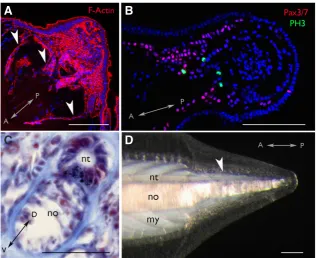
Fig. 2. Tail regeneration in adult B. lanceolatum. (A). Blastema tail bud viewed by confocal microscopy (stage 2). F-Actin staining (red) using Phalloidin demarcates cellular membranes. White arrowheads indicate myoseptal boundaries (B). Elongating regenerate (stage 3) viewed by confocal microscopy. Pax3/7+ and dividing phospho-histone 3 (PH3+) cell populations are shown in red and green, respectively. DAPI labelled nuclei are false-coloured in blue. (C). Transverse section through regenerating tail stained with Mallory trichrome (nuclei, red; connective tissue, blue). The black speckles in the floor of the neural tube are pigment granules normally associated with photoreceptors. (D). Mature regenerate viewed under polarising light (15 weeks post-amputation; stage 4). Differentiated notochord cells are birefringent, and new muscle fibres can clearly be distinguished (white arrowhead). The Anterior-Posterior (A-P) and Dorso-Ventral (D-V) axes are indicated in each panel. nt: nerve cord; no: notochord; my: myosepta. Scale, 100 mm.
C
B
D
A
anterior to the original cut results in enhanced re-generation speed (Somorjai et al., 2012b), but that amputations anterior to the anus progressively result in poorer regenerative responses (Somorjai et al., 2012a), in spite of similar surface areas, suggest that a number of variables must be involved. The age of the animal, which can be roughly inferred in wild animals based on known population demographics, nevertheless has clear effects on regenerate quality (Somorjai et al., 2012a), and is another important factor that requires further study.
Cirrus regeneration
chordin is expressed in new notochord cells, suggesting a role in differentiation. The neural differentiation marker soxB2, the ortho-logue of vertebrate sox17/21, is also expressed in the regenerating ependymal tube (Somorjai et al., 2012a).
A third study has recently taken a global look at tail regeneration by analysing transcriptomes of the blastema and the unamputated tail (Dailey 2017). As expected, all previously identified players using the candidate approach were recovered (Dailey 2017). A large number of additional genes were also identified from the Wnt, BMP, Notch, FGF and RTK signalling pathways, as well as
epimorphic process in amphioxus, as blastema cells show consider-able proliferation by phospho-histone H3 immunostaining, as does the extending nerve cord (Somorjai et al., 2012a; Fig. 2B). The latter further demonstrates that the nerve cord undergoes active neurogenesis during regeneration. Interestingly the extent of this proliferative activity is significantly lower in older animals, and may account for their reduced capacity to regenerate a complete tail. In contrast to tail regeneration, Kaneto and Wada (2011) report that regeneration of the oral cirri occurs via tissue remodelling, as no increase in proliferation was observed. However, it cannot
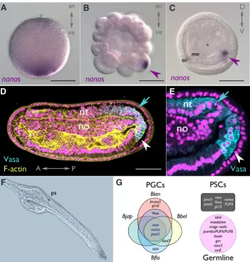
Fig. 3. Evidence for amphioxus stem cell populations. (A-C). nanos transcripts are segregated to the vegetal pole in eggs (A), and inherited by one to a few cells (magenta arrowheads) by PGCs (primitive germ cells) from morula (B) to gastrula (C) stages in cephalochordate embryos (here B. lanceolatum). (D) These cells also express Vasa protein (white arrowheads), as does the posterior neural tube (cyan arrows) at mid-neurula stages [magnification in (E)], as seen by confocal microscopy. (F) Surviving twin of pair of 2-day larvae resulting from dissociation experiment at the 2-cell stage. Note morphological abnormalities in elongation and head/oral structures. (G) Summary of proposed core molecular markers (transcripts and protein) asymmetrically labelling PGCs from studies in four cephalochordate species (Blan: B. lanceolatum; Bbel: B. belcheri; Bjap: B. japonicum; Bflo: B. floridae). A number of these also mark the tailbud, a possible posterior stem cell (PSC) pool (grey box). Other markers may also play a role in the germline based on expression in B. floridae and in silico prediction in A. lucayanum (magenta box). The animal-vegetal (an-ve), Anterior-Posterior (A-P) and Dorso-Ventral (D-V) axes are indicated in each panel where appropriate. Cyan: Vasa; yellow: F-actin; magenta: DAPI labelled nuclei. ec: ectoderm; me: mesendoderm; *: early blastopore; nt: neural tube; no: notochord; ps: pigment spot. Scale, 50 mm.
G
B
C
F
A
D
E
germline and pluripotency factors (Dailey 2017; see below), many of which have neither been characterised during normal development nor regeneration. However, one of the identified blastema transcripts, Sp5, is expressed in the amphioxus pos-terior growth zone and is a target of Wnt/b
-catenin signalling (Dailey et al., in press). Greater sequencing depth using Illumina in more replicates and at additional regenera-tion stages are currently underway (Dailey and Somorjai, unpublished). Nevertheless, this initial pilot study has opened up new avenues for understanding the molecular basis of tail regeneration in amphioxus, and provides a comparative framework for identifying processes shared with both ambulacrarians and vertebrates.
Cirrus regeneration
Oral cirrus regeneration has been used as a model for understanding the evolu-tion of skeletogenesis (Kaneto and Wada 2011). Markers for a number of genes im-plicated in vertebrate chondrogenesis and osteogenesis including collagens FCol1 and FCol2, SPARC, soxE and runx were expressed specifically in the tips of the re-generating skeletal rods. Amphioxus in fact possesses two SPARC genes, conserved since the eumetazoan ancestor, both of which are expressed in the notochord dur-ing embryonic development (Bertrand et al., 2013). Combined with expression data in other chordates, this leads the authors to suggest that co-expression of SPARC-like and Collagen proteins in mesenchymal cells was one of the key steps to skeletal evolution (Bertrand et al., 2013). Indeed, a SPARC gene and runx2 are expressed in prospective scleroblast progenitors during scale regeneration in the goldfish, a collagenous dermal skeleton component (Iimura et al., 2012). It will be interesting to determine whether or not any of these SPARC and runx genes are expressed during tail regeneration, most specifically in notochord precursors.
be excluded that progenitors contributing to the regenerate may have migrated from elsewhere, or that there may exist different populations of slow or fast cycling stem cells. BrdU pulse-chase labelling may give additional insight into proliferation kinetics. Al-together, these studies support data in other systems suggesting that proliferation-dependent and -independent mechanisms of regeneration may occur in different tissues in the same organism.
Evidence for amphioxus stem cells
Primordial germ cells
Germ cells, by virtue of being able to generate a whole new organism in the next generation, and their close affinity with em-bryonic stem cells, may be considered stem cells par excellence. In metazoans, two different mechanisms of germline specification have been described: inductive and determined (or ¨preformation¨). In the inductive mechanism, germ cells are specified by signalling, usually relatively late during embryogenesis, while in the determined mode, segregation of germ plasm already in the ooctye determines germ cell fate (Whittle and Extavour 2017).
Although cephalochordates are generally reported as using an inductive mode, recent data in four amphioxus species (all Bran-chiostoma) suggest rather an inherited mechanism for primordial germ cell (PGC) formation (Wu et al., 2011; Zhang et al., 2013; Dailey et al., 2016). At the two-cell stage, a number of highly con-served germline transcripts including nanos (Fig. 3A-C) and vasa -as well as Vasa protein- are asymmetrically distributed into a single cell, and inherited by few progeny. At the late gastrula stage, up to 8 Vasa+ cells can be identified in B. belcheri (Wu et al., 2011). In neurula stages, these PGCs are nestled posteriorly in endoderm of the tailbud (Fig. 3D, E), and remain as a cluster near the anus in premouth larval stages in all species examined (Wu et al., 2011; Zhang et al., 2013; Dailey et al., 2016).
Given the remarkable conservation of developmental gene expression in Branchiostoma (Somorjai et al., 2008; Yong et al., 2017), comparisons across species, including in silico predictions in Asymmetron lucayanum (Yue et al., 2015) allow a tentative reconstruction of a core set of germline-associated genes in cephalochordates (Fig. 3G). This likely includes nanos, piwil1, vasa, bruno2 and pl10 and a number of Tudor-related genes, which are asymmetrically localised to the vegetal cortex. However, it may also include PUF-domain containing genes, mago nashi or maelstrom, which are more broadly expressed (Yue et al., 2015); these may play roles in germline maintenance or differentiation.
As marker expression is independent of the germline specifica-tion mechanism utilised (i.e. nanos, see Fresques et al., 2016), we cannot currently exclude the possibility that other cephalochordate genera utilise non-determinative mechanisms (eg Asymmetron), or that inductive mechanisms may not operate under particular conditions. For instance, removal of PGCs at late stages may permit inductive mechanisms to re-specify germ cells, as seen in the solitary ascidian Ciona intestinalis when the larval tail is am-putated (reviewed in Kawamura et al., 2011). However, at least in the earliest cleavage stages, inductive mechanisms do not appear to be operating. Recent blastomere dissociation experiments at the two-cell stage, similar to those performed by Tung (reviewed in Yan 1999), suggest that while morphologically broadly normal twins can be recovered (Wu et al., 2011 and see Fig. 3F), one likely lacks germline as assessed by germ marker expression, and the
posterior is malformed (Wu et al., 2011). Taken together, studies of PGC determination in amphioxus such as these will begin to shed light on the nature of stem cell regulation in this taxon.
Somatic progenitors
Reports in a number of animals suggest that the germ-soma divide is not strict, and that mechanisms used to maintain the PGCs/ germline may also be utilised in somatic stem cells, supporting the existence of an ancestral multipotency programme already in the last common ancestor of metazoans (Juliano et al., 2010; Fierro-Constain et al., 2017). Thus, many “germline”-associated genes are also expressed in pluripotent somatic progenitors, and can be re-activated during regenerative processes.
In amphioxus, comparative studies suggest the existence of a posterior progenitor/stem cell pool located in the larval tailbud, the growth zone responsible for posterior elongation. Both transcripts and protein of one of the key germline determinants in the PGCs, vasa, localise to this domain, as do nanos and piwil1 and piwil2 (Wu et al., 2011; Zhang et al., 2013; Dailey et al., 2016). Vasa plays a general role in regulating translation in the soma (Poon et al., 2016) and nanos2 has recently been shown to transiently repress translation in PGCs via inhibition of eIF1a in sea urchins (Ouhlen et al., 2017). Thus, we might expect expression of such genes in the adult regenerating tail. Indeed, transcriptomic data from the blastema suggest that “germline” genes are in fact acting as general “stemness factors” in amphioxus (Dailey 2017).
While some data exist in adult amphioxus concerning signalling pathways expressed during regeneration (outlined above), the contribution of stem cells to the process is poorly understood due to current technical limitations. A single study in B. lanceolatum has identified a pool of dividing progenitors positive for Pax3/7 in and around the tail blastema, which appear to decline during differentiation and ageing (Somorjai et al., 2012; Fig. 2B). Pax3/7 expressing muscle satellite-like cells with a role in regeneration have also been identified in Parhyale hawaiensis, a crustacean, suggesting that shared mechanisms for muscle regeneration existed in the common ancestor of bilaterians (Konstainides and Averof 2014). However, the contribution of such Pax3/7+ cells to the amphioxus regenerate -and whether or not the mechanisms employed in amphioxus and vertebrates are indeed homologous- requires further study. Recent research in two salamanders, the axololt and the newt, suggests that muscle dedifferentiation and satellite cell contributions may differ across species (Sandoval-Guzman et al., 2014). However, the differences observed between the two could also reflect changes in cellular mechanisms and resident stem cell activation during ontogeny (Tanaka et al., 2016). Either way, these have important implications: the first suggests the evolution of an alternate strategy to achieve the same regenera-tive outcome within urodeles, while the latter has a clear impact on our approach to regenerative medicine. Understanding how amphioxus regenerates muscle may help polarise our views on ancestral mechanisms in chordates.
Outlook
understanding of how amphioxus fits into the broader spectrum of animal regeneration generally, and chordate and vertebrate regeneration more specifically, contributes to our understanding of the diversity of mechanisms utilised by animals to repair inju-ries and replace lost tissues. This in turn has potential biomedical implications: the blastema is effectively a tumour, but undergoing controlled growth, differentiation and patterning. Insight gained into how complex tissue regeneration is regulated in adult amphi-oxus, and how this potential declines with age, may help inform us about degenerative disease and ageing-associated stem cell misregulation in humans.
Based on studies in other chordates, one can make a number of predictions about amphioxus tail regeneration. As demonstrated in amphibians (Gargioli and Slack 2004; Kragl et al., 2009), no-tochord, muscle and nerve cord lineages are likely defined early in amphioxus, with little to no lineage switching. However, addi-tional distant sources of progenitors may also contribute to the tail blastema in cephalochordates. For instance, branchial sac stem cells in the solitary ascidian Ciona intestinalis contribute to distal regeneration (Jeffery 2015). Moreover, in some colonial ascid-ians, small pieces of vessel containing blood cells are sufficient to regenerate an entire organism (Rinkevich et al., 1995), and the endostyle, homologue of the vertebrate thyroid gland, acts as a stem cell niche (Voskoboynik et al., 2008). Although circulating cells are lacking in cephalochordates, amphioxus blood vessels do contain coelomocytes or amoebocytes with some migratory and endocytic capabilities (Rhodes et al., 1982; Monahan-Earley et al., 2013). Further, amphioxus have a well-developed endostyle within the floor of the branchial basket, which could act as a niche due to its close apposition to the circulatory system there, in addition to more local signals originating from the wound epithelium to the underlying tail blastemal cells. The embryonic tailbud is a source of numerous signals in amphioxus (i.e. Wnt, BMP etc.; Bertrand et al., in press) that might regulate proliferation/differentiation of the posterior stem cell pool, similarly to what occurs during posterior regenerate outgrowth. Regeneration and developmental research in amphioxus will therefore have to progress side-by-side within a comparative framework; highlighting differences is as important as identifying commonalities among systems. Ultimately, research in amphioxus, an invertebrate with conserved chordate anatomy and simpler genome, provides an attractive foil and complement to regeneration and stem cell studies in vertebrates.
Acknowledgements
I would like to thank Nicholas Holland for 10 o’clock espresso breaks at the kiosk, Russian translations, and fine wine; Linda Holland for tips on amphioxus care and numerous suppers; and reviewers for constructive criticism that improved the manuscript. Experimental work in my lab is currently funded by the Wellcome Trust ISSF grant 204821/Z/16/Z, the European Union Horizon 2020 research and innovation programme under grant agreement numbers 654428 (“CORBEL”) and 730984(“ASSEMBLE+) and the RS MacDonald Charitable Trust. I apologise to the many authors whose primary literature could not be cited due to space restrictions.
References
ANDREWS EA (1893). An undescribed acraniate, Asymmetron lucayanum. Stud Biol
Lab Johns Hopkins Univ 5: 213–247.
BELY AE and NYBERG KG (2010). Evolution of animal regeneration: re-emergence of a field. Trends Ecol Evol 25: 161-170.
BERT MP (1867). On the anatomy and physiology of amphioxus. Annals Mag. Nat.
Hist. 20: 302-304.
BERTRAND S, LE PETILLON Y, SOMORJAI I, and ESCRIVA E (2017). Developmental cell-cell communication pathways in the cephalochordate amphioxus: actors and functions (doi: 10.1387/ijdb.170202sb)
BERTRAND S, FUENTEALBA J, AZE A, HUDSON C, YASUO H, TORREJON M, ESCRIVA H and MARCELLINI S (2013). A dynamic history of gene duplications and losses characterizes the evolution of the SPARC family in eumetazoans.
Proc Biol Sci 280: 20122963.
BIBERHOFER R. (1906) Über Regeneration bei Amphioxus lanceolatus (1906). Arch
EntwMech Org 22: 15–17.
BONE Q (1992). Protochordates. J Mar Biol Assoc UK 72: 952-953.
BROCKES JP and KUMAR A (2008). Comparative aspects of animal regeneration.
Annu Rev Cell Dev Biol 24: 525-549.
CARNEVALI MD (2006). Regeneration in echinoderms: repair, regrowth, cloning.
Invertebrate Surviv J 3: 64-76
CZARKWIANI A, FERRARIO C, DYLUS DV, SUGNI M and OLIVERI P (2016). Skeletal regeneration in the brittle star Amphiura filiformis. Front Zool 13: 18.
DAILEY SC, FEBRERO PLANAS R, ROSSELL ESPIER A, GARCIA-FERNANDEZ J and SOMORJAI I (2016). Asymmetric distribution of pl10 and bruno2, new members of a conserved core of early germline determinants in cephalochordates.
Front Ecol Evol 3: 156.
DAILEY SC (2017). Evolutionary developmental and genomic insights from a tail regeneration transcriptome of the cephalochordate Branchiostoma lanceolatum.
PhD Thesis. University of St Andrews, Scotland, UK.
DAILEY SC, KOZMIKOVA I, SOMORJAI I (2017). Amphioxus Sp5 is a member of a conserved Specificity Protein complement and is modulated by Wnt/b-catenin signalling (doi: 10.1387/ijdb.170205sb).
FENG J, LI G, LIU X, WANG J, and WANG YQ (2014). Functional analysis of the promoter region of amphioxus b-actin gene: a useful tool for driving gene expres-sion in vivo. Mol Biol Rep 41: 6817-6826.
FIERRO-CONSTAÍN L, SCHENKELAARS Q, GAZAVE E, HAGUENAUER A, ROCHER C,ERESKOVSKY A, BORCHIELINI C, RENARD E (2017). The conservation of the germline multipotency program, from sponges to vertebrates: A stepping stone to understanding the somatic and germline origins. Genome Biol Evol 9: 474-488. FRANZ V (1925). Morphologische und ontogenetische Akranierstudien über Darm,
Trichter, Zölomderivate, Muskulatur- und Bindegewebsformationen. Jenaische
Zeitschrift für Naturwissenschaft 61: 407-468.
FRESQUES T, SWARTZ SZ, JULIANO C, MORINO Y, KIKUCHI M, AKASAKA K, WADA H, YAJIMA M and WESSEL GM (2016). The diversity of nanos expression in echinoderm embryos supports different mechanisms in germ cell specification.
Evol Dev 18: 267-278.
GAWRILUK TR, SIMKIN J, THOMPSON KL, BISWAS SK, CLARE-SALZLER Z, KIMANI JM, KIAMA SG, SMITH JJ, EZENWA VO and SEIFERT AW (2016). Comparative analysis of ear-hole closure identifies epimorphic regeneration as a discrete trait in mammals. Nat Commun 7: 11164.
GARGIOLI C and SLACK JM (2004). Cell lineage tracing during Xenopus tail regen-eration. Development 131: 2669-2679.
GEMBERLING M, BAILEY TJ, HYDE DR and POSS KD (2013). The zebrafish as a model for complex tissue regeneration. Trends Genet 29: 611-620.
HAAS BJ and WHITED JL (2017). Advances in decoding axolotl limb regeneration.
Trends Genet 33: 553-565.
HENRY JJ and TSONIS PA (2010). Molecular and cellular aspects of amphibian lens regeneration. Prog Retin Eye Res 29: 543-555.
IIMURA K, TOHSE H, URA K, and TAKAGAKI Y (2012). Expression patterns of runx2,
sparc, and bgp during scale regeneration in the goldfish Carassius auratus. J Exp Zool B Mol Dev Evol 318: 190-198.
IGAWA T, NOZAWA M, SUZUKI DG, REIMER JD, MOROV AR, WANG Y, HENMI Y and YASUI K (2017). Evolutionary history of the extant amphioxus lineage with shallow-branching diversification. Sci Rep 7: 1157.
JEFFERY WR (2015). Distal regeneration involves the age dependent activity of bran-chial sac stem cells in the ascidian Ciona intestinalis. Regeneration (Oxf) 2: 1-18. JULIANO CE, SWARTZ SZ and WESSEL GM (2010). A conserved germline
KANETO S and WADA H (2011). Regeneration of amphioxus oral cirri and its skeletal rods: implications for the origin of the vertebrate skeleton. J Exp Zool B Mol Dev
Evol 316: 409-417.
KAWAMURA K, TIOZZO S, MANNI L, SUNANAGA T, BURIGHEL P, and DE TOMASO AW (2011). Germline cell formation and gonad regeneration in solitary and colonial ascidians. Dev Dyn 240: 299-308.
KONSTANTINIDES N and AVEROF M (2014). A common cellular basis for muscle regeneration in arthropods and vertebrates. Science 343: 788-791.
KOZMIKOVA I and KOZMIK Z (2015). Gene regulation in amphioxus: An insight from transgenic studies in amphioxus and vertebrates. Mar Genomics 24 Pt 2: 159-166. KRAGL M, KNAPP D, NACU E, KHATTAK S, MADEN M, EPPERLEIN HH and TANAKA EM (2009). Cells keep a memory of their tissue origin during axolotl limb regeneration. Nature 460: 60-65.
KÜRN U, RENDULIC S, TIOZZO S and Lauzon RJ (2011). Asexual propagation and regeneration in colonial ascidians. Biol Bull 221: 43-61.
LEHOCZKY JA, ROBERT B and TABIN CJ (2011). Mouse digit tip regeneration is medi-ated by fate-restricted progenitor cells. Proc Natl Acad Sci USA 108: 20609-20614. MONAHAN-EARLEY R, DVORAK AM and AIRD WC (2013). Evolutionary origins of
the blood vascular system and endothelium. J Thromb Haemost 11 Suppl 1: 46-66. NOGUEIRA AF, COSTA CM, LORENA J, MOREIRA RN, FROTA-LIMA GN, FURTADO
C, ROBINSON M, AMEMIYA CT, DARNET S and SCHNEIDER I (2016). Tetrapod limb and sarcopterygian fin regeneration share a core genetic programme. Nat
Commun 7: 13364.
NUSBAUM J (1905). Vergleichende Regenerationsstudien. Ueber die Regeneration der Polychäten Amphiglene mediterranea Leydig und Nerine cirratulus Delle Chiaje. Z Wiss Zool 79: 222-307
OULHEN N, SWARTZ SZ, LAIRD J, MASCARO A and WESSEL GM (2017). Transient translational quiescence in primordial germ cells. Development 144: 1201-1210. OULION S, BERTRAND S, BELGACEM MR, LE PETILLON Y and ESCRIVA H
(2012). Sequencing and analysis of the Mediterranean amphioxus (Branchiostoma
lanceolatum) transcriptome. PLoS One 7:e36554.
PEGETA VP (1992) The regenerative capacity of the tail section of the cephalochordate (in Russian). Vestn Zool [Zoological Herald] (Kiev) 1: 74-76.
POON J, WESSEL GM and YAJIMA M (2016). An unregulated regulator: Vasa expres-sion in the development of somatic cells and in tumorigenesis. Dev Biol 415: 24-32. POSS SG and BOSCHUNG HT (1996). Lancelets (Cephalochordata:
Branchiosto-matidae): how many species are valid? Israel J Zool 42: Suppl 13–66. PROBST G (1930). Regenerationsstudien an Anneliden und Branchiostoma
lanceo-latum (Pallas). Rev Suisse Zool 37: 343–352.
PUTNAM NH, BUTTS T, FERRIER DE, FURLONG RF, HELLSTEN U, KAWASHIMA T, ROBINSON-RECHAVI M, SHOGUCHI E, TERRY A, YU JK, BENITO-GUTIÉRREZ EL, DUBCHAK I, GARCIA-FERNÀNDEZ J, GIBSON-BROWN JJ, GRIGORIEV IV, HORTON AC, DE JONG PJ, JURKA J, KAPITONOV VV, KOHARA Y, KUROKI Y, LINDQUIST E, LUCAS S, OSOEGAWA K, PENNACCHIO LA, SALAMOV AA, SATOU Y, SAUKA-SPENGLER T, SCHMUTZ J, SHIN-I T, TOYODA A, BRONNER-FRASER M, FUJIYAMA A, HOLLAND LZ, HOLLAND PW, SATOH N, and ROKHSAR DS (2008). The amphioxus genome and the evolution of the chordate karyotype. Nature 453: 1064-1071.
RHODES CP, RATCLIFFE NA and ROWLEY AF (1982). Presence of coelomocytes in the primitive chordate amphioxus (Branchiostoma lanceolatum). Science 217: 263-265.
RINKEVICH B, SHLEMBERG Z and FISHELSON L (1995). Whole-body protochordate regeneration from totipotent blood cells. Proc Natl Acad Sci USA 92: 7695-7699. RINKEVICH Y, LINDAU P, UENO H, LONGAKER MT and WEISSMAN IL (2011).
Germ-layer and lineage-restricted stem/progenitors regenerate the mouse digit tip. Nature 476: 409-413.
SANDOVAL-GUZMÁN T, WANG H, KHATTAK S, SCHUEZ M, ROENSCH K, NACU E, TAZAKI A, JOVEN A, TANAKA EM and SIMON A (2014). Fundamental differences in dedifferentiation and stem cell recruitment during skeletal muscle regeneration in two salamander species. Cell Stem Cell 14: 174-187.
SEIFERT AW, KIAMA SG, SEIFERT MG, GOHEEN JR, PALMER TM and MADEN M (2012). Skin shedding and tissue regeneration in African spiny mice (Acomys).
Nature 489: 561-565.
SEIFERT AW and VOSS SR (2013). Revisiting the relationship between regenerative ability and aging. BMC Biol 11: 2.
SILVA JR, MENDES EG and MARIANO M (1995). Wound repair in the Amphioxus (Branchiostoma platae), an animal deprived of inflammatory phagocytes. J
Invertebr Pathol 65: 147-151.
SILVA JRMC, MENDES EG and MARIANO M (1998). Regeneration in the amphioxus (Branchiostoma platae). Zoologischer Anzeiger 237: 107-112.
SLACK JM (2017). Animal regeneration: ancestral character or evolutionary novelty?
EMBO Rep pii: e201643795.
SOMORJAI I, BERTRAND S, CAMASSES A, HAGUENAUER A and ESCRIVA H (2008). Evidence for stasis and not genetic piracy in developmental expression patterns of Branchiostoma lanceolatum and Branchiostoma floridae, two amphi-oxus species that have evolved independently over the course of 200 Myr. Dev
Genes Evol 218(11-12): 703-713.
SOMORJAI IML, SOMORJAI RL, GARCIA-FERNÀNDEZ J and ESCRIVÀ H (2012a). Vertebrate-like regeneration in the invertebrate chordate amphioxus. Proc Natl
Acad Sci USA 109: 517-522.
SOMORJAI IML, ESCRIVÀ H and GARCIA-FERNÀNDEZ J (2012b). Amphioxus makes the cut—Again. Commun Integr Biol 5: 499-502.
TANAKA HV, NG NC, YANG YU Z, CASCO-ROBLES MM, MARUO F, TSONIS PA and CHIBA C (2016). A developmentally regulated switch from stem cells to dedifferentiation for limb muscle regeneration in newts. Nat Commun 7: 11069. UNGUEZ GA (2013). Electric fish: new insights into conserved processes of adult
tissue regeneration. J Exp Biol 216: 2478-2486.
VORONTSOVA MA and LIOSNER LD (Eds.) (1960). Asexual Propagation and
Regeneration. Pergamon, Oxford.
VOSKOBOYNIK A, SOEN Y, RINKEVICH Y, ROSNER A, UENO H, RESHEF R, ISHIZUKA KJ, PALMERI KJ, MOISEEVA E, RINKEVICH B and WEISSMAN IL (2008). Identification of the endostyle as a stem cell niche in a colonial chordate.
Cell Stem Cell 3: 456-464.
VOSS GJ, KUMP DK, WALKER JA and VOSS SR (2013). Variation in salamander tail regeneration is associated with genetic factors that determine tail morphology.
PLoS One 8:e67274.
WANG YB, CHEN SH, LIN CY and YU JK (2012). EST and transcriptome analysis of cephalochordate amphioxus-past, present and future. Brief Funct Genomics 11: 96-106.
WHITTLE CA and EXTAVOUR CG (2017). Causes and evolutionary consequences of primordial germ-cell specification mode in metazoans. Proc Natl Acad Sci
USA 114: 5784-5791.
WU HR, CHEN YT, SU YH, LUO YJ, HOLLAND LZ and Yu JK (2011). Asymmetric localization of germline markers Vasa and Nanos during early development in the amphioxus Branchiostoma floridae. Dev Biol 353: 147-159.
YAJIMA M and WESSEL GM (2015). Essential elements for translation: the germline factor Vasa functions broadly in somatic cells. Development 142, 1960–1970. YONG LW, BERTRAND S, YU JK, ESCRIVA H and HOLLAND ND (2017).
Conserva-tion of BMP2/4 expression patterns within the clade Branchiostoma (amphioxus): Resolving interspecific discrepancies. Gene Expr Patterns 25-26: 71-75. YUE JX, YU JK, PUTNAM NH and HOLLAND LZ (2014). The transcriptome of an
amphioxus, Asymmetron lucayanum, from the Bahamas: a window into chordate evolution. Genome Biol Evol 6: 2681-2696.
YUE JX, LI KL and YU JK (2015). Discovery of germline-related genes in Cepha-lochordate amphioxus: A genome wide survey using genome annotation and transcriptome data. Mar Genomics 24: 147-157.
YUE JX, KOZMIKOVA I, ONO H, NOSSA CW, KOZMIK Z, PUTNAM NH, YU JK, and HOLLAND LZ (2016). Conserved noncoding elements in the most distant genera of cephalochordates: the Goldilocks principle. Genome Biol Evol 8: 2387-2405. ZHANG QJ, LUO YJ, WU HR, CHEN YT and Yu JK (2013). Expression of germline
markers in three species of amphioxus supports a preformation mechanism of germ cell development in cephalochordates. EvoDevo 4: 17.
Planaria: an animal model that integrates development, regeneration and pharmacology
Oné R. Pagán
Int. J. Dev. Biol. (2017) 61: 519-529
Reptile genomes open the frontier for comparative analysis of amniote development and regeneration
Marc Tollis, Elizabeth D. Hutchins and Kenro Kusumi
5 yr ISI Impact Factor (2013) = 2.879
Int. J. Dev. Biol. (2014) 58: 863-871
Regeneration in spiralians: evolutionary patterns and developmental processes
Alexandra E. Bely, Eduardo E. Zattara and James M. Sikes Int. J. Dev. Biol. (2014) 58: 623-634
From the American to the European amphioxus: towards experimental Evo-Devo at the origin of chordates
Jordi Garcia-Fernàndez, Senda Jiménez-Delgado, Juan Pascual-Anaya, Ignacio Maeso, Manuel Irimia, Carolina Minguillón, Èlia Benito-Gutiérrez, Josep Gardenyes, Stéphanie Bertrand and Salvatore D’Aniello
Int. J. Dev. Biol. (2009) 53: 1359-1366 https://doi.org/10.1387/ijdb.072436jg
The germ line and somatic stem cell gene Cniwi in the jellyfish Podocoryne carnea.
Katja Seipel, Nathalie Yanze and Volker Schmid Int. J. Dev. Biol. (2004) 48: 1-7
FGF signalling and blastema growth during amphibian tail regeneration