Triploidy – the breakdown of monogamy
between sperm and egg
HEY-JOO KANG and ZEV ROSENWAKS*
The Center for Reprodutive Medicine and Infertility, Weill Cornell Medical Center, New York, USA
ABSTRACT The advent of assisted reproductive technology (ART) has taught us a great deal about human fertilization patterns. Thirty years of experience with IVF and cultivation of early embryos has provided a unique view into the mechanisms of normal and aberrant human fertilization. Here we review the different types of triploidy following conventional in vitro fertilization and intracytoplasmic sperm injection, as well as the mechanisms giving rise to digynic and dispermic fertilization. Additionally, the role of the centrosome in triploidy, the genetic analysis of triploid embryos and the potential for therapeutic enucleation are explored. Lastly, we review our own clinical experience with human fertilization patterns following > 20,000 treatment cycles of assisted reproduction.
KEY WORDS:
fertilization, triploid, dispermic, digynic, IVF , ICSI
Introduction
Triploidy is responsible for approximately 15% of chromo-somally-caused human reproductive losses (Dyban et al., 2008). Although triploidy is one of the most frequent chromosomal errors in cleavage and implantation failure, resulting pregnancies have been observed with survival up to several months following term delivery (Rosenbush et al., 2002). Until recently, information was scarce regarding the conditions governing the ability of a triploid embryo to survive past fertilization. The advent of assisted repro-ductive technology (ART) has permitted study of triploid fertiliza-tion patterns extending beyond epidemiologic data to more pre-cise large scale in vitro genetic analysis of human embryos.
Direct visualization of human zygotes at the pronuclear stage does not always portend the exact fate of an individual embryo. Tri-pronuclear embryos derived from the same couple have been observed to follow a variety of developmental paths. Some fail to enter syngamy and initial cleavage, while others may cleave into 2 blastomeres and an extrusion mass (Fig. 1). A subset of embryos will form three blastomeres following the first cleavage to give rise to morphologically normal embryos that are indistin-guishable from chromosomally competent embryos (Veeck, 1999) (Fig. 2). This explains why it is critical to separate bipronuclear (2PN) zygotes from 1PN and 3PN zygotes 18 hours post-fertiliza-tion in order to avoid transferring chromosomally abnormal em-bryos on day 3. 1PN zygotes have also been observed to progress beyond the embryonic stage and to successfully implant. Certain
BIOLOGY
www.intjdevbiol.com*Address correspondence to: Zev Rosenwaks. The Center for Reproductive Medicine and Infertility, Weill Cornell Medical Center, New York, NY 10021, USA. e-mail: [email protected]
Published online: 4 July 2008
0214-6282/2008/$35.00
© UBC Press Printed in Spain
Abbreviations used in this paper: ART, assisted reproductive technology; IVF, in vitro fertilization.
factors may predispose to an increased rate of triploidy including: advanced maternal age, supraphysiologic estradiol levels, oo-cyte post-maturity, severe oligospermia and chromosomal abnor-malities in the parental gamete.
Here, we will review the mechanisms behind in vivo and in vitro triploid formation. We will also detail the genetic constitution of triploid embryos and the tools necessary for mitotic competence. Finally, we will summarize our own observations and detail potential causes of triploid embryos and their impact on IVF success.
Mechanism of triploidy formation
produce diandric triploids predominantly through dispermic fertili-zation (Macas et al., 1996; Egozcue et al., 2002).
The third mechanism for triploidy gives rise to digynic embryos containing two maternal pronuclei. Retention of the second polar body during meiosis II is the most common cause of digynic triploid embryos but they may less frequently result from retention of the first polar body during meiosis I. Penetration of a binucleate oocyte by a single spermatozoon will result in a digynic, triploid embryo. Triploid zygotes following IVF with intracytoplasmic sperm injection (IVF/ICSI) are typically found to be digynic in origin via retention of the second polar body.
“Giant” oocytes constitute a distinct class of gametes that are predisposed to form triploid embryos. These gametes have ap-proximately twice the volume of normal oocytes and are tetraploid before meiosis. They can result either from a failure of cytoplas-mic division (with normal nuclear division) or cytoplascytoplas-mic fusion of two oogoni (Austin, 1960). These mechanisms explain the binucleate state of immature giant cells prior to fertilization (Rosenbusch et al., 2002). Giant embryos have been observed following fertilization of giant oocytes in the human (Munne et al., 1994).
Pre and post-fertilization cytogenetic analysis of giant oocytes has revealed two possible fertilization pathways. As the oocyte matures, the two haploid chromosome sets can combine during formation of the meiotic spindle, resulting in a single diploid chromosome set and a diploid first polar body; fertilization by a single spermatozoon will lead to a haploid male and a diploid
female pronucleus. Morphologically, these are technically 2PN embryos, but the chromosomal content is consistent with trip-loidy. The second scenario arises if the binucleate state is maintained resulting in extrusion of two haploid first polar bodies and two maternal haploid sets within the oocyte. Monospermic fertilization in this case will result in three haploid pronuclei (Veeck, 1999). These genetically abnormal embryos can be identified at the fertilization check and excluded from transfer.
Natural barriers to triploid formation
The human female reproductive tract is the initial defense against dispermic fertilization. Although 200-300 million sperm are deposited into the vagina during coitus, only a few hundred ultimately reach the oviduct. In the case of conventional IVF, oocytes are exposed to much higher concentrations of sperm, leading to higher rates of triploidy compared to the in vivo state (Ducibella, 1996). However, intrinsic oocyte mechanisms also help to minimize dispermic fertilization.
The most important line of defense against triploidy is the zona pellucida (ZP). The zona pellucida is composed of three glycopro-teins (ZP1, ZP2, and ZP3) that interact via non-covalent bonds to create the latticework of the extracellular membrane (Wasserman, Fig. 1 (Left). Various fates of tripronuclear zygotes. One (upper right)
has failed to accomplish first cleavage; another (upper left) has two blastomeres and an extrusion mass; one (lower right) has three multi-nucleated blastomeres. (Courtesy of Dr. Lucinda Veeck, An atlas of Human Gametes and Conceptuses, Parthenon Publishing Group 1998).
Fig. 2 (Right). Developmental path of a 2PN and 3PN embryo. (A) A 2PN and 3PN fertilized oocyte. (B) The same zygotes during first cleav-age; notice the tripolar spindle of the 3PN zygote. (C) Diploid and triploid embryos on day 3 after harvest. The similar morphological appearance of the embryos on day 3 compared to 2PN embryos requires that ploidy be determined before the first cleavage. (Courtesy of Dr. Lucinda Veeck).
B
1988). ZP2 and ZP3 are thought to be the structural backbone of the membrane, while ZP1 interconnects the two proteins. Null mice deficient in ZP2 or ZP3 are infertile because they lack a zona pellucida entirely, while those deficient in ZP1 have a loosely bound ZP membrane and are sub-fertile (Wasserman, 2004). Immediately following sperm binding to the ZP3 protein receptor, intracellular calcium increases via the IP3/DAG pathway, which results in the release of cortical granules (Kline et al., 1994). The exact mechanism of oocyte activation is not completely under-stood. There is some evidence in the murine model that suggests cytoplasmic calcium increases as a consequence of the release of a “sperm factor” following spermatozoon-egg fusion (Dale et al., 1985; Stice et al., 1990; Swann et al., 1990). Others contend that this phenomenon occurs via a sperm-receptor coupled G-protein receptor pathway (Kline et al., 1988; Miyazaki et al., 2006; Williams et al., 1992). Ultimately, the release of cortical granules leads to a zona reaction, or the hardening of the extracellular layer. This zona reaction prevents additional sperm from pen-etrating the zona pellucida, thus avoiding polyspermy.
The human centrosome and the fate of digynic and
dispermic embryos
The explanation as to why some triploid embryos fail to develop past the pronuclear stage while others survive to full term lies in the mechanisms of normal cellular division and chromosomal segregation. As a cell prepares to divide, several steps lead to the equal partitioning of genomic material into two daughter cells. During prophase, the replicated chromosomes condense; in metaphase they line up along the equatorial plate, and in anaphase, one full set of chromosomes migrate toward the spindle’s poles. The mitotic spindle is a key factor in this process and the spindle is composed of microtubles that are nucleated by the centrosome. The centrosome was initially described independently by Boveri and van Beneden in 1887.Each centrosome is composed of two centrioles, each made up of 9 rods of microtubles and surrounded
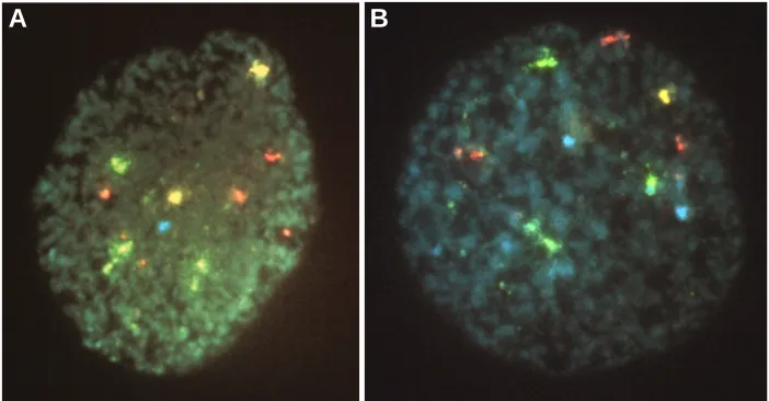
Fig. 3. Fluorescent in situ hybridization analysis of a blastomere from a triploid embryo. (A) A 69 XXY blastomere; (B) a 69 XYY blastomere. Note the presence of four copies of chromosome 18 in the dispermic fertilized blastomere, a result of abnormal chromosome division due to the presence of two functional centromeres. (Yellow = X, Blue = Y, Red = chromosome 13/21, Green = chromosome 18) (Courtesy of Dr. Gianpiero Palermo).
by pericentriolar material (Palermo et al., 1997). The centrosome nucleates the microtubules, and in doing so it also helps organize the cytoskeletal elements. Cor-rect mitotic division of a cell requires the two centrioles to split apart and nucleate a second centriole at a right angle to the original template centriole (Boveri 1887). Each newly formed pair then migrates to opposite poles and cell division may then proceed through metaphase, anaphase, and telophase. If the centriole fails to replicate itself, the bipolar mitotic spindle cannot form, thus ending that particular cell line.
During syngamy, the female centriole is inactive (Santhananthan et al., 1996). It is therefore the male centriole that is re-sponsible for the initial embryonic cell division (Santhananthan et al., 1998). The male centriole replicates to form the bipo-lar microtubule spindle necessary for faith-ful genomic partitioning in early euploid
cell development. The need for paternal inheritance is a key reason why parthenogenic activation of the oocyte usually does not proceed with normal cell division because it lacks a functional centrosome. If purified paternal centrosomes are injected into an activated egg, the egg can then divide (Glover et al., 1993; Palermo et al., 1997).
The functional importance of the paternal centrosome explains why the fate of a digynic embryo differs from that of dispermic embryos. Because the female centrosome is inactive, a digynic triploid embryo has the normal number of sperm-derived bipolar centrioles from the single spermatozoon. Hence, the incidence of chromosomal mosaicism in the development of an early embryo originating from a digynic triploid is low (Palermo et al., 1997). On the other hand, the dispermic triploid contains two pairs of active centrioles in one ovum. In 50% of these cases, the resultant product is a tripolar spindle producing a disorganized chromo-some distribution within each blastomere, leading to gross aneu-ploidy (Golubovsky, 2003) (Fig. 3). This difference may explain why in vivo digynic triploids survive longer than dispermic concep-tions (Hasegawa et al., 1999) as well as why dispermic concep-tions disproportionately lead to the formation of anembryonic molar pregnancies (Daniel et al., 2001).
Studying the chromosomal constitution of triploid embryos formed from conventional IVF and IVF/ICSI helps shed light on both the origin of the extra pronucleus as well as its mitotic competence. Since ICSI involves the introduction of a single spermatozoon into an oocyte, thus precluding dispermic fertiliza-tion, the resultant triploid embryos should be overwhelmingly digynic as a result of failed second polar body extrusion. Conven-tional IVF should produce a higher proportion of dispermic triploid embryos with an allotment that mimics in vivo rates of triploid conceptions - although naturally occurring digynic embryos still exist (McFadden et al., 2000).
FISH analysis of triploid embryos for the sex chromosomes and chromosome 18 support these theories. All of the blas-tomeres from embryos developing from tripronuclear oocytes
obtained after ICSI were analyzed and 100% were either XXX181818 or XXY181818 with no XYY181818 (indicating dis-permy) found. Of these, only 16% were triploid mosaics - mosaics having at least one complete triploid blastomere - supporting the evidence that one active paternal centromere is essential for a regular first mitotic cell division. In triploid embryos resulting from conventional IVF, there was a high preponderance of dispermic triploids (XYY181818) (Staessen et al., 1997). Greater than 60% of these embryos are mosaic or complex mosaic indicative of a chaotic chromosome distribution initiated at the first cell division due to the presence of 2 active paternal centrosomes. The duplication of each centrosome results in four active centrosomes, rather than the usual two, thus preventing the 69 chromosomes from aligning at the metaphase plate for equal distribution into daughter cells. However, it should be noted that some triploid embryos from either ICSI or IVF can sporadically demonstrate triploid cells indicating regular chromosome segregation. Albeit this occurs more frequently in embryos fertilized with ICSI as compared with conventional insemination.
Enucleation procedures
The incidence of triploidy in conventional IVF averages 4% per cycle and at least one triploid prezygote is found in 60% of all IVF cycles (Escriba et al., 2006). Therefore, the potential implications for therapeutic enucleation of triploid fertilized oocytes in order to obtain diploid zygotes are considerable. Conservatively, this could translate into a 5-20% increase in the number of embryos available for transfer or cryopreservation. However, the safe correction of a triploid embryo into a heteroparental diploid con-ceptus depends upon several factors, ranging from accurate identification of the accessory pronucleus to the technical aspects of the enucleation procedure itself.
The first reported attempt at microsurgical enucleation of tripronuclear human zygotes was by Rawlins et al. in 1988. They describe pretreatment of three dispermic zygotes with cytochala-sin B - a cytoskeletal relaxing agent - and removal of one male pronucleus identified by the larger size and associated sperm tailpiece. In this report, one embryo entered syngamy but cleav-age did not occur (Rawlins et al., 1988). Since that time, there have been three reports of microsurgical enucleation procedures producing cleavage stage embryos in humans (Gordon et al., 1989; Malter et al., 1989; Palermo et al., 1994), and one case report of a 46XY live birth following this procedure (Kattera et al., 2003).
The initial study following Rawlins’ work compared the out-come of enucleation procedures with and without the cytoskeletal relaxing agent cytochalasin. There appeared to be a 100% embryo survival rate when the relaxing agent was used, and no survival without cytochalasin (Gordon et al., 1989).Later that same year, another group reported a 36% survival rate after an enucleation procedure using zona-drilled oocytes without the use of relaxing agents (Malter et al., 1989).Despite an improvement in survival rates, cytochalasin can potentially cause increased fragmentation, delayed cleavage and instability of the meiotic spindle via disruption of the cytoskeletal organization of the oocytes (Hoppe et al., 1977; Pickering et al., 1988). It would therefore appear that attempts should be made to enucleate tripronuclear embryos using biopsy techniques similar to those
utilized for preimplantation genetic diagnosis (PGD) in lieu of using cytochalasin (Fig. 4).
Although the potential for therapeutic enucleation is encourag-ing, one must consider the practical drawbacks. First, the selec-tion of the extra pronucleus is based upon three inexact criteria: detection of pronucleus-associated sperm tails, the typically larger male pronucleus, and the greater distance of the pronucleus from the second polar body relative to the female pronucleus. Because these criteria are subjective, they have an intrinsic error rate. Confirmation that the correct pronucleus has been removed would require PGD with fluorescent in situ hybridization (FISH). This would involve yet another biopsy of the abnormally fertilized embryo. Each additional blastomere biopsy decreases an embryo’s implantation potential. Alternately, forgoing PGD and proceeding directly to embryo transfer carries with it a risk of transferring a diandric embryo, resulting in a molar gestation and the potential for malignancy. Incorrect removal of a male pronucleus in a digynic zygote following ICSI would fail to produce a viable embryo due to the lack of a functional centrosome and the uniformly maternal genetic material.
Clinical observations of triploidy
and possible etiologic factors responsible for the formation of triploid embryos.
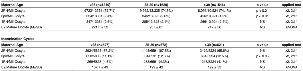
All IVF cycles from January 2004 to January 2007 were collected and analyzed. Within this time period, there were 6,335 total IVF cycles at our center, of which 4,515 required ICSI and the remaining 1,820 were conventional insemination cycles. Our data confirms that ICSI is associated with a significant overall reduc-tion in triploidy rates across all age groups compared to insemi-nation cycles (2.9% vs. 12.0% respectively, p<0.01). This under-scores the notion that the most common mechanism for naturally occurring triploid embryo formation is dispermic fertilization of a haploid oocyte, a process that is largely circumvented by ICSI.
Results were analyzed by maternal age groups: <35, 35-39, and >39 years old. The incidence of 3PN formation was signifi-cantly higher in the >39 maternal age group regardless of the method of fertilization, although 4.17% of all mature oocytes following ICSI and 12.46% of all mature oocytes following conven-tional IVF were found to be triploid (Table 1). This lends support to the theory that oocyte function declines with advancing mater-nal age and is critical in allowing normal fertilization.
The plasticity of the oocyte membrane during injection of a single spermatozoon has been noted to impact oocyte survival and fertilization patterns. Sudden breakage of the oolema in the absence of the usually observed oolema funnel seen upon insertion of the injection needle has been associated with a higher incidence of digynic embryos in the same cohort. This phenom-enon often portends a decreased survival rate in the normally fertilized embryos of the same cycle (Palermo et al., 1996).
This is not to imply that oocyte quality is solely attributable to maternal age. The method of stimulation can also affect oocyte quality and have an impact on fertilization success. Higher estra-diol (E2) levels during an IVF cycle resulting from aggressive doses of exogenous gonadotropins or from a prolonged duration of ovarian stimulation can impair the oolemma membrane (Gelety et al.; Sachs et al., 2000). Further evidence for the effect of a higher E2 level on oocyte quality comes from IVF cycles where “coasting” has been applied. Coasting is a technique used to decrease the risk of ovarian hyperstimulation syndrome. It is applied to rescue IVF cycles where there is an unexpectedly robust response to controlled ovarian stimulation; exogenous gonadotropins are withheld while continuing the GnRH agonist. The oocytes of hyperstimulated cycles have an increased rate of 3PN and 1PN formation and are noted to be morphologically
substandard compared to age-matched controls (Kang et al., 2006). The pregnancy rates from these cycles are largely unaf-fected, however, due to the preponderance of younger, better prognosis patients who are at risk for hyperresponse (Kang et al., 2006). Younger patients are more likely to have at least a few remaining 2PN fertilized embryos that are chromosomally nor-mal. When cycles requiring “coasting” were analyzed separately, our results were consistent with this notion, with higher rates of triploidy formation across all age groups (Data not shown).
The sperm source is yet another factor to consider when evaluating abnormal fertilization patterns. As noted earlier, men with abnormal semen parameters are more likely to have diploid sperm penetrating a haploid oocyte. Male factor infertility requir-ing the use of testicular or epididymal sperm may be causative factors behind the slightly lower rate of 2PN formation in the <35 age group (72.7% vs. 74.5%, p<0.01), although there was no noted increase in the rate of 3PN embryos.
Methods to improve normal fertilization
Understanding how to create a favorable hormonal milieu for fertilization should preclude any need for enucleation procedures in IVF cycles. As evident from our coasting data, oocytes exposed to high estrogen environments have an increased tendency to form triploid embryos following insemination. Although one can-not change the impact of age on oocyte quality, our experience suggests that oocyte membrane integrity is best served by de-creasing both the amount and duration of gonadotropin stimula-tion. One effective method to temper the estrogen level in an IVF cycle is the step-down approach, where the dose of exogenous gonadotropin is sequentially reduced once follicular recruitment has been achieved (Davis et al., 1996). In general, careful consideration and monitoring of the stimulation protocol during IVF may positively affect oocyte quality.
Conclusion
Assisted reproductive technology has improved our under-standing of human fertilization patterns. There is an exquisitely delicate balance that needs to be maintained between heteroparental chromosomes in order for an embryo to gain mitotic competence. With respect to incidence, In vitro rates of triploid embryo formation may not mimic in vivo rates as the
TABLE 1
NORMAL, 3PN, AND 1PN FORMATION IN ICSI AND INSEMINATION CYCLES BY MATERNAL AGE GROUP
ICSI Cycles
Maternal Age <35 (n=1349) 35-39 (n=1620) >39 (n=1546) p value applied test
2PN/MII Oocyte 9722/13361 (72.7%) 9,932/13,325 (74.5%) 8,093/10,924 (74.1%) p < 0.01 x2, 2d.f.
3pn/MII Oocyte 324/13361 (2.4%) 346/13,325 (2.6%) 456/10,924 (4.2%) p < 0.01 x2, 2d.f.
1PN/MII Oocyte 347/13361 (2.6%) 285/13,325 (2.1%) 266/10,924 (2.4%) NS x2, 2d.f.
E2/Mature Oocyte (M±SD) 221.3 ± 52 237 ± 61 242 ± 50 NS ANOVA
Insemination Cycles
Maternal Age <35 (n=527) 35-39 (n=672) >39 (n=621) p value applied test
2PN/MII Oocyte 3924/5835 (67.2%) 4448/6591 (67.5%) 3429/5224 (65.6%) NS x2, 2d.f.
3pn/MII Oocyte 650/5835 (11.1%) 834/6591 (12.6%) 651/5224 (12.5%) p < 0.05 x2, 2d.f.
1PN/MII Oocyte 269/5835 (4.6%) 283/6591 (4.3%) 216/5224 (4.1%) NS x2, 2d.f.
conditions for fertilization in each are quite different. For example, in the instance of conventional IVF there are thousands of additional sperm exposed to each oocyte compared to the natural environment within the fallopian tube. With ICSI, there is only one spermatozoon directly introduced into the oocyte. In reality, in vivo rates of triploidy most likely fall somewhere between these two scenarios – more than 2.4% but less frequent than 12.6% of all fertilized oocytes. It should additionally be stressed that in both cases the treated population is comprised of infertile couples who may harbor biologic or genetic polymorphisms which may predis-pose to abnormal fertilization patterns.
Overwhelmingly, most triploid embryos will not achieve viabil-ity as they cannot overcome developmental checkpoints. One cannot discount the possibility of a teleologic mechanism behind triploid fertilization as a way to prevent chromosomally incompe-tent oocytes from progressing to the point of implantation. As triploid fertilization is only one of the many events that can go awry in an early conceptus, the creation and development of a chromo-somally competent embryo and its faithful mitotic replication into a human fetus is indeed an impressive feat of nature.
References
ADASHI E, ROCK J, ROSENWAKS Z. (1996). Reproductive Endocrinology, Surgery, and Technology; In vitro fertilization (pp. 2325-2326) Lippincott-Raven
Publishers (1996).
BOVERI T. uber die Befrynchtung der Eier von Ascaris megalocephala Fischer, Jena, Germany.
DALE B, DEFELICE L, EHRENSTEIN G. (1985) Injection of a soluble sperm extract into sea urchin eggs triggers the cortical reaction. Experimentia 41:1068-1070.
DANIEL A, WU Z, BENNETTS B et al. (2001) Karytype, phenotype, and parental
origin in 19 cases of triploidy. Prenat Diagn 21:1034-1048.
DUCIBELLA T. (1996) The cortical reaction and development of activation compe-tence in mammalian oocytes. Hum Reprod Update 2: 29-42.
ESCRIBA M, MARTIN J, RUBIO C et al. (2006) Heteroparental blastocyst
produc-tion from mivrosurgially corrected tripronucleated embryos. Fertil Steril
86:1601-1607.
GELETY TJ, BUYALOS RP The influence of supraphysiologic estradiol levels on human nidation. J Assist Reprod Genet. 12: 406-412
GLOVER DM, GONZALEZ C, RAFF J. (1993) The Centrosome. Scientific Ameri-can: 62-68.
GORDON JW, GRUNFELD L, GARISSI GJ. (1989) Successful microsurgical removal of a pronucleus from tripronuclear human zygotes. Fertil Steril
52:367-372.
HASEGAWA T, HARADA N, IKEDA K et al. (1999) Digynic triploid infant surviving
for 46 days. An J Med Genet 87:306-310.
HOPPE PC, ILLMENSEE K. (1997) Microsurgically produced homozygous diploid uniparental mice. Proc. Natl Acad Aci USA 74:5657-5661.
KANG H, D. REICHMAN, H.C. LIU et al. (2006) FSH levels predict success in
hyperresponders: How much “coasting” can a follicle withstand? Fertility and Sterility 86:S71.
KANG H, Q.V. NERI, A. WANG. (2006) Withdrawal of FSH during controlled ovarian superovulation impairs fertilization with ICSI. Fertility and Sterility 86: S320.
KATTERA S, CHEN C. (2003) Normal birth after microsurgical enucleation of tripronuclear human zygotes: Case report. Hum. Reprod. 18:1319-1322.
KLINE D, SIMONCINI L, MANDEL G et al. (1998) Fertilization events induced by
neurotransmitters after injection of mRNA in Xenopus eggs. Science
241:464-467.
KLINE JT, KLINE D. (1994) Regulation of intracellular calcium in the mouse egg: evidence for inositoltriphosphate-induced calcium release, but not
calcium-induced calcium release. Biol Reprod 50:193-203.
MALTER HE, COHEN J. (1989) Embryonic development after microsurgical repair of polyspermic human zygotes. Fertil Steril 52:373-380.
MCFADDEN DE, LANGLOIS S. (2000) Paternal and meiotic origin of triploidy in the embryonic and fetal periods. Clin Genet 58:192-200.
MERCAN R, OEHNINGER S, MUASHER SJ et al. (1998) Impact of fertilization
history and semen parameters on ICSI outcome. J Assist Reprod Genet. 15:
39-45.
MIYAZAKI S, ITO, M. (2006) Calcium signals for egg activation in mammals. J Pharmacol Sci. 100: 545-52.
MUNNE S, ALIKANI M, COHEN J. (1994) Monospermic polyploidy and atypical embryo morphology. Hum Reprod 9: 506-510.
OEHNINGER S, VEECK L, LANZENDORF S et al. (1995) Intracytoplasmic sperm
injection: achievement of high pregnancy rates in couples with severe male factor infertility is dependent primarily upon female and not male factors. Fertil Steril. 64: 977-981.
PALERMO G, MUNNE S, COHEN J. (1994) The human zygote inherits its mitotic potential from the male gamete. Hum. Reprod.,9: 1220-1225.
PALERMO GD, ALIKANI M, BERTOLI M et al. (1996) Ooclema characteristics in
relation to survival and fertilization patterns of oocytes treated by intracytoplas-mic sperm injection. Hum. Reprod.11:172-176.
PALERMO GD, COLOMBERO LT, ROSENWAKS Z. (1997) The human sperm centrosome is responsible for normal syngamy and early embryonic develop-ment. Review of Reproduction 2: 19-27.
PALERMO GD, MUNNE S, COLOMBERO et al. (1994) The human zygote inherits
its mitotic potential from the male gamete. Hum Reprod. 9:1220-1225.
PALERMO GD, MUNNE S, COLOMBERO LT et al. (1997) Genetics of abnormal
human fertilization. Hum Reprod 10:20-27.
PICKERING SL, JOHNSON MH, BRAUDE PR et al. (1988) Cytoskeletal
organiza-tion in fresh, aged and spontaneously activated human oocytes. Hum Reprod.
3: 978-989.
RAWLINS RG, BINOR Z, RADWANSKA E et al. (1988) Microsurgical enucleation
of tripronuclear human zygotes. Fertil Steril 50: 266-72.
SACHS A, POLITCH J, JACKSON K et al. (2000) Factors associated with the
formation of triploid zygotes after intracytoplasmic sperm injection. Fertil Steril
73:1109-1114.
SANTHANANTHAN AH, RATNAM SS, NG SC et al. (1996) The sperm centriole: it’s
inheritance, replication and perpetuation in early human embryos. Hum. Reprod.
11:345-356.
SANTHANANTHAN AH. (1998) Paternal centrosomic dynamics in the early human development and infertility. J Assist Reprod Genet 15:129-138.
SPANDORFER S, AVRECH OM, COLOMBERO LT et al. (1998) Effect of parental
age on fertilization and pregnancy characteristics in couples treated by intracy-toplasmic sperm injection. Hum. Reprod. 13:334-338.
STAESSEN C, VAN STEIRTEGHEM. (1997) The chromosomal constitution of embryos developing from abnormally fertilized oocytes after intracytoplasmic sperm injection and conventional in-vitro fertilization. Hum Reprod 12:321-327.
STICE SL, ROBL JM. (1990) Activation of mammalian oocytes by a factor obtained by rabbit sperm. Mol Reprod Dev 25:272-280.
SWANN K. (1990) A cytosolic sperm factor stimulates repetitive calcium increases and mimics fertilization in hamster eggs. Development 110:1295-1302.
VAN BENEDEN E, NEYT A. Nouvelles recherches sur la fecundation et la division mitotique chez l’Ascaride megalocephale. Bull Acad. Roylae Belgique 14 3eme
serie:215-295.
WASSERMAN PM, JOVINE L, LITSCHER E et al. (2004) Egg-sperm interactions
at fertilization in mammals. Eur J Obstet Gynecol 115S: S57-S60.
WASSERMAN PM. (1998) Zona pellucida glycoproteins. Annu Rev Biochem
57:415-42.
Related, previously published Int. J. Dev. Biol. articles
See our recent Special Issue Developmental Biology in Poland edited by Tarkowski, Maleszewski and Kloc at: http://www.ijdb.ehu.es/web/contents.php?vol=52&issue=2-3
See our recent Special Issue Ear Development edited by Fernando Giraldez and Bernd Fritzsch at: http://www.ijdb.ehu.es/web/contents.php?vol=51&issue=6-7
Allorecognition mechanisms during ascidian fertilization
Yoshito Harada and Hitoshi Sawada Int. J. Dev. Biol. (2008) 52: 637-645
Glycobiology of fertilization in the pig
Edda Töpfer-Petersen, Mahnaz Ekhlasi-Hundrieser and Miroslava Tsolova Int. J. Dev. Biol. (2008) 52: 717-736
Mammalian fertilization:the egg’s multifunctional zona pellucida
Paul M. Wassarman and Eveline S. Litscher Int. J. Dev. Biol. (2008) 52: 665-676
Regionalized calcium signaling in zebrafish fertilization
Dipika Sharma and William H. Kinsey Int. J. Dev. Biol. (2008) 52: 561-570
2006 ISI **Impact Factor = 3.577**
Defective calcium release during in vitro fertilization of maturing oocytes of LT/Sv mice
Karolina Archacka, Anna Ajduk, Pawel Pomorski, Katarzyna Szczepanska, Marek Maleszewski and Maria A. Ciemerych
Int. J. Dev. Biol. (2008) 52: doi: 10.1387/ijdb.072397ka
Early mammalian embryo: my love. An interview with Andrzej K. Tarkowski
Marek Maleszewski and Andrzej K. Tarkowski Int. J. Dev. Biol. (2008) 52: 163-169
Enhanced development of porcine embryos cloned from bone marrow mesenchymal stem cells
Hai-Feng Jin, B. Mohana Kumar, Jung-Gon Kim, Hye-Jin Song, Yeon-Ji Jeong, Seong-Keun Cho, Sivasankaran Balasubramanian, Sang-Yong Choe and Gyu-Jin Rho Int. J. Dev. Biol. (2007) 51: 85-90
Preimplantation development of manipulated mouse zygotes fused with the second polar bodies: a cytogenetic study.
S V Evsikov and A V Evsikov Int. J. Dev. Biol. (1994) 38: 725-730
Strife in the germ line.
C F Graham