Regulation and function of Spalt proteins
during animal development
JOSE F. DE CELIS
1and ROSA BARRIO
21Centro de Biología Molecular “Severo Ochoa”. Universidad Autónoma de Madrid and Consejo Superior de Investigaciones Científicas, Madrid and 2Functional Genomics, CIC bioGUNE, Bizkaia Technology Park, Derio, Spain.
ABSTRACT The genes of the spalt (sal) family play fundamental roles during animal develop-ment. The two members of this family in Drosophila, spalt (sal) and spalt-related (salr) encode Zn-finger transcription factors that link the Decapentaplegic (Dpp)/BMP signalling pathway to the patterning of the wing. They are regulated by the Dpp pathway in the wing disc, and they were shown to mediate some of the morphogenetic activities of the Dpp/BMP4 secreted ligand. The sal genes were initially found by virtue of mutations that produce homeotic transformations in the head and tail of the Drosophila embryo. Since then, a number of other requirements have been associated to these genes in Drosophila, including morphogenesis of the respiratory system, cell fate specification of sensory organs and the differentiation of several photoreceptor cells, among others. Vertebrate sal orthologues (spalt-like/sall) have also important developmental roles during neural development and organogenesis, and at least two human sall genes are linked to the genetic diseases Townes Brocks Syndrome (TBS; SALL1 ) and Okihiro Syndrome (OS; SALL4). In this review, we will summarize the main characteristics of the sall genes and proteins, pointing out to the similarities in their developmental roles during Drosophila and vertebrate development.
KEY WORDS:
spalt, gene regulation, organogenesis, embryonic development
The Sall protein family
Sall proteins are zinc finger transcription factors present from C. elegans, which harbours only one member of the family, to vertebrates, which generally present four spalt genes (sall1-4).
The Drosophila genome contains two paralogues, spalt (sal) and
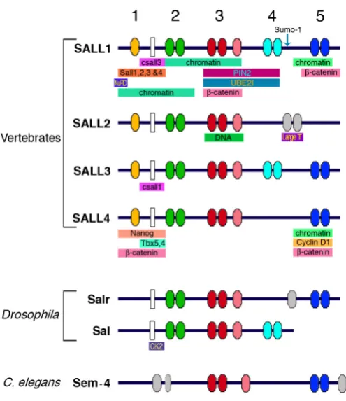
spalt-related (salr) which form part of a gene complex (Kuhnlein et al., 1994; Barrio et al., 1996). The more characteristic feature of Sall proteins is the presence of several zinc finger domains scattered along the protein (Fig. 1). Zinc finger domain 1 corre-sponds to the C2HC class, and it is only present in the vertebrate homologues. The rest of the domains (2-5) correspond to C2H2 zinc fingers arranged in pairs. The doublets are connected by a H/ C link conserved throughout evolution, and the second finger from each pair contains a characteristic domain called Sal-box that is present in other zinc finger transcription factors. The third finger domain contains an associated finger, also highly conserved among orthologs. Another important domain characteristic of these proteins is a Glutamine rich region (polyQ), present from Drosophila to humans, which might be involved in protein-protein
BIOLOGY
www.intjdevbiol.com*Address correspondence to: Jose F. de Celis. Centro de Biología Molecular “Severo Ochoa”. Universidad Autónoma de Madrid and Consejo Superior de Investigaciones Científicas. Cantoblanco. Madrid 28049. Spain. e-mail: [email protected] or Rosa Barrio. Functional Genomics, CIC bioGUNE, Bizkaia Technology Park. Building 801A, E-48160 Derio. Spain. e-mail: [email protected]
Final author-corrected PDF published online: 27 October 2008.
ISSN: Online 1696-3547, Print 0214-6282 © 2008 UBC Press
Printed in Spain
Abbreviations used in this paper: BMP, bone morphogenetic protein; Dpp, decapentaplegic; OS, Okihiro Syndrome; TBS, Townes Brocks Syndrome. interactions among members of the family and between Sall and other proteins. The four orthologues vertebrate proteins, Sall1-4, display differences in the finger distribution, being Sall2 the more
distant member of the family (Fig. 1; Kohlhase et al., 1996;
Hollemann et al., 1996; Kohlhase et al., 1999a; Onuma et al., 1999; Ott et al., 1996; Buck et al., 2000; Ott et al., 2001; Ma et al., 2001; Kohlhase et al., 2002a; Ma et al., 2006). The nematode Sall protein, named Sem-4, shares common features with their homo-logues, like the finger domains 3 and 5 (Fig. 1; Basson and Horvitz, 1996; Photos et al., 2006). For a recent phylogenetic analysis of the Sall family, and a comprehensive update on the nomenclature of vertebrate orthologues, see a recent review by Sweetman and Munsterberg (2006).
this review we will summarize different aspects of Sall proteins and genes biology, with emphasis in their modes of regulation, their functions in proliferation and transcription, their develop-mental roles in different organisms and their association with several human genetic diseases.
Regulation of sall gene expression
Most of what is known about the regulation of sall expression derives from studies in Drosophila sal and salr genes and in some vertebrate sall members. A common aspect is that the expression of sall genes depends on the activity of several signal transduction pathways (Table 1). In particular, the Wnt, FGF, Shh, EGFR and BMP pathways participate in the activation of sall expression in different tissues and, in some cases, it has been shown that Sall proteins are key mediators of the function of these pathways during organogenesis and cell differentiation. The regulation of sal and salr in Drosophila has been studied extensively, and a number of tissue specific enhancers have been characterized
Fig. 1. Schematic representation of the main conserved domains present in Sall proteins. Coloured ovals numbered 1 to 5 represent the zinc finger domains from vertebrate, Drosophila and C. elegans Sall homologues. White rectangles represent the polyQ regions. The arrow in Sall1 indicates the sumoylation site described for this protein. Coloured horizontal bars below each protein indicate the Sall-interaction domains with other proteins. Vertebrate data were collected from human, mouse, chicken and frog homologues (Bohm et al., 2007; Kiefer et al., 2002; Kiefer et al., 2003; Koshiba-Takeuchi et al., 2006; Lauberth and Rauchman, 2006; Ma et al., 2006; Netzer et al., 2001; Netzer et al., 2002; Netzer et al., 2006; Li et al., 2001; Li et al., 2004; Onai et al., 2004; Sakaki-Yumoto et al., 2006; Sato et al., 2004; Sweetman et al., 2003; Trott et al., 2001; Wu et al., 2006; Yamashita et al., 2007).
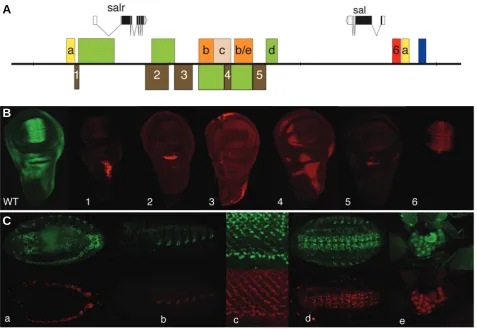
(Wagner-Bernholz et al., 1991; Kuhnlein et al., 1997; Chen et al., 1998; Barrio et al., 1999; de Celis et al., 1999; Guss et al., 2001; Barrio and de Celis, 2004). In this organism, the sal and salr transcription units are separated by 50kb of non-coding DNA containing regulatory sequences. sal is expressed during embry-onic development in a variety of tissues, including the cellular blastoderm, posterior spiracles, trachea, oenocytes and cells in the central and peripheral nervous system (Fig. 2). The regions where salr is expressed overlap in all these tissues, except in the early blastoderm where salr is not expressed (Barrio et al., 1996).
During larval development, sal andnsalr are expressed in the
same cells in the wing, eye-antenna and haltere imaginal discs, as well as in the ring gland and central nervous system (Fig. 2). The
structure of the sal and salr regulatory regions shows many
similarities with those of other Drosophila gene complexes, such
as the achaete-scute and Iroquois complexes (Ruiz-Gomez and
Modolell, 1987; Gomez-Skarmeta et al., 1996). Thus,
tissue-specific enhancers are scattered in the 50 Kb intergenic region and also in the 5´and intronic regions of both genes (Fig. 2). The expression of the sal and salr transcripts is regulated by separate and, in some cases, shared cis-regulatory elements (Fig. 2). Enhancers that direct the expression of sal in the blastoderm, wing and tracheae are some of the best characterized so far (Kuhnlein et al., 1997; Barrio and de Celis, 2004; Chen et al., 1998).
The detailed analysis of sal/salr regulatory elements in the wing disc showed an even greater complex organization, in that inde-pendent enhancers control the expression in different territories such as the wing pouch, thorax, hinge and pleura (Fig. 2; Barrio et al., 1999; de Celis et al., 1999). Interestingly, the expression in the thorax is also controlled by multiple elements affecting specific sub-domains. The organization of modular regulatory regions implies that the territories of sal and salr expression are, from the regulatory point of view, a mosaic of cell populations where different combinations of factors are responsible for the activation of each gene in different groups of cells. The expression of sal genes in the wing pouch is directly regulated by the Dpp pathway,
acting through sal and salr independent enhancers. The Dpp
pathway activates sal expression in a central domain that is
broader than the dpp expression territory through a genomic
region of 453 bp localized 5’ of the sal transcript (Barrio and de Celis, 2004). This enhancer integrates positive inputs mediated by the Dpp effectors Mad/Medea with the repressor activity of Brinker. The mechanism of repression by Brinker does not rely on competition with Mad–Medea overlapping sites, but on the exist-ence of adjacent binding sites for Brinker and Mad/Med (Barrio and de Celis, 2004). Additional factors such as the T-box tran-scription factor Optomotor blind, the trithorax protein Ash2, the activator complex Vestigial/Scalloped and the repressor Groucho are also involved in the regulation of sal in the wing blade (Guss et al., 2001; del Alamo Rodriguez et al., 2004; Angulo et al., 2004; Winter and Campbell, 2004; Hasson et al., 2005). The enhancer regulating salr expression in the wing blade has not yet been identified.
The regulation of sall genes expression in organisms other
than Drosophila is less documented. However, some of the
stage in the frog (Onai et al., 2004). Interestingly, Xsall2 and human SALL1 modify the response to Wnt signalling, although Xsall2 antagonises Wnt signalling in vivo (Onai et al., 2004), and human SALL1 promotes Wnt signalling in cell culture assays (Sato et al., 2004). The function of Xsall2 is essential for the expression of the Pax6, Otx2, and Bf-1 genes in the forebrain/ midbrain region, and for the repression of the caudal genes En2, Pax2, Wnt1 and Gbx2. Xsall2 is also required for anterior expres-sions of two antagonistic effectors of Wnt signalling, GSK3 and Tcf3 (Onai et al., 2004).
The expression patterns of sall family genes and the analysis of their regulation indicates that Sall function can not be univer-sally assigned to specific signalling pathways, but rather that Sall has been adopted by different signalling pathways in different developmental contexts. Similarly, it appears that orthologues, as determined by degrees of conservation of sall coding sequences, do not imply similarities of expression patterns.
Function of Sall proteins in gene regulation
The genetic approach to study sal function in Drosophila
identified a number of developmental processes in which sal and salr are involved. In addition, this approach also allowed in some instances to place sal and salr into genetic hierarchies, in which both upstream and downstream elements to sal/salr were identi-fied. Some of these aspects will be considered latter when addressing the specific roles of sal/salr in Drosophila tracheal and limb development. However, very few data are available in flies about the molecular mechanisms of Sal function, and no compre-hensive analysis of Sal/Salr partners and target genes has been carried out yet. Thus, a direct interaction with DNA has only been shown in the case of Salr, which is able to bind an AT-rich sequence in the chorion gene s15 promoter with the central zinc finger domain 3 (Table 2; Shea et al., 1990; Barrio et al., 1996).
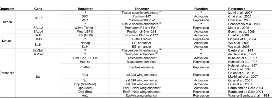
Organism Gene Regulator Enhancer Function References
? Tissue-specific enhancers (1) ? Izumi et al., 2007
SIX1 Position -947 Activation Chai et al., 2006
WT1 Position -2000 to +1 Repression Chai et al., 2006 SALL1
? Tissue-specific enhancers (2) ? Pennacchio et al., 2006
SALL2 Wilms Tumor-1 Promotors P1 and P2 (3) Repression Ma et al., 2006
Human
SALL4 Wnt (LEF1) Position -249 to -218 Activation Boehm et al., 2006
Sall1 Shh (GLI3) Position -1344 to -1137 Activation Hu et al., 2006
Sall3 ? T-DMR region Repression Ohgane et al., 2004
Nanog ES enhancer Activation Wu et al., 2006
Mouse
Sall4
Sall4 ES enhancer Activation Wu et al., 2006
Sal/Salr ? Tissue-specific enhancers (4) ? Barrio et al., 1999
Sal/Salr ? Wing disc enhancers (5) ? de Celis et al., 1999
Bcd, Cad, Tll, Hb Blastoderm enhancer Activation Kuhnlein et al., 1997 Hkb, Kr Blastoderm enhancer Repression Kuhnlein et al., 1997
Kni/Knir Trachea enhancer Repression Kuhnlein et al., 1997 Chen et al., 1998
Ubx sal 328 wing enhancer Repression Galant et al., 2002 Makhijani et al., 2007 Sc sal 328 wing enhancer Activation Guss et al., 2001 Dpp (Mad/Med) sal 328 wing enhancer Activation Guss et al., 2001
Dpp (Med) EcoRI-NdeI wing enhancer Activation Barrio and de Celis 2004 Dpp (Brk) EcoRI-NdeI wing enhancer Repression Barrio and de Celis 2004 Drosophila
Sal
Antp Eye/Antenna enhancer Repression Wagner-Bernholz et al., 1991 TABLE 1
REGULATORY REGIONS AND DIRECT REGULATORS IDENTIFIED FOR SALL GENES
Only the regulators shown to interact directly with sall promoters or enhancers are included in this Table. In some cases, the enhancers have been isolated, but the regulators are unknown and they
are indicated by question marks. (1) Tested in chicken: Prosencephalon and anterior neural ridge. (2) Tested in mouse: Forebrain, midbrain, hindbrain, neural tube, limb, eye, dorsal root ganglia, somites, nose, branchial arc, genital tubercle, trigeminal nerve, heart, neural crest mesenchyme, melanocytes and cranial nerve. (3) Tested by reporter activity. (4) Embryonic (central nervous system, peripheral nervous system, oenocytes, trachea, gut, epidermis and larval (wing, haltere, eye, CNS, leg, ring gland) enhancers. (5) Wing blade, hinge and thorax. Data were compiled from the references indicated in the right-hand column.
aspect in the regulation of vertebrate sall genes is the involvement of signalling pathways in different developmental systems. For example, the expression of Xenopus Xsall4 within the interdigital spaces suggests that BMP proteins are involved in regulating its expression in these territories (Neff et al., 2005). Similarly, the Msall3 gene from Medaka fish is expressed in most places where Hedgehog signalling is active, and Hedgehog regulates the expression of the gene at the midbrain-hindbrain organizer region (Koster et al., 1997). In this territory, FGF signalling is required to
activate Msall3 expression in response to Shh during Medaka
development, and this regulatory relationship is also observed during the growth of the optic vesicle (Carl and Wittbrodt, 1999). The FGF pathway, now in collaboration with Wnt signalling, is also required for the activation of csall1 expression in chicken limb buds, where csall1 is expressed in the apical ectodermal ridge and in the underlying distal mesenchyme (Farrell and Munsterberg, 2000). In these cells, a combination of Wnt3a and Wnt7a with FGF4 and FGF8, which are expressed in the apical ectodermal ridge, regulates csall1 expression, whereas BMP function is also required to activate csall1 in mesenchymal cells of the proximal limb (Capdevila et al., 1999; Farrell and Munsterberg, 2000). A recent analysis of the human SALL4 promoter region identified 367 bp located upstream of the ATG which sequence is extremely conserved in several vertebrates sall4 genes. The observation that this region contains consensus-binding sites, which integrity is required for promoter activity in cell culture assays, for LEF/ TCF, a transcription factor mediating the response to canonical Wnt signalling, implies a direct effect of TCF on SALL4 expression (Bohm et al., 2006). Regulatory relationships between Wnt
sig-nalling and sal are also observed in Drosophila and Xenopus.
Thus, wingless, a Drosophila Wnt homologue, induces sal
2002; Sweetman et al., 2003; Netzer et al., 2006). First, the N-terminal part of the protein contains a 12 amino acids sequence that is able by itself to confer repression capacity and to interact with the Histone Deacetylase Complex NuRD (Kiefer et al., 2002; Lauberth and Rauchman, 2006). This interaction can be modified by phosphorylation of Sall1 (Lauberth et al., 2007). The NuRD-interaction domain is also found in other Sall homologues, includ-ing C. elegans Sem-4, and in transcription factors not related to the Sall family, but it is not present in the Drosophila Sal homo-logues. In the cases of human and murine SALL2/Sall2 and SALL4/Sall4, alternative spliced forms have been described that lack this repression domain that would function independently of the NuRD repression complex, although the functional role of these alternative forms is still unexplored. The N-terminal part of the Sall1 shows localization to heterochromatin foci when fused to a nuclear localization signal, suggesting an association be-tween transcriptional repression and protein location (Kiefer et al., 2002; Sato et al., 2004).
Similarly, CK2 kinase is the only protein reported to interact with Drosophila Sal (Trott et al., 2001). However, the biological rel-evance of these interactions has not yet been explored.
In contrast to the paucity of data concerning Sal molecular function in Drosophila, a wealth of data identifying Sall protein-protein interactions, Sall subcellular localization and Sall tran-scriptional effects are stemming from the analysis of vertebrate sall genes (Table 2). In what follows we will summarise some of the interactions identified for the sall genes and proteins 1, 2 and 4, which taken together suggest that the variety of processes requiring Sall function can be accounted by the diversity of protein-protein and protein-DNA interactions in which Sall pro-teins are engaged (see Fig. 1).
Human SALL1 has been described as a transcriptional repres-sor in a number of experimental settings, most of them involving the regulation of heterologous promoters fused to reporter genes, and presents two possible mechanisms of repression (Nishinakamura et al., 2001; Netzer et al., 2001; Kiefer et al.,
Fig. 2. Genomic structure of Drosophila sal genes and their regulatory modules. (A) Schematic representation of the sal-salr gene complex, showing the coding regions as black boxes, the non-coding RNA as empty boxes and the introns as connecting lines between boxes. Arrowheads indicate the direction of transcription. The coloured boxes above and below the genomic DNA (black line) represent regulatory modules identified in the sal complex (Kuhnlein et al., 1997; Chen et al., 1998; Barrio et al., 1999; de Celis et al., 1999; Barrio and de Celis, 2004). Yellow boxes correspond to regulatory regions driving reporter expression in the trachea (A), brown boxes in the wing imaginal disc (1-5), orange in the oenocytes (B) and the oenocytes and the ring gland (B/E), light brown in the eye imaginal disc (C), red in the wing blade (6) and blue in the blastoderm. (B) Expression of Sal in the wing imaginal disc (WT, green), and expression of β-Gal (red) in imaginal discs bearing reporter constructs for the regulatory regions shown in panel A as brown boxes with numbers 1-5 and red box with number 6. (C) Each pair of pictures represent focal planes through Drosophila embryos showing the expression of Sal (above and in green in all pictures) and the expression of β-Gal driven by reporter constructs (below and in red). The letters in each picture correspond to the same letter code in panel A: the trachea (a), the oenocytes (b), the photoreceptors in the eye imaginal disc (c), the central nervous system (d) and the ring gland (e).
B
C
The second repression mechanism is independent of the Histone Deacetylase Complex and requires the central region of the protein including the finger domains 2 and 3 (Netzer et al., 2001; Netzer et al., 2006). This region also shows localization in heterochromatin foci in murine cells. In addition, SALL1 can interact with PIN2, an isoform of telomere-repeat binding factor 1
(TRF1) (Netzer et al., 2001). TRF1/PIN2 binds to telomeres,
suggesting a mechanism of repression for SALL1 by association to pericentromeric heterochromatin. Yet another region of the protein located in the C-terminal fingers has been described as important for the interaction with heterochromatin. This domain is particularly well conserved from Drosophila to humans and it has been reported to bind the major satellite DNA (Table 2; Yamashita et al., 2007).
Even though controversy exists about the identity of the do-main involved in Sall-DNA interactions and the existence of different repression domains, it is interesting to speculate that Sall proteins might recruit remodelling factors to heterochromatin. In this context, Sall1 is able to bind to β-catenin and activate synergistically a reporter construct responding to the Wnt signal-ling pathway (Sato et al., 2004). However, the domain of Sall1 that co-activates this reporter does not coincide with the β-catenin binding domain, but with the heterochromatin localization do-main, indicating that Sall1 localization, and not its interaction with
β-catenin, is the mediator of the interactions between Sall1 and the Wnt signalling pathway. In vivo, the role of human SALL1 as a transcriptional repressor has been shown during steroidogen-esis in adrenal gland, where Sall1 represses the expression of the enzymes 11-hydroxylase and aldosterone synthase, involved in the glucocorticoid and mineralocorticoid biosynthetic pathways under the modulation of Angiotensin II (Romero et al., 2007). In contrast, murine Sall1 is necessary for the activation of some kidney mesenchymal markers, consistent with its role in ureteric bud invasion (Nishinakamura et al., 2001). As in the case of the activation of Wnt signalling, the up-regulation of these genes might not be direct.
Sall protein interactions
The subcellular localization and transcriptional capacity of Sall proteins might be conditioned by posttranslational modifications.
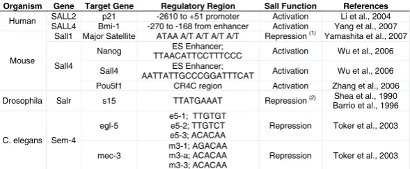
Organism Gene Target Gene Regulatory Region Sall Function References SALL2 p21 -2610 to +51 promoter Activation Li et al., 2004 Human
SALL4 Bmi-1 -270 to -168 from enhancer Activation Yang et al., 2007 Sall1 Major Satellite ATAA A/T A/T A/T A/T Repression (1) Yamashita et al., 2007
Nanog ES Enhancer;
TTAACATTCCTTTCCC Activation Wu et al., 2006
Sall4 ES Enhancer;
AATTATTGCCCGGATTTCAT Activation Wu et al., 2006 Mouse
Sall4
Pou5f1 CR4C region Activation Zhang et al., 2006
Drosophila Salr s15 TTATGAAAT Repression (2) Shea et al., 1990 Barrio et al., 1996
egl-5
e5-1; TTGTGT e5-2; TTGTCT e5-3; ACACAA
Repression Toker et al., 2003 C. elegans Sem-4
mec-3
m3-1; AGACAA m3-a; ACACAA m3-3; ACACAA
Repression Toker et al., 2003 TABLE 2
TARGET DNA SEQUENCES BOUND BY SALL PROTEINS
Only the sequences bound by Sall proteins are included. (1) Repression is inferred but not proved. (2) Repression is inferred, as Salr is not expressed at the same time than S15. Data were compiled from the references indicated in the right-hand column.
lular localization of cSall1, which is retained in the cytoplasm in presence of cSall3 through protein-protein interactions via the
conserved polyQ domains (Sweetman et al., 2003). The
conser-vation of the polyQ region in Sall proteins opens the possibility of interactions among all the paralogues, which could to be impor-tant for the biological activity of the proteins.
The protein-protein interactions of Sall4 during embryonic development have also been studied in mouse and zebrafish limb development. In mice, Sall4 interacts with Tbx5, a T-box tran-scription factor involved in limb development, regulating the formation of the forelimb through the activation of FGF10 in a feed-forward mechanism (Koshiba-Takeuchi et al., 2006). In the hindlimb, an analogous interaction occurs with Tbx4, a factor necessary for hindlimb development. The interaction with Tbx5 seems to be important for the activation of Gja5 in the heart where, at the same time, Sall4 interferes with the capacity of Tbx5 to activate Nppa. How Sall4 can achieve its role as transcriptional activator and repressor, and how this is related to its capacity to bind heterochromatin and promote the methylation of histones remains unclear.
Sall proteins in stem cell and cancer biology
Murine Sall1 has a role in maintaining cellular pluripotency and proliferation. Thus, renal primordial cells in the ureteric bud epithelium and metanephric mesenchyme are able to produce nephrons and collecting ducts when induced from pluripotent embryonic stem cells. Only cells expressing high levels of Sall1 can reconstitute a three-dimensional kidney structure in an organ culture setting, indicating that renal progenitors with multipotent capacity require Sall1 (Osafune et al., 2006; Yamamoto et al., 2006). In these cells, Sall1 is not required for generation or differentiation of renal progenitors but for their proliferation or survival (Osafune et al., 2006). Sall1, expressed in embryonic stem cells, seems to contribute to the activation of Oct4 (Zhang et al., 2006) and Sall1a is necessary for the activation of FGFR2 downstream of Tbx5 during zebrafish pectoral fin development (Harvey and Logan, 2006). Whether this activation capacity is direct or indirect remains to be investigated.
Mouse and human Sall2 and SALL2 genes have been reported
as tumour suppressors in several conditions. Thus, Sall2 was Thus, human SALL1 interacts with UBE2I, the
subcel-identified in a large screen looking for targets of the Large T
antigen from the highly oncogenic mouse polyoma virus (Li et
al., 2001). The interaction with Sall2 is important to suppress viral DNA replication and the growth of the virus (Li et al., 2001). Moreover, the presence of Sall2 in ovarian cancer cells inhibits their growth rate and their capacity to form colonies in soft agar. Some human ovarian carcinoma cell lines express low levels of SALL2 which, when re-introduced, results in a substantial reduction in the capacity of these cells to grow as tumours in nude mice. The control of cell growth and proliferation by SALL2 could be determined by its direct activation of p21 and Bax (Table 2; Li et al., 2004).
Human SALL2 is also necessary for the activation of a number of genes expressed after serum deprivation, a situation in which there is inhibition of cell growth. These genes are repressed in many types of prostate, blood and lung cancers, and their repression can predict the increased risk of cancer progression and death in human breast cancers (Table 3; Liu et al., 2007). SALL2 is considered as an “early response gene” and it is necessary for the repression of the “middle response
genes” that become super-induced when SALL2 is silenced,
being unclear whether the activation and repression exerted by SALL2 on these genes is direct (Liu et al., 2007). SALL2 is also downregulated in other tumour types, like some lung
carcino-mas and adenocarcinoma of colon and prostate (Ma et al.,
2001; Li et al., 2002). In contrast to the cases indicated above,
where, as expected for a tumor suppressor, SALL2 is
down-regulated, SALL2 is upregulated in Wilm´s Tumors and in
Synovial Sarcoma cases (Table 3; Li et al., 2002; Nielsen et al., 2003). The molecular mechanisms underlying the roles of SALL2 as a tumour suppressor in certain types of cancers and its upregulation in sarcomas are still unknown.
Murine Sall4 mRNA is inherited maternally and is abundant
in the mice zygote. These transcripts are degraded during the two-cell stage. Zygotic transcription occurs after the four-cell
stage, after which Sall4 mRNA levels continue to increase to
the blastocyst stage (Zhang et al., 2006). The effects of Sall4 deficiency were studied using knockout mice and knockdown embryos (Zhang et al., 2006; Elling et al., 2006; Sakaki-Yumoto et al., 2006; Koshiba-Takeuchi et al., 2006; Warren et al., 2007). Homozygous mutant mice die during peri-implantation stages, due to lack of proliferation of the inner cell mass. In
addition, Embryonic Stem Cells (ESC) derived from Sall4-null
embryos proliferate poorly with no aberrant differentiation, and no embryonic nor extraembryonic endoderm stem cell lines can
be established from Sall4 mutant blastocysts (Elling et al.,
2006; Sakaki-Yumoto et al., 2006). The role of Sall4 on ESC
maintenance can be achieved through its interaction with Nanog, a homeodomain transcription factor identified as a protein able to sustain pluripotency in murine ESCs. The complex Sall4-Nanog could regulate the transcription of genes necessary for self-renewal, such as Sox2 and Oct4, in addition to their own transcription, constituting a regulatory circuit (Table 2 and Fig. 3; Wu et al., 2006). Similarly to Oct4, the reduction in Sall4 expression results in re-specification of ESCs to the trophoblast lineage, and this change is related to the expansion of Cdx2 expression (essential to the trophectoderm lineage) into the Inner cell mass of the blastocyst (Zhang et al., 2006; Elling et al., 2006). The co-occupancy of Nanog binding sites by the complex Nanog-Sall4 results in the activation of Nanog down-stream genes by the over-expression of Sall4 (Wu et al., 2006). In this experimental setting, the up-regulation of the trophecto-derm lineage markers CDX2, HAND1 and GATA6 observed in the absence of human SALL4 could be indirect, occurring
through the loss of POU5F1 expression (Zhang et al., 2006).
In concordance with its role in preserving the pluripotency of
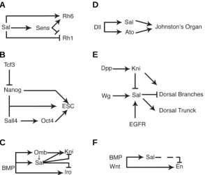
Fig. 3. Schematic representation of genetic regulatory circuits in which Sall proteins and genes are involved during development. (A) Regulation by Sal and Senseless (Sens) of rhodopsin gene expression (Rh6 and Rh1) during the differentiation of the photoreceptor cell R8 (modified from Domingos et al., 2004a). (B) Regulation of sal expression by Distal-less, and requirement of Sal and Atonal in the formation of the Drosophila auditory organ, the Johnston’s organ (modi-fied from Si Dong et al., 2003). (C) Regulation of Oct4 by Sall4, and requirement of Nanog, Oct4 and Sall4 during Embryonic stem cell maintenance and Epiblast development (modified from Zhang et al. (2006) and Pereira et al. (2006). (D) Regula-tion of sal expression by Wg, EGFR and Dpp signalling during trachea development and its function in the specification of the dorsal trunk (modified from Kühnlein and Schuh (1996); Chen et al. (1998) and Chihara and Hayashi (2000)). (E) Regulatory interactions between the Dpp (BMP) downstream transcrip-tion factors Sal, Kni, Omb and Iro during Drosophila wing blade development (de Celis and Barrio, 2000; del Alamo Rodriguez et al., 2004; Cook et al., 2004). (F) Regulatory interactions occurring during Butterfly eye spot formation involving Sal, Engrailed and the candidate eyespot signalling molecules BMP and Wnt homologues. The dashed line indicates that Sal only represses En expression in some species, but not in others, generating either concentring rings or nested domains of Sal and En expression. Modified from Brunetti et al. (2001) and Monteiro et al. (2006).
B
C
D
E
stem cells in mice, the Xenopus homologue, Xlsall4, was identified in a subtracted limb regeneration screen (King et al., 2003). Xlsall4 transcripts are expressed during the early and middle phases of limb development and also in the fore- and hindlimb during regeneration-competent stages, suggesting that its activity could maintain blastema cells in an undifferen-tiated state (Neff et al., 2005). Similarly, the chicken homologue csal4 seems to keep neural crest cells in an undifferentiated stage (Barembaum and Bronner-Fraser, 2004). All these verte-brate homologues are expressed in the growing tail tip region rich in undifferentiated cells (Kohlhase et al., 2002a; Barembaum and Bronner-Fraser, 2004; Neff et al., 2005). The expression of
human and murine SALL4/Sall4 during adulthood is restricted
to testis and ovaries (Kohlhase et al., 2002a; Kohlhase et al., 2002b). Furthermore, microarray analysis shows that in the
ovaries of newborn mice mutant for Nobox, a homeobox gene
expressed in oocytes and required during oogenesis, Sall4 is
drastically downregulated, coinciding with a rapid loss of post-natal oocytes (Choi et al., 2007).
The lack of proliferation observed in Sall4 null mutant mouse cultured blastocysts and embryos in vivo (Sakaki-Yumoto et al., 2006) might be related to the inefficient G1/S transition ob-served in ESCs, which could be linked to the interaction of Sall4 with CyclinD1 (Bohm et al., 2007). A possible role of Sall4 in promoting cell proliferation could also be related to the
expres-sion of human SALL4 in certain type of tumours. Accordingly,
SALL4 is upregulated in acute myeloid leukaemia (Table 3). The constitutive expression of SALL4 may enable leukaemic blasts to acquire stem cell properties, such as self-renewal and/ or lack of differentiation, and become leukaemia stem cells (Ma et al., 2006; Cui et al., 2006). This is probably achieved through the activation of the Wnt/β-catenin signalling pathway, as shown by the up-regulation of the Wnt targets c-Myc and CyclinD1 in
leukaemic cells where SALL4 is over-expressed (Ma et al.,
2006), or by the activation of the polycomb gene Bmi-1, which plays an essential role in regulating adult, self-renewing he-matopoietic stem cells and leukaemia stem cells (Yang et al., 2007). The activation of Bmi-1 is associated to increased levels
of histone methylation in the Bmi-1 promoter, but the
mecha-nism relating the over-expression of SALL4 and the
hypermethylation of histones is still unknown (Yang et al.,
2007). A different role for SALL4 during tumourigenesis might be achieved through its role as a “caretaker” of chromosomal stability, which could be related to the capacity of SALL4 to bind to heterochromatic regions through its most C-terminal finger
pair (Sakaki-Yumoto et al., 2006; Bohm et al., 2007). Human
SALL4 is epigenetically silenced in colorectal cancer aneuploid cells where SALL4 promoter is more frequently hypermethylated than in diploid cells (Habano et al., 2007). Thus, the absence of SALL4 might influence tumourigenesis by destabilization of c h r o m o s o m e s , b u t i t s u p r e g u l a t i o n m i g h t i n f l u e n c e tumourigenesis by promoting proliferation.
Sal proteins in cell specification and morphogenesis
sall genes are required for multiple developmental pro-cesses, suggesting that they engage in a variety of interactions and modify the expression of target genes in a context-depen-dent manner. We have attempted to classify these processes
into several categories that include sall invertebrate and verte-brate members, and will discuss in more detail some represen-tative examples.
Cell fate assignment
The Drosophila sal and salr genes, and also several
mem-bers of the sall family in other organisms, participate in a variety of cell-fate decisions during development, controlling the distinc-tion between alternative cell fates or the implementadistinc-tion of a particular program of cell differentiation. Examples of the former are the function of the sem-4 ortologue in C. elegans during the specification of touch receptor neurons (Mitani et al., 1993), and the function of Drosophila sal genes in the formation of the oenocytes and strech receptors (Rusten et al., 2001; Elstob et al., 2001). In the first case Sem-4 regulates, by repression, the
expression of the Hox gene egl-5 and the LIM homeobox gene
mec-3. These interactions are direct, because Sem-4 binds to a common motif present in the mec-3 and egl-5 promoters (Table 2; Toker et al., 2003). Sem-4 also regulates the expression of the
Hox genes lin-39 and, in the absence of sem-4, the secondary
vulval cell lineage is not correctly specified (Grant et al., 2000). The relationships between Sal and Hox functions in the specifica-tion of cell identities is a common aspect of Sal proteins also observed in Artemia and Drosophila, although the interactions
between sal and Hox genes vary in different developmental
systems. Thus, the Artemia sal orthologue is expressed in the pre-segmental growth zone and in the segments that emerge from this zone (Copf et al., 2006). The loss of sal function, caused by RNA interference, results in a variety of homeotic transformations associated with the de-repression of different Hox genes in the corresponding segments, indicating that Sal regulates Hox gene expression (Copf et al., 2006). Because Artemia sal is expressed in all segments, and the observed homeotic transformations in knockdown animals are variable and stochastic, it was suggested that Sal function is related to the maintenance of spatial domains of Hox expression acting in transcriptional repression by chroma-tin modifications (Copf et al., 2006).
In contrast to this role in the maintenance of Hox expression,
the Drosophila sal gene acts downstream of different Hox genes
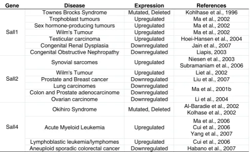
Gene Disease Expression References
Townes Brocks Syndrome Mutated, Deleted Kohlhase et al., 1996 Trophoblast tumours Upregulated Ma et al., 2002 Sex hormone-producing tumours Upregulated Ma et al., 2002 Wilm's Tumour Upregulated Ma et al., 2002 Testicular carcinoma Upregulated Hoei-Hansen et al., 2004 Congenital Renal Dysplasia Downregulated Jain et al., 2007 Sall1
Congenital Obstructive Nephropathy Downregulated Liapis, 2003 Synovial sarcomes Upregulated Niesen et al., 2003
Subramaniam et al., 2006 Wilm's Tumour Upregulated Liet al., 2002 Prostate and Breast cancer Downregulated Liu et al., 2007
Lung carcinomes Downregulated
Colon and Prostate adenocarcinome Downregulated Ma et al., 2001b Sall2
Ovarian carcinome Downregulated Li et al., 2004 Okihiro Syndrome Mutated, Deleted Al-Baradie et al., 2002
Kolhase et al., 2002
Acute Myeloid Leukemia Upregulated
Ma et al., 2006 Cui et al., 2006 Yang et al., 2007 Lymphoblastic leukemia/lymphomes Upregulated Cui et al., 2006 Sall4
Aneuploid sporadic colorectal cancer Downregulated Habano et al., 2007 TABLE 3
SALL PROTEINS INVOLVED IN HUMAN DISEASES
in the haltere, labial and antennal imaginal discs. The distinction between wing and haltere relies in the function of the Ultrabithorax
(Ubx) Hox gene. Among several other target genes, Ubx directly
repress sal expression in the haltere, suppressing the positive input of Dpp on sal and contributing to the differences between these two structures (Weatherbee et al., 1998). Similarly, the Hox proteins Proboscipedia and Sex combs reduced direct the devel-opment of the proboscis by repressing sal expression in the labial disc (Abzhanov et al., 2001). In the antennal disc sal also acts downstream of genes specifying segmental identity, but its ex-pression is activated rather than repressed by the combination of Distal-less and Homothorax (Dong et al., 2000). Interestingly,
reminiscent to the loss of hearing associated to human SALL1
mutations (see below), loss of sal and salr in the antennal disc causes a severe reduction in the major Drosophila auditory organ, the Johnston’n organ, and is associated with deafness (Dong et al., 2003). Finally, Drosophila Sal proteins also have homeotic functions independent of Hox genes during embryogenesis, act-ing to promote head versus trunk development (Jurgens, 1988). The function of sal genes in specifying cell types does not always relies in their relationships with Hox genes. A clear example of a direct role of Sal proteins in cell differentiation occurs
during Drosophila eye development, where Sal influences the
formation of the R3, R4, R7 and R8 photoreceptors (Fig. 3A). Thus, Sal is required for the specification of R7 and the expression of R7 specific markers, the terminal differentiation of R8 and the regulation of photoreceptor specific rhodopsins, the correct speci-fication of the R3/R4 pair of cells and establishment of planar cell polarity. Finally, Sal expression needs to be repressed latter in these cells to inhibit their transformation to R7 fate (Mollereau et al., 2001, Domingos et al., 2004a; Domingos et al., 2004b). A similar function in cell-fate specification can be operative in many cell populations during neural system development, because sal
and sall genes are expressed predominantly in the developing
nervous system in a variety of organisms. An interesting example of Sal functions in cell fate decisions is the formation of a particular type of sensory organs in Drosophila, where Sal operates as a switch between two cell types induced by EGFR activity, the oenocytes and the precursors of the pentascolopodial sensory organ. This organ is formed by five sensory units derived from five
chordotonal organ precursors (COPs; Gould et al., 2001). The
oenocytes form around the most dorsal COP and express high levels of Sal. The absence of Sal results in the lack of oenocytes accompanied by the formation of extra COPs, indicating that Sal is necessary to promote oenocyte formation and to restrict the number of COPs at the same time (Rusten et al., 2001; Elstob et al., 2001). This role of Sal is reminiscent of the role of Sall4 in the decision between inner cell mass and trophoblasts in the mouse (Elling et al., 2006).
Regional specification
Another common aspect of sall functional requirements in
different organisms occurs during the subdivision of a cell popu-lation into smaller developmental units, which we refer to as “regional specification”. This feature of sall function was first identified for the Drosophila sal and salr genes during the growth and patterning of the wing imaginal disc, an epithelial tissue that differentiate during metamorphosis the fly wing and thorax. The Sal/Salr proteins act in the wing blade as transcription factors
conferring regional identity to the central part of the wing, linking the activity of the secreted molecule Dpp to pattern formation (de Celis et al., 1996). Thus, sal and salr are expressed in a central domain of cells in the wing region of the disc, where they participate to the patterning of the wing blade (Fig. 3E). The Dpp pathway directly regulates the expression of sal and salr in this territory, and they direct the localisation of characteristic wing pattern elements, the veins, by regulating the expression of the vein-specific genes of the knirps and Iroquois gene complexes (Fig. 3E; de Celis and Barrio, 2000). In the case of the Iroquois genes, Sal/Salr repress their expression in all cells not exposed to Hedgehog signalling, confining Iroquois expression to the posterior L5 provein territory. The relationship between Sal/Salr and the knirps genes is more complex, because their expression is activated in the domain where Sal/Salr levels are lower in anterior cells, and repressed by higher levels of Sal/Salr in the rest of the wing (de Celis and Barrio, 2000). In addition to its pattern-promoting function, Sal and Salr are also required for cell viability, cell proliferation and epithelial integrity of the cell population where they are expressed (de Celis et al., 1996; Milan et al., 2002).
Several vertebrate Sall proteins are also expressed in the growing limbs, where they could also function to provide territorial identities to mesenchymal cell populations. Xenopus Xsall4 is expressed in developing hind- and forelimbs in a dynamic tempo-ral and spatial pattern that first is confined to the distal half of the limb bud, later is excluded from proximal-posterior and anterior regions of the bud, and finally becomes restricted in the future autopod to six interdigital domains (Neff et al., 2005). In chicken, csall1 and csall2 are also expressed in developing limbs (Farrell and Munsterberg, 2000; Farrell et al., 2001). The expression of csall1 is observed continuously through the distal limb mesen-chyme and the apical ectodermal ridge (Capdevila et al., 1999; Farrell and Munsterberg, 2000). In contrast, csall2 displays a dynamic temporal and spatial pattern of expression that is differ-entially regulated in wing and leg primordia, being in both cases detected mainly in the posterior-distal mesenchyme (Farrell et al., 2001). In zebrafish, sall1a and sall4 are expressed in developing limb-like structures, the pectoral fins (Camp et al., 2003). The expression of sall4 is first detected through the fin bud mesen-chyme, and as its development proceeds, sall4 transcripts are accumulated at the distal tip of the fin. Loss-of-function experi-ments using sall4 morpholinos showed that this gene is required for the outgrowth of pectoral fins and the formation of its distal
structures (Harvey and Logan, 2006). The gene sall1a is
thumbs, as well as polydactyly are characteristic abnormalities of TBS and OS (Kohlhase et al., 1998; Kohlhase et al., 2002b; Al-Baradie et al., 2002 and see below).
A conceptually similar function of Sall proteins during regional specification is observed during the development of eyespots in the wings of butterflies. Eyespots are pigmentation patterns characteristic of many butterflies and moth wings. The formation of the eyespot is controlled from its centre, the focus, which induces surrounding cells to acquire different colour fates. In Bicyclus anynana, the Sal homolog is expressed in the focus from its onset, and later in several concentric rings outside the focal region (Brunetti et al., 2001; Monteiro et al., 2006). Interestingly, the Engrailed homolog is expressed in an outer ring outside the domain of Sal expression, suggesting that regulatory interaction between Sal and Engrailed orthologs participate in the elabora-tion of gene expression domains. This interacelabora-tion is reminiscent to the repression of Iroquois expression by Sal observed in the Drosophila wing, and in both case leads to the creation of adjacent domains of gene expression (Fig. 3E-F).
Organogenesis
During organogenesis, cells from distinct origins, or with differ-ent developmdiffer-ental programs, must be integrated to form func-tional structures. The activity of sall genes is required in several internal organs such as the heart and kidney in vertebrates and
the tracheae (respiratory tubes) in Drosophila. A conserved
feature among vertebrates is the expression of sall genes during
the development of the kidney. Thus Xenopus Xsall4b and
zebrafish sall1a, are expressed in the pronephric ducs, and
chicken csall3 is expressed in the mesonephros (Onuma et al., 1999; Farrell et al., 2001; Camp et al., 2003). The function of sall during kidney development has been mainly studied using Sall1 knockout mice. The development of the vertebrate metanephros implies mutual inductive interactions between the ureteric bud and the metanephric mesenchyme. In this manner, the invasion of the mesenchyme by the ureteric bud epithelia, and its accom-panying branching morphogenesis to form the collecting ducts and urethra, is induced by the mesenchyme, and reciprocally, the ureteric bud induces mesenchymal aggregation around the bud tip and mesenchyme-to-epithelial conversion to form the renal
vesicle (Dressler, 2006). The Sall1 mice gene is exclusively
expressed in the metanephric mesenchyme prior to bud invasion, and this expression is maintained in the mesenchyme condensing around the ureteric bud tips. The function of Sall1 is required to promote ureteric bud invasion, which failure causes a subsequent collapse of tubule differentiation by the mesenchyme. In this manner, in Sall1-null mice the metanephric mesenchyme and the ureteric bud are formed, but the bud fails to invade the mesen-chyme (Nishinakamura et al., 2001). FGF signalling could regu-late the expression of Sall1 in the early metanephric mesen-chyme, as double mutant FGFR1/FGFR2 mice display renal aplasia and the expression of Sall1 is absent from the rudimentary mutant metanephric mesenchyme (Poladia et al., 2006). It is not clear what is the exact role of Sall1 in the mesenchyme, because direct targets activated or repressed by Sall1 in this tissue have not yet been identified. In contrast to the requirement of Sall1 during vertebrate kidney development, the function of Drosophila sal genes is not operative in the fly kidney equivalent, the Malpighian tubules, even though the formation of this structure
also includes interactions between ectodermal epithelial buds
and mesenchymal mesodermal cells (Denholm et al., 2003).
The formation of the Drosophila tracheal system involves a number of cellular activities similar to vertebrate kidney formation, such as oriented cell migration, branching morphogenesis and inductive signalling from independent tissues (Metzger and Krasnow, 1999; Affolter and Shilo, 2000). Trachea formation is initiated from ectodermic placodes that invaginate into the under-lying mesoderm and undertake a complex branching pattern to form a three-dimensional network of tubes. Loss of sal function results in a variety of phenotypes including the formation of ectopic placodes and the lack of the dorsal trunk (Kühnlein and Schuh, 1996). The first phenotype suggest an early role of Sal in suppressing tracheal fate, whereas the loss of the dorsal trunk is due to faulty cell specification within the tracheal placodes (Kühnlein and Schuh, 1996; Franch-Marro and Casanova, 2002). The failure to form the dorsal trunk in sal mutants, caused by the lack of antero-posterior migration and fusion into a trunk of the dorsal trunk primordia, is reminiscent of the requirement of Sall1 in promoting ureteric bud invasion, although during tracheal devel-opment the requirement of Sal is cell autonomous in the migrating cells. Wnt and EGFR signalling induce and maintain, respec-tively, the expression of Sal in the dorsal part of all tracheal placodes, in a region that initially encompasses the primordia of the dorsal branch and the dorsal trunk (Fig. 3D; Chihara and Hayashi, 2000). Latter, Sal expression is restricted to the dorsal trunk primordia, where it is present after the connection between the posterior and anterior dorsal trunk branches from adjacent placodes (Kühnlein and Schuh, 1996; Wappner et al., 1997; Chen et al., 1998). The downregulation of Sal in the dorsal branch primordia is mediated by repression of Knirps, acting directly on a sal regulatory element (Chen et al., 1998). The repression of sal expression by Knirps is a requisite for normal dorsal branch morphogenesis. In this manner, Sal and Knirps became ex-pressed to adjacent territories, the primordia of the dorsal branch and the dorsal trunk, which will follow different developmental
fates (Fig. 3; Chen et al., 1998; Franch-Marro and Casanova,
2002).
Sall genes in disease
Human SALL1 mutations are associated to TBS, an autosomal
dominant group of malformations characterized by imperforate anus, triphalangeal and supernumerary thumbs, dysplastic ears and sensorineural hearing loss (Kohlhase et al., 1998; Surka et al., 2001; reviewed by Powell and Michaelis, 1999). So far, 56
family mutations in SALL1 associated to TBS disorders are
haploinsuffi-ciency is not enough to cause the severe classical TBS symptoms (Borozdin et al., 2006). Confirming the role of Sall1 in kidney formation, SALL1 expression is reduced in patients with congeni-tal dysplastic kidneys, a major cause of renal failure in infants (Jain et al., 2007), as well as in congenital obstructive nephropa-thy, a common disease affecting foetuses and young children (Table 3; Liapis, 2003). Mice homozygous for Sall1 show kidney agenesis and die in the perinatal period. The abnormal kidneys result from incomplete ureteric bud outgrowth, deficient mesen-chyme tubule formation and apoptosis of the mesenmesen-chyme
(Nishinakamura et al., 2001; reviewed by Nishinakamura and
Osafune, 2006). However, in contraposition to the dominant effect shown in human TBS patients, heterozygous Sall1 mutants do not show any phenotype. Interestingly, the expression in mice of truncated Sall1 lacking all the double zinc fingers but preserving the N-terminal part of the protein, recapitulate remarkably all the abnormalities found in human TBS, supporting the idea of TBS being caused by the dominant negative effect of truncated SALL1 proteins (Kiefer et al., 2003).
Mutations in SALL4 are involved in the autosomal dominantly inherited human OS (Al-Baradie et al., 2002; Kohlhase et al., 2002b). This malformation syndrome is characterized by radial defects of the upper limbs and by Duane anomaly, a rare form of strabismus, also associated with hearing loss. There is large intra- and interfamilial variability in the clinical features of patients with SALL4 mutations and patients can be miss-diagnosed, being the mutational analysis of SALL4 important for the interpretation of the symptoms (Kohlhase et al., 2002b; Brassington et al., 2003; Borozdin et al., 2004; Kohlhase and Holmes, 2004; Kohlhase et al., 2005). In contrast to SALL1 mutants causing TBS, the muta-tions founded in SALL4-related syndromes do scatter along the gene, indicating that the clinical features are caused by loss-of-function and haploinsufficiency, rather that by a dominant nega-tive effect of truncated proteins (Borozdin et al., 2004).
Some OS patients also show severe growth retardation, also seen in patients affected by TBS that might indicate pituitary dysfunctions associated with SALL4 mutations (Kohlhase et al.,
2005; Miertus et al., 2006). A plausible explanation for the
features shared by OS and TBS is that SALL4 can interact with SALL1. Thus, the C-terminally truncated SALL1 protein produced in TBS patients could dimerise with SALL4, interfering with the binding of SALL4 to heterochromatin in a dominant-negative
manner (Sakaki-Yumoto et al., 2006). Therefore, some
pheno-types observed in SALL1 truncations could be explained by the reduction of SALL4 function. Homozygous Sall4 mutant mice dye during peri-implantation stages due to lack of proliferation of the inner cell mass (Zhang et al., 2006; Elling et al., 2006; Sakaki-Yumoto et al., 2006; Warren et al., 2007). Interestingly heterozy-gous Sall4 mice reproduce most of the features of the OS. This syndrome can also be reproduced in zebrafish, where Sall4 is not required for the initiation of development but for outgrowth of the pectoral fins primordia (Harvey and Logan, 2006). The zebrafish model allows distinguishing between features typical of OS ver-sus Holt-Oram Syndrome, caused by mutations in the T-box TBX5, demonstrating that these models are extraordinary valu-able to understand the clinical consequences of SALL mutations. In contrast to SALL1 and 4, mutations in SALL2 and 3 have not
been associated to any genetic syndrome, although SALL3 maps
in the chromosomal region associated to the 18q Deletion
Syn-drome characterized by mental retardation, short stature, hypoto-nia, hearing impairment, and foot deformities (Kohlhase et al.,
1999a), and SALL2 maps to a chromosomal region related to
haploinsufficiency in some ovarian carcinomas (Kohlhase et al.,
1996). Murine Sall2 is dispensable for normal development,
showing no effects in the tissues where it is expressed. Moreover, Sall2 removal does not exacerbate the kidney defects caused by Sall1 mutation. Despite its classification as a tumour suppressor gene, homozygous mutant mice did not show spontaneous tu-mour formation for more than 1 year after birth (Sato et al., 2003).
The most prominent expression domain of Sall2 is the brain,
raising the possibility for a function in this organ (Kohlhase et al., 2000). However, no behavioural defects or any other anomalies were reported.
Sall3 deficient mutant mice present malformation in organs necessary for normal feeding behaviour, such as the palate, the epiglottis, the tongue, and the corresponding cranial nerves. Homozygous animals die shortly after birth because their inability to feed properly, but the heterozygotes are fertile and indistin-guishable from wild type (Parrish et al., 2004). In a similar way to Sall4, Sall3 could also be required during the specification of embryonic versus throphoblast stem cells (Ohgane et al., 2004).
Concluding remarks
The understanding of Sall proteins function and sall genes regulation is still incomplete, but the use of different experimental models and the combination of biochemical and genetic ap-proaches is unravelling many significant aspects of their biology. The existence of many Sall interacting proteins and the likely variety of Sall mechanisms of transcriptional regulation confers a great versatility to Sall function. Similarly, it is expected that the existence of multiple cis-regulatory regions in sall genes is a general trend, contributing to the deployment of sall expression in multiple developmental contexts under the regulation of a diver-sity of transcriptional regulators. These two characteristics most likely determine the multiple requirements identified for Sall function during multicellular development and the variety of tis-sues where they are expressed. Future research avenues into Sall biology will certainly include the identification of additional Sall-interacting proteins, the analysis of Sall posttranscriptional modifications and their functional consequences, and the study of the molecular mechanism of transcriptional regulation. The iden-tification of Sall downstream genes, and the characterisation of their mode of regulation are expected to contribute fundamentally to the understanding of the biological requirements of Sall during animal development.
Acknowledgements
Program (MEC) and is recipient of grants from the Spanish Ministry of Education (BFU2005-00257), the Department of Industry, Tourism and Trade of the Government of the Autonomous Community of the Basque Country (Etortek Research Programs 2005/2006) and from the Innova-tion Technology Department of the Bizkaia County.
References
ABZHANOV, A., S. HOLTZMAN and T. C. KAUFMAN (2001) The Drosophila
proboscis is specified by two Hox genes, proboscipedia and Sex combs reduced, via repression of leg and antennal appendage genes. Development
128: 2803-2814.
AFFOLTER, M., and B. Z. SHILO (2000) Genetic control of branching morphogen-esis during Drosophila tracheal development. Curr Opin Cell Biol 12: 731-735.
AL-BARADIE, R., K. YAMADA, C. ST HILAIRE, W. M. CHAN, C. ANDREWS et al.
(2002) Duane radial ray syndrome (Okihiro syndrome) maps to 20q13 and results from mutations in SALL4, a new member of the SAL family. Am J Hum Genet 71: 1195-1199.
ANGULO, M., M. COROMINAS and F. SERRAS (2004) Activation and repression activities of ash2 in Drosophila wing imaginal discs. Development 131:
4943-4953.
BAREMBAUM, M., and M. BRONNER-FRASER (2004) A novel spalt gene
ex-pressed in branchial arches affects the ability of cranial neural crest cells to populate sensory ganglia. Neuron Glia Biol 1: 57-63.
BARRIO, R., and J. F. DE CELIS (2004) Regulation of spalt expression in the
Drosophila wing blade in response to the Decapentaplegic signaling pathway. Proc Natl Acad Sci USA 101: 6021-6026.
BARRIO, R., J. F. DE CELIS, S. BOLSHAKOV and F. C. KAFATOS (1999) Identification of regulatory regions driving the expression of the Drosophila spalt
complex at different developmental stages. Dev Biol 215: 33-47.
BARRIO, R., M. J. SHEA, J. CARULLI, K. LIPKOW, U. GAUL et al. (1996) The spalt-related gene of Drosophila melanogaster is a member of an ancient family,
defined by the adjacent, region-specific homeotic gene spalt. Dev Genes Evol
206: 315-325.
BASSON, M., and H. R. HORVITZ (1996) The Caenorhabditis elegans gene sem-4 controls neuronal and mesodermal cell development and encodes a zinc
finger protein. Genes Dev 10: 1953-1965.
BOHM, J., F. J. KAISER, W. BOROZDIN, R. DEPPING and J. KOHLHASE (2007) Synergistic cooperation of Sall4 and Cyclin D1 in transcriptional repression.
Biochem Biophys Res Commun 356: 773-779.
BOHM, J., C. SUSTMANN, C. WILHELM and J. KOHLHASE (2006) SALL4 is directly activated by TCF/LEF in the canonical Wnt signaling pathway. Biochem Biophys Res Commun 348: 898-907.
BOROZDIN, W., D. BOEHM, M. LEIPOLDT, C. WILHELM, W. REARDON et al.
(2004) SALL4 deletions are a common cause of Okihiro and acro-renal-ocular syndromes and confirm haploinsufficiency as the pathogenic mechanism. J Med Genet 41: e113.
BOROZDIN, W., K. STEINMANN, B. ALBRECHT, A. BOTTANI, K. DEVRIENDT et al. (2006) Detection of heterozygous SALL1 deletions by quantitative real time
PCR proves the contribution of a SALL1 dosage effect in the pathogenesis of Townes-Brocks syndrome. Hum Mutat 27: 211-212.
BOTZENHART, E. M., G. BARTALINI, E. BLAIR, A. F. BRADY, F. ELMSLIE et al.
(2007) Townes-Brocks syndrome: twenty novel SALL1 mutations in sporadic and familial cases and refinement of the SALL1 hot spot region. Hum Mutat 28:
204-205.
BRASSINGTON, A. M., S. S. SUNG, R. M. TOYDEMIR, T. LE, A. D. ROEDER et al. (2003) Expressivity of Holt-Oram syndrome is not predicted by TBX5
genotype. Am J Hum Genet 73: 74-85.
BRUNETTI, C. R., J. E. SELEGUE, A. MONTEIRO, V. FRENCH, P. M. BRAKEFIELD
et al. (2001) The generation and diversification of butterfly eyespot color
patterns. Curr Biol 11: 1578-1585.
BUCK, A., L. ARCHANGELO, C. DIXKENS and J. KOHLHASE (2000) Molecular cloning, chromosomal localization, and expression of the murine SALL1 ortholog Sall1. Cytogenet Cell Genet 89: 150-153.
BUCK, A., A. KISPERT and J. KOHLHASE (2001) Embryonic expression of the
murine homologue of SALL1, the gene mutated in Townes—Brocks syndrome. Mech Dev 104: 143-146.
CAMP, E., R. HOPE, R. D. KORTSCHAK, T. C. COX and M. LARDELLI (2003) Expression of three spalt (sal) gene homologues in zebrafish embryos. Dev Genes Evol 213: 35-43.
CAPDEVILA, J., T. TSUKUI, C. RODRIQUEZ ESTEBAN, V. ZAPPAVIGNA and J. C. IZPISUA BELMONTE (1999) Control of vertebrate limb outgrowth by the proximal factor Meis2 and distal antagonism of BMPs by Gremlin. Mol Cell 4:
839-849.
CARL, M., and J. WITTBRODT (1999) Graded interference with FGF signalling reveals its dorsoventral asymmetry at the mid-hindbrain boundary. Develop-ment 126: 5659-5667.
CHEN, C. K., R. P. KUHNLEIN, K. G. EULENBERG, S. VINCENT, M. AFFOLTER
et al. (1998) The transcription factors KNIRPS and KNIRPS RELATED control
cell migration and branch morphogenesis during Drosophila tracheal
develop-ment. Development 125: 4959-4968.
CHIHARA, T., and S. HAYASHI (2000) Control of tracheal tubulogenesis by Wingless signaling. Development 127: 4433-4442.
CHOI, Y., Y. QIN, M. F. BERGER, D. J. BALLOW, M. L. BULYK et al. (2007)
Microarray Analyses of Newborn Mouse Ovaries Lacking Nobox. Biol Reprod.
77: 312-319.
COOK, O., B. BIEHS and E. BIER (2004) brinker and optomotor-blind act
coordi-nately to initiate development of the L5 wing vein primordium in Drosophila. Development 131: 2113-2124.
COPF, T., N. RABET and M. AVEROF (2006) Knockdown of spalt function by RNAi
causes de-repression of Hox genes and homeotic transformations in the crustacean Artemia franciscana. Dev Biol. 298: 87-94.
CUI, W., N. R. KONG, Y. MA, H. M. AMIN, R. LAI et al. (2006) Differential expression
of the novel oncogene, SALL4, in lymphoma, plasma cell myeloma, and acute lymphoblastic leukemia. Mod Pathol. 19: 1585-1592.
DE CELIS, J. F., and R. BARRIO (2000) Function of the spalt/spalt-related gene
complex in positioning the veins in the Drosophila wing. Mech Dev 91: 31-41.
DE CELIS, J. F., R. BARRIO and F. C. KAFATOS (1996) A gene complex acting downstream of dpp in Drosophila wing morphogenesis. Nature 381: 421-424.
DE CELIS, J. F., R. BARRIO and F. C. KAFATOS (1999) Regulation of the spalt/ spalt-related gene complex and its function during sensory organ development
in the Drosophila thorax. Development 126: 2653-2662.
DEL ALAMO RODRIGUEZ, D., J. TERRIENTE FELIX and F. J. DIAZ-BENJUMEA (2004) The role of the T-box gene optomotor-blind in patterning the Drosophila
wing. Dev Biol 268: 481-492.
DENHOLM, B., V. SUDARSAN, S. PASALODOS-SANCHEZ, R. ARTERO, P. LAWRENCE et al. (2003) Dual origin of the renal tubules in Drosophila:
mesodermal cells integrate and polarize to establish secretory function. Curr Biol 13: 1052-1057.
DOMINGOS, P. M., S. BROWN, R. BARRIO, K. RATNAKUMAR, B. J. FRANK-FORT et al. (2004a) Regulation of R7 and R8 differentiation by the spalt genes. Dev Biol 273: 121-133.
DOMINGOS, P. M., M. MLODZIK, C. S. MENDES, S. BROWN, H. STELLER et al.
(2004b) Spalt transcription factors are required for R3/R4 specification and establishment of planar cell polarity in the Drosophila eye. Development 131:
5695-5702.
DONG, P. D., J. CHU and G. PANGANIBAN (2000) Coexpression of the homeobox genes Distal-less and homothorax determines Drosophila antennal identity. Development 127: 209-216.
DONG, P. D., S. V. TODI, D. F. EBERL and G. BOEKHOFF-FALK (2003)
Drosophila spalt/spalt-related mutants exhibit Townes-Brocks’ syndrome
phe-notypes. Proc Natl Acad Sci USA 100: 10293-10298.
DRESSLER, G. R. (2006) The cellular basis of kidney development. Annu Rev Cell Dev Biol 22: 509-529.
ELLING, U., C. KLASEN, T. EISENBERGER, K. ANLAG and M. TREIER (2006) Murine inner cell mass-derived lineages depend on Sall4 function. Proc Natl Acad Sci USA. 103: 16319-16324.