ON THE COMPONENTS OF SEGREGATION DISTORTION IN
DROSOPHILA MELANOGASTER. 11. DELETION MAPPING AND
DOSAGE ANALYSIS OF THE
SD
LOCUS'
JOHN G. BRITTNACHER AND BARRY GANETZKY
Loborotory of Genetics, University of Wisconsin, Madison, Wisconsin 53706 Manuscript received August 16, 1982
Revised copy accepted December 17,1982
ABSTRACT
Segregation distorter (SD) chromosomes are preferentially transmitted to offspring from heterozygous SD/SD' males owing to the induced dysfunction of the SD+-bearing sperm. This phenomenon involves at least two major loci: the Sd locus whose presence is necessary for distortion to occur and the Rsp locus which acts as the site of Sd action. Several additional loci on SD chromosomes enhance distortion.-In a previous study deletions were used to map the Sd locus and to determine some of its properties. We have extended this analysis with the isolation and characterization of 14 new deletions in the Sd region. From our results we conclude (1) SD chromosomes contain a single Sd locus located in region 37D2-6 of the salivary gland chromosome map. Deletion of this locus in any of three SD chromosomes now studied results in complete loss of ability to distort a sensitive chromosome; (2) the reduced male fecundity observed in many homozygous SD or SDJSD, combinations is due at least in part to the action of the Sd locus. The fecundity of these males can be substantially increased by deletion of one Sd locus. Thus, it is the presence of two doses of Sd rather than the absence of Sd' that produces the lowered male fecundity in SD homozygotes: (3) Sd behaves as a neomorph, whereas S d + , if it exists at all, is amorphic with respect to segregation distortion; (4) these results support a model in which the Sd product is made in limiting amounts and the interaction of this product with the Rsp locus causes sperm dysfunction. The Sd product appears to act preferentially at Rsp" (sensitive-Responder) but may also act at Rsp' (insensitive-Responder).
EGREGATION distorter (SD) is a naturally occurring meiotic drive system
S
located on the second chromosome in Drosophila melanogaster (SANDLER, HIRAIZUMI and SANDLER 1959). Males heterozygous for a n SD chromosome can transmit this chromosome virtually to all of their offspring owing to the dysfunction of those sperm bearing the non-SD homologue (HARTL, HIRAIZUMIand CROW 1967; NICOLETTI, TRIPPA and DEMARCO 1967; NICOLETTI 1968; TOKU-
YASU, PEACOCK and HARDY 1972a,b). Generally, this distortion is measured in terms of a
k
value which is defined as the proportion of SD-bearing progeny among the total offspring. At the ultrastructural level, sperm dysfunction is first manifest as a failure of normal chromatin condensation in half the spermatidsThis paper is dedicated to the memory of GIOVANNI TRIPPA in recognition of his seminal contributions to the study of segregation distortion and whose untimely passing w e deeply regret.
660 J. G . RRITTNACHER A N D B. G A N E T Z K Y
produced by SD heterozygotes (TOKUYASU, PEACOCK and HARDY 1977;
HAUSCHTECK-JUNGEN and HARTL 1978).
Previous genetic analysis of SD chromosomes established that they contain two loci primarily responsible for the distortion phenomenon: the Sd locus, whose presence is necessary to cause distortion (SANDLER and HIRAIZUMI 1960; HIRAIZUMI and NAKAZIMA 1967). and the Rsp (Responder) locus which behaves as the site where Sd exerts its effect. Chromosomes carrying a sensitive allele of Rsp (Rsp') are subject to segregation distortion, whereas those bearing a n insensitive allele (Rsp ') segregate normally from an SD chromosome (HARTL 1973, 1974). In addition, a number of modifiers that alter the degree of segrega- tion distortion are scattered throughout the genome. Two enhancers with major effects have been located on SD chromosomes closely linked to the Sd and Rsp loci (MIKLOS 1972; GANETZKY 1977; SHARP 1977; HIRAIZUMI, MARTIN and ECK-
Much previous work has involved attempts to dissect S D chromosomes by recombination which resulted in the initial identification and charactcrization of the Sd and Rsp loci (see HARTL and HIRAIZUMI 1976 for a review). The performance of such experiments and their interpretation have been compli- cated by the presence of inversions and lethal and sterile mutations on SD chromosomes. In addition, Sd, Rsp and the two major enhancers all lie within a genetic region about 3.4 map units in length, spanning the centric heterochro- matin of chromosome 2. This has made it yery difficult to identify and charac- terize the individual loci in their natural background. Partly for this reason, questions still exist concerning the number, location and function of the com- ponents of SD chromosomes 25 years after they were first discovered.
An alternative approach to recombinational dissection of SD chromosomes is to delete individually each of the loci involved in segregation distortion while leaving the rest of the chromosome intact (GANETZKY 1977). This approach was used to map the physical location of Sd to salivary chromosome region 37D- 38B; Rsp to the proximal heterochromatin of 2R: and a major enhancer, E ( S D ) ,
to the proximal heterochromatin of 2L. These deletions were also useful in determining some properties of these loci (GANETZKY 1977; SHARP 1977). The work presented here extends this approach to a more detailed mapping and analysis of the Sd locus itself. The results indicate that SD chromosomes have a single Sd locus located in salivary region 37D2-6 which is primarily responsible for distorted segregation ratios in heterozygotes and for partial sterility of SD
homozygotes. This sterility appears to depend on the number of copies of Sd present rather than homozygosity per se. Our results indicate that the presence of Sd' does not affect male fecundity or the degree of distortion; thus, Sd" appears either to have no function or one different from Sd. These results are discussed in respect to recent ideas concerning the genetic structure of Sd chromosomes and their mechanism of action.
STRAND 1980).
MATERIALS A N D METHODS
with SD-Romo,bw. Except where otherwise indicated, all k values given are measured using this chromosome as a tester.
SD-Romo,bw was produced by recombination between SD-Romo (=SDR I of NICOLETTI and
TRIPPA 1967) and cn bw. It has a k = 0.88 and is homozygous viable and female fertile. The chromosome carries a recessive male sterile mutation separable from the Sd locus that we mapped to the salivary chromosome region 38C6-10 using the deletions of WRIGHT, HODCETTS and SHERALD (1976). GANETZKY (1977) and deletions recovered in this study. Sterility caused by this mutation is covered by Dp(2;Y)G.
SD-72, recovered from natural populations in Madison, WI (SANDLER, HIRAIZUMI and SANDLER 19591, k > 0.99, carries a pericentric inversion, In(2LR)39D;42A1 and a paracentric inversion in ZR, In12R)NS=In(ZR)5ZAZ-B2;56FS-13.
SD-5, recovered in Madison (SANDLER, HIRAIZUMI and SANDLER 1959), k > 0.99 carries two nonoverlapping paracentric inversions in 2R, In(ZR)45C-F;49A and In(2R)NS.
SD-NH2 w as collected in Ohdate, Japan (HrRAIzuMl and NAKAZIMA 1965). k > 0.99. carries the same inversions a s SD-72 with a n additional inversion, In(ZR)55E;60E, imposed on In(2R)NS.
SD-Mad wa s collected in Madison in 1979 by RAYLA G. TEMIN, k = 0.99. It carries the same inversions as SD-72 but unlike SD-72 is fully viable and fertile in both sexes when homozygous.
Dp(2;Y)G is a n insertional translocation of chromosome region 3 6 A 4 0 into the Y chromosome (see LINDSLEY and GRELL 1968).
Euchromotic deletions and 1ethaIs:The euchromatic deletions of WRIGHT, HODGETTS and SHERALD (1976) and GANETZKY (1977) and the lethals of WRIGHT, BEWLEY and SHERALD (1976) were used to determine genetically the breakpoints of the newly induced deletions. T. R. F. W R i G H T supplied
additional deletions with breakpoints in the Sd region, and P. GAY sent additional lethal mutations used in complementation mapping.
For a complete description of the markers used in this study, see LINDSLEY and GRELL (1968). Nomenclature: In naming the deleted SD chromosomes, we consiGred it important to provide information about the nature of the deletion as well as the SD chromosome on which it wa s induced since all SD chromosomes may not he strictly equivalent. For each deleted chromosome we write the SD chromosome of origin followed by a superscript. Deletions that remove Sd are designated with a n R (for revertant) In the superscript Followed by an isolation number. The other deletions contain either pr or hk in the superscript to indicate which of these loci is deleted. T o conform with this nomenclature, a previous Sd deletion named SDR"'-37 is now referred to as SD-SR3'. The deficiency associated with each deleted chromosome is given a corresponding designation in accord with standard nomenclature.
We use the name SD-Romo rather than SD R
'
since the latter leads to confusion because R is used to refer to revertants and has also been used to denote recombinant derivatives of SD chromosomes, i.e., R(SD)-I of SANDLER, HIRAIZUMI and SAWDLER (1959).Screen for deletions in the Sd region: SD-Roma,bw/cn bw, SD-R/cn bw and SD-Mad males were irradiated with 3000-4000 rad of X rays and mated to rdo hk pr cn or b pr It pk cn females. After 3
days, the irradiated males were discarded. The F, males were screened for the appearance of hk [hooked bristles) or pr (purple eyes). The putative deleted chromosomes were extracted with a balancer and examined in complementation tests with other deletions an d lethals in the region to determine the presence and extent of any newly induced lesion. In cases in which the genetic tests indicated a deletion, salivary gland chromosomes were examined to confirm these results and to determine breakpoints.
Tests for the ability to distort: To test whether the SD chromosomes carrying newly induced deletions retained the ability to distort, ten males heterozygous for the deletion a n d the cn bw tester chromosome were singly mated to two cn bw females. Each male and two females were brooded 5 days, transferred to new food vials, and brooded another 7 days before being discarded. Offspring from each vial were counted through day 16. Differences in genetic background were minimized by
repeated backcrosses to cn bw females for all chromosomes tested.
662 J. G. BRITTNACHER AND B . GANETZKY
Thus, departures from k = 0.50 in heterozygous females can he attributed to differences in relative viabilities of the chromosomes in question. The progeny produced by SD/cn bw females mated to cn bw males were counted following the same protocol as cited prcviously. In a n effort to keep larval density constant, we still used two females per vial. The k values, corrected for viability, will be given +- two standard errors of the mean based on thc empirical variance hut do not include the added variance introduced by the viability estimates.
Tests of male fecundity in SD homozygotes: The fecundity of males of the genotype SD,/SD, and Df(Sd)SD,/SD, was determined in a series of experiments using a protocol similar to that used to determine k values. The major difference was that 20 males were tested instead of ten. We observed that when brooded for a shorter period of time (e.g., MARTIN and H I R A I Z U M I 1979; H I R A I Z U M I , MARTIN and ECKSTRAND 1980) it is more difficult to distinguish males that are sterile from those with low fecundity or those with low fecundity from those that are normal. Males that gave no offspring were not included in the fecundity estimates. When possible, k values for one SD vs. the other were determined with the appropriate female controls.
RESULTS
Deletion mapping of Sd
In a previous screen for SD-revertants from a collection of X-irradiated SD-5 chromosomes (GANETZKY 1977), one revertant was recovered which was asso- ciated with a cytologically visible deletion in the salivary chromosome region 37D2-38B1. Because this chromosome, SD-5R37, caused no distortion, it was argued that reversion of SD and the cytological lesion were the consequence of the same X-ray-induced event and that the Sd locus was located within the confines of the deletion. These results were not entirely conclusive, however, because several other revertants were recovered whose behavior was identical with that of SD-5R37 but which lacked any newly generated cytological lesion at the base of 2L. Thus, the possibility remained that reversion of SD in SD-5R37 was independent of the deletion.
In the present experiments, we generated a collection of new deletions at the base of 2L in SD chromosomes in order to assign a definite location to the Sd locus. The deletion in SD-5R37 is flanked on the left by the hk locus at 37B9-13
(WRIGHT et al. 1981) and on the right by the pr locus at 38B4-6 ( W R I G H T ,
HODGETTS and SHERALD 1976; this work). To produce new deletions in this region we examined X-irradiated SD chromosomes for the loss of p r + or hk'
(see MATERIALS AND METHODS). Recovery of these deletions was, therefore, not
dependent on their behavior with respect to segregation distortion. We then examined the ability of each deletion generated in this way to distort the cn bw tester chromosome.
We recovered a total of 14 deletions: 11 uncovered pr, two uncovered hk and one uncovered both hk and pr. The cytological extent of each deletion is shown in Figure 1 and listed in Table 1. The excess of pr deletions relative to hk
deletions appears significant since 11 of the 1 4 deletions were recovered from flies heterozygous for the rdo
hk
pr cn chromosome which is capable of detecting both hk a n d pr deletions (see MATERIALS AND METHODS). Whether theTABLE 1
The SD chromosomes and their derivatives containing X-ray-induced deletions ~
Chromosome Deletion Breakpoints k
SD-72 0.993 f 0.008
SD-7Zhk18 Df(ZLjhk18 Df(ZL)36E4-6;37B9-13 0.978 f 0.015 SD-72PR' Df(2L)prZI Df(ZL)37E3-F1;38CS-10 1.aoo 0.000 SD-72Pe6 Df(ZL)pr26 Df(ZL)37D5-6;38C8-10 0.974 f 0.015
SD-Mad 0.989 f 0.005
SD-MadP6' Df(2L)pr69 Df(ZL)38B1-2;38C5-6 0.994 f 0.004 S D - M C I ~ ~ ~ ~ Df(2L)SdR'7 Df(ZL)37Dl-2;38Cl-Z 0.573 -t 0.054
S D - R ~ r n o p ' ~ ~ , bw Df(ZLIpr47 Df(ZL)38Bl-2;38C1-2 0.828 f 0.039 SD-MadRm Df(2L)SdR6' Df(ZL)37B3-7;38E3-5 0.485 f 0.018'
SD-Roma, bw 0.884 f 0.037
SD-Romahk3', bw Df(2L)h k39 Df(ZL)3SF6-37A1;37D1-2 0.845 f 0.041 SD-RomF4', bw Df(2L)pr49 Df(ZL)38B3-6;38C6-10 0.809 f 0.072 SD-RomrfA5, bw Df(2L)pr65 Df(ZL)38A3-5;38D3-5 0.763 f 0.083 SD-Romrf6', bw Df(ZL)pr67 Df(ZL)38A5-8;39C-E 0.843 f 0.044 SD-RomaR2, bw Df(2L)SdRZ Df(ZL)37D1-2;38DZ-E1 0.537 f 0.048
S D - R O ~ ~ ~ ' ~ , bw Df(2L)SdRI4 Df(ZL)37D1-2;38Cl-Z 0.512 f 0.028 S D - R O ~ ~ ~ ~ ~ , bw Df(2L)SdR57 Df(ZL)37D1-2;38Cl-Z 0.504
*
0.032The k values are the proportion of S D offspring from males heterozygous with the cn bw tester chromosome corrected for viability and reported f 2 S.E.
Dp(2;Y)G;SD-MadRm/cn bw males were tested: no correction for viability was made.
similar results with SD' chromosomes suggesting this difference is not associ- ated with SD chromosomes.
The k values of the unirradiated SD chromosomes and the deleted chromo- somes derived from them are presented in Table 1. The k values of the deleted chromosomes fall into two discrete groups: (1) k values that do not depart significantly from the unirradiated nondeleted SD chromosomes and (2) k
values close to the 0.50 expected for complete revertants. In no case did one of
the deleted chromosomes exhibit a n intermediate k value that would be indic- ative of residual distortion. No revertants were recovered from the SD-72
chromosome, although the structurally similar SD-Mad chromosome yielded two revertants. Three remaining revertants were all induced in the SD-Roma,bw chromosome.
Comparison of the k values with the breakpoints of the deletions shows that deletions extending no further to the right than 37D2 (e.g., SD-Romahk3', S D - 72hk1s) or to the left than 37D6 (e.g., SD-72pR6, SD-RomaPfl7) do not have a n appreciable effect on k value. Coversely, all five deletions with k values close to 0.50 include the region 37D2-37D6. These results argue strongly that the Sd locus is located in the chromosome interval 37D2-37D6, and that deleting Sd results in the complete loss of ability to distort. These conclusions are in agreement with the previous analysis of SD-5R37 (GANETZKY 1977).
664 J. G . BRITTNACHER AND B. CANETZKY
u u - n w a t i a .
-
FECURE l.--Cytogenetic. representation of thc salivary gland chromosame region 36C-39F show- ing the extent of deletions in the S D chroniosomes recovered in this study. Deletions shown with solid ham reverted the SD phenotype; deietioris shown with open bars did not. The deletions S D - 5"" and SD-72"' from GANETZKV (1977) are shown for refrrence. The marker ms IS the recessive mate-sterile mutation on the SD-Roma chromosome. Thc map of this region is based on BRIDGES
(194.2). and the locattons of many of the markers are from WRIGHT, HODGETTS and SHERALD (1967) and T. R. E. WRIGHT (personal ccm"nicaiiun).
The resulting complementation map is shown in Figure 2. The complementation tests further resolve the left endopoint of the five Sd deletions examined. SD-
RornaE57 is lethal in heterozygous combination with @)37Cf indicating that its breakpoint extends further to the left than the other revertants, SD-MadRT7 does
not overlap 1(2)37Cf but is lethal when heterozygous with S D - R O ~ ~ ~ ~ ~ ~ and four other deletions extending into 37Dl-2 from the left. The existence of another
lethal Complementation group deleted by SD-MadR77 located to the right of
&2)37Cf and distal to the breakpoints of the other revertants is suggested by these results. Such a complementation group has not yet been defined by any EMS-inducled mutations. The left endpoint of the three remaining revertants cannat be distinguished genetically or cytologically. These complementation
Sd
1
37Cf
*
E19 (OE30 E 1 0 6 ) ( E l E 1 0 3 1VA14, VA16, E71,130
VA20 2
4 I
VA6, VA8, E55
1
SD-RomaR57
SD-MadR77
SD-RomaR2, SD-RomaRI4, $D- !jRa7
FIGURE 2,-Complementation map of the Sd region. The lethals shown are representative alleles from WRIGqT, BEWLEY and SHERALD (1976) and WRIGHT et al. (1981) except OE30 which is from P.
GAY (personal communication). They are in the region 37C7 to 37E1 based on lack of complemen- tation with Df(2L)VAIS. which removes 37C7 and extends to the right, and with Df(ZL)158, which removes 37E1 and extends to the left. The extent of deletions in SD+ chromosomes from WRIGHT, HODGETTS and SHERALD (1967) and T. R. F. WRIGHT (personal communication) are shown with lines. Deletions in SD chromosomes that reverted the SD phenotype are shown with solid bars; deletions that did not are shown with open bars. Deletions that gave identical complementation results are listed on the same line or bar. Lethals that could not be ordered are enclosed in parentheses. An extra lethal complementation group not identified by aD EMS-induced allele is marked with a star.
breakpoint in SD-72pr26. Based on both the cytological and complementatisn analyses of these chromosomes, we believe that this region extends from 37D2 to 37D6.
Dosage analysis of male fecundity
It has been previously reported that males bearing two SD chromosomes (e.g., SD-S/SD-72) in some instances have significantly reduced fecundity when compared with SD/SD+ males (HARTL 1973;
KETTANEH
and HARTL 1980). Fur- thermore, with the use of recombinant SD chromosomes, HARTL (1973) was able to map this phenotype to the Sd locus. The partial sterility in such males may be due to the absence of S d + (HARTL 1973) or, alternatively, to the presence of two doses of Sd (GANETZKY 1977). The distinction between these alternatives is important to the proposed models of SD.Experiments to distinguish between these possibilities are not as straightfor- ward as they might seem. What we really want to know is the proportion of dysfunctional sperm; however, fecundity is only an indirect measure of this at best. Discrepancies exist in the literature as to whether or not certain S D
genotypes are fertile or sterile as males (see HARTL and HIRAIZUMI 1976). Some of these differences can be attributed to the lack of standardized procedures for measurements of fecundity and to varying uses of the labels, “fertile” and “sterile” (cf. HARTL 1973; GANETZKY 1977; HIRAIZUMI, MARTIN and ECKSTRAND
666 J. G. BRITTNACHER AND B. GANETZKY
We measured male fecundity as the number of offspring per fertile male when brooded with two females over a 12-day period. Under these conditions, SD/cn bw males produce 120 to 180 offspring. This provided a reasonable compromise between expediency and accuracy. We have found these measure- ments to be fairly reproducible for males of a particular genotype and suffi- ciently reliable to reveal consistent and significant differences in fecundity. In our experiments, infertility of various SD genotypes appears to involve produc- tion of few offspring rather than none at all.
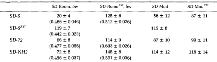
The fecundity of males bearing two intact SD chromosomes is compared in Table 2 with the fecundity of males that are genetically identical except that the Sd locus is deleted from one of the SD chromosomes. The results with SD-
5 and SD-Roma,bw are most clear-cut. For SD-5/SD-Roma,bw males only about
20 offspring/male were produced. However, when Sd was deleted from either SD-5 or SD-Roma,bw the total number of progeny increased six-fold. Because fecundity can be increased in these males by deleting the Sd locus, it appears that it is the dosage rather than homozygosity of Sd that leads to lowered fecundity.
Similar but less striking changes in fecundity were observed with other combinations of SD chromosomes. There was about a two-fold increase in fecundity of SD-Roma,bw/SD-72 and SD-Roma,bw/SD-NHZ males when the Sd locus was deleted from the SD-Roma,bw chromosome (Table 2). Fecundity of SD-5/SD-Mad males also increased about two-fold when Sd was deleted from the SD-5 chromosome, although deleting Sd from the SD-Mad chromosome has little, if any, effect on fecundity.
The fecundity of SD-Mad/SD-72 and SD-Mad/SD-NHB males was higher than for the other combinations of intact SD chromosomes examined. Since SD-Mad is a strongly distorting chromosome it appears that fecundity effects in homozygotes and drive strength need not be directly correlated. In particular, SD-Mad has little effect on the fecundity component such that one dose and two doses of Sd for this chromosome are equivalent. HARTL (1973) reported similar differences among various SD chromosomes with respect to male fecundity in SD,/SD, combinations.
We conclude from these experiments that, for combinations of SD chromo- somes that produce a significant reduction in male fecundity, this phenotype is associated with the Sd locus and the dosage of Sd rather than homozygosity per se is responsible. However, two doses of Sd in intact SD chromosomes do not invariably lead to reduction in male fecundity; the fecundity depends on the particular SD chromosomes involved.
TABLE 2
The fecundity of SDJSD, and Df(Sd)SDi/SDj males
SD-Roma, bw SD-RO~U'"~, bw SD-Mod S D - M C I ~ ~ ' ~
S D - 5 20 f 4 125 f 6 56 k 12 87 rt 11
SD-5R37 119 f 7 115 & 8
S D- 72 66 k 8 114 f 9 87 & 10 99 rt 11
SD-NHP 72 rt 8 145 rt 8 114 f 12 116 f 1 4
(0.466 rt 0.046) (0.442 f 0.023) (0.477 f 0.056) (0.496 2 0.037)
(0.512 f 0.026)
(0.603 & 0.026) (0.501 f 0.036)
The fecundity is the number of offspring per fertile male using our standard k protocol. When determined, k values giving the proportion of the SD-Roma chromosome are reported in parentheses.
dissimilar subunits should be able to complement (FINCHAM 1966). However, the SD chromosomes studied by HARTL carried recessive lethals preventing the analysis of male fecundity in true homozygotes. MARTIN and HIRAIZUMI (1979) used recombinant SD chromosomes that were lethal free to show that Sd,Rsp"/ Sd,Rsp" males showed no apparent reduction in fertility.
Two of the SD chromosomes used in this study are viable as homozygotes, enabling us to examine the fecundity of males homozygous or hemizygous for the Sd locus carried by these chromosomes. As shown in Table 3, the fecundity of homozygous SD-Mad males does not depart noticeably from SD-Mad/cn bw males (128 f 7), and this level of fecundity did not increase in SD-Mad/SD- MadR77 males. The high level of fecundity in such males obviously cannot be attributed to intracistronic complementation.
Males homozygous for SD-Roma,bw (and carrying a duplication that covers an unrelated male-sterile mutation-see MATERIALS AND METHODS) are fertile but have reduced fecundity. The fecundity of such males shows a significant increase when one of the chromosomes is deleted for Sd, whereas adjacent deletions that do not remove the Sd locus do not alleviate the reduced fecundity. Since the only difference between SD-Roma,bw males homozygous or hemi- zygous for the Sd locus is the number of copies of Sd, the increased fecundity of the hemizygote again must be attributed to dosage effects rather than to any type of subunit interaction.
668 1. G . BRITTNACHER A N D B . GANETZKY
TABLE 3
The fecundity of homozygous SD moles
Male gcnotype Copies of Sd Offspring/male SD-Roma, bw/SD-Roma, b w
SD-Romo, bw/SV-Romahk'9, b w SD-Roma, bw/SD-Romyh', b w SD-Roma, bw/SV-RomoWL, b w SD-Roma, bw/SV-Roma"', h w SD-Roma, bw/SD-RomuRS', h w SD-RomoK2, bw/SD-Roma"*, b w SD-Roma"', bw/SD-RomaK", b w SD-RomaK14, bw/SD-RomaH", b w SD-Mad/SV-Mad
SD-Mad/SD-MadR'-
2 2 2 1 1 1 0 0 0 2 1
65 -C 9
78 4 11 79 f 11 95 f 4 108 f 9 115 f 5
123 f 6 132 & 15
62 f 8 117 f 12 107 f 6
The fecundity is the number of offspring per fertile male from our standard k protocol.Al1 SD- Roma males had Dp(2;Y)G which covers the male sterile located near pr.
procedure for measuring male fecundity is probably insufficient to detect differences due to a single dose of Sd (HARTL, HIRAIZUMI and CROW 1967; NICOLETTI, TRIPPA and DEMARCO 1967); two doses of Sd increase the level of sperm dysfunction to a point where lowered fecundity becomes apparent.
One interpretation of the lowered fecundity of S D , / S D , males is that each S D
chromosome renders the sperm carrying its homologue dysfunctional (HARTL
and HIRAIZUMI 1976; KETTANEH and HARTL 1980). This mutual dysfunction
hypothesis implies that each S D chromosome is insensitive to its o w n action but sensitive to the action of a different S D chromosome. Our results do not support this interpretation. If mutual dysfunction were occurring, then signifi- cant departures in k value from 0.50 would be expected when one of the Sd loci is deleted. Specifically, in Table 2, the SD-Roma, I J W / S D - ~ ~ ~ ~ males would have a k value much higher than 0.50 (in favor of S D - R o m a , b w ) , whereas for SD- R o m a R"/SD-5, S D - R o m a R57/SD-72 and S D - R O ~ ~ ~ ~ ~ / S D - N H ~ it would be much less than 0.50 (in favor of the nondeleted S D chromosome). Clearly, this did not occur-the SD chromosome deleted for Sd was not distorted by the intact S D
chromosome.
DISCUSSION
The location of the Sd locus
recombination frequency. As the present study illustrates, most of these prob- lems can be eliminated by using deletions to map the S D elements.
Analysis Qf our deletions that revert the SD phenotype indicates that a single major component, the Sd locus, is responsible for causing segregation distortion. This component is located in region 37D2-6 of the salivary chromosome map which corresponds to about 2-54.1 on the standard recombination map [by interpolation between hk (2-53.9) and p r (2-54.5)].
Based on the recovery of recombinants between the markers
b
(2-48.5) and cn(2-57.5), TANZARELLA et al. (1972) reported a map position for Sd of 2-52.9 f 1.0. Although the actual mapping data are not provided, the locus mapped by these workers probably corresponds to the one we have localized.
More recently, HARTL (1980) has suggested that the unequal recovery of reciprocal recombinants from a derivative of SD-36 (an SD-5 type) could be explained by the existence of a second Sd locus “inseparable from pr and perhaps even to its right.” Our results provide no support for this interpretation because the ability of an SD chromosome to distort is not affected by euchro- matic deletions at the base of 2L which do not remove the Sd locus and segregation of S D chromosomes with the Sd locus deleted do not deviate from Mendelian expectations. Deletions of Sd have now been generated in three structurally different SD chromosomes (SD-5, SD-Roma and SD-Mad), and all fail to distort a sensitive homologue. Thus, if the chromosome analyzed by HARTL (1980) does have two copies of Sd, it is most probably a peculiar feature of that chromosome and not a general feature of SD chromosomes.
The nature of the Sd locus
The results of GANETZKY (1977) and the more extensive data reported here argue that Sd behaves as a neomorph (MULLER 1950), whereas Sd+ is the null condition with respect to segregation distortion. Operationally, we are unable to distinguish Sd’ from a deletion of Sd by its effects either on
k
value or male fecundity. Additional evidence supporting this view comes from experiments showing that increasing the dosage of Sd’ by means of a duplication has no effect on k value or fecundity in Sd/Sd+/Sd+ or Sd/Sd/Sd+ males (B. GANETZKY and M. E. ULBER, unpublished observations).It has been known for some time that the Sd locus is responsible for the reduced fecundity observed in many SDJSD, males (HARTL 1973). However, we find the fecundity of homozygous S D or SD,/SD, males increases after the deletion of Sd. This argues that the low fecundity was not due to the absence of an Sd* function in Sd homozygotes but results from the presence of two copies of Sd. The implication is that the Sd product actively causes some deleterious effect in its interaction with Rsp leading to sperm dysfunction. Furthermore, the Sd product must be made in limiting amounts relative to its target of action such that the effect of two doses of Sd is distinguishable from a single dose.
670 J. G . BRITTNACHER AND B. GANETZKY
(PEACOCK, TOKUYASU and HARDY 1972; KETTANEH and HARTL 1980). These observations suggest a mechanism of mutual dysfunction in which each SD
chromosome in a n SD,/SD, male could cause the dysfunction of sperm carrying the other SD (KETTANEH and HARTL 1980). An alternative view expressed by PEACOCK and MIKLOS (1973) is that the reduced fecundity of SD,/SD, males may not depend directly on effects of the SD system itself.
Our observation that deleting the Sd locus can alleviate the reduced fecundity in SD,/SD, males indicates that the original low fertility is a concomitant of the
S D system and, thus, supports the mutual dysfunction hypothesis. However, this hypothesis must be somewhat modified. If each S D chromosome in a n SD,/
SO, male were causing the dysfunction of the spermatid receiving the other SD chromosome, then the increased fecundity observed when Sd is deleted in one of the S D chromosomes should result in preferential recovery of the nondeleted
S D chromosome. We observed a n equal recovery of the deleted and nondeleted chromosomes. Thus, each SD chromosome in S D , / S D , males contributes to its own dysfunction as well as to that of its homologue. This further suggests that insensitivity to distortion of a chromosome bearing Rsp' is relative rather than absolute. Under certain conditions, Sd can apparently act even at its own Rsp' locus to cause dysfunction, although it acts preferentially at Rsp". An alternative possibility is that reduced fecundity in SD,/SD, males results from a n effect of Sd that does not involve the Rsp locus at all.
The mechanism of distortion
HARTL (1973) presented the first explicit molecular model for the mechanism of SD action. He proposed that the Sd product was a multimeric regulatory protein whose binding at the Rsp site was necessary for a chromosome to be included in a functional gamete. Subsequently, GANETZKY (1977) proposed a n alternative model in which the binding of a n Sd product at the Rsp locus initiated the event that led to dysfunction. GANETZKY ascribed no function to
Sd+ and further proposed that the Sd product was made in limiting amounts relative to available binding sites. Our observations that deletion of Sd reverts the S D phenotype and that males bearing one vs. two doses of Sd have differences in fecundity support the latter model.
The suggestion that the Sd product is made in limiting amounts follows from other considerations as well. Our data indicate that a n SD chromosome is not actually immune to its own effect but that non-Mendelian recovery of a n SD
chromosome when segregating from a sensitive homologue is due to preferential action of Sd at Rsp" rather than Rsp'. If the Sd product is made in unlimited amounts, then in Sd Rsp'/Sdf Rsp' males, excess Sd product could bind at Rsp' leading to the dysfunction of the spermatid receiving this chromosome. But this event would limit the fitness advantage that the SD chromosome might other- wise enjoy. It is more likely to suppose that the Sd locus is expressed in such a way that dysfunction of a sensitive homologue occurs without exceeding a threshold that could lead to self-distortion.
referred to as M(SD) that enhanced k values of a n S D chromosome and proposed that binding of the product of this locus at the Rsp site was necessary for normal sperm development and that the Sd product interfered with this binding causing dysfunction. Their results can also be explained in the context of our model where the various enhancers of SD such as E(SD) (GANETZKY 1977) and M(SD) control the amount of Sd product made.
The high fecundity of homozygous SD-Mad males is somewhat unusual. In previous cases in which it has been possible to test true SD homozygotes they have usually had low fecundity (HARTL and HIRAIZUMI 1976). Several SD chromosomes have been found to be quite fecund in S D , / S D , combinations (cf.
TRIPPA, LOVERRE and CICCHETTI 1980), although many others are not very fecund in such combinations (HARTL 1973). The reason for the fecundity differences among SD chromosomes is not clear. It may be due to allelic variation at the Sd and Rsp loci or it may involve differences in the presence of or response to various modifiers of Sd (cf. HIRAIZUMI, MARTIN and ECKSTRAND 1980). In any case, the fecundity of males homozygous or hemizygous for Sd would appear to rule out the possibility of intracistronic complementation at the Sd locus.
Finally, earlier views of segregation distortion (e.g., HARTL 1973; HIRAIZUMI, MARTIN and ECKSTRAND 1980) imply that Sd represents a mutation in a system that is involved in the normal control of sperm development. However, there is no direct evidence that S d + or any of the components of segregation distortion subserve a normal role in spermatogenesis. The fact that Sd' behaves as a n amorph with respect to segregation distortion indicates that if there is a n S d + function it is probably qualitatively different from that of Sd. Sd is similar to other dominant neomorphic mutations in that the severity of its effect depends on the number of mutant alleles and that it is revertible by deletion. Thus, Sd could represent a mutant allele in which a normal gene is inappropriately expressed in place, in time or in amount. It is also possible that Sd represents a foreign element inserted in the Drosophila genome and S d + is the absence of this element. This notion does not rule out the likely possibility that SD operates by parasitizing existing mechanisms that regulate communcation between hom- ologues and control gene expression at a chromosomal level. The most striking feature about SD chromosomes is the remarkable regularity a n d efficiency with which they exert their effect. Thus, they provide a unique opportunity to elucidate the way these regulatory mechanisms normally operate.
We thank JAMES F. CROW, WILLIAM ENGELS, DAN LINDSLEY, TERRY LYTTLE, MARTA MARTHAS, and
RAYLA G. TEMIN for helpful discussion and for comments on the manuscript. We also thank ROBERT
KREBER for technical assistance and T. R. F. WRIGHT and P. GAY for generously sharing unpublished data and providing us with stocks. This work was supported in part by Public Health Service grant GM07131 to the Laboratory of Genetics and by National Science Foundation grant PCM-8102325. This is paper number 2603 from the Laboratory of Genetics.
LITERATURE CITED
BRIDGES,'P. N., 1942
FINCHAM, J. R. S., 1966
A new map of the salivary gland 2L-chromosome of Drosophila melanogaster. J. Hered. 33: 403-408.
672 J. G . BRITTNACHER AND B. G A N E T Z K Y
GANETZKY, B., 1977 On the components of segregation distortion in Drosophila melanogaster. Genetics 86: 321-355.
HARTL, D. L., 1973 Complementation analysis of male fertility among the segregation distorter chromosome in Drosophila melanogaster. Genetics 73: 613-629.
HARTI,, D. L., 1974 Genetic dissection of segregation distortion. 1. Suicide combinations of SD genes. Genetics 76: 477-486.
recombinants. Genetics 96: 685-696.
HARTL, D. L., 1980 Genetic dissection of Segregation distortion. 111. Unequal recovery of reciprocal
HARTL, D. L. and HIRAIZIJMI, Y., 1976 Segregation distortion. pp. 615-666. In: The Genetics of Drosophila melanogaster, Vol. l h , Edited by E. NOVITSKI and M. ASHBURNER. Academic Press, New York.
HARTI., D. L., H I R A I Z ~ J M I , Y. and J. F. CROW, 1967 Evidence for sperm dysfunction a s the mechanism of segregation distortion in Drosophila melanogaster. Proc. Natl. Acad. Sci. USA 5 8 2240- 2245.
HAUSCHTECK-JURGEN, E. and D. L. HARTL, 1978 DNA distribution in spermatid nuclei of normal and segregation distorter males of Drosophila melanogaster. Genetics 89: 15-35.
H I R A I Z I J M I , Y., D. W. MARTIN and I. A. ECKSTRAND, 1980 A modified model of segregation
S D in a natural population of D. melanogaster in Japan.
Deviant sex ratio associated with sr!gregation distortion in
Ultrastructural analysis of spermiogenesis in segregation
LINDSLEY, D. L. and E. H. GRELL, 1968 Genetic variations of Drosophila melanogaster. Carnegie
MARTIN, D. W. and Y. H I R A I Z I J M I , 1979 On tha models of segregation distortion in Drosophila melanogaster. Genetics 93: 423-435.
MIKLOS, G. L. G., 1972 An investigation of the components of Segregation distorter systems in distortion in Drosophila melunogaster. Genetics 95: 693-706.
HIRAIZUMI, Y. and K. NAKAZIMA, 1965 Drosophila Inform. Serv. 40: 72.
HiRAIzuMi, Y. and K. NAKAZIMA, 1967
Drosophilo melunogaster. Genetics 55: 681-697.
distorter males of Drosophila melbnogaster: the homozygotes. Genetics 96: 665-684.
Inst. Wash. Puhl. 627.
KETTANEH, N. P. and D. L. HARTL, 198C
Drosophila melanogaster. Genetics 7 0 405-418.
MULLER, H. J., 1950
NICOLETTI, B., 1968
NICOLETTI, B. and G. T R I P P A , 1967 Osservazioni citologiche s u di un nuovo caso di "Segregation Distortion" (SD) in una popolazione naturalc di Drosophila melanogaster. Atti Assoc. Genet. Ital. 12: 361-365.
Evidence of the precision of genetic adaptation. Harvey Lect. 43: 165-229
11 controllo genetic0 della meiosi. Atti Assoc. Genet. Ital. 13: 1-71.
NICOLETTI, B., G. TRIPPA and A. DEMARCO, 1967 Reduced fertility in SD males and its bearing on
Meiotic drive in Drosophila: new interpretations of the segregation distortion in Drosophila melunogaster. Atti Acad. Naz. Lincei 43: 383-392.
Segregation Distorter and sox chromosome systems. Adv. Genet. 17: 361-409.
Spermiogenesis and meiotic drive in Drosophila. pp. 247-268. In: Edinburgh Symposium on the Genetics of the Spermatozoon, Edited by R. A. BEATTY and S. GLLJECKSOHN-WAELSCH. Bogtrykkeriet Forum, Copenhagen. SANDLER, L. and Y. HIRAIZIJMI, 1960 Meiotic drive in natural populations of Drosophila melano-
gaster. V. O n the nature of the SD rcgion. Genetics 45: 1671-1689.
SANDLER, L., Y. H I R A I Z ~ J M I and I. SANnI.ER. 1959 Meiotic drive in natural populations of Drosophiltr melanogaster. I. The cytogenetic basis of segregation-distortion. Genetics 44: 232-250.
SHARP, C. B., 1977 The location and properties of some of the major loci affecting the segregation PEACOCK, W. J. and G. L. G. MIKLOS, 1973
distortion phenomenon in Drosophila melanogaster. M S c . Thesis. University of British Colum- bia, Vancouver, British Columbia, Canada.
Localizzazione genetics e caratter- istiche funzionali del gattore SD-RI in Drosophila melanogaster. Atti Assoc. Genet. Ital. 17:
TANZARELLA, C., V. SPANO, A. MICHELI and B. NICOLETTI, 1972
108-109.
TOKUYASU, K. T., W. J. PEACOCK and R. W. HARDY, 1972a Dynamics of spermiogenesis in Drosophila melonogaster. I. Individualization process. Z. Zellforsch. Mikrosk. Anat. 124: 479- 506.
Dynamics of Spermiogenesis in
Dynamics of spermiogenesis in Drosophila melanogaster. VII. Effects of Segregation distorter (SD) chromosome. J. Ultrastruct. Res. 5 8
TOKUYASU, K. T., W. J. PEACOCK and R. W. HARDY, 1972b
TOKUYASU, K. T., W. J. PEACOCK and R. W. HARDY, 1977
Drosophila melanogaster. 11. Coiling process. Z. Zellforsch. Mikrosk. Anat. 127: 492-525. 96-107.
TRIPPA, G., A. LOVERRE and R. CICCHETTI, 1980 Cytogenetic analysis of an SD chromosome from a natural population of Drosophila melanogoster. Genetics 95: 399-412.
WRIGHT, T. R. F., W. BEERMAN, 1. L. MARSH, C. P. BISHOP, R. STEWARD, B. C. BLACK, A. D. TOMSETT and E. Y. WRIGHT, 1981 The genetics of dopa decarboxylase in Drosophila melanogaster. IV. The genetics and cytology of the 37810-37131 region. Chromosoma (Berl.) 83: 45-58.
The genetics of dopa decarboxylase in Drosophila melanogaster. 11. Isolation and characterization of dopa decaryoxylase mutants and their relationship to the alpha-methyl dopa hypersensitive mutants. Genetics 84: 287-310.
WRIGHT, T. R. F., R. B. HODGETTS and A. F. SHERALD, 1976 The genetics of dopa decarboxylase in Drosophila melanogaster. I. Isolation and characterization of deficiencies of the structural locus and the alpha-methyl dopa hypersensitive locus. Genetics 8 4 267-285.
Corresponding editor: T. C. KAUFMAN