PSEUDOINVERSIONS IN THE CHROMOSOME
OF ESCHERICHIA COLI K-12
N. GLANSDORFFI
Laboratoire de Microbiologie, Facultk des Sciences, Universitk de Bruxelles, et Institut de Recherches du C.E.R.I.A., Bruxdles 7 , Belgique
Received August 8 , 1966
HE following observations are mainly methodological; they concern the use of bacterial conjugation in genetic topography. However, as will be shown hereafter, they are by-products of investigations on the genetic control of arginine biosynthesis in Escherichia coli and have precise (although limited) implications for the treatment of this question.
Four of the eight sti-uctural genes of the latter pathway are tightly clustered in the order argE-C-B-H ( GLANSDORFF 1965) ; Figure
1
gives the gene-enzyme correspondance, as defined by M a s , MAAS, WIAME and GLANSDORFF (1964). Both enzymatic determinations and genetic evidence suggest that argB and H(and probably C ) form an operon, the fate of argE being uncertain ( GLANSDORFF and SAND 1965). It was thus expected that chromosomal rearrangements which would modify the spatial relationships of the arg loci should also alter the regula- tion o i their expression.
In this last respect, we became interested by the E . coli strain AB1206, isolated
by PITTARD. LOUTIT and ADELBERG (1963) ; this organisrii harbours an episome
thr leu
%rg argG slr
A B C
glutamate-N-acetyl --N-acetyl- -N-acetyl- g l u t a m a t e g l u t a m y l - g l u t a m a t e
phosphate semi-aldehyde
D E F G H
*-acetyl- -0rnithlne-citrulline -arginino--arginrne
o r n i t h i n e succinate
FIGURE 1.-The gene-enzyme relationships for arginine biosynthesis in E . coli K-12 (outline
of the enzymic steps and map of the corresponding arg loci). For abbreviations, see MATERIAL and METHODS. argD has been mapped i n E . coli W only (VOGEL et al. 1963).
Chargk de Recherches du “Fonds National Belge de la Recherche Scientifique”
of the F' type (a unit of replication composed of the fertility factor F and a seg- ment of the bacterial chromosome; JACOB and ADELBERG 1959). This episome
(F,,) carries a segment 'which represents about 10% of the chromosome (PITTARD
and RAMAKRISHNAN 1964); F,, can be considered as a small bacterial chromo- some, analogous to the genetic material of an Hfr strain. The point which is relevant to our discussion is that F,, was said to carry a transposition of the gene
argH which specifies argininosuccinate lyase, the last enzyme of arginine bio-
synthesis (PITTARD et al. 1963). However, a more recent report stated that the transposition involves the locus responsible for acetylornithinase biosynthesis
(argE; BAUMBERG, BACON and VOGEL 1965) ; surprisingly, from the observations of the latter authors as well as froin our own (see further), it became clear that the regulation of the whole arg cluster was not altered i n AB1206.
After a n extensive reexamination of the F,, episome, we have found that its topography is identical with that of the same genetic region in other strains. However, AB1206, as well as other E. coli donor strains, gives rise to pseudo- transpositions or pseudoinversions when: (1 ) The markers investigated are very close to the leading end of the donor chromosome; at 37"C, the transfer of the segment extending from this point to the pseudoinverted markers does not seem to demand more than 1 minute. (2) The order of the genes is deduced solely from the determination of the curves which reflect the kinetics of their transfer (i.e. the kinetics of formation of separated classes of recombinants, from matings inter- rupted at regular intervals by mechanical means (WOLLMAN and JACOB 1959) or by treatment of the donor strain with a bacteriophage (HAYES 1957).
Possible interpretations of this phenomenon are discussed. A preliminary ac- count of this work has been given previously (GLANSDORFF 1966).
MATERIALS A N D METHODS
Genetical and enzymological techniques; The media and the techniques used in the mating and transduction experiments have been described previously (GLANSDORFF 1965). References for measurements of enzyme activities are as follows. The symbol used to represent the genetic determinant of each of these enzymes is indicated in parentheses. ATP: cy-N-acetyl-L-glutamate 5-phosphotransferase (argB) : BAICH and VOGEL (1962). a-N-acetyl-L-glutamate y-semialdehyde:
NADP oxydoreductase (phosphorylating) (argC) : GLANSDORFF and SAND (1965). L-ornithine a-N-acetylornithine lyase (argE) : VOGEL and BONNER 1956. L-argininosuccinate arginine lyase
(argH) : RATNER, ANSLOW and PETRACK 1953.
Abbreviations used. arg = arginine; glu = glucose (designates a gene involved in phos- phoenolpyruvate carboxylation) ; his = histidine; ilva = isoleucine and valine; leu = leucine;
met = methionine; pro = proline; pur = purine; rha = rhamnose; str = streptomycin; thi =
thiamine; thr = threonine; T6 = phage T6; r = resistance; s = sensitivity; F- = female, re- cipient; Hfr = male, donor. Capital letter: nomenclature of gene lcci according to DEMEREC
(1956).
Strains; The basic material of this work is the strain AB1206 (F', his, pro, thi, T 6 S , str'),
supplied by DR. ADELBERG. PA373 (F-, thr, leu, thi, his, metA, argH, T68, strr), PA374 (F-, thr, leu, thi, his, metE, argH, T68, str'), P i 0 (Hfr, thr, leu, thi, T68, strs), P72 (Hfr, thr+, leu+, thi, met& T68, str8) and the transducting phage 363 were obtained from DR. F. JACOB. AT1 1-31
(Hfr, thr, leu, thi, purD, T68, str') and A T l S 5 6 (Hfr, thr, leu, thi, T68, str') were obtained from R. LAVALLE. The rest of the strains either have been described previously (GLANSDORFF
PSEUDOINVERSIONS IN E. coli K- I 2 51
rha, arg and ilua mutants isdated in the course of this work were induced by N-methyl- N'-nitro-N-nitrosoguanidine, following the method recommended by ADELBERG, MANDEL and CHEN (1965). Penicillin was used to kill wild-type cells surviving the mutagen treatment, as described by GORINI and KAUFMAN (1960).
EXPERIMENTS
The first section describes the ,wild-type topography of the genetic markers considered in this study; then follows a recall of the properties of AB1206, with some comments on the use of this strain in transductions. The secolid section presents determinations of enzyme activities which reflect the activity of the genes involved in the apparent rearrangement undergone by the F,, episome. Data concerning enzymes E and H have been reported by BAUMBERG et al.
(1965), but in a different context and leading to different conclusions, since these authors consider AB1206 as carrying a transposition of the argE locus. The third and fourth sections contain the detailed genetic analysis of AB1206, Hfr
strains P10 and P72, carried out by matings and transductions.
1. Topography of the segment investigated-properties of AB1206: The seg- ment of the E. coli chromosome which is relevant to this study is represented in Figure 2. The main part of this map is based on the data recently reviewed by
TAYLOR and THOMAN (1964). Additional information for the met-arg region has
been included (GLANSDORFF 1965). The site of rhamnose mutations was until now uncertain with rcspect to the closest markers; one rhamnose mutation iso- lated in the course of this work has been mapped on the following basis (GLANS-
DORFF 1966): rha lies on the left of metB (see Figure 3 ) ; rha is cotransducible
with metB (30% cotransduction) ; rha is cotransducible with metE ( 1
%
cotrans- duction); metB and metE are not cotransducible (less than 0.01%). rha thus lies between metE and metB.The episome F,,, harboured by the strain AB1206, has originated from the
Hfr AB313. It contains about 10% of the bacterial chromosome, and extends from
ilua to the right of the arg region. As shown hereafter, its origin lies between
argH and purD. T h e episome is represented (Figure 2) after its original descrip- tion (PITTARD et al. 1963), with a transposition of the argH locus to the left of
I I
episome I
;r C
F i l v a met€ a r g H m e t 8 a y € I ?
N. GLANSDORFF
me&
U
4 J16 20
FIGURE 3.-Transfer of arg, glu, met, rha and ilva alleles by Hfr PI0 and F'AB1206. The
donor is indicated in parentheses; the genotypes of the recipients are: Crosses ( 1 ) and
(e),
m t E ,ilua. Crosses ( 3 ) and (4), glu, metB, metE. Cross ( 7 ) , argE, metB. Crosses ( 5 ) and (6) , metB, rha. Crosses (8) and (9), argH, glu, metB. Abscissa: time (minutes). Ordinate: recombinants (percent input donor strain). Differential supplementation of selective media with homocysteine or vitamin B,, was used to distinguish metEf from metB+ recombinants, respectively.
metB. The sequence of chromosomal transfer of Hfr P10 is also represented (see
section 3 ) .
The interpretation of transduction carried out with AB1206 as donor rests on the assumption, strongly supported by the work of PITTARD and RAMAKRISHNAN
(1964), that the genes carried by F,, are deleted from the chromosome of AB1206. The origin of the transducing particles produced from AB1206 and carrying
met, glu or arg alleles, is thus episomic and does not contain chromosomal
material. This was confirmed by three lines of evidence:
(1) The cotransduction of purD and metB, which amounts to 3% when a
(metB, purD) strain is transduced by a wild-type donor strain, is not observed
when AB1206 is used as the donor (less than 0.01
%) .
Moreover, matings per- formed with the same recipient strain show that AB1206 does not transfer thePSEUDOINVERSIONS I N E . coli K - 1 2 53
conclusion that metB is on the episome and purD on the chromosome. As F14 carries the glu and arg genes, the right limit of the material which is absent from the chromosome and carried by the episome must lie between the arg cluster and
purD. The chromosonial deletion of AB1206 thus seems to correspond exactly to the episomal markers of F14. Although the genetic constitution of AB1206 shows that its formation has involved several events (PITTARD et al. 1963), it is conceivable that the chromosomal deletion is an immediate consequence of the formation of F,, from the chromosome of the Hfr AB313. This possibility should be considered in relation to the hypothesis proposed by BRODA, BECKWITH and SCAIFE (1964) for the formation of F-prime episomes.
(2) Recessive ilva mutations harboured by F,, render AB1206 dependent on isoleucine and valine ( PITTARD and RAMAKRISHNAN 1964). Similarly, we could isolate argB and rha mutants from this strain. It was shown, by transfer of the mutated F,, to F- strains, that these mutations are recessive.
( 3 ) When an argB mutant of AB1206 is used as donor to transduce hetero- allelic argB recipients, the yield of arg+ recombinants is very low: 0.2 to 1.0% of the number of wild-type transductants recovered f o r a met o r glu allele present in the argB recipient. This means that the phage particles obtained from the mu- tant do not contain a wild-type allele of argB which could have been present in the chromosome, although unexpressed.
2. Enzymatic study of AB1206: Previous studies suggested that, from the cluster of arg loci ( E , C , B, H ) situated on the right side of gZu, at least two ( B
and H ) are joined in an operon, with a probable B + H polarity (GLANSDORFF
and SAND 1965). It was thus essential to examine the expression of the argH
locus in AB1206, where it was supposed to be transposed (PITTARD et al. 1963). Specific activities of argininosuccinate lyase (coded by argH) and of the other enzymes specified by the arginine cluster, were determined in AB1206. P4X,
and argG or A derivatives of both strains grown under conditions of derepression in a chemostat. Table 1 shows that there is no significant difference between the two families of strains in a range of variation which goes from maximal re-
TABLE 1
Specific activities of the enzymes specified by the loci argE, C , B and H in P4X and AB1206, under various conditions;
Strain
P4XargA P4X P4X
AB121 OGargG
AB1206 AB1206
Enzyme activity (&moles/hr/mg protein Acetylorni-
thine lyase Condition (locus a r g E )
Oxydore- ductase (locus argC)
Phospho- transferase (locus argB)
chemastat 31 without arginine 13.6
with arginine 2.4 chemostat 39 without arginine 13.8
with arginine 2.8
2.1 0.7 0.07 2.8 0.09 2.5 0.9 0.1 2.9 0.1 Arginmosuc- cinate lyase (locus a i g H )
-~ ~ 3.6 1.5 0.15 5.7 1.5 0.18 ~~~~ ~~
pression to nearly complete derepression. Faced with the difficulty of interpret- ing these results in terms of a transposition of argH when considering this locus probably belongs to an argB + H operon, we were led to an extensive reexamina-
tion of AB1206, from a genetical point of view.
3 . The genetic analysis of AB1206 by mating and transduction experiments:
To determine the order of transfer of several genetic markers from AB1206 to F-
recipients, we used HAYES' technique of T6 interrupted matings (1957). T6' derivatives were thus prepared from the required F- strains. As AB1206 is pro-
and all our F- strains pro+, we submitted AB1206 to a double counterselection: T6 killing and absence of proline in the different selective media. In crosses per- formed with Hfr P10, we used T6 and streptomycin for the counterselection of the donor. The various crosses performed are represented in Figure 3 .
As numerous workers have found (see TAYLOR and THOMAN 1964, for a re- view) the kinetics of formation of recombinants is not as a rule a straight line, but a continuous curve which sometimes resembles a broken line, with two successive slopes. The intersection of these curves with the abscissa is not always easy to determine with precision; nevertheless, we conclude from Figure 3 that the sequence of markers injected by Hfr P10 is:
origin-cluster arg-glu-met B-rha-metE-ilua
and when AB1206 is used as donor this becomes:
origin-metB-glu-cluster arg-rha-metE-ilua
Thus, at first sight the metBtzrg segment seems to have undergone an inversion in AB1206.
This point was examined with particular attention, in view of the theoretical bearing that the occurrence of inversions should have on our representation of the organisation of the bacterial chromosome (see MARGOLIN 1965 and SANDER-
SON 1965).
From the inversion o i the met-arg segment in the F,, episome we should expect the following consequences: (1) The linkage relationships of the metB, glu and
arg alleles should not be modified within the limits of the inversion. (2) The linkage relationships of the same markers with a locus outside the inverted zone should be modified (suggested by R. LAVALLE). ( 3 ) The pairing of F,, with a complete chromosome within the limits of the inversion should lead to the re- versal of the order of chromosomal transfer which this episome usually induces (PITTARD and ADELBERG 1964); thus the kinetic analysis of a cross between a
PSEUDOINVERSIONS IN E. coli K - I 2 55
means (WOLLMAN and JACOB 1959) but does not often appear to have been carried out in more recent works; it had not yet been applied to AB1206.
Points (1) and (2) have been tested by transduction.
( a ) Had the argH locus undergone a transposition leaving a deletion at its normal site, then it should be revealed by transduction of the metB, glu and arg
alleles carried by F,,, in various recipient strains. For example, a transduction between a (glu, argH’1 recipient and AB1206 should give no arg+ organisms among the glu+ recombinants, and no or very few urg+ recombinants, depending of the ability of the transposed fragment to synapse with the argH region of the recipient. Were the usual linkage relationships respected ( GLANSDORFF 1965), about 90% of the glu+ recombinants should be arg+; this is, indeed, the result which is found (see Table 2). With (glu, argC) or (glu, argB) recipients, a donor carrying a transposed argH locus should give a low number of arg+ re- combinants among the glu+ class, because argH, B and C are strongly linked in normal strains. The same reasoning would hold if argE was the transposed locus. In fact. again 90 to 95% a r g f recombinants are found among the glu+ class. Table 2 also shows that in each case, the data are paralleled by the scores of glu+
recombinants among the arg+ class.
The fourth transduction shown in Table 2 ( m e t B , g l u ) x AB1206, confirms the absence of the urg- class which should have been contributed by the donor, if carrying a transposed argH. This experiment also shows that the cotransduction index normally found for metB and glu (about 50%) is conserved when AB1206 is the donor.
These results do not show any structural modification within the limits of the
metB-argH segment carried by AB1206, with respect to the topography which has been worked out with other E. coli strains.
(b) Unexpectedly, the results reported in Table 3 show that the distances between metB, arg and rha (outside of the apparent inverted zone) remain roughly the same, whether the cross is performed between “normal” strains, between derivatives of AB1206, or between a “normal” strain and AB 1206. These results contradict the hypothesis of an inversion and give a first indication that the order of the genes could be the same in AB1206 and PIO.
(c) The same conclusion arises from the testing of point ( 3 ) . When the episome F,, was introduced in a F- strain possessing a complete chromosome,
TABLE 2
Transduction analysis of the metB-glu-argH region, with AB1206 as the donor
Number of unselected versus selected recombinants Recipient arg+/glu+ glu+/arg+ met+/glu+ glu+/met+
glu-2, argH-2 109/119 37/39
glu-2, argB-1 108/119 32/36
glu-2, argC-1 104/111 28/29
- ? A
2
B
-
a.h
4
$ 5
b .;
.3
.;
.':
4
e-k
.s
s
M
p
d
4;
8
2 ;
8
'i
8;
s i
O.!.$ ;
, x c
B
Y P 2 E:*
- I .?A- Q &
w
F9 I t
Q t
% <
g
. U U
E: h t, .U
F
3
d s 5-
oi% x
B
s
0 c .- + to .- v1 .- " E e" 2 8 8 . 22 : 2 E g S E 2 9 z c-
$ 69"
ss
g!g
U Ms
U - Y i Q 0 L C + k 5.5:a
PSEUDOINVEHSIONS I N E. coli K- I 2 57 and the latter transferred to appropriately marked recipient strains, we could not find any reversal of the direction of this transfer.
(d) Conclusive evidence that the order of transfer of AB1206 is indeed the same as that of P10, could be obtained from the analysis of the genetic constitu- tion of recombinants taken at early times during interrupted matings between recipient strains and either AB1206 or P10 (see Table 4). The progressive in- crease of met+ among arg+ recombinants, whether P10 or AB1206 is used as the donor, shows that the apparent inversion of the me-arg segment is an artefact of the methodology applied previously.
We have seen that the arg cluster is very close to the origin of the F,, episome. Indeed, argH and purD are cotransducible, but F,, does not carry the purD
locus. Formally speaking, the pseudoinversion effect can be attributed to an influ- ence of the origin on the integration of donor genes situated in its immediate vicinity (less than 1 or even 0.5 minute of transfer time). The closer a gene would be to the origin, the less frequently should it appear in the progeny of the cross. The fact that the percent of arg+ among met+ recombinants is two times lower when AB1206 is used as the donor rather than P10 gives a direct measure- ment of this restriction. Conceivable mechanisms of such a phenomenon will be discussed at the end of this paper, together with evidence already available in this topic (Low 1965; GLANSDORFF 1966).
From a restriction exerted by the origin, we would expect that the curves relating to arg+ and met+ recombinants should follow the real order of the genes only at their early beginning. As the restriction effect of the origin does not seem to influence the integration of loci distal to metB, the curves should intersect in the very first minutes of mating. This crossing of curves should probably not be demonstrable with certainty by the numeration techniques used in this kind of experiment; its visible consequence should thus consist in pseudo- transpositions or pseudoinversions, depending on the number of markers investi- gated. A tendency for the curves to cross could in fact be observed in experiments where interruptions were performed at very short intervals; it was also more apparent when the speed of the transfer was lowered by a shift from 37°C to 32°C. The results, however, cannot be considered demonstrative.
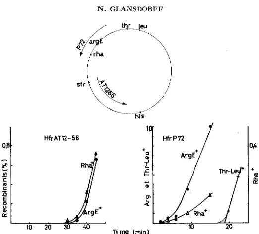
Our interpretation was strengthened by the occurrence of a second pseudo- inversion which could be related to the same restrictive effect. We had mapped the rha locus between m t E and metB; the kinetics of transfer of the rha+ and
argE+ alleles to a rha-, argE recipient by the Hfr AT1256 is shown in Figure 4. Figure 4 also represents the transfer of the same alleles by Hfr P72, whose lead- ing end was known to lie between metE and m t B (Hfr No. 5 in JACOB and WOLLMAN 1961). Although the succession of the curves seems to point out the order arg-rha in this last mating, it is clear from the genetic analysis of arg+
and rha+ recombinants that both Hfrs inject the chromosome in the same order,
rha-arg; however, a strong restriction operates on the integration of rha+ from P72 into the recipient strain (see Table 5 ) . The distance separating rha and the origin of P72 is of the same order of magnitude as the argH-FI4 origin interval.
FIGURE 4.-Transfer of argE, rha and thr-leu alleles by Hfr AT12-56 and Hfr P72. The genotype of the recipient is, in both cases, rha, nrgE, thr, leu.
Indeed, rha is cotransducible with metE (about 1
%
),
but the latter locus is not injected early by P72. We could show in additional experiments that the restric- tion operating onr h
does not affect the locus metF; however the cotransduction percent of metF and rha approaches 20%; metF and argE are about 40%cotransducible.
DISCUSSION
The main conclusion of this work has to be set forth on a methodological ground. When a genetic marker is very close to the leading end of the chromo- some which Hfr and F’ strains transfer during conjugation, the frequency of
TABLE 5
Genetic analysis of rha+ and argf recombinants recovered from interrupted matings performed between an (rha, argE) E- strain, either Hfr ATi2-56 or Hfr P72
F-(rha, arg) XAT12-5G
Time of arg+ rhat
interruption . _ _ ~
(min) rha+ ”/, a m + %
F- (rha, arg) X P72
Time o f arg+ r h d
Interruption
(min) rha+ % arg+ %
30 18/23 78 8/36 22 35 47/62 76 32/80 40
40 62/80 78 32/78 41 45 62/80 78 41/76 54 50 68/79 86 45/77 59
4 7/61 11.4 2/21 9.5 6 4/32 12.5 13/49 26.5 9 8/119 6.7 21/64 32.8 15 6/118 5.1 62/120 51.7
PSEUDOINVERSIONS I N E. coli K - 1 2 59
integration of this marker in the progeny of the cross is significantly reduced as compared to that of more distal markers. This phenomenon has an important consequence when two or more very proximal markers are investigated. Sup- pose these markers are a, b, c, injected in that order by the donor strain; we have seen that the determinable parts of their kinetics of transfer will appear in the order c, b, a, giving thus the picture of a pseudoinversion (pseudoinversion or pseudotransposition in the case of two markers). The real order of transfer has to be deduced obligatorily from the analysis of the genetic constitution of several classes of recombinants.
The idea that the leading end of the chromosome might restrict marker integra- tion in its vicinity has already been considered by Low (1 965), but without em- phasis on pseudoinversion effects; obviously, the latter can only appear when F-
strains carrying several proximal markers are used in the mating experiments, such as those reported by PITTARD et al. (1964) and in the present paper. At present. several hypotheses can be advanced to explain the restriction operating on the integration of proximal genes. Low (1965) and ourselves (1966), in a preliminary report of this work, had independently advanced the following ex- planation: the restriction imposed by the origin should merely reflect a relation between the distance which separates a gene from the origin, and the crossing over frequency within this interval. Indeed, as in bacterial crosses, the genetic material contributed by the donor is limited to a fragment ( a fortiori when the mating is interrupted for the purpose of gene mapping) the formation of a recombinant requires at least two genetic exchanges, one of them occurring be- tween the selected marker and the chromosomal origin. However, a recent quan- titative analysis of linkage between bacterial genes makes such a simple explana- tion unlikely (VERHOEF and DE HAAN 1966; DE HAAN and VERHOEF 1966). In
agreement with the present results, the latter authors have presented convincing evidence that the proximity of the origin has no influence on the integration of markers so close to the origin as to be transferred 1 or 2 minutes after it (the proA locus, injected early by Hfr R4). Were the present results ascribable only to the distance (origin-argH or rha) then one would expect the influence of the origin operating up to markers situated 10 minutes of transfer time from the origin. The model presented by the latter authors may offer an explanation for the short range effect that we observe; the data supporting the model are strongly consistent with the idea that a n obligate crossing over occurs between the Hfr and the F- chromosomes at the level of the origin; it is conceivable that this crossing over could occur not only at the very point of origin, but also in a segment situated in the immediate vicinity of it. The source of another explanation, also compatible with the model of DE
HAAN
and VERHOEF, can be found in the paper by FULTON( 1965) on continuous chromosomal transfer during bacterial conjugation. The
low integration frequency of a pur marker injected early by HfrC has led FULTON
60
has until now precluded any simple means of testing this hypothesis by genetical experiments. The question thus remains unanswered. Experiments are now in progress to distinguish between the possibilities mentioned.
Our last conclusion concerns the genetic control of arginine biosynthesis in
E. coli. A problem had originated from the apparent transposition of the locus
argH (PITTARD et al. 1963) or argE (BAUMBERG et al. 1965), without any con- sequence on the control of their expression, nor on the expression of argC and B,
which are their neighbours in the argECBH cluster of which at least argB and H form an operon (GLANSDORFF and SAND 1965). The problem now disappears, since the chromosome rearrangements previously described are methodological artefacts. Moreover, we have yet unpublished evidence that the effective trans- position of argH a short distance from its site of origin makes the expression of this gene constitutive (SAND and GLANSDORFF)
.
Lastly, we should recall that the locus argE might well constitute a unit of expression independent of argC, B andH , although the present work shows that this suspicion can no more be based on the properties of strain AB1206; it has been shown, indeed, that the synthesis of enzyme E does not appear strictly coordinated with that of enzymes C, B and H
(see BAUMBERG et al. [1965] for enzymes E and H; GLANSDORFF and SAND [ 19651 for enzymes E, C, B and H)
.
The most generous gift of the strain AB1206 by DR. E. A. ADELBERG and the strains provided by DR. F. JACOB and R. LAVALLE are gratefully acknowledged. I wish to thank PROFESSOR P. G.
DE HAAN and C. VERHOEF for their invaluable criticism and discussions. My gratitude also goes
to PROFESSOR J. M. WIAME for his interest in this work, to R. LAVALLE for many stimulating discussions and to G. SAND for his assistance in the determinations of enzyme activities.
SUMMARY
The restriction which the chromosomal origin of E. coli males exerts on the integration of very proximal markers in the progeny of crosses, leads to apparent pseudoinversions of the markers investigated in the usual type of kinetic analysis applied to chromosomal transfer. Previously reported chromosomal rearrange- ments which had been said to disorganize a cluster of arginine loci without alter- ing its regulation, are methodological artefacts of that type.
LITERATURE CITED
ADELBERG, E. A., M. MANDEL, and G. C. C. CHEN, 1965 Optimal conditions for mutagenesis by N-methyl-N'-nitro-N-nitrosoguanidine in Escherichia coli K-12. Biochem. Biophys. Res. Commun. 18: 788-795.
BAICH, A., and H. J. VOGEL, 1962 N-Acetyl-y-glutamokinase and N-acetyl-7-semialdehyde dehydrogenase: repressible enzymes of arginine biosynthesis in Escherichia coli. Biochem. Biophys. Res. Commun. 7: 491-496.
Individually repressible enzymes specified by clustered genes of arginine synthesis. Proc. Natl. Acad. Sci. U.S. 53: 1029-1032.
The characterization of a new type of F-prime factor in Escherichia coli K-12. Genet. Res. 5 : 4 8 9 4 4 .
BAUMBERG, S., D. F. BACON, and H. J. V O G ~ , 1965
PSEUDOINVERSIONS I N E. coli K - I 2 61
DE HAAN, P. G., and C. VERHOEF, 1966 Genetic recombination in Escherichia coli. 11. Calcula- tion of incorporation frequency and relative map distance by recombinant analysis. Mutation Res. 3: 111-117.
DEMEREC, M., 1956 Terminology and nomenclature. Carnegie Inst. Wash. Publ. 612: 1-4.
FULTON, C., 1965 Continuous chromosome transfer in Escherichia coli. Genetics 52 : 55-74. GLANSDORFF, N., 1965 Topography of cotransducible arginine mutations in Escherichia coli
K-12. Genetics 51: 167-179. - 1966 Le contr6le gknktique des biosynth6ses de l'arginine et du carbamoylphosphate chez Escherichia coli. Thesis, University of Brussels, Belgium.
GLANSDURFF, N., and G. SAND, 1965 Coordination of enzyme synthesis in the arginine pathway of Escherichia coli K-12. Biochim. Biophys. Acta 108: 308-311.
GORINI. L., and H. KAUFMAN, 1960 Selecting bacterial mutants by the penicillin method. Science 131: 604-605.
The kinetics of the mating process in Escherichia coli. J. Gen. Microbiol. 16:
HAYES, N., 1957 97-1 19.
JACOB, F., and E. A. ADELBERG, 1959 Transfert de caractkres g6nktiques par incorporation au
Sexuality and the Genetics of Bacteria. Academic Press,
Low recombination frequency for markers very near the origin in conjugation facteur sexuel dEscherichia coli. Compt. Rend. 249: 189-191.
JACOB, F., and E. WOLLMAN, 1961 New York.
Low, B., 1965
in E. coli. Genet. Res. 6: 469-473.
MAAS, W. K., R. MAAS, J. M. WIAME, and N. GLANSDORFF, 1964 Studies on the mechanism of repression of arginine biosynthesis i n Escherichia coli. I. Dominance of repressibility in zygotes. J. Mol. Biol. '8: 359-364.
MARGOLIN, P., 1965 Bipolarity of information transfer from the Salmonella typhimuriun
chromosome. Science 147: 166-1458,
PITTARD, J., and E. A. ADELRERG, 1964 Gene transfer by F' strains of Escherichia coli K-12. 111. An analysis of the recombination events occurring in the F male and in the zygotes. Genetics 49: 995-1007.
Gene transfer by F strains of Escherichia coli K-12. I. Delay in initiation of chromosome transfer. J. Bacteriol. 85: 1394-1401.
Gene transfer by F' strains of Escherichia coli K-12.
IV. Effects of a chromosomal deletion on chromosome transfer. J. Bacteriol. 88: 367-373.
Biosynthesis of urea. VI. Enzymatic cleavage
Information transfer in Salmonella typhimurium. Prw. Natl. Acad.
The genetic map of Escherichia coli K-12. Genetics
Genetic recombination in Escherichia coli. I. Relation
PITTARD, J., J. S. LOUTIT, and E. A. ADELBERG, 1963
PITTARD, J., and T. RAMAKRISHNAN, 1964
RATNER, S., N. P. ANSLOW, and B. PETRACK, 1953
SANDERSON, K. E., 1965
TAYLOR, A. L., and M. S. THOMAN, 1964
VERHOEF, C., and P. G. DE HAAN, 1966
VOGEL, H. J., D. F. BACON, and A. BAICH, 1963 of argininosuccinic acid. J. Biol. Chem. 204.: 115-125.
Sci. U.S. 53: 1335-1343.
50: 659-677.
between linkage of unselected markers and map distance. Mutation Res. 3: 101-110.
Induction of acetylornithine 8-transaminase during pathway-wide repression. pp. 293-300. Symposium on Informational Macromolecules Edited by H. J. VOGEL, V. BRYSON and J. 0. LAMPEN. Academic Press, New York.
Acetylornithinase of Escherichia coli: partial purification and some properties. J. Biol. Chem. 218: 97-106.
VOGEL, H. J., and M. BONNER, 1956