INVESTIGATIONS ON LATERALIZATION OF FUNCTION IN THE DISCONNECTED HEMISPHERES OF MAN
Thesis by Robert David Nebes
In Partial Fulfillment of the Requirements for the Degree of
Doctor of Philosophy
California Institute of Technology Pasadena, California
1971
Acknowledgements
I would like to express
my
deep appreciation to Dr. Roger Sperry for his patient guidance and counsel, and for sharing with me his perceptive insights into the signifi-cant questions in the field of psychobiology.I wish also to thank Drs. Joseph Bogen and Evelyn Lee -Teng for their helpful comments and suggestions on parts of this worke
A special word of gratitude is due those individuals who served as subjects for these experiments: without their earnest cooperation, this study would not have been possible.
iii Abstract
The effect of long standing cerebral damage upon the pattern of functional lateralization revealed by division of the forebrain commissures was investigated in a young conunissurotomy patient with birth injury to the somato-sensory region of his left hemisphere. Results from a battery of sensory - motor tasks showed that, unlike pre-vious conunissurotomy cases, the major hemisphere of this subject had access to somesthetic information from the ipsi-lateral as well as the contraipsi-lateral hand, thus allowing him to name objects out of sight in his left hand, and to use this hand to tactually find items, the pictures or names of which had been visually presented to only the left hemisphere. The most plausible explanation for these excep-tional cross integrative abilities would be the presence of a left sided ipsilateral somesthetic projection, which,in ·compensation for the subject's early brain damage, has
strengthened into a functional system. Additional evidence for compensatory reorganization in this boy was found in his minor hemisphere, which exhibited an enhanced capacity for
expressive language, being capable of transcribing printed words into script6 and, upon occassion, of writing the name
of an object.
v
Table of Contents
I. General Introduction
II. The Commissurotomy Syndrome in a Patient with Birth Injury to the Left Hemisphere
A. Introduction B. Case History
c.
General Procedure·n.
Tests for Compensatory Reorganization of the Somesthetic Systemle Introduction
2o Results
a) Verbal Identification of
1 4 4 6 8 10 10 13
Stimuli in the Left Hand 13 b) Written Identification of
Stimuli in the Left Hand 16 c) Verbal and Written Identification
of Stimuli in the Left Visual
Field 18
d) Localization of Left Hand
Stimulation 18
e) Tactual Cross Matching 20 f) Visuo - tactile Matching 21 g) Visuo - visual Matching 23 3., Discussion
Eu Tests for Minor Hemisphere Expressive Language
1. Introduction
2e Procedure 3u Results
a) Writing to Printed Words in the Left Visual Rield
b) Writing to Object Pictures in the Left Visual Field
4. Discussion
III. Hemispheric Dominance for the Perception of
Page
31 .35 36
Part - Whole Relations 39
Ao Introduction 39
B. Subjects 41
c.
Arc - Circle Matching 431. Introduction 43
2. Method 44
3. Results 51
a) Conunissurotomy Patients 51 b) Right Occipital Lesion Patient 65 c) Normal Control Subjects 65 d) Comparison of Right Handed and
Left Handed Normal Subjects 70
4. Discussion 72
D. Figural Unification 82
lo Introduction 82
2e Method 83
3 411 Results 88
4(ll Discussion 93
Ee Perceptual Organization of Dot Patterns 96 le Introduction
2 o. Method 3e Results
vii
4. Discussion F. General Discussion
Page
105 107
References 109
Behavioral testing of both animals and human beings in whom the neocortical commissures have been surgically
divid-ed has establishdivid-ed to a large degree the role of these structures in unifying the higher processes of the two cerebral hemispheres (1,2,3,4). Section of these large interhemispheric fiber tracts has been found to abolish normal integration of the two halves of the sensory world, leaving each hemisphere aware only of the contralateral sensory field. The left hemisphere thus perceives visual stimuli only if they fall in the right half visual field, and tactual stimuli only if they contact the right side of the body. Since in most human beings the left hemisphere alone possesses language, following commissurotomy the patient is asphasic for those events cccurring in the left sensory field; these stimuli are, however, perceived by the right hemisphere as can be demonstrated by various non-verbal testsa While later research (5,6,7) showing the underlying unity of the brain, especially in its primative orienting functions (8), has modified the above picture, the general conclusions as to the independence of higher functions, such as learning, memory, perception etc, in the separated hemispheres remains unchanged.
experiments to investigate problems such as compensatory ~e98anization and hemispheric specialization, in which
. .F
separate testing of the two hemispheres is a distinct advantage.
Shifts in the laterality of various functions follow-ing early unilateral cortical damage would be easily detec-table in the commissurotomized patient as a retention of unusual cross integrative abilities, or as a variation from the normal pattern of hemispheric dominance. Inde-pendent examination of each hemisphere might further reveal the form and strategy taken by such compensatory reorganiza-tione
Human c~mmissurotomy patients are also an especially fine preparation for investigating the lateral speciali-.zation of cerebral function. Most studies of this question
have compared the performances of individuals with damage restricted to one or the other hemisphere. This produces grave problems in matching the two unilateral lesion groups for size and locus of lesion, as well as for age, sex, etc. By contrast, in commissurotomy patients both hemispheres are intact and available for independent testing, allowing
comparison in a· single individual of the two sides of the brain~ Since both hemispheres are from the same person,
is the possibility of determining directly the competency of each of their hemispheres on a task. In studies
involv-ing humans with unilateral injury this competency is infer-red from the patient1
s failure on a particular test. If, for example, subjects with right hemisphere damage do worse than those with injury to the left hemisphere, the
right side of the brain is inferred to be more essential for that task than the left. This sort of reasoning can, however, prove dangerous as has been pointed out by Semmes
(9). If both hemispheres are equally competent on a test, but the neural substrate involved is more focally organized on one side than the other, damage to that hemisphere will be more apt to cause a severe deficit,· thus producing an
appearance of superiority. This danger of
4.
II. The Commissurotomy Syndrome in a Patient with Birth Injury to the Left Hemisphere.
A. Introduction
The conunissurotomy syndrome,as established to date1has been based largely on several select cases with little
pre-existent brain damage. While all commissurotomy patients have had severe epilepsy, and thus some brain disfunction, the reported cases showed, prior to surgery, no outstanding sensory or motor deficits which would obscure the pure
symptoms of the the cerebral disconnection. The ways in which compensation for long standing brain injury might change the functional consequences of this surgery, thus have not been investigated.
The plasticity of the young mammalian brain in response to injury has been amply demonstrated in both sensory and motor systemso Large cortical lesions having devastating
tactual discriminations (17). More complex functions show a similar relation between time of injury and severity of the aftermath. Infant monkeys with damage to the posterior association cortex do not show the visual learning deficits seen in adults with an identical lesion (18). The same hold true for the frontal association cortex and delayed response performance {19).
In man1the compensatory reorganization which occurs after early injury to large areas of the cortex is revealed by hemispherectomy, where removal of an entire hemisphere atrophied from infancy· produces few of the sensory or motor deficits seen to follow ablation of a hemisphere injured at maturity (20,21,22)e Even the capacity for language,
normally restricted to the left hemisphere of most right handers, can, if the major hemisphere is damaged before age 15, shift to the normally mute right hemisphere (23).
Cases of agenesis provide the most dramatic examples of compensation, for although a portion of the brain is missing at birth due to a developmental or genetic error, the person displays none of the symptoms which would follow loss of this structure as an adulte A man with agenesis of the cerebellum thus earned his living washing windows in high rise buildings (24), while a girl born without a corpus callosum failed to show any of the cross integrational
left hemispheric injury dating from birth made possible an investigation of the manner in which compensation had
afEectedthe deconnection syndrome as seen in earlier cases. These experiments were concerned mainly with manual
func-tions/ as the pre-existent damage was to the cortical repre-sentation of the patient's right hand. The language
capacities of the minor hemisphere were, however, also of interest as any damage to the major hemisphere might cause some s~ift in laterality of the language processes.
B. Case History
A.A.us birth was a difficult one, necessitating a forceps delivery fourteen hours after labor was induced because of toxemia. At the age of four months he had two convulsions associated with fever, but was thought to be developing normally until age five and one-half when general-ized convulsions began to recur. These often started with
"spasms0 or a "drawing up" of the right arm. The EEG indicated generalized abnormalities more marked over the left hemisphereo The convulsions continued, and despite medical treatment became progressively worse over the next eight years~ A fractured clavicle~ and a number of head injuries were sustained in attacks during this period.
The operation included division of the entire co!p..ls callosum and anterior commissure with presumed section of the hippo-campal commissure. The massa intermedia was not visualized. The surgery was difficult, requiring interruption of two
large bridging veins from the frontal cortex. Postoperative-ly, substantial right hemisphere edema occured1 leaving the subject with a mildly spastic left leg and a positive
Babinski sign. His left arm, however, showed recovery to approximately the preoperative level. Since the operation he has suffered occassional episodes of right arm numbness and incoordination often associated with speech arrest.
Preliminary testing two years after surgery revealed A.A.es right hand to be subnormal in several respects. Not only was its two point threshold raised above that of the left hand which was normal, but also the direction in which the first joint of one of his right fingers was moved by the examiner was often reported incorrectly. There was no
deficit in either hand in the discrimination of pressure as tested by the von Frey hairs. In simple tactile tests where the patient had to blindly retrieve from among many objects an item which he had been told to find, or which he had previously felt, the right hand was usually less successful than the left. Despite this sensory deficit A.A. was right handed for most activities.
a.
fairly complex tasks.c.
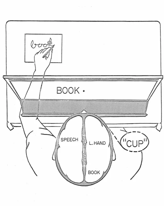
General ProcedureThe testing procedures were, in general, similar to those used for studying integrational deficits in previous patients with section of the forebrain commissures (1,2). Most of the tests were carried out in a standard set-up
(Figure 1) in which the subject was seated at a table in front of a projection screen of translucent plexiglas that served also to shield from sight the top of the table, the examiner, and the testing equipment. In the center of the screen at eye level was a black spot upon which the subject centered his gaze during tachistoscopic presentation of visual material. The patient could reach under the screen through a fringe to perform various manual tasks hidden from sight. To minimize auditory cues during tactile test-ing, the stimuli were placed behind the screen on a thick towel. This experimental arrangement allowed for controlled lateralized testing of different sensory modali~ies, and for separate motor performance by the two hands with vision
excluded~
Unless otherwise stated1the subject was allowed in advance of the actual trials to identify by sight and touch, and to name aloud,all of the objects, words, or pictures to be used in a given test. In the case of visual stimuli this
~
0
..µ
(1) (/)
CT\
c:
•r-f
I
" ..µtJl (1)
8
I
I
,...;
ro S-1
I
(1)
I
c:
(1) (.!)
•
10.
of the projected images subtended a visual angle of approxi-mately ten degrees.
In preliminary testing it was noted that, when requir-ed toidentify stimuli in the left sensory field, the
subject would often silently mouth, over and over, the names of the possible choices. To eliminate this as a source of peripheral cross cueing, mouthing was prohibited in the
,tests to be reported, even to the point of having the patient hold his tongue between his teeth.
Further procedural details for specific tests are described below in context.
D. Tests for Compensatory Reorganization of the Somesthetic System
let Introduction
The main cortical representation for sensa-tions of touch and kinesthesis from one half of the body lies in the contralateral hemisphere. The second order
neurons from both the ventral spinoth.alamic tract, mediating coarse touch, and the dorsal funiculi, mediating discrimina-tive touch and kinethesis, cross the midline and rise to the contralateral thalamus from which the third order neurons theri project to the post central gyr~s.
In view of the predominantly unilateral nature of the somesthetic projection it is not surprising that fine
transfer between the forepaws of animals in whom the neocor-tical commissures have been cut (26,27,28,29}. Commissuro-tomized humans show similar incapacities for cross localiz-ing points on different halves of the body, and for repro-ducing with one hand, positions imposed by the examiner upon the other (30,31). The disjunction of the cortical representations for the two halves of the body is especially evident in the inability of the human patients to talk about somesthetic stimuli on their left sidee The left, speaking hemisphere is thus ignorant of sensory events in the body half whose somatic representation is in the right hemisphere.
There is, however, behavioral evidence that somesthetic information is not totally restricted to the contralateral side of the brain. Respiratory responses conditioned to tactile stimulation of one paw of a split brain cat transfer-ed to stimulation of the contralateral paw (32). A monkey could blindly coordinate his two hands so as to drop a grape from one to the other, even after division of the fore and midbrain commissures, and the cerebellum (6).
12.
ipsilateral body parts could be demonstrated even after total removal of the other hemisphere, and thus can not be attributed to an intercortical relay (37). In the cat two types of ipsilateral potential have been found. Although one of these is abolished by callosal section, the other, slower, one survives division of all forebrain and diencepha-lic commissures. This longer latency potential arrives
simultaneously at both hemispheres, suggesting a bilateral projection system (38).
The active role played by the ipsilateral tactile representation in the normal functioning of the brain has recently been shown by an experiment in which ablation of the somatosensory cortex on one side of a monkey's brain caused a deficit in his performance on a tactual discrimina-tion with the ipsilateral hand {39).
Since the ipsilateral systems have such a significant function in the normal brain, they undoubtedly would be of even greater value in compensating for early damage to the primary projection areas. The amount of sensation remaining after hemispherectomy depends to a great extent on the time of the original lesion. If an injury, such as a tumor, occurs at maturity, then following removal of an entire hemisphere the person usually has no sensation below the elbow in the contralateral arm (21). If on the other hand, the original lesion dates from birth, as in infantile
can describe coins, objects and even skin writing in his
left hand (20). Since identification and naming require
cortical processing i t seems certain that the left hemisphere
in these hemiplegics received tactual input from the left
hand, probably through ipsilateral pathways.
The results of the following tests of somesthesis on a
commissurotomy subject with early injury to the
somatosen-sory region of his left hemisphere reveals a pattern of
compensatory reorganization which would not have been
evi-dent with the commissures intact.
2. Results
a) Verbal Identification of Stimuli in the
Left Hand. With his left hand screened from sight the
subject was asked to feel and to verbally describe or name
objects placed in his hand one at a time by the examiner.
The simplest task involved stereognostic discriminations
based separately on size, weight, or surface texture. For
each of these tests a set of three cylin~ers was used. In the set varying in weight the cylinders were wood with lead
inserts; all had the same height ·( 2~") and diameter ( 1~11 ),
but weighted 100~ 150, or 200 gms. The size discrimination involved three wood cylinders all 311
high but 111 , 1~
11
or 211
in diameter. The subject was not allowed to lift these last
stimuli in order that weight would not be a cue. For the
texture discrimination, metal cylinders 211
14.
a smooth, lightly or heavily knurled surface were used. In
this task the cylinders were gently rubbed across the
subject's finger tips by the examiner. In all of these
tests the patient was shown, and allowed to feel the three
stimuli before the actual trials began, and during the test
had only to state how the cylinder out of sight in his hand
compared to the other two of the given set. In size
discrim-ination for example, he had only to say largest, smallest or
medium. The subject's verbal reports for all three types of
tactual discrimination made out of sight with the left hand
were correct well above the chance level (Table I). When
testing was extended to.verbal identification of simple shapes (a round versus a square wooden rod, both 3~11 long and 3/4" in diameter) A.A. correctly identified which one
was in his left hand 22 of 24 times (p < .001).
Under conditions where he did not see or name the test
items in advance, the subject was able to give good verbal
descriptions of common household objects, such as a spoon,
pencil or cup, placed in his left hand. He could describe
these items in terms of their size, texture, ·material, etc. For example, he characterized a quarter as being "rou~d,
thin and made of metal". An oval bar of soap he called 1
"smooth, hard, and rounded""
as "soft and made of cloth".
A cotton glove was reported
In this test where the objects
were totally unspecified in advance he was occasionally able
Table I.
Verbalization of Stimuli in the Left Hand
Three Cylinders of 16/27 p
<
.005Different Texture
Three Cylinders of 24/30 p
< .
001 Different .WeightThree Cylinders of 13/13 p
<
.001Different Diameter
Two Conunon Objects 62/84 p
<
.001Three Conunon Objects 103/129 p
<
.0001Three Plastic Numbers 12/14 p
<
.01Four Common Objects 59/74 p
<
.0001Four Wooden Shapes 13/21 p
<
.001Nine Conunon Objects 12/56 p ) .OS
Touch on One of 30/70 p
<
.001equilateral triangle, by describing aloud their tactile characteristics. His accuracy on this task was below that of his own right hand or of normal control subjects, but was well above that of the subordinate hand of other
commis-surotomy patients.
Since A.A. was generally unable to identify stimuli in his left hand when he had no prior knowledge of the objects to be given, experiments were conducted in which this infor-mation was provided. To discover the limits of his left hand naming capacities both the number and similarity of the stimuli were varied over several sessions.
Results of tests using from two to six choices demon-strated that A.A. could verbally identify well above chance which item the examiner placed in his left hand for tactual inspection (Table I). The somesthetic sensitivity possible under these conditions is seen in a series in which five centimeter high plastic letters (C,H,M,P,S,T) randomly presented to his left hand were correctly named 8 of 16 times (p <.002). When the number of items exceeded nine, even prior knowledge as to their identity was not sufficient to increase the subject's accuracy above the chance level.
The scores with his right hand on the preceding test were6 as a rule, somewhat above those of the left, despite
the right hand's sensory impairment.
Left lifillf!e
hemisphere speech in this patient, his ability to identify
in writing items felt by the left hand was examined. In
these tests the subject was asked to write with his right
hand the name of an object presented to the left hand,
instead of speaking i t aloud. Both the paper and the
right hand were out of sight behind the screen. The written
scores obtained under these conditions were quite similar
to those for verbal reports of stimuli in the left hand.
In detecting whether the rough or smooth metal cylinder had
been lightly drawn across his left fingers1his written
answers were correct 15 of 15 times ( p <.02). When four
wooden shapes (square, triangle, cross, and circle, with a
diameter or side of 211
and a thickness of ~") were
indivi-dually presented to the left hand in random order, he wrote
the correct name 9 out of 18 times ( P< a02). The
identi-ties of five common household objects (fork, pen, cup, comb,
and key) randomly placed in his left hand were correctly
written with the right hand 14 of 20 times ( p < .0001).
When the left instead of the right hand was used to write
the answers in the preceding test, he was correct 8 of 11
times (p ~ vOOl)e Six of the nine errors made by the two
hands in this last test involved one object (the fork), all
presentations of which were incorrectly identified. At
the end of this session i t was discovered that the subject
had forgotten this particular stimulus was among the five
18.
identify objects in the left hand, A.A. was, as on those involving verbal reports, markedly superior to previous commissurotomy cases.
c) Verbal and Written Identification of Stimuli in the Left Visual Field. In order to determine
whether under the present experimental ccnditions the subject's ability to name objects in the left sensory field was
con-fined to the tactual modality, object pictures were present-ed by tachistoscopic flash to one or the other visual field, and A.A_ instructed to say, or write out blindly with his right hand the correct name. Under these conditions he was able to name, either verbally or in writing, only those
pictures presented in the right half field of vision. Neither the number of stimuli nor prior knowledge as to their iden-tity made any difference in the results. His inability to identify stimuli presented in the left half visual field was quite comparable to other commissurotomy patients. In brief, A.A. was often able to say or write the names of test objects when they were presented tactually to the left hand, but not when the same objects were presented visually
as pictures in the left half visual field.
d) Localization of Left Hand Stimulation. The subject extended his hands, palm upwards with fingers
pressure from a blunt plastic stylus. He could always
report verbally the onset of contact with either hand, the
pressure thresholds for the right and left hands not being
noticeably different in this respect. When asked to say
which of eight spots on his left arm and hand (lower arm,
wrist, palm and the ends of the five fingers) had been
lightly touched_, his performance was well above chance
{21/80, p
<
.001), as i t also was when just the five fingerswere tested {30/70, p
<
.01). The right hand scores on theselatter tasks were somewhat better than those of the left,
but were also subnormal· {12/20, p <.0001).
In order to compare the ability of each hemisphere to
cross localize touch1a test was given in which, with both
hands screened from view, the subject was instructed to move
the finger on his left hand that corresponded to the one
touched by the examiner on his right, and vice versa. It
was found that A.A. could perform this task from the left
to the right hand but not in the reverse direction, from
the right to the left. When a finger on his left hand was
touched he correctly moved the corresponding finger on his
right hand 21 of 49 times {p-< .001). When,however, the
right hand was stimulated, his performance with the left did
not rise above chance {10/44, p
=
.38). A good deal ofperseveration was also evident in this latter situation.
The observed ability of this subject to verbally
20.
arm, though well below that of normals, was much better than has been demonstrated in any other corrunissurotomy
patient. Previous patients also have not been able to cross localize touch in either direction.
e) Tactual Cross Retrieval. In this test an object was placed in one of the subject's hands for
tactile examination, after which it was removed and scrambled among an array of other test items for retrieval by the
oppos"ite hand. This entire process was carried out with both hands screened from view, and with controls for audi-tory cues. Significant scores for cross retrieval in both directions were obtained for the three sets of cylinders described under verbal testing. When the right hand was required to retrieve from among the three cylinders of a set the one which the left hand had felt, the scores were 10/15 (p
<
.01) for size discrimination, 14/22 (p<
.01) for weight discrimination and 11/15 (p=
.002) for roughness discrimination. With left handed retrieval of cylindersfelt by the right hand he was correct 10 of 15 times (p <"·01) on the size,14 of 24 times (p
<
.02) on the weight, and 13 of 18 times (p<
.001) on the texture ..When common household objects were used or items, like wooden blocks or plastic letters, that varied only in their
shapes, A.A. was not able to perform the cross retrievals with any significant success. Even with three objects
qualities no reproducible positive results were obtained.
f) Visuo-tactile Matching. Since the
results of visual testing with this subject were identical with those of previous cases, in that he was unable to
verbally identify left field stimuli1it seemed reasonable to assume that each half field projected to only the
contra-lateral hemisphere. This allowed independent testing of each hemisphere's ability to use tactile information from the
right and left hands.
The patient was instructed to retrieve by touch from among an array of objects behind the screen the item that matched a picture flashed tachistoscopically to one or the other visual field. In early tests, pictures of 15 common household objects (key, spoon, pencil, cork, coin etc.) were used, with the articles themselves set in scrambled order behind the screen for tactual inspection. Under these condi-tions A.A., like previous patients, had no difficulty find-ing the correct items with the hand ipsilateral to the half field receiving the visual stimulus. When, however, requir-ed to use the hand contralateral to the field in which the picture appeared, the subject performed successfully in one direction but not in the other. While with his left hand he was able to find objects pictured in the right
Visuo· ..,, Tactile Matchin2.·
LVF-Left Hand LVF-Right·Hand RVF-Right Hand RVF-Left Hand
Picked
NamedPicked
Named Picked Named P·ic'ked I Named·Pictures of · 8/15
---
4/24---
17/21---
8/1515 Objects p
<
.0001---
N.S.---
p <.0001---
p <.0001Printed Names 6/15
---
4/23---
13/22---
13/23of is· Objects p <.002
---
N.s.---
p <.0001---
p<
.0001Pictures of 15 13/15 2/15 2/16 2/16 12/14 12/14 14/15 . 14/15
"'
"'
•
Objects p
< •
0001 · N.s.
N.S. N.S. p<
.0001 p <.0001 p<.0001 p<
.0001Pictures of 6 18/19 4/19 5/21 5/21 16/21 21/21 .. 20/~0 29/30 Wooden Shapes P< .001 N.S. N.S. · N.S. p
<
.0001. p < .000.1 p < .001 p <.001Pictures of 6 6/7 3/7
1/7
1/7 8/11 10/11 5/10 9/10Plastic Letters p < .001 N.S.
N.s.
N.s. p <.001 p < .0001 p < .015 P.< .0001their pictures were flashed to the two half fields. Here as before, the hand ipsilateral to the field of presentation was
readily used to find the named objects, while in the cross retrieval situation the left hand alone could retrieve items named in the contralateral half field.
In further testing when the subject was requested after each retrieval to name aloud objects he had seen, A.A. correc-tly identified only those pictures flashed to the right visual fielde Despite this inability to name objects in the left field he was, as in previous tests, able to find the correct stimuli with his left hand. Upon flashing the picture to the right half field he could retrieve the object with the left as well' as the right hand, and could verbally name it
(Table II.). The results of the foregoing naming and retrie-val tasks were the same whether the stimuli were fifteen common objects, six wooden shapes, or six plastic letters. In these tests A.A., alone of the commissurotomy cases report-ed to date, has shown an ability to do crossreport-ed intermodal matching, using the left hand to find-objects whose identity had been visually revealed to only the left hemisphere.
24.
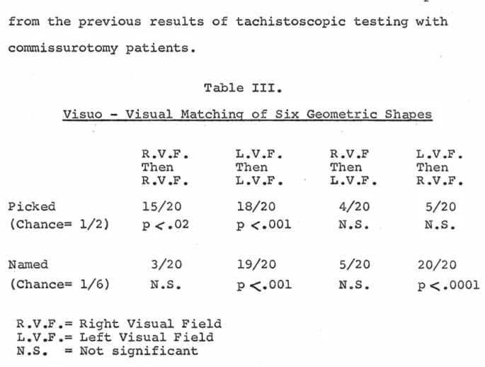
later by the flash presentation in the same or opposite field of two of these shapes vertically arranged, one above and one below the level of fixation. One of these latter two stimuli was identical to that seen in the first presentation. The subject was asked to point to the place on the screen where the matching form had appeared, and then to name it. Only when both presentations fell in the same visual half field was he able to point out the correct shape above the chance level. He correctly named the figure solely on those trials where the first presentation was to the right visual field (Table III.). This failure to match stimuli between the two half visual fields ls what would be expected
from the previous results of tachistoscopic testing with commissurotomy patients.
Table III.
Visuo - Visual Matching of Six Geometric Shapes
R. V .F. L. V .F. R.V.F L .V .F.
Then Then Then Then
R .V .F. L. V .F. L. V .F. R.V.F.
Picked 15/20 18/20 4/20 5/20
(Chance= 1/2) p <.02 p
<
.001 N.S. N.S.Named 3/20 19/20 5/20 20/20
(Chance= 1/6) N .. S. p <.001 N .. S. p
< ..
00013. Discussion
The combined results of cross integrational tests given this subject point to the presence in his left hemi-sphere of an unusually strong sensory representation of the left hand. Although the amount of useful somesthetic infor-mation received by A.A.'s major hemisphere from this hand was
less than would be normally obtained across the callosum, it qualitatively exceeded that found in any other commissurotomy patient. This subject could describe both the location and somesthetic qualities of stimuli out of view in his left
hand. This information as to size, weight, texture, material and shape was sufficient to allow actual identification of the object if the number of alternatives was limited, and their identities known to the subject beforehand. His lack of success with a larger number of choices could be due either to the crudity of the data with which the left hemisphere had to make its discriminations, or to the difficulty of remem-bering all the possible alternatives • . If the major
26.
visual - tactile matching where the left hemisphere
success-fully distinguished up to 15 items through the lef_t hand, a
much smaller-requirement was· placed upon the ipsilateral
system, as in this case the major hemisphere was provided with
the item's identity, and had only to search with the left
hand for
a
set of somesthetic characteristics it hadpre-viously learned was unique among the choices to that object.
While the above results s~gge_st that the left
hemi-phere has access to tactual information from the left hand,
there is no evidenca that the minor hemisphere has a similar
access to somesthesis from the right hand. Successful cross
localization of touch occurred in one direction only, from
the left to the right hand. This can be understood either
as the right hemisphere possessing an exceptional amount of
ipsilateral motor control not shared by the major hemisphere,
or as the left hemisphere alone receiving tactile input from
its ipsilateral hand. Similar·undirectional results with
visualtactile matching, where only the left hemisphere
-left hand combination was successful, settles this question
in. favor of an ipsilateral somesthetic system.
The only results inconsistent with the proposed model
are those for tactual cross _retreival.. Although objects of
varying size, weight or texture which were felt by one hand
could be retrieved by the other, those differing in shape.
could not. This failure may be attributable to the subject's
shapes has been shown to be a more difficult task than is visual - tactile matching (40). Since A.A. could cross retrieve items varying in simple somesthetic qualities, his failure with shape, a stimulus characteristic more normally ~xamined through vision, may reflect the difficulty of this type of matching for someone of such lowered tactile capa-cities.
There are several other possible interpretations for the data obtained from A.A., but none account for all the results as well as does the proposed left sided ipsilateral tactual system. Speech in the minor hemisphere while conceivably explaining the naming of objects in the left hand, can not be the basis of the cross localization of touch, or the
increased intermodal matching. If peripheral o~ subvocal
cross cueing of answers between the hemispheres were involved, then visual as well as tactual information should cross. This, however, was not the case as was shown by the failure of the subject to name left field stimuli, or to match shapes
between the visual fields. This latte.r result demonstrates that the success of the left hemisphere - left hand combina-nation in visual - tactile.matching must be due to the major hemisphere receiving information from the left hand, and not to the right hemisphere learning the identity of the stimulus in the right visual field.
somesthetic projection, the amount of information his major hemisphere receives from the left hand seems to be substan-tially less than in A.A •• Although this patient could say aloud which of two shapes lay out of sight in his left hand, if asked to write the name, he performed at chance unless given feedback as to the correctness of his answers. It thus appears that his major hemisphere received sufficient information to distinguish the two shapes, but not enough to decide which was which without knowledge as to the accuracy of his replies (7). It should also be noted that L.B. was only thirteen when he underwent conunissurotomy, and thus any ipsilateral abilities he possesses may, .like A.A.' s, be a result of compensation. This possibility is strengthened by the failure of an adult conunissurotomy patient to show any left hand naming on identical tests (7).
The main issue remaining concerns the course that
compensatory readjustment has taken in this subject. Results obtained from lesions in immature animals would lead one to expect that A.A.'s right hand would gain an increased repre-sentation in the right hemisphere after its primary projec-tion in the left had been injured. Exactly the opposite was found. In both visual -tactile matching and cross localiza-tion of touch the right hemisphere showed no ability to utilize tactual information from the right hand, but rather it was the damaged left hemisphere which exploited its
of subsidiary damage from head injuries or edema, it is more likely a reflection of basic brain organization, as it has been shown that, in man, the left hand has a higher
probabil-ity than does the right of possessing a functional bilateral representation (41). Since compensation probably occurs through stengthening of existing pathways, this would predis-pose alteration in favor of a left sided system. Compensa-tion would thus provide the left hemisphere with increased sensory information from the left hand offsetting the lose of tactual capacity caused by the birth injury.
This sort of reorganization would only be detected after division of the commissures allowed demonstration of cross manual abilities far above those seen in the. typical commissurotomy patient.
E. Tests for Minor Hemisphere Expressive Language
1. Introduction
The association between right sided paralysis and aphasia has been known since biblical
times-"If I forget thee
o
Jerusalem, let my right hand forget her cunning. If I do not remember thee let my tongue cleave to the roof of my mouth ••• "(42).right hemisphere for expressive language, while proven for some left handers (43,44), remains unclear for those
persons in whom the left hemisphere is dominant. A right handed adult whose major hemisphere was removed due to a tumor was capable of comprehending some written and spoken language, but not of producing any substantial amount. himself
(45 .• 46). The right hemisphere of reported conunissurotomy subjects show a similar capacity for comprehension, and incapacity for expression (47,48,49). There is, however, good evidence that the right hemisphere participates in the normal acquisition of expressive language in children, and has a potential for developing speech in the presence of
left hemisphere damage (50). The earlier in life this injury to the major hemisphere occurs, the more likely is the right hemisphere to acquire verbal skills (23,51). Agenesis of the corpus callosum depriving the two hemispheres of their normal interaction also appears to induce the development of language in the minor hemisphere (52).
In view of these shifts in laterality produced by early cortical insult, the capacity of A.A.'s minor hemisphere for expressive language was investigated to determine how it
might differ from that of the typical commissurotomy patient.
2. Procedure
were confined to tachistoscopically presented vi.sual material.
Earlier visual tests had demonstrated that, under the present
experimental conditions, A.A. could not verbally identify
stimuli in the left visual field, therefore in these tests
he was required instead to blindly write out his answers.
Before each session began all stimuli to be used, printed
words or object pictures, were shown to the subject in free
view, and he was asked to say their names aloud. The same
'
stimuli were then exposed_ tachistoscopically with both the
order of their presentation and the alternation between the
visual fields randomized. After a stimulus had been flashed,
the subject wrote his answer with one or the other hand on a
pad of paper out of sight, behind the screen, and then named
aloud the word he had written. In all cases A.A. spontaneously
wrote in script rather than printing his answers.
3. Results
a) Writing to Printed Words in the Left
Visual Field. In the first task, the ·visual stimuli were
ten to fifteen short common printed nouns (cup, pen, key,
et.)e When these were projected to the right half visual
field the results were similar to previous cases and to
normals; the subject was able to write the correct word with
either the right (18/22, p <.01) or left hand (16/24, p<.01),
and could always name what he had written. When the stimuli
32.
t~e right hand exhibited the deficits seen in the other
cormnissurotomy patients; the written answers were never
correct (0/24),and his verbal responses always mirrored the
written ones, thus demonstrating major hemisphere guessing.
By contrast, his performance with the left, subordinate, hand
was altogether different from previous subjects. Of the
thirty-nine presentations of printed nouns to the left field,
he wrote in script with his left hand the correct word twelve
times.
On
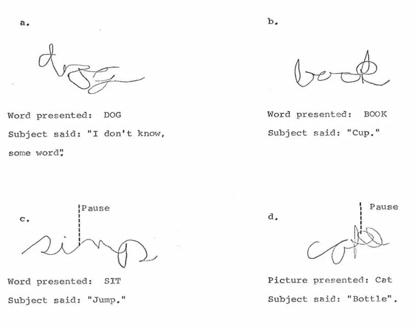
ten of these occassions he then either could notname, or misnamed the word he had just written, suggesting
minor hemisphere writing (Figure 2). The words correctly
written but misnamed were : "cup", "comb~' "dog", "key", · 11eye11
(twice), 11
book11
(twice), and "cat" (twl.ce) (Figure 3 a &
bi
.Theresponses by his left hand to the rest of the left field
presentations consisted of incorrect answers which he could
later. always verbalize, indicatinc~· that in these instances
the major hemisphere was doing the writing.
When the stimuli were printed verbs rather than nouns,
again it was only the left field -left hand combination that
yielded results divergent from the typical commissurotomy
syndrome. Of the twelve presentations to the left field,
the left hand wrote two possibly correct answers. In the
first case the word presented with "lie"; he wrote "li",
stopped, added 11
n", and said "run". In the second case the
word was "sit": he wrote "si", stopped, added "mp", and
said "jump" (Figure 3c • ) • Both "jump 11
and 11
run11
BOOK·
Figure 2. An example of left hand writing to a left field
presentation, followed by incorrect
verbaliza-tion of the answer given. The written word
a.
Word presented: DOG
Subject said: "I don't know,
some word'~
:Pause
I
C,. I
I I
/LA)
.
~
/
Word presented: SIT
Subject said: "Jump."
Word presented: BOOK
Subject said: 11
Cup."
d.
I
: Pause
I I f\ I .
1··r·~\;\
/
/._u
~
)~)~\
L /
(
} -
v
·- ..7'-"'Picture presented: Cat
Subject said: 11
Bottlen.
Figure 3. Illustrations of writing by the left hand after presentation of words or pictures in the left visual field.
w
J::.
the subject. to be possible choices on this test. On the
other trials the major hemisphere apparently dominated the
left hand respt;>nse throughout, and only incorrect answers~
which he could later say, were obtained.
b) Writing to Object Pictures in the Left
Visual Field. In this task A.A. was required to write out
the names of fifteen common objects the pict~res of which were
flashed to one or the other visual field. Most of the
pictures were of articles the printed names of which had been the
stimuli in the first task. When these were flashed in the
right field, as ·expected, he was very successful with either
the right (16/16) or the left (38/43) h_and. When the stimuli
.. were introduced in the left half field the right hand wrote
the correct answer only 2 of 15 times. Using the left hand,
of 54 tachistoscopic exposures of pictures to the left visual
field, the subject wrote the correct name six times, but on
only two of these occasions did he then fail to name what he
had written. Both of these exceptional successes involved a
picture of a Siamese cat greatly resembling the family pet.
The first time. when asked what he had written he tried to
peer over the screen, and only after being prevented,
admit-ted that he did not know. In the second case he wrote "cat"
.
'
stopped~ said ••no that's wrong", added two loops (Figure 3dl
and then said •oottle". On all other presentations to the
left visual field he wrote an incorrect answer, and then
pictures of other breeds of cat did not elicit a correct
written response, however, major hemisphere interference
was exceptionally great in these particular sessions.
4. Discussion
The preceding results demonstrate that A.A.
could transcribe into script with his left hand printed words
seen only by his minor hemisphere. The subject's inability
to then verbally name the word just written by his own left
hand makes i t clear that the major hemisphere did not
parti-cipate in this writing. These examples of minor hemisphere
writing cannot be viewed as mere copying of visual shapes,
for while the stimuli were printed, the subject's answers
were always in cursive script. Rather, this performance
required, on the part of the right hemisphere, both
compre-hension of the printed symbols, and an ability to transform
them into an equivalent form.
The common tendency of the major hemisphere to
super-cede the minor's command of the left hand after a left field
presentation can be seen in the frequent writing of incorrect
words which could then be verbalizeQ. Transfer of motor
control from the minor to the major hemisphere occurred
several times in the middle of an answer already correctly
begun by the right hemisphere. Outward signs of this shift
were a cessation of writing often accompanied by some excla~
mation of the effect that what he had written was wrong: the
word which he could later verbalize.
Since all responses fully written by the minor hemis-phere were correct, it seems reasonable to suppose that on those trials in which the right hemisphere was unsure of the word, or was hesitant in beginning to write, the major hemisphere seized control of the left hand and imposed its own guess.
The left hemisphere was even more intrusive when the visual stimuli were pictures of objects rather than their names. The only two examples of minor hemisphere writing under these conditions occurred with the picture of a cat resembling the patient's pet. The role of emotional ties in this performance is not clear as other pictures with emotional overtones elicited no right hemisphere writing. These two instances, however, were definitely not random
responses, as the subject only on:e wrote 11
cat" to an inappro-priate picture.
Due to interference by the major hemisphere it was not possible to obtain a true measure of the capacity of A.A.'s minor hemisphere for expressive writing. This, however, was not the sole limiting factor on its performance, but rather the language skills of his right hemisphere seemed basically inadequate to produce the name of a picture. While there was greater major hemisphere interference with pictures than
38.
There is no reason to believe the major hemisphere would
have intruded more often with the one type of stimulus than
with the other6 unless the right hemisphere had shown itself
particularly deficient in handling pictures.
While surpassing all previous commissurotomy cases,
except for L.B. (53), by having the motor patterning
neces-sary to write words, A.A.'s minor hemisphere fell short of
infant left heroispherectomy cases in that it was unable to
initiate writing of a name upon seeing the object itself.
This deficiency seemed to be mainly one of ascertaining the
correct word. since A.A.'s right hemisphere, like those of
previous cases (50), could recognize and pick out the name
of an object i t had se~n or felt. Therefore, this subject's
right herni~phere knew how to write but.not what to write,
being incapable of itself creating the correct symbol.
Zn summary, while A.A. has, in his right hemisphere an
increased aptitude for language, it is qualitatively less
than is seen in cases in which the left hemisphere was
total-ly damaged ear1y in life. This is probably a reflection of
the continued functional presence,.in this subject, of the
left hemi?phere language centers, who~e activity would tend
to inhibit the development of language in his minor
III. Minor Hemispheric Dominance for the Perception of Part - Whole Relations
A. Introduction
The role played by man's right hemisphere in
complex mental activities was, until recently, greatly
under-estimated. The dramatic nature of the language deficits
which follow left hemisphere damage, plus the verbal
charac-ter of most of the testing procedures of the time contributed
to the concept that the left hemisphere was the sole or
dominant seat of all higher brain processes: the right
hemi-sphere at best was an automaton possessing no special
func-tions. The left hemisphere was even proposed to be the sole
possessor of consciousness (54).
The development in the 1930's of test batteries such as
the W.A.I.S. (55), which examined many diverse mental
opera-tions, demonstrated that, while left cerebral injury did
affect verbal test scores, defects on nonverbal or
perfor-mance tasks were more likely to follow ·damage to the right
hemisphere (56,57). Since that time performance deficits
such as dressing apraxia (58), some types of drawing
disabil-ity (59,60,61), and constructional apraxia (62,63) have been
associated with the right, rather than the left, hemisphere.
In the past ten years the interest of many investigators
has turned to the perceptual aspects of hemispheric
40.
the left hemisphere and verbal material , and has linked the right hemisphere to the perception of a large variety of non-verbal stimuli, such as visuospatial relations (64,65,66,67), faces (68,69,70), nonsense shapes (71,72,73), and incomplete figures (74,75,76). Even such widely divergent functions as stereopsis (77), visual hallucinations (78), and the recog-nition of melodies (79) have been said to reside mainly in the right side of the brain.
There have been several attempts to characterize the psychological properties common to these tests on which per-formance is effected more by damage to one hemisphere than to the other. The left hemisphere has been said to handle best tasks in which the stimuli are verbal, verbalizable (71,
74), or familiar (73), the right, those having nonsense, meaningless (72), or visually complex discriminanda (71).
Other hemispheric dichotomies have been based on postu-lated differences in the type of perceptual processing employ-ed by the two sides of the braine This distinction between the left and right hemispheres has been described as:
symbolic versus visuospatial (80), associative versus apper-ceptive (81), propositional ver sus appositional (82), and
analytic versus _gestalt (83). All these classifications imply that the· organization and processing of data by the right hemisphere is in terms of complex wholes, with a predisposi-. tion for perceiving the total rather than the parts. By
analyze input~ abstracting out the relevant details and
associating these with verbal symbols.
If the minor hemisphere does concern itself.mainly with
the overall stimulus con~iguration, then it ought to excell
. .
on those operations necessary to form this type of percept,
·such as generating from incomplete data a concept of the
whole, or detecting the organization present in an array due
to the in.terre1ationship of ·its elements. ··In order to test
this prediction., tasks were designed to examine the relative
abilities of the.two hemispheres to perceive the whole
inher-ent in the part or parts of a stimulus.
B. Subiect$
The seven commissurotomy patients used in these
. studies were op1erated on · from three to five years before ·
testing in order to relieve epilepsy not controlled by
medi-cation. The surgery by Dr. P.J. Vogel and his staff at the
White Memorial. Hospital involved complete section of the
corpus c.allosum1 anterior and hippocampal cornmissures (84, 85).
Except for R.M.. and
c.c.,
these individuals now lead fairlynormal lives in their own homes. Before surgery all seven
patients considered themselves right handed. This was
con-firmed during· the present expe~iments by the Harris Test of
Lateral Dominance (86), which also revealed them to be,
except for R.M.Jright eye dominant. None of these subjects
or air study. The approach to the callosum in every case was accomplished by retraction of the right hemisphe~e. Evidence for preoperative brain damage in each individual
is as follows:
A.A.'s case history was given in the previous section.
L.B.,a seventeen year old boy,presented prior to surgery, no lateralizing signs or symptoms, his EEG abnormalities
always being generalized. Post-operatively, a few seizures restricted to the left side of the body occurred, indicating a possible right Rolandic lesion.
c.c.,
an eighteen year old boy, evidenced symptoms,including turning of the head to the right and speech arrest, characteristic of an anterior occipital focus in the left hemisphere.
N.G., a thirty-seven year old woman, had EEG indications of a left temporal focus: evidence for a right central
lesion also existed, consisting of a one centimeter wide Rolandic calcification as well as a left side numbness pre-ceding some of her preoperative convulsions.
R.M., a thirty year old man, had no reliable localizing signs either before or after surgery. He is the only patient whose generalized convulsions were not helped by this
oper-ation.
43.
slowing of the right temporal EEG was present, as was an
intermittent 1eft hypesthesia. One year after her
commissur-otomy a ventriculo-jugular shunt was implanted through a
right parieta1 burr hole_. Revision of this shunt has been
necessary three ti.mes.
R.Y •• a forty-six year old man, suffered generalized
seizures probably dating from a childqood head injury. A
visua1 aura often preceded his attacks; according to Mullan
and Penfield (78), the chances are ten to one that this
repre-sents a right hemispheric focus.
:en
a11 but A.A. andc.c.,
therefore, it is the righthemisphere which is more liable to disfunction from
extra-C·allosal damage or from any residual subictal abnormalities.
:rn
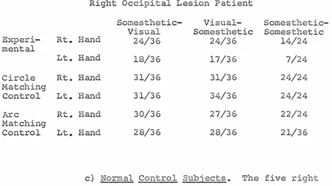
addition to the conunissurotomy patients, some testing ·was carried out on a fifty-five year old man (H.D.) in whom
the right occipital and posterior parietal lobes had been
removed due to an abcess. Prior to surgery ~.D. had been a
draftsman, but in the year since his operation he has been
unable to return to work due to left field blindness ang an
inabi1ity to recognize persons by their faces (prosopagnosia).
·
·
c.
Arc-Circle Matching1. :Introduction
· Previous studies of right hemispheric function
in human_ beings have involved mainly visual stimuli, especially
44.
exist in the strategies by which the two hemispheres
organ-ize and process perceptual data, it should be evident also
in other sensory modalities; likewise complex stimuli should
not be a necessity if the mental manipulations required are
performed better by one side of the brain than the other.
The present experiment was designed to test the ability
of individuals to handle simple part-whole relationships.
Subjects were asked to judge from tactual or visual
examina-tion of an arc, the siee of the circle from which it had come.
Since the stimuli were arcs and circles differing only in
their size, and thus in their rate of curvature, complicating
factors such as novelty, ·complexity, and verbalizability
should not obscure the part-whole nature of the problem.
Besides comparing the independent perceptual capacities
for this task of the right and left hemisphere of
commissuro-tomy patients, the present tests was also used to examine a
prediction made by a recent theory of hemispheric
speciali-zation (87), to the effect that left handed normal subjects
would be inferior to right handers on ·tests requiring minor
hemisphere performance.
2. · Methods
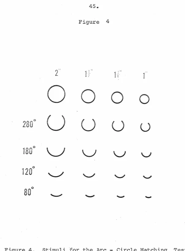
The stimuli for this experiment were made from
plexiglas rings of four different sizes: 211
, 1~11, 1~11 , and 111 in inner diameter (Figure 4). For each size there was a set
Figure 4
2"
I IIL
1+'1
0
0
0
0
280
°
u
u
u
u
180
°
\ J
v
v
1
2
0
°
80°
of completeness: 280°, 180°, 120°, and 80°; all had the same
wall thickness (1/8") and height ( 1/811
) . Each was individually
mounted on a 3"x 311
card.
In the first session with each individual,special sets
of stimuli (1 3/4" and 2~") were used to demonstrate the
geometrical relationship existing between the arcs and
com-plete rings of the two sizes. The subject was encouraged to
superimpose different arcs on the rings to see how they fit.
I t was emphasized that the length of an arc alone could not
reveal the size of the circle from which i t had come, but,
rather i t was the amount of curvature over the given length
which was important. None of the subjects had any apparent
difficulty grasping this concept.
The individual was next instructed that he would be
presented with a series of arcs, and for each one he was to
pick out the size of circle of which that arc was a segment.
Each person was given the test in three different forms.
The first two of these required intermodal matching, as the
arc was presented in one modality and .the choices in another;
the third was totally intramodal.
In the first form, Somesthetic - Visual, (Figure Sa) the
subject reached beneath a screen and felt an arc, while
simul-taneously looking at three sizes of ring. When he had made
his decision as to which one the segment was from, he
with-drew his hand and pointed to i t .
Figure 5
b.
c.
/
-;---fyr--
\
,
@
a.
Figure 5. The Three Forms of the Arc - Circle Matching
Test - a) Somesthetic Visual, b) Visual
arc was presented in free view, while the rings were arranged
behind the screen for tactual inspection. In this case the
subject indicated his choice by tapping the correct ring.
The third form, Somesthetic - Somesthetic, (Figure Sc)
had both the arc and the rings hidden from view, with no
restriction on the number of times the subject could shuttle
between them for comparison.
In the second and third forms of the test the
arrange-ment of the choices was changed after every trial; the
dis-position,ho~ver, for any one arc was identical for the
right and left hands. In all thr~e test forms the various
arcs were presented to both hands in the same predetermined
random order.
The exact sizes of the rings used in the three forms of
the test depended on each individuals ability. On the
Somesthetic - Visual (S - V} and Somesthetic - Somesthetic
(S - S) forms all subjects were given circles differing by
one quarter inch - 1~11, l~ia and 111
• In the Visual -
Somesthe-tic (V - S) procedure both L.B. and R.Y. performed at chance
with these sizes, and were, therefore, retested with rings
varying by one half inch - 211
, 1~
11
and 111
•
In all forms of the test, somesthetic examination of the
I
stimuli was limited to the index finger of either hand. The
subject's arm rested on the table and only finger and some
wrist movement was allowed. Before either hand was given any