Published Ahead of Print 16 April 2014.
10.1128/JCM.00565-14.
2014, 52(6):2144. DOI:
J. Clin. Microbiol.
Kuile and Steven R. Meshnick
Muna Affara, Kephas Otieno, Simon Kariuki, Feiko O. ter
Eusebio Macete, Clara Menendez, Pau Cisteró, Fanta Njie,
Ayola Akim Adegnika, Carlo Severini, Michela Menegon,
Prakash Narayan Singh, Martha A. Clark, Jaco J. Verweij,
Ndam, Happy Mkali, Grace Mwangoka, Neena Valecha, Jai
Hilaire M. Kenguele, Smaïla Ouédraogo, Nicaise Tuikue
Steve M. Taylor, Alfredo Mayor, Ghyslain Mombo-Ngoma,
Detect Plasmodium Species
Trial Consortium for PCR Protocols To
A Quality Control Program within a Clinical
http://jcm.asm.org/content/52/6/2144
Updated information and services can be found at:
These include:
REFERENCES
http://jcm.asm.org/content/52/6/2144#ref-list-1
at:
This article cites 27 articles, 12 of which can be accessed free
CONTENT ALERTS
more»
articles cite this article),
Receive: RSS Feeds, eTOCs, free email alerts (when new
http://journals.asm.org/site/misc/reprints.xhtml
Information about commercial reprint orders:
http://journals.asm.org/site/subscriptions/
To subscribe to to another ASM Journal go to:
on June 11, 2014 by University of Liverpool Library
http://jcm.asm.org/
Downloaded from
on June 11, 2014 by University of Liverpool Library
http://jcm.asm.org/
PCR Protocols To Detect
Plasmodium
Species
Steve M. Taylor,a,bAlfredo Mayor,c,dGhyslain Mombo-Ngoma,e,f,g,hHilaire M. Kenguele,e,iSmaïla Ouédraogo,j,k,l
Nicaise Tuikue Ndam,m,nHappy Mkali,oGrace Mwangoka,oNeena Valecha,pJai Prakash Narayan Singh,pMartha A. Clark,q
Jaco J. Verweij,r*Ayola Akim Adegnika,e,hCarlo Severini,sMichela Menegon,sEusebio Macete,dClara Menendez,c,dPau Cisteró,c
Fanta Njie,tMuna Affara,tKephas Otieno,uSimon Kariuki,uFeiko O. ter Kuile,vSteven R. Meshnickb
Division of Infectious Diseases and International Health, Duke University Medical Center, Durham, North Carolina, USAa; Department of Epidemiology, Gillings School of Global Public Health, University of North Carolina, Chapel Hill, North Carolina, USAb; Barcelona Centre for International Health Research, Barcelona, Catalonia, Spainc; Centro de Investigação em Saúde de Manhiça, Maputo, Mozambiqued; Centre de Recherches Médicales de Lambaréné, Lambaréné, Gabone; Département de Parasitologie, Université des Sciences de la Santé, Libreville, Gabonf; Ngounié Medical Research Centre, Fougamou, Gabong; Institute for Tropical Medicine, University of Tubingen, Tubingen, Germanyh; Département de Biologie, Université des Sciences et Techniques de Masuku, Franceville, Gaboni; Laboratoire de Parasitologie, Faculté des Sciences de la Santé, Cotonou, Beninj; Centre Hospitalier Universitaire Yalgado Ouédraogo, Ouagadougou, Burkina Fasok; Département de Santé Publique, Unité de Formation et de Recherche en Sciences de la Santé, Université de Ouagadougou, Ouagadougou, Burkina Fasol; Institut de Recherche pour le Développement, Faculté des Sciences Biologiques et Pharmaceutiques, Paris, Francem; Centre d’Etude et de Recherche sur le Paludisme associé à la Grossesse et à l’Enfance, Faculté des Science de Santé, Université d’Abomey-Calavi, Cotonou, Beninn; Ifakara Health Institute, Bagamoyo, Tanzaniao; National Institute of Malaria Research, Dwarka, New Delhi, Indiap; Department of Microbiology and Immunology, University of North Carolina, Chapel Hill, North Carolina, USAq; Department of Parasitology, Leiden University Medical Center, Leiden, The Netherlandsr; Department of Infectious, Parasitic and Immunomediated Diseases, Istituto Superiore di Sanità, Rome, Italys; Medical Research Council Laboratories, Banjul, Gambiat; Kenya Medical Research Institute/Centers for Disease Control and Prevention, Kisumu, Kenyau; Malaria Epidemiology Unit, Department of Clinical Sciences, Liverpool School of Tropical Medicine, Liverpool, United Kingdomv
Malaria parasite infections that are only detectable by molecular methods are highly prevalent and represent a potential transmission
reservoir. The methods used to detect these infections are not standardized, and their operating characteristics are often unknown. We
designed a proficiency panel of
Plasmodium
spp. in order to compare the accuracy of parasite detection of molecular protocols
used by labs in a clinical trial consortium. Ten dried blood spots (DBSs) were assembled that contained
P. falciparum
,
P. vivax
,
P. malariae
, and
P. ovale
; DBSs contained either a single species or a species mixed with
P. falciparum
. DBS panels were tested
in 9 participating laboratories in a masked fashion. Of 90 tests, 68 (75.6%) were correct; there were 20 false-negative results and 2
false positives. The detection rate was 77.8% (49/63) for
P. falciparum
, 91.7% (11/12) for
P. vivax
, 83.3% (10/12) for
P. malariae
,
and 70% (7/10) for
P. ovale
. Most false-negative
P. falciparum
results were from samples with an estimated
<
5 parasites per
l
of blood. Between labs, accuracy ranged from 100% to 50%. In one lab, the inability to detect species in mixed-species infections
prompted a redesign and improvement of the assay. Most PCR-based protocols were able to detect
P. falciparum
and
P. vivax
at
higher densities, but these assays may not reliably detect parasites in samples with low
P. falciparum
densities. Accordingly,
for-mal quality assurance for PCR should be employed whenever this method is used for diagnosis or surveillance. Such efforts will
be important if PCR is to be widely employed to assist malaria elimination efforts.
M
olecular detection methods for malaria parasites,
includ-ing PCR assays, detect low-level malaria parasitemias that
are missed by microscopy and rapid diagnostic tests (RDTs). In
many settings, these “submicroscopic” infections usually far
outnumber patent infections (1). Because they are not
identi-fied by routine clinical or research diagnostics and therefore
remain untreated, they constitute a reservoir of parasites for
ongoing transmission (2). Thus, efforts to reduce transmission
and eliminate malaria may need to use PCR in order to
elimi-nate the malaria reservoir (3).
PCR-based methods to detect malaria parasites are diverse in
design and operation. These factors have the potential to
intro-duce substantial variability in the operating characteristics of
methods (4). The definition of “submicroscopic” parasites is
fur-ther complicated by inconsistencies between operators in
assess-ing parasites by microscopy (5). Collectively, these microscopic
and molecular considerations can produce inconsistencies in
measurement and undermine the generalizability of findings
re-lated to PCR-detectable parasites.
Quality control procedures have been endorsed by the WHO for
parasite detection by both microscopy (6) and RDTs (7), likely owing
to their clinical use. Although RDTs have been advanced as adequate
tools to capture the submicroscopic parasite reservoir (8), their
sen-sitivity of parasite detection is generally lower than that achieved by
PCR methods or by expert microscopy (9). Therefore, PCR methods
are increasingly employed for parasite detection in research studies,
and there exist nascent efforts to standardize quantification (10) and
reporting (11) of assays. Nevertheless, the comparative, qualitative
performance of PCR assays used to detect the major species of malaria
parasites is largely unknown.
Received28 February 2014 Returned for modification24 March 2014
Accepted8 April 2014
Published ahead of print16 April 2014
Editor:M. J. Loeffelholz
Address correspondence to Steve M. Taylor, [email protected]. * Present address: Jaco J. Verweij, Laboratory for Medical Microbiology and Immunology, St. Elisabeth Hospital, Tilburg, The Netherlands.
Copyright © 2014, American Society for Microbiology. All Rights Reserved. doi:10.1128/JCM.00565-14
on June 11, 2014 by University of Liverpool Library
http://jcm.asm.org/
The Malaria in Pregnancy Consortium is a global consortium
of research groups conducting clinical studies investigating the
impacts of current and novel interventions to prevent
pregnancy-associated malaria (http://www.mip-consortium.org/). Two
fac-tors necessitated the evaluation of PCR assays in this complex
clinical trial consortium: (i) the use of multiple laboratories with
various methodologies for molecular parasite detection, and (ii)
the requisite need to quantify the impact on birth outcomes of the
low-level parasitemias that are typically observed in pregnancy.
Therefore, we assembled a “proficiency panel” of
Plasmodium
parasites and distributed this panel to associated molecular
labo-ratories for masked testing. Herein, we describe the design of the
panel and the results of testing at 9 molecular laboratories from
four continents.
MATERIALS AND METHODS
Panel design.A panel of 10 dried blood spots (DBSs) was designed that contained the four most common human malaria parasites:P. falciparum, P. vivax,P. ovale, andP. malariae. We designed the panel to address several common issues with molecular detection methods: (i) the ability to detect all four of the most common human malaria species in Africa; (ii) the sensitivity ofP. falciparumdetection, taking into account three different parasite densities (5%, 0.5%, and trace); (iii) the ability to discriminate species within mixed-species infections that would most likely includeP. falciparum(thus, we included samples of each of the other species mixed withP. falciparum).
Additionally, we included a DBS with uninfected whole blood as a control. The sources of each parasite are summarized inTable 1.
Panel assembly.For DBSs withP. falciparum,P. falciparumline 3D7 (MRA-102, MR4; ATCC, Manassas, VA) was cultivated in continuousin vitroculture in O⫺human red blood cells (RBCs) to 5% to 13% parasite density (as confirmed by light microscopy), centrifuged to remove serum and concentrate RBCs, and split with uninfected fresh whole blood col-lected in a tube with EDTA to obtain the targeted parasite densities. Be-cause of variations in the density of cultivated parasites and hematocrits, varied final densities ofP. falciparum3D7 were considered approximate. For DBSs withP. falciparummixed with other species,P. vivax,P. ovale, or P. malariaewas added to the 3D7 aliquot.
ForP. vivax, a 500-l specimen of 40,000 parasites/l at 50% hemat-ocrit was used. This was diluted with fresh uninfected whole blood with or without additionalP. falciparumstrain 3D7 from culture to obtainP. vivax-containing DBSs. ForP. malariaeandP. ovale, we blotted fresh venous blood from admitted patients onto filter paper, with or without additionalP. falciparumstrain 3D7 from culture, to obtain DBSs.
All DBSs consisted of 50l of blood aliquoted from the single prepa-rations as described above. These were placed onto prelabeled Whatman 3MM filter paper in duplicate and left to air dry overnight. All DBSs were produced in a laminar flow hood. The 10 DBSs that comprised each panel were placed into a single sealable plastic bag with desiccant and separated within by standard weighing paper.
Panel internal quality control.Genomic DNA (gDNA) was extracted within 2 weeks of preparation and storage at 4°C from each DBS of a single set of 10 DBSs after punching a 5-mm hole from each DBS by using a QIAamp DNA minikit. gDNA samples were tested forP. falciparum,P. malariae,P. ovale, and the human gene glyceraldehyde 3-phosphate de-hydrogenase (gapdh), using duplex real-time PCR assays targeting the Plasmodium18S rRNA gene (12). Cycle thresholds (CT) were set manually
by personnel masked to input gDNA. Samples were also tested in a sepa-rate SYBR green real-time PCR assay targeting onlyP. vivax(13); this 25 l reaction mixture consisted of 12.5l of SYBR green master mix, 900 nM (each) primers targeting theP. vivax18S rRNA, and 5l of template. In order to estimate the quantities ofP. falciparumparasites in the samples, gDNA samples were tested in aP. falciparum-specific real-time PCR assay targeting the single-copy geneP. falciparumlactate dehydroge-nase (Pfldh) (14). The reactions were performed with a set of 10 standards ofP. falciparum3D7 gDNA from 0.1 ng/l to 5⫻10⫺5ng/l, which were used to generate a standard curve and estimate parasite quantities in the unknowns.
All real-time PCRs were prepared in a laminar flow hood with filtered pipette tips, tests were performed in duplicate, and samples were tested on plates that included appropriate negative and positive controls.
Distribution of panels.We offered the panel to collaborating labora-tories within the Malaria in Pregnancy Consortium and to others who requested it. The panels were posted at room temperature and received within 7 days of sending. Each filter paper was labeled only 1 to 10. A letter accompanying the panel informed recipients of the following: “The sam-ples representP. falciparum,vivax,ovale, andmalariae, some in mixed fashion, and some as pure species. All samples were prepared with whole parasites, so they should accommodate any genes targeted by your molec-ular assays.” Therefore, recipients were blinded to the constituents of the DBSs. The panels were tested per each laboratory’s standard procedures for parasite detection.
Collaborating laboratories were located in Gambia, Benin, Gabon, Kenya, Tanzania, Mozambique, India, The Netherlands, and Italy. After the collaborating laboratory tested the panel, feedback was provided on the accuracy of results to both the laboratory staff and the principal inves-tigator of the associated clinical study. The detection rate was defined as the number of correct species detected divided by the number of species tested for; therefore, these rates were computed only for species that were
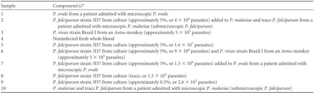
TABLE 1Constituents of the proficiency panel
Sample Component(s)a
1 P. ovalefrom a patient admitted with microscopicP. ovale
2 P. falciparumstrain 3D7 from culture (approximately 5%, or 4⫻106parasites) added toP. malariaeand traceP. falciparumfrom a patient admitted with microscopicP. malariae(submicroscopicP. falciparum)
3 P. vivaxstrain Brazil I from anAotusmonkey (approximately 5⫻105parasites)
4 Noninfected fresh whole blood
5 P. falciparumstrain 3D7 from culture (approximately 5%, or 1.6⫻107parasites)
6 P. falciparumstrain 3D7 from culture (approximately 5%, or 9⫻106parasites) andP. vivaxstrain Brazil I from anAotusmonkey (approximately 5⫻105parasites)
7 P. falciparumstrain 3D7 from culture (approximately 5%, or 1.5⫻106parasites) added toP. ovalefrom a patient admitted with microscopicP. ovale
8 P. falciparumstrain 3D7 from culture (trace, or 1.5⫻105parasites)
9 P. falciparumstrain 3D7 from culture (approximately 0.5%, or 2.6⫻105parasites)
10 P. malariaeand traceP. falciparumfrom a patient admitted with microscopicP. malariae(submicroscopicP. falciparum)
aAll samples included whole blood, either from an infected patient (samples 1, 2, 7, and 10) or uninfected whole blood from a single donor (samples 3, 4, 5, 6, 8, and 9). Estimated
quantities of input 3D7 were computed from input parasitemias and output quantitation.
PlasmodiumPCR Quality Control
June 2014 Volume 52 Number 6 jcm.asm.org 2145
on June 11, 2014 by University of Liverpool Library
http://jcm.asm.org/
targeted by the laboratory’s assay. False-positive results were defined as the detection of a species that was absent; false-negative results were de-fined as the failure to detect a species that was present.
Ethics statement.The collection and use ofPlasmodium parasites from patients at the University of North Carolina Hospital were approved by the UNC Institutional Review Board.
RESULTS AND DISCUSSION
Panel assembly and quality control.
We produced 20 panels of 10
DBSs each. The constituents of each panel are outlined in
Table 1.
Internally, a panel was first tested with real-time PCR assays that
collectively detected all 4
Plasmodium
species that are known to be
transmitted in Africa as well as a human gene. In this testing,
human DNA was detected in all 10 DBSs. All four species were
detected from each DBS on which they were known to be present,
in both mono- and mixed-species samples. The DBS with only
uninfected blood was positive only for the human control gene.
There were no false-positive results.
A second assay capable of estimating
P. falciparum
quantity
was also 100% sensitive and specific, although the two samples
containing “trace”
P. falciparum
returned discordant results
be-tween the two replicates. Based upon standard curves, this assay
returned quantity estimates of 0.5 parasites/
l of gDNA (sample
8) and 2 parasites/
l of gDNA (sample 10) for these two trace
P.
falciparum
samples. Because each DNA extraction yielded 50
l of
gDNA from approximately 20
l of blood, we estimated that these
samples contained approximately 1.25 and 5 parasites/
l of whole
blood, respectively.
Results by
Plasmodium
species.
We distributed the panel to 9
collaborating molecular laboratories for testing. The protocols
employed by these labs are outlined in
Table 2. These laboratories
were blinded to the true constituents of each DBS. Overall, 68 of
90 (75.6%) results were correct (Table 3). Of the 22 incorrect
results, 20 were false-negative results and 2 were false positives.
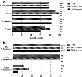
The detection rate was 77.8% (49/63) for
P. falciparum
, 91.7%
(11/12) for
P. vivax
, 83.3% (10/12) for
P. malariae
, and 70% (7/
10) for
P. ovale
(Fig. 1). Among
P. falciparum
samples, the
detec-tion rate for
P. falciparum
was 77.8% both for monospecies
sam-ples (21/27) and for multispecies samsam-ples (28/36). Among
P.
falciparum
monospecies samples, the detection rate was 100%
(9/9) for those with 5% parasite density, 100% (9/9) for those with
0.5% density, and 33.3% (3/9) for those with trace parasites (Fig.
1). Therefore, overall, the diverse protocols employed were largely
able to accurately detect
P. falciparum
when present at densities
that are typically identified in clinical infections in monospecies
infections and when mixed with other
Plasmodium
spp.
Detection of
P. falciparum
was reduced at the lowest density. In
the sample containing only trace amounts of
P. falciparum
from
in
vitro
culture, only 3/9 labs detected the parasite. Similarly, in the
sample with microscopically detected
P. malariae
and
submicro-scopic
P. falciparum
, only 1/9 labs detected
P. falciparum
. This low
detection rate (22.2%; 4/18) of trace
P. falciparum
suggests that
some PCR protocols require optimization to improve sensitivity
and that this sensitivity should be routinely quantified and
re-ported, as has been suggested for real-time PCR protocols (15).
Among
P. vivax
samples, the detection rate was 100% (6/6) for
monospecies
P. vivax
and 83.3% (5/6) for
P. vivax
when mixed
with
P. falciparum
. Only 6 of the 9 labs included methods to detect
P. vivax
in their protocols. The only false-negative
P. vivax
result
originated from lab B, which failed to report any mixed infections.
TABLE2 Protocols employed to detect Plasmodium spp. parasites at an internal laboratory and 9 collaborating laboratories Lab gDNA extraction PCR chemistry No. of cycles Positive criterion Gene target No. of replicates No. of targets Species targeted Reference(s) UNC QIAamp DNA minikit Real-time PCR with TaqMan probes 40 CT ⬍ 40 Plasmodium 18S rRNA 2 Duplex/monoplex P. falciparum , P. vivax , P. malariae , P. ovale 22 UNC QIAamp DNA minikit Real-time PCR with TaqMan probe 40 CT ⬍ 40 P. falciparum lactate dehydrogenase 2 Monoplex P. falciparum 14 A QIAamp DNA blood kit Nested PCR 30/30 Agarose gel electrophoresis Plasmodium 18S rRNA 1 Monoplex P. falciparum , P. vivax , P. malariae 25 , 26 B QIAamp DNA minikit Real-time PCR with TaqMan probe 40 CT ⬍ 40 Plasmodium 18S rRNA 2 Duplex P. falciparum , P. vivax , P. malariae , P. ovale 25 , 26 C Chelex protocol Real-time PCR with TaqMan probes 40 CT ⬍ 40 Plasmodium 18S rRNA 2 Multiplex P. falciparum , P. vivax , P. malariae , P. ovale 27 D QIAamp DNA minikit Real-time PCR with XS probes 50 CT ⬍ 50 Plasmodium 18S rRNA 2 Multiplex P. falciparum , P. vivax , P. malariae , P. ovale 28 , 29 E QIAamp DNA minikit Real-time PCR with TaqMan probes 40 CT ⬍ 40 P. falciparum 18S rRNA 2 Monoplex P. falciparum 30 F Invitrogen PureLink genomic DNA kit Real-time PCR with TaqMan probes 50 CT ⬍ 45 Plasmodium 18S rRNA 2 Multiplex P. falciparum , P. vivax , P. malariae , P. ovale 13 , 27 G QIAamp DNA minikit Real-time PCR with TaqMan probe 40 CT ⬍ 40 P. falciparum 18S rRNA 2 Monoplex P. falciparum 25 H QIAamp DNA minikit Real-time PCR with TaqMan probe 40 CT ⬍ 40 P. falciparum 18S rRNA 2 Monoplex P. falciparum 30 J QIAxtractor Nested PCR 24/29 Capillary gel electrophoresis Plasmodium 18S rRNA 1 Monoplex P. falciparum , P. vivax , P. malariae , P. ovale 22 , 23
on June 11, 2014 by University of Liverpool Library
http://jcm.asm.org/
After revising their workflow, both
P. falciparum
and
P. vivax
were
successfully detected. Therefore, the diverse protocols employed
in these labs were largely able to detect
P. vivax
in mono- and
mixed-species parasitemias.
P. ovale
had the lowest detection rate of the four species (70%),
though it was assayed the least frequently, in only 10 tests. The
three false-negative results occurred in labs that reported other
false negatives; therefore, it is unclear if the errors resulted from
laboratory procedures or from an inability to detect the dimorphic
P. ovale
(16).
There were only 2 false-positive results: one lab reported
P.
falciparum
in the negative DBS, and another lab reported
P.
fal-ciparum
in the
P. vivax
monospecies sample. Among the 20
false-negative results, 14 resulted from the failure to detect
P.
falcipa-rum
in trace quantities either in a monospecies sample or mixed
with
P. malariae
.
Results by laboratory.
Accuracy ranged from 100% (Lab A) to
50% (Labs B and F). In five of the labs (C, E, G, H, and J), the only
incorrect results were the failure to detect
P. falciparum
in trace
amounts, either alone or when mixed with
P. malariae
.
Detection of mixed infections was challenging. Lab B returned 5
incorrect results: one false positive (
P. falciparum
detected in an
uninfected sample) and four false negatives (all in mixed-species
infections). These results prompted discussions with the
labora-tory head and with a reference laboralabora-tory regarding the protocol
used. The real-time PCR protocol consisted of a single set of
Plas-modium
primers multiplexed with four species-specific probes,
suggesting that, in mixed-species infections, only the dominant
species was being amplified and detected. This competitive
inhi-bition can reduce the sensitivity of parasite PCR assays (17). The
protocol was modified to include species-specific forward
prim-ers, and a fresh panel was retested with the updated protocol.
Using this updated protocol, the assay was newly able to detect
P
vivax
,
P. ovale
, and
P. malariae
when each was mixed with
P.
falciparum
, thus improving the assay’s ability to detect mixed
in-fections (Lab D). The updated protocol subsequently went live in
Labs B and D.
Future directions and recommendations.
Our observations
suggest several practices that will help to assure high quality in
molecular detection protocols.
(i) A DBS panel of
Plasmodium
spp. should be tested and
in-terpreted by standard protocols by routine laboratory staff
blinded to sample constituents.
(ii) This testing should be repeated at planned intervals or
when procedures are altered by the addition of new reagents,
hardware, or personnel.
(iii) For protocols that are employed to detect low-level,
“sub-microscopic” parasitemias to aid in malaria elimination efforts,
the lower limit of detection of the assay should be formally
quan-tified and reported. This can be achieved by using a panel of
P.
falciparum
samples across a range of parasite densities.
PCR methods and laboratories vary in their sensitivities and
specificities to detect malaria parasites. Most protocols tested in
this study consistently detected the major species
P. falciparum
and
P. vivax
at densities that typically manifest symptoms.
How-ever, sensitivities and specificities of
P. falciparum
detection were
much more variable for low-level and submicroscopic
para-sitemias. Submicroscopic infections are important because they
constitute reservoirs of parasites that can sustain transmission
(18) and are targeted by elimination campaigns (2). Additionally,
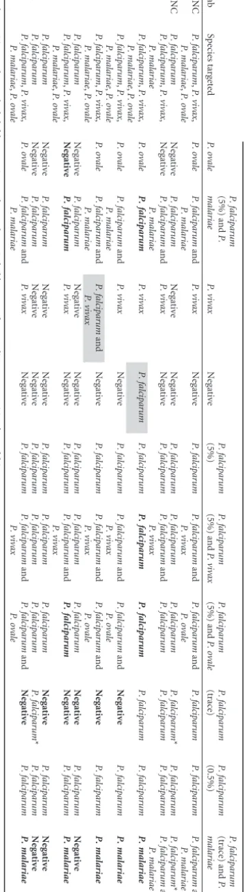
TABLE 3 Results of molecular testing of the proficiency panel at the source laboratory and 9 collaborating laboratories Lab Species targeted Species detected when sample contained a : P. ovale P. falciparum (5%) and P. malariae P. vivax Negative P. falciparum (5%) P. falciparum (5%) and P. vivax P. falciparum (5%) and P. ovale P. falciparum (trace) P. falciparum (0.5%) P. falciparum (trace) and P. malariae UNC P. falciparum , P. vivax , P. malariae , P. ovale P. ovale P. falciparum and P. malariae P. vivax Negative P. falciparum P. falciparum and P. vivax P. falciparum and P. ovale P. falciparum P. falciparum P. falciparum and P. malariae UNC P. falciparum Negative P. falciparum Negative Negative P. falciparum P. falciparum P. falciparum P. falciparum * P. falciparum P. falciparum * A P. falciparum , P. vivax , P. malariae Negative P. falciparum and P. malariae P. vivax Negative P. falciparum P. falciparum and P. vivax P. falciparum P. falciparum P. falciparum P. falciparum and P. malariae B P. falciparum , P. vivax , P. malariae , P. ovale P. ovale P. falciparum P. vivax P. falciparum P. falciparum P. falciparum P. falciparum P. falciparum P. falciparum P. malariae C P. falciparum , P. vivax , P. malariae , P. ovale P. ovale P. falciparum and P. malariae P. vivax Negative P. falciparum P. falciparum and P. vivax P. falciparum and P. ovale Negative P. falciparum P. malariae D P. falciparum , P. vivax , P. malariae , P. ovale P. ovale P. falciparum and P. malariae P. falciparum and P. vivax Negative P. falciparum P. falciparum and P. vivax P. falciparum and P. ovale Negative P. falciparum P. malariae E P. falciparum Negative P. falciparum Negative Negative P. falciparum P. falciparum P. falciparum Negative P. falciparum Negative F P. falciparum , P. vivax , P. malariae , P. ovale Negative P. falciparum P. vivax Negative P. falciparum P. falciparum and P. vivax P. falciparum Negative P. falciparum P. malariae G P. falciparum Negative P. falciparum Negative Negative P. falciparum P. falciparum P. falciparum Negative P. falciparum Negative H P. falciparum Negative P. falciparum Negative Negative P. falciparum P. falciparum P. falciparum P. falciparum * P. falciparum Negative J P. falciparum , P. vivax , P. malariae , P. ovale P. ovale P. falciparum and P. malariae P. vivax Negative P. falciparum P. falciparum and P. vivax P. falciparum and P. ovale Negative P. falciparum P. malariae a False-positive results are shaded; false-negative results are shown in bold. *, one of two replicates was positive for P. falciparum .
PlasmodiumPCR Quality Control
June 2014 Volume 52 Number 6 jcm.asm.org 2147
on June 11, 2014 by University of Liverpool Library
http://jcm.asm.org/
quantitative PCR methods are increasingly used to measure
par-asite clearance in drug efficacy studies (19).
Our study had several limitations. First, we included a limited
number of strains of each species owing to restricted availability,
and genetic diversity of the targets could have produced false
neg-atives. Nevertheless, we did include reference standards of both
P.
falciparum
and
P. vivax
, and all assays used by collaborating labs
targeted common, conserved sequences and were previously
val-idated against multiple
P. falciparum
strains. Additionally, as
noted above, we included only a single
P. ovale
sample of
indeter-minate geographic origin, which may not reflect global species
diversity owing to the recent recognition of its dimorphism.
Fi-nally, with only 10 DBSs, we did not comprehensively test the
limits of detection and the quantitative performance of the assays.
Future efforts should include a greater range of parasite densities
to assist in defining this measure for molecular parasite assays.
In order to allow comparability between sites, PCR assays
should be subject to strict ongoing quality control programs. Such
efforts can utilize new panel preparations, such as those described
here, including preparations of parasites obtained from central
repositories, such as the Malaria Research and Reference Reagent
Resource Center (MR4) or a
P. falciparum
preparation available
through the UK National Institute for Biological Standards and
Control, which has been endorsed by the WHO as a standard
quantity of
P. falciparum
DNA (10). Notably, the WHO currently
sponsors external quality assurance programs for the molecular
detection of influenza virus (20) and HIV drug resistance
muta-tions (21). Similar centralized efforts may be required if molecular
detection assays for malaria parasites become increasingly vital for
malaria control programs.
ACKNOWLEDGMENTS
This work was supported by the Malaria in Pregnancy Consortium, which is funded through a grant from the Bill & Melinda Gates Foundation to the Liverpool School of Tropical Medicine and the European & Developing Countries Clinical Trials Partnership. Tests at Istituto Superiore di Sanità in Rome were performed in the framework of the PregVax Consortium; the PregVax project received funding from the European Union Seventh Frame-work Programme (FP7/2007–2013) under grant agreement 201588.
We thank John Barnwell (Centers for Disease Control and Prevention, Atlanta, GA) for providing theP. vivaxDNA, Melissa Miller and Peter Gilligan (University of North Carolina) for assisting with clinical speci-men collection, and MR4 for providing us with the 3D7 strain ofP. fal-ciparumthat was originally contributed by D. J. Carucci. We appreciate the input provided by two reviewers within the Malaria in Pregnancy Consortium. We also thank Mireia Piqueras and Azucena Bardaji (both of the Barcelona Centre for International Health Research) for their assis-tance with trial coordination and the lab technicians who participated in the study process at each participating laboratory.
FIG 1Detection rate at nine collaborating laboratories of eachPlasmodiumspp. (A) andP. falciparumat different parasite densities (B). The detection rate was defined as the number of correctly detected species divided by the number ofPlasmodiumspp. for which the samples were tested. 5%, 0.5%, and trace indicate the approximate parasite density of the sample tested; based upon testing in a real-time PCR assay, the “trace” specimens included between 1.25 and 5 parasites/l of whole blood (see Results). (A) Results for each of the four species tested. (B) Results only forP. falciparumat the three approximate densities included in the panel. NA, not tested. Values to the right of the graph are the number of positive tests per total number of tests.
on June 11, 2014 by University of Liverpool Library
http://jcm.asm.org/
All authors declare that they have no relationships that may constitute a conflict of interest.
REFERENCES
1.Okell LC, Ghani AC, Lyons E, Drakeley CJ. 2009. Submicroscopic infection in Plasmodium falciparum-endemic populations: a systematic review and meta-analysis. J. Infect. Dis.200:1509 –1517.http://dx.doi.org
/10.1086/644781.
2.Okell LC, Bousema T, Griffin JT, Ouedraogo AL, Ghani AC, Drakeley CJ.2012. Factors determining the occurrence of submicroscopic malaria infections and their relevance for control. Nat. Commun.3:1237.http:
//dx.doi.org/10.1038/ncomms2241.
3.Mosha JF, Sturrock HJ, Greenhouse B, Greenwood B, Sutherland CJ, Gadalla N, Atwal S, Drakeley C, Kibiki G, Bousema T, Chandramohan D, Gosling R.2013. Epidemiology of subpatent Plasmodium falciparum infection: implications for detection of hotspots with imperfect diagnos-tics. Malaria J.12:221.http://dx.doi.org/10.1186/1475-2875-12-221.
4.Alemayehu S, Feghali KC, Cowden J, Komisar J, Ockenhouse CF,
Kamau E.2013. Comparative evaluation of published real-time PCR as-says for the detection of malaria following MIQE guidelines. Malaria J.
12:277.http://dx.doi.org/10.1186/1475-2875-12-277.
5.O’Meara WP, McKenzie FE, Magill AJ, Forney JR, Permpanich B,
Lucas C, Gasser RA, Jr, Wongsrichanalai C.2005. Sources of variability in determining malaria parasite density by microscopy. Am. J. Trop. Med. Hyg.73:593–598.http://www.ajtmh.org/content/73/3/593.long. 6.World Health Organization.2009. Malaria microscopy quality assurance
manual. World Health Organization, Geneva, Switzerland.http://www
.who.int/malaria/publications/atoz/mmicroscopy_qam/en/. Accessed 4
April 2014.
7.World Health Organization.2010. Methods manual for laboratory quality con-trol testing of malaria rapid diagnostic tests. World Health Organization, Geneva, Switzerland.http://www.wpro.who.int/malaria/NR/rdonlyres/461C306D-D720
-43CA-A476-1A250EC3C26A/0/rdt_laboratory_qc_testing_meth_man_v6.pdf.
Accessed 4 April 4 2014.
8.Sutcliffe CG, Kobayashi T, Hamapumbu H, Shields T, Mharakurwa S, Thuma PE, Louis TA, Glass G, Moss WJ.2012. Reduced risk of malaria parasitemia following household screening and treatment: a cross-sectional and longitudinal cohort study. PLoS One7:e31396.http://dx.doi
.org/10.1371/journal.pone.0031396.
9.Abba K, Deeks JJ, Olliaro P, Naing CM, Jackson SM, Takwoingi Y, Donegan S, Garner P.2011. Rapid diagnostic tests for diagnosing un-complicated P. falciparum malaria in endemic countries. Cochrane Data-base Syst. Rev.2011(7):CD008122.http://dx.doi.org/10.1002/14651858
.CD008122.pub2.
10. Padley DJ, Heath AB, Sutherland C, Chiodini PL, Baylis SA. 2008. Establishment of the 1st World Health Organization international stan-dard for Plasmodium falciparum DNA for nucleic acid amplification tech-nique (NAT)-based assays. Malaria J.7:139.http://dx.doi.org/10.1186
/1475-2875-7-139.
11. Saunders N, Zambon M, Sharp I, Siddiqui R, Bermingham A, Ellis J, Vipond B, Sails A, Moran-Gilad J, Marsh P, Guiver M, HPA Microbi-ology Services Division.2013. Guidance on the development and valida-tion of diagnostic tests that depend on nucleic acid amplificavalida-tion and detection. J. Clin. Viriol.56:260 –270.http://dx.doi.org/10.1016/j.jcv.2012
.11.013.
12. Taylor SM, Juliano JJ, Trottman PA, Griffin JB, Landis SH, Kitsa P, Tshefu AK, Meshnick SR.2010. High-throughput pooling and real-time PCR-based strategy for malaria detection. J. Clin. Microbiol.48:512–519.
http://dx.doi.org/10.1128/JCM.01800-09.
13. Veron V, Simon S, Carme B.2009. Multiplex real-time PCR detection of P. falciparum, P. vivax and P. malariae in human blood samples. Exp. Parasitol.121:346 –351.http://dx.doi.org/10.1016/j.exppara.2008.12.012. 14. Rantala AM, Taylor SM, Trottman PA, Luntamo M, Mbewe B, Maleta K, Kulmala T, Ashorn P, Meshnick SR.2010. Comparison of real-time PCR and microscopy for malaria parasite detection in Malawian pregnant women. Malaria J.9:269.http://dx.doi.org/10.1186/1475-2875-9-269. 15. Bustin SA, Benes V, Garson JA, Hellemans J, Huggett J, Kubista M,
Mueller R, Nolan T, Pfaffl MW, Shipley GL, Vandesompele J, Wittwer CT.2009. The MIQE guidelines: minimum information for publication of quantitative real-time PCR experiments. Clin. Chem.55:611– 622.http:
//dx.doi.org/10.1373/clinchem.2008.112797.
16. Sutherland CJ, Tanomsing N, Nolder D, Oguike M, Jennison C,
Pukrit-tayakamee S, Dolecek C, Hien TT, do Rosario VE, Arez AP, Pinto J, Michon P, Escalante AA, Nosten F, Burke M, Lee R, Blaze M, Otto TD, Barnwell JW, Pain A, Williams J, White NJ, Day NP, Snounou G, Lockhart PJ, Chiodini PL, Imwong M, Polley SD.2010. Two nonrecom-bining sympatric forms of the human malaria parasite Plasmodium ovale occur globally. J. Infect. Dis.201:1544 –1550.http://dx.doi.org/10.1086
/652240.
17. Bialasiewicz S, Whiley DM, Nissen MD, Sloots TP. 2007. Impact of competitive inhibition and sequence variation upon the sensitivity of ma-laria PCR. J. Clin. Microbiol.45:1621–1623.http://dx.doi.org/10.1128
/JCM.02145-06.
18. Karl S, Gurarie D, Zimmerman PA, King CH, St Pierre TG, Davis TM.2011. A submicroscopic gametocyte reservoir can sustain malaria transmission. PLoS One6:e20805.http://dx.doi.org/10.1371/journal.pone.0020805.
19. Beshir KB, Sutherland CJ, Sawa P, Drakeley CJ, Okell L, Mweresa CK, Omar SA, Shekalaghe SA, Kaur H, Ndaro A, Chilongola J, Schallig HD, Sauerwein RW, Hallett RL, Bousema T.2013. Residual Plasmodium falciparum parasitemia in Kenyan children after artemisinin-combination therapy is associated with increased transmission to mosquitoes and par-asite recurrence. J. Infect. Dis.208:2017–2024.http://dx.doi.org/10.1093
/infdis/jit431.
20. World Health Organization.2012. WHO External Quality Assessment Project for the detection of influenza virus type A by PCR. World Health Organization, Geneva, Switzerland.http://www.who.int/influenza/gisrs
_laboratory/external_quality_assessment_project/en/. Accessed 4 April
2014.
21. Parkin N, Bremer J, Bertagnolio S.2012. Genotyping external quality assurance in the World Health Organization HIV drug resistance labora-tory network during 2007–2010. Clin. Infect. Dis.54(Suppl 4):S266 –S272.
http://dx.doi.org/10.1093/cid/cir992.
22. Rougemont M, Van Saanen M, Sahli R, Hinrikson HP, Bille J, Jaton K.2004. Detection of four Plasmodium species in blood from humans by 18S rRNA gene subunit-based and species-specific real-time PCR assays. J. Clin. Microbiol.42:
5636–5643.http://dx.doi.org/10.1128/JCM.42.12.5636-5643.2004.
23. Snounou G, Viriyakosol S, Jarra W, Thaithong S, Brown KN.1993. Identification of the four human malaria parasite species in field samples by the polymerase chain reaction and detection of a high prevalence of mixed infections. Mol. Biochem. Parasitol.58:283–292.
24. Johnston SP, Pieniazek NJ, Xayavong MV, Slemenda SB, Wilkins PP, da Silva AJ.2006. PCR as a confirmatory technique for laboratory diag-nosis of malaria. J. Clin. Microbiol.44:1087–1089.http://dx.doi.org/10
.1128/JCM.44.3.1087-1089.2006.
25. Hermsen CC, Telgt DS, Linders EH, van de Locht LA, Eling WM, Mensink EJ, Sauerwein RW.2001. Detection of Plasmodium falciparum malaria parasites in vivo by real-time quantitative PCR. Mol. Biochem. Parasitol.118:247–251.http:
//dx.doi.org/10.1016/S0166-6851(01)00379-6.
26. Andrews L, Andersen RF, Webster D, Dunachie S, Walther RM, Bejon P, Hunt-Cooke A, Bergson G, Sanderson F, Hill AV, Gilbert SC.2005. Quantitative real-time polymerase chain reaction for malaria diagnosis and its use in malaria vaccine clinical trials. Am. J. Trop. Med. Hyg.73:
191–198.http://www.ajtmh.org/content/73/1/191.long.
27. Shokoples SE, Ndao M, Kowalewska-Grochowska K, Yanow SK.2009. Multiplexed real-time PCR assay for discrimination of Plasmodium spe-cies with improved sensitivity for mixed infections. J. Clin. Microbiol.
47:975–980.http://dx.doi.org/10.1128/JCM.01858-08.
28. Wiria AE, Prasetyani MA, Hamid F, Wammes LJ, Lell B, Ariawan I, Uh HW, Wibowo H, Djuardi Y, Wahyuni S, Sutanto I, May L, Luty AJ, Verweij JJ, Sartono E, Yazdanbakhsh M, Supali T.2010. Does treatment of intestinal helminth infections influence malaria? Background and methodology of a longitudinal study of clinical, parasitological and im-munological parameters in Nangapanda, Flores, Indonesia. ImmunoS-PIN Study. BMC Infect. Dis.10:77.http://dx.doi.org/10.1186/1471-2334
-10-77.
29. Muller-Stover I, Verweij JJ, Hoppenheit B, Gobels K, Haussinger D, Richter J.2008. Plasmodium malariae infection in spite of previous anti-malarial medication. Parasitol. Res. 102:547–550. http://dx.doi.org/10
.1007/s00436-007-0804-4.
30. Mayor A, Serra-Casas E, Bardaji A, Sanz S, Puyol L, Cistero P, Sigauque
B, Mandomando I, Aponte JJ, Alonso PL, Menendez C. 2009.
Sub-microscopic infections and long-term recrudescence of Plasmodium fal-ciparum in Mozambican pregnant women. Malaria J.8:9.http://dx.doi
.org/10.1186/1475-2875-8-9.
PlasmodiumPCR Quality Control
June 2014 Volume 52 Number 6 jcm.asm.org 2149
on June 11, 2014 by University of Liverpool Library
http://jcm.asm.org/