R E S E A R C H
Open Access
Small Noncoding RNA Modulates Japanese
Encephalitis Virus Replication and Translation in
trans
Yi-Hsin Fan, Muthukumar Nadar, Chiu-Chin Chen, Chia-Chen Weng, Yun-Tong Lin and Ruey-Yi Chang
*Abstract
Background:Sequence and structural elements in the 3’-untranslated region (UTR) of Japanese encephalitis virus (JEV) are known to regulate translation and replication. We previously reported an abundant accumulation of small subgenomic flaviviral RNA (sfRNA) which is collinear with the highly conserved regions of the 3’-UTR in JEV-infected cells. However, function of the sfRNA in JEV life cycle remains unknown.
Results:Northern blot and real-time RT-PCR analyses indicated that the sfRNA becomes apparent at the time point at which minus-strand RNA (antigenome) reaches a plateau suggesting a role for sfRNA in the regulation of antigenome synthesis. Transfection of minus-sense sfRNA into JEV-infected cells, in order to counter the effects of plus-sense sfRNA, resulted in higher levels of antigenome suggesting that the presence of the sfRNA inhibits antigenome synthesis.Trans-acting effect of sfRNA on JEV translation was studied using a reporter mRNA containing the luciferase gene fused to partial coding regions of JEV and flanked by the respective JEV UTRs.In vivoandin vitrotranslation revealed that sfRNA inhibited JEV translation.
Conclusions:Our results indicate that sfRNA modulates viral translation and replicationin trans.
Background
Japanese encephalitis virus (JEV), a member of the Fla-viviridae family, is a major zoonotic agent. Pigs and birds are the principal viremic hosts and mosquitoes are responsible for the transmission between these verte-brates to human [1]. In humans, JEV causes acute meningomyeloencephalitis with high mortality rate [2]. The JEV genome is a single-stranded positive sense RNA of about 10,976-nts that encodes a single large open reading frame (ORF) flanked by a 95-nucleotide (nt) long 5’ untranslated region (UTR) and a 585-nt long 3’UTR with no poly A tail. Cap-dependent transla-tion of the JEV ORF results in a polyprotein which is co- and post-translationally processed by viral as wells as host proteases to yield three structural proteins (C, prM, and E), and seven nonstructural proteins (NS1, NS2A, NS2B, NS3, NS4A, NS4B and NS5)[3].
As with all positive-sense RNA viruses, JEV RNA replication begins with the synthesis of negative-strand
antigenome, which serves as a template for the synthesis of progeny positive-strand genomic RNA. The asym-metric RNA replication leading to 10- to 100-fold excess of positive strands over negative strands which was observed in Kunjin virus and dengue virus (DENV), and in JEV infected cells [4-7]. In addition to genome and antigenome, flaviviruses produce a small subgenomic RNA (named sfRNA) representing highly conserved regions of the 3’-UTR [8-11]. The sfRNA is more abun-dant in JEV-infected mosquito cells than mammalian cells and the molar ratio of sfRNA to genomic RNA can range from 0.25 to 5.14 [8]. The abundant accumulation of this RNA suggests that it may play an important role in viral life cycle. Using West Nile virus (WNV) as a model, Pijlman et al. demonstrated that the sfRNA is a product of incomplete degradation of viral genome by cellular ribonuclease XRN1 and is essential for virus-induced pathogenicity [11]. It was reported that a pseu-doknot structure at the 3’-UTR is responsible for stalling XRN1 from degrading the RNA further, which results in sfRNA [9,10].
* Correspondence: [email protected]
Department of Life Science and Institute of Biotechnology, National Dong Hwa University, Hualien 97401, Taiwan
The 3’-UTR of flaviviruses has been shown to serve various important functions such as translation, replica-tion, and encapsidation [12-17]. There are several func-tional motifs in the 3’-UTR including the conserved sequences (CS motifs), cyclization motifs, pseudoknot structure, and the 3’-stemloop (3’-SL) motif. Two pairs of cyclization motifs were reported. Hahn et al. first reported that the 5’ and 3’ conserved sequences are complementary to each other and potentially form a cyclization structure [18]. The second pair of cyclization motif is the upstream of AUG codon (5’UAR), which is complementary to the sequences located in the 3’-UTR (3’UAR). The UAR cyclization motifs have been shown to be required for replication in DENV and WNV [19-23]. In addition, Friebe and Harris identified another element located downstream of the AUG (designated 5’DAR) also involved in DENV replication and possibly genome cyclization [24]. Although these conserved sequences were found in JEV, the functions of these motifs have not been characterized in detail. Yun et al. analyzed the 3’-UTR of JEV and defined it into six domains [25]. By constructing serial deletion mutants, they demonstrated that the two 3’-proximal domains are sufficient for RNA replication, while the other four domains are dispensable but required for maximal repli-cation efficiency suggesting the cis-acting sequences required for JEV replication might be slightly different from those other flaviviruses. In addition to RNA-RNA interactions, numerous studies have been shown that 3’ -UTR interacts with both viral and cellular proteins, and is required for RNA synthesis and translation [15,26-36]. In JEV, viral NS3 and NS5 proteins as well as cellular Mov 34 and La proteins have been shown to bind to the 3’-SL and play roles in viral replication [37-39]. Pre-viously we showed that the cellular protein GAPDH binds more efficiently to the 3’ end of minus-strand RNA than to the 3’-SL of plus-strand RNA, suggesting a role for promoting asymmetric RNA replication [40].
The presence of such essential motifs in sfRNA and its abundance in infected cells present a compelling indica-tion of a possible funcindica-tion that prompted us to elucidate its role in the viral life cycle at the cellular level. We found that high levels of sfRNA accumulates in the cytoplasm during the late stages of viral life cycle sug-gesting that sfRNA may inhibit either viral translation or minus-strand synthesis or both. To test this, plus- or minus-strand forms of the sfRNA were separately trans-fected in virus-intrans-fected cells and the effects on genome and antigenome accumulation were measured. The effect of sfRNA on JEV translation was determined by co-transfecting plus or minus sense of sfRNA with a luciferase reporter RNA forin vivotranslation studies in cultured cells and a rabbit reticulocyte lysate assay sys-tem was used forin vitrotranslation studies. Our results
indicated that the sfRNA inhibits antigenome synthesis and also down regulates viral translationin trans.
Results
sfRNA localizes to the cytoplasm along with a major proportion of genomic RNA while a small proportion of genomic RNA is localized in the nucleus
Replication of flavivirus RNA takes place mainly in the cytoplasm, however, the major replicase proteins NS3 and NS5 were also found to localize within the nucleus [41]. To determine subcellular distribution of the sfRNA, which is important toward to understanding its function, total RNA from the nuclear and cytoplasmic fractions from both uninfected and infected BHK-21 cells were subjected to Northern blot analysis using 3JEV10950(-) oligonucleotide probe that detects genome and the sfRNA (Figure 1A). The results showed that the sfRNA and the genomic RNA could be detected in the total RNA extract and the cytoplasmic fraction (Figure 1A, lanes 4 and 5). In the nuclear fraction, only a few genomic RNA was detected, whereas the sfRNA was not detected at all (Figure 1A, lane 6). These signals were not detected in any of the fractions of the uninfected cells (Figure 1A, lanes 1 to 3). Mitochondrial 12S rRNA was used as a cytoplasm specific subcellular marker to exclude the possibility of the JEV genomic RNA detected in the nuclear extract coming from cytoplasmic contamination during fractionation. As shown in Figure 1B, the 12S rRNA was detected in the total RNA extract and cytoplasmic fraction (lanes 1, 2, 4, 5) but not in the nuclear fraction (lanes 3, 6), indicating that the subcellu-lar fractionation of nucleus versus cytoplasm was accu-rate and efficient.
Time course study of viral genome and antigenome synthesis in JEV-infected cells
threshold cycle (Ct value) in the real-time RT-PCR assay was determined (data not shown). The amount of intra-cellular genome or antigenome per cell was calculated by dividing the copy number of genomic or antigenomic RNA by the number of cells counted at each time point. As shown in Figure 2C and 2D, the genomic RNA was 4.64 × 10³ copies per cell at 24 h postinfection, and its abundance continued to increase throughout the 48-h infection period, except for a slight diminution at 44 h postinfection. The RNA levels reached to 2.91 × 10⁵ copies per cell at 48 h postinfection. The accumulation of the antigenome followed the same trend as the geno-mic RNA throughout the experimental period but the amount was less by one to two orders of magnitude than that of genomic RNA. From 24 to 48 h postinfec-tion there was a 63-fold increase in genome accumula-tion whereas only a 15-fold increase for the antigenome indicating that the increasing rate of genomic RNA accumulation is nearly four times faster than that of the antigenomic RNAs (Figure 2D). Interestingly, as shown in Northern analysis the sfRNA becomes apparent at the time point when antigenome reaches a plateau sug-gesting a role for sfRNA in the regulation of minus strand synthesis.
The presence of the sfRNA inhibits antigenome synthesis To elucidate the possible function of the sfRNA during viral replication, plus- and minus-strand forms of the sfRNA was separately transfected in virus-infected cells and the effects on genomic and antigenomic
accumulation were measured. The intention for the transfection of minus-strand sfRNA into JEV-infected cells was to counter the effects of the naturally occur-ring plus-strand sfRNA and observe the outcome. BHK-21 cells were infected with JEV at an MOI of 0.01 and
in vitro transcribed plus- or minus-strand forms of the sfRNA were transfected separately at 28 h postinfection, at which time point the sfRNA was detected by North-ern blot in mammalian cells (Figure 2A, lane 4). Cyto-plasmic RNAs were extracted and examined with Northern blotting usingin vitrotranscribed DIG-labeled riboprobes to detect plus- and minus-strands, respec-tively. The results showed that transfection of the (+) sfRNA did not affect genomic or antigenomic RNA accumulation compared to mock transfection (Figure 3A and 3B, lanes 3, 6, 9), while transfecting of the (-) sfRNA increased amount of antigenome synthesis at 48 h postinfection (Figure 3B, lane 10). Interestingly, an RNA band slightly higher than the antigenome was observed when transfecting of (-)sfRNA (Figure 3B, lanes 4, 7, 10). This higher molecular weight molecule was not detected when transfecting with (+)sfRNA or mock transfected cells. The input (-)sfRNA was tran-scribed from SP6 promoter of the Nco I-linearized pGEMT/JEV10450-10976 resulting in extra 75 nts at the 5’-end and 15 nts at the 3’ end derived from vector sequences. These extra sequences could have primed cellular sequences during the amplification step result-ing in an unexpected band. To generate a (-)sfRNA with precise sequences complementary to the authentic
mtRNA
sfRNA
genome
A
1 2 3 4 5 6 1 2 3 4 5 6
B
Uninf.
T
C
N
T
C
N
48 hpi
Uninf.
T
C
N
T
C
N
48 hpi
genome
sfRNA
gttttgggagccttacttgtgctgatgcttgggggcatcacttacactgatttggcgaggtatgtggtgctagtcgctgctgctttcgca.... tagtgatgaacatgcttagc
NS1 NS2a
SalI
pGEMT/JEV3642-3821
JEV3642(+) JEV3705(-) JEV3726(-) JEV3821(-)
JEV3650(+)
Nco I
B
A
RNA copies
/ cell
20 24 28 32 36 40 44 48 hpi 106
105
104
103
102
genome
-genome
C
128 244
127 102
29 62
31 (+)/(-) ratio
15X 2.28x103
5.2x10² 1.73x10³
1.24x10³ 1.28x10³
3.61x10² 1.48x10²
-genome/cell
63X 2.91x105
1.27x10⁵ 2.20x10⁵
1.26x10⁵ 3.73x10⁴
2.25x10⁴ 4.64x10³
genome/cell
48 hr/24 hr 48 hr
44 hr 40 hr
36 hr 32 hr
28 hr 24 hr
1 2 3 4 5 6 7 8 9
Uninf
.
22 h 24 h 28 h 32 h 36 h 40 h 44 h 48 h
D
18S rRNA
sfRNA genome sfRNA
Fold increase
of RNA
E
F
Quantitation of antigenome
0.5 1.0 1.5 2.0 2.5 3.0 3.5 4.0 4.5 5.0
Quantitation of genome
0.2 0.4 0.6 0.8 1.0 1.2 1.4 1.6
M (-)sfRNA M (-)sfRNA M (-)sfRNA
29 h 38 h 48 h
Fold increase
of RNA
M (-)sfRNA M (-)sfRNA M (-)sfRNA
29 h 38 h 48 h
C
1 2 3 4 5 6 7
Uninf. 29 38 48 29 38 48 hpi Mock (-)sfRNA Plus-strand Detection
1 2 3 4 5 6 7
18S rRNA Transfected (-)sfRNA Uninf. 29 38 48 29 38 48 hpi
Mock (-)sfRNA Minus-strand Detection
D
Transfected (-)sfRNA
A
genome
29 38 48
Uninf.
Transf.
Plus-strand Detection
1 2 3 4 5 6 7 8 9 10
18S rRNA
29 38 48
Uninf.
hpi Minus-strand Detection
1 2 3 4 5 6 7 8 9 10
B
-genome (-)
(+)
M M (+)(-) M (+)(-) M(+)(-) M (+)(-) M(+)(-)sfRNA
-genome
sfRNA, we then constructed pUC18/JEV(-)10976-10454 and used for generating (-)sfRNA without extra sequences at the termini. Transfecting of the (-)sfRNA did not have significant influence on the accumulation of genome (Figure 3C), while the amount of antigenome increased compared to mock transfection (Figure 3D). These experiments were repeated at least three times and the amount of RNA on each lane was quantitated by densitometry, normalized to 18S rRNA, and com-pared to mock transfection (Figure 3E and 3F). The results showed that transfection of (-)sfRNA did counter the effects of sfRNA resulting in higher levels of antige-nome (Figure 3D and 3F). These results suggest that the presence of the sfRNA plays a role in the inhibition of antigenome synthesis.
The sfRNA inhibits translation
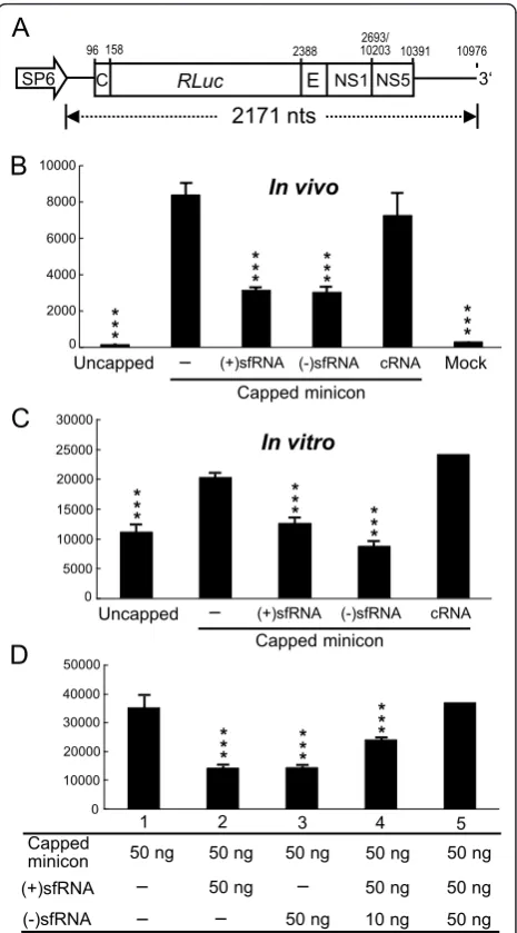
It has been shown that the 3’-UTR possesses many highly ordered secondary structures involved in viral translation most of which are also present in sfRNA. To test whether the presence of sfRNA affects viral transla-tion, a JEV minicon containingRenillaluciferase repor-ter gene constructed with authentic JEV 5’and 3’UTRs and part of the coding sequences (Figure 4A; described in Materials and Methods) was used.In vitrotranscribed minicon RNA was cotransfected with either (+)sfRNA, (-)sfRNA, or control RNA (cRNA) and effects on repor-ter translation were measured at 8 h posttransfection. Interestingly, transfecting either (+)sfRNA or (-)sfRNA but not control RNA inhibited luciferase translation dri-ven by JEV 5’UTR (Figure 4B). Consistent within vivo
translation assay, in vitro translation of luciferase was inhibited by the addition of (+)sfRNA as well as (-) sfRNA in rabbit reticulocyte lysate (Figure 4C). In order to confirm if the (-)sfRNA could counter the effects of (+)sfRNA, both sense and antisense sfRNA were added simultaneously into thein vitrotranslation mix to mea-sure their effect on minicon translation. As shown in Figure 4D, the inhibitory effect of (+)sfRNA on transla-tion was rescued by the additransla-tion of (-)sfRNA. When equimolar amounts of plus-sense and minus-sense sfRNA were added to the reaction, translation was restored to control (minicon only) levels.
Discussion
The discovery of sfRNA in flaviviruses has generated considerable interest in its generation, localization, and its possible function in flavivirus infected cells. In this study, we have shown that JEV sfRNA is localized in cytoplasm along with the genomic RNA (Figure 1A). Although the biogenesis of JEV sfRNA has not yet been studied, it has been reported that in WNV and YFV, sfRNA is a product of incomplete degradation of viral genome by cellular ribonuclease XRN1 and is
co-localized to the P-body in the cytoplasm [11]. Consistent with their report we found that JEV sfRNA is also loca-lized to the cytoplasm along with the genomic RNA. On the other hand, we also found that a few genomic RNA is also localized to the nucleus consistent with reports on the presence of flaviviral proteins and flaviviral RNA
NS5
96 2388 102032693/ 10391
C
Uncapped (+)sfRNA (-)sfRNA cRNA
Capped minicon
Uncapped (+)sfRNA (-)sfRNA cRNA Mock
10000
Figure 4Effect of sfRNA on viral translation in cultured cells andin vitro. A. A diagram of the JEV minicon RNA is shown. B. JEV minicon RNAs were transcribedin vitrowithout or with capped analog and then transfected alone (-) or cotransfected with (+) sfRNA, (-)sfRNA, or control RNA (cRNA). TheRenillaluciferase activity of cell lysates was determined at 8 h posttransfection. C.Renilla
luciferase assays of JEV minicon RNA translated in rabbit reticulocyte lysate in the presence of (+)sfRNA, (-)sfRNA or control RNA. D. The effect of different amounts of (+)sfRNA or (-)sfRNA onin vitro
translation of minicon RNA in rabbit reticulocyte lysate. TheRenilla
replication in the nuclear fraction [41-43]. The presence of abundant sfRNA in the cytoplasm of infected cells hints at possible roles of the sfRNA in cytoplasmic events during infection, namely viral RNA replication and translation.
The RNA synthesis of plus-strand RNA viruses is asymmetrical meaning that positive-sense genomic RNA strands are generated in excess over minus sense antige-nome and the ratio is about 10:1 to 100:1. Northern ana-lysis from a previous study showed that the ratio of plus-to-minus strands at 8 h postinfection was 3:1 which rapidly increased thereafter to 11.7:1 by 18 h postinfec-tion in porcine kidney cells infected with JEV at an MOI of 10 [7]. In this study, we describe the kinetics on the synthesis of JEV genome and antigenome in BHK-21 cells at an MOI of 0.01 using strand specific oligonucleo-tides for real-time RT-PCR. Our results indicated that the ratio of plus-to-minus strands during 24-48 h postin-fection was in the range of 29:1 to 244:1 during inspec-tion period (Figure 2D). Interestingly, we found that the time point at which the antigenome accumulation reaches a plateau coincides with the appearance of sfRNA as shown in our results from Northern and real-time RT-PCR analyses. It would be ideal to show the time course of antigenome and sfRNA accumulation together but since it was impossible to distinguish sfRNA accumulation from genomic RNA accumulation in real-time RT-PCR experiments, we employed Northern analy-sis which clearly distinguishes the two (Figure 2A). The time course of antigenome, on the other hand, real-time RT-PCR is much more sensitive than Northern analysis especially during the early time points (Figure 2C). Thus, we compare the same amounts of RNA under the same condition by these two different methods.
The artificial addition of (-)sfRNA (by transfection) at 28 h postinfection countered the effects of naturally occurring sfRNA thereby increasing the accumulation of antigenome (Figure 3D and 3F) indicating that sfRNA could negatively interfere with antigenome synthesis. The addition of (-)sfRNA may not only anneal to the naturally occurring plus-sense sfRNA but also to the 3’-UTR of the genomic RNA. However, the probability of the minus-sense sfRNA to bind to the plus-minus-sense sfRNA is more than its binding to the genomic 3’-UTR because of (i) the binding strength of same size shorter complementary RNA should be greater than the binding of a short RNA sequence to a large RNA polynucleotide akin to a highly complementary primer dimer and a PCR template. This was observed in ourin vitroluciferase assays (Figure 4) where the individual addition of either only (+)sfRNA or (-)sfRNA reduced translation but the addition of both (+) sfRNA (50 ng) and (-)sfRNA (10 ng) into the reaction partially restored translation and the addition of more (-) sfRNA (50 ng) restored translation to control levels
probably because the plus-sense and the minus-sense sfRNA hybridized to form duplex RNA thereby prevent-ing sfRNA from interferprevent-ing with translation; (ii) cycliza-tion of the genome could render the 3’-UTR inaccessible to the minus-sense sfRNA. Curiously, when (-)sfRNA is transfected, the amount of naturally occurring plus-sense sfRNA does not decrease (Figure 3A and 3C) and is simi-lar to that of mock, indicating that naturally occurring plus-sense sfRNA is either not degraded or that the rate of sfRNA generation (RNA turnover) is very high.
The presences ofcis-acting sequences including promo-ters, enhancers, and repressors that aid in the in regulation of minus-strand synthesis have been reported in many plus-strand RNA viruses [44-47] and these elements may conse-quently contribute to asymmetrical RNA synthesis. Viral or host proteins may also contribute to asymmetric replication
in trans[48-50]. Several RNA motifs within 5’and 3’-UTR, for instance, 5’-CS/CS1, 5’-UAR/3’-UAR, and 5’DAR/ 3’DAR are involved in RNA interactions. These RNA-RNA interactions have been demonstrated to be required for viral replication. In DENV, a stemloop A (SLA) has been identified at 5’end of the genome which was shown to be required for long-range RNA-RNA interaction and the recruitment of virus RdRp which is then transferred to the initiation site present in the 3’-UTR in order to promote minus-strand RNA synthesis [51]. In addition, the balance between circular and linear forms of the DENV genome is crucial for viral replication [52]. Thus, the presumable mechanism of the suppression of antigenome synthesis by JEV sfRNA may be due to the interruption of genome cycli-zation by its complementarity to the 5’-end elements of JEV genome (Figure 5A). Since the sfRNA is in high molar excess it could also be assumed that free sfRNA (sfRNA not bound to the 5’of JEV genome) could further reduce anti-genome synthesis through a second mechanism by compet-ing for viral and host proteins that would otherwise bind to the 3’UTR and promote antigenome synthesis (Figure 5B). We hypothesis that sfRNA through itstrans-acting function could be one of the factors contributing to the asymmetry in JEV RNA replication.
translation in trans, as depicted in Figure 5B, proteins binding to 3’-UTR elements are essential to promote viral translation. Transfection of (+)sfRNA sequesters proteins binding to the 3’-UTR of genome and reduces translation, while if (-)sfRNA is transfected it could bind to the 3’-UTR of the genome and prevent the host pro-teins from binding the 3’UTR (competes with host fac-tors binding to the 3’UTR). In addition, transfection of (-)sfRNA could also prevent the interaction of the 3’ and 5’regions of the genome and that too could reduce translation. Thus, transfecting of either (+) or (-)sfRNA reduces translation. Furthermore, the sfRNA could titrate the host factors and even the newly synthesized viral proteins like RdRp thereby drastically reducing minus-strand RNA synthesis that results in the afore-mentioned asymmetry in RNA accumulation.
Conclusions
As seen in our results (Figures 2A, 3A and 3C), sfRNA is present in great abundance in the late stages of the viral replication cycle and that sfRNA interferes and impairs both antigenome synthesis and JEV translation (Figures 3 and 4). It could be thought that the JEV
genomic RNA produced during the late stages of the viral replication cycle is bound for packaging and should not be used as templates for antigenome synthesis or for translation and the presence of sfRNA is suspected to compete against the translation and antigenome synthesis. From our data, we conclude that sfRNA could be the switch (a trans-acting riboswitch) that shuts down both antigenome synthesis and JEV translation thereby promoting only genomic RNA synthesis that needs to be packaged and released for the next infec-tious cycle.
Materials and methods Cells and viruses
Baby hamster kidney (BHK-21) cells were grown in RPMI 1640 medium supplemented with 2% fetal bovine serum (FBS) (Gibco-BRL) at 37°C. JEV strain RP9, a var-iant of NT109 isolated originally fromCulex tritaenior-hynchuswas used in this study [53].
Construction of plasmids
pGEMT/JEV3642-3821 plasmid (Figure 2B) used for making RNA standard in real-time RT-PCR was
Genome cyclization aids in
antigenome synthesis. Genome cyclization disrupted and antigenome synthesis reduced. sfRNA
Proteins binding to 3’-UTR elements
promotes translation/replication. Proteins sequestered by sfRNA and reduced translation/replication.
NS3 La
NS3 La NS3 La
NS3 La
3’ 3’
3’
3’
A
B
Mov34
NS5 NS5 Mov34
Mov34 NS5
Mov34 NS5
Figure 5Model depicting the role of sfRNA in flaviviral RNA replication and translation. A. The diagrammatic representation of flaviviral cyclization, through the cyclization elements, which promote antigenome synthesis. The sfRNA disrupts genome cyclization by interacting with the 5’cyclization elements and thereby reduces antigenome synthesis. B. Flaviviral genome contains protein-binding elements in the 3’-UTR. Viral and host proteins bind to their respective RNA elements in the 3’-UTR and promote translation and replication. The presence of sfRNA
generated by cloning the 180-nt PCR product amplified from JEV cDNA with the JEV3642(+) (nt 3642-3662) and JEV3821(-) (nt 3802-3821) primers into pGEMT-easy vector (Promega). To generate pGEMT/JEV10450-10976 construct, we followed the same method as for pGEMT/JEV3642-3821 plasmid, except that JEV10450(-) (nt 10450-10476) and JEV10950(+) (nt 10950-10976) primers were used for PCR. PCR products were ampli-fied from sfRNA with primers containing T7 promoter at 5’end and a unique restriction site at the 3’end, then cloned into pUC18 plasmid to generate pUC18/JEV(+) 10450-10976, and pUC18/JEV(-)10976-10454 for making (+)sfRNA and (-)sfRNA respectively. Sequences of each construct were confirmed by sequencing.
RNA preparation and Northern blot analysis
RNA extraction and Northern analyses were done as described previously [8]. Briefly, total RNA was extracted with Trizol (Invitrogen) or REzol™ C&T reagent (Protech). Cytoplasm/Nucleus fractionation was done using Cytoplasmic & Nuclear RNA Purification kit (Norgen) according to manufacturer’s instruction. Approximate 2.5 μg or 7-10 μg of cytoplasmic RNA were used per lane in formaldehyde-agarose gel electro-phoresis for the detection of plus- or minus-strand, respectively. To label oligonucleotide probe, approxi-mately 100-pmol of oligonucleotide was 3’tailed with Digoxigenin (DIG)-ddUTP using a DIG Oligonucleotide 3’-End Labeling Kit (Roche Molecular Biochemicals). For labeling sfRNA probe, 1μg of linearized DNA was used forin vitro transcription with DIG RNA labeling mix (Roche Molecular Biochemicals). Hybridization was done at 54°C for oligonucleotide probes and 68°C for riboprobes. DIG luminescent detection of the viral spe-cific bands was done according to the manufacturer’s instructions (Roche Molecular Biochemicals).
Synthetic oligonucleotides and accession number
JEV genome nucleotide positions correspond to those for JEV RP9, GenBank accession number AF014161. 3JEV10950(-) oligonucleotide (5’-AGATCCTGTGTT CTTCCTCACCACCAG-3’) detects RNA containing the very 3’-terminal 27 nts of the JEV genome. 18S rRNA(-) oligonucleotide (5’ -GCACTTACTGGGAATTCCTCG-3’) location corresponds to mouse 18S rRNA, GenBank accession number X00686. Mitochondria 12S rRNA(-) oligonucleotide (5’-AAGGCCAGGACCAACCT-3’) was synthesized according to GenBank accession number NC_005089.
Real-time RT-PCR
The method used for real-time RT-PCR assay was as described previously [54]. Thein vitrotranscripts of posi-tive-sense RNA were generated from T7 transcription
(Promega) of theSalI-linearized pGEMT/JEV3642-3821 and the minus-sense transcripts were transcribed from SP6 promoter of theNcoI-linearized pGEMT/JEV3642-3821 (as diagramed in Figure 2B). The amount of purified RNA was measured by spectrophotometry and the copy number was calculated based on the concentration mea-sured and its molecular weight. The known amounts of RNAs were serially diluted 10-fold (1.78 × 10¹¹to 1.78 × 10⁵copies) and subjected to real-time RT-PCR using the one-step RT-PCR master mix reagent kit following the manufacturer’s instructions (Applied Biosystems). Oligo-nucleotides JEV3642(+) and JEV3821(-) were used as pri-mers for binding to minus and plus-strand RNA, respectively, during RT step carried out at 48°C for 30 min. The PCR amplification conditions were 95°C for 10 min, followed by 40 cycles of 95°C for 15 sec and 60°C for 1 min with primers JEV3650(+), JEV3726(-) and Taq-Man probe JEV3705(-) (sequences and binding positions are illustrated in Figure 2B). The assay was performed on an ABI 7000 Sequence Detector using TaqMan One-Step RT-PCR master mix to analyze the emitted fluorescence during amplification (Applied Biosystems). A linear equa-tion of known amounts of RNA to CT value was determined.
The intracellular plus- or minus-strand RNAs in JEV-infected cells were determined by using strand specific primers during RT step as described above. Cytoplasmic RNAs were extracted at the indicated time points post-infection. RNA from each time point was diluted to a concentration of 100 ng/μl and 10 ng/μl, respectively, and subjected to real-time RT-PCR together with the known amount of in vitrotranscripts. The amount of intracellular genome or antigenome per cell was deter-mined by dividing the copy number by the numbers of cells counted at each time point postinfection.
RNA transfection
Luciferase assay
JEV minicon (kindly provided by Dr. Yi-Ling Lin) con-tains a Renilla luciferase (Rluc) fused in-frame to the JEV coding regions as a single ORF in the following order; the core (nt 96-158), Rluc (933 nts), E (nt 2388-2477), NS1 (nt 2478-2693), and NS5 (nt 10203-10391), and this ORF unit was flanked by the authentic JEV 5’ -and 3’-UTRs (Figure 4A). The Rluc-reporter plasmid was transcribed using a Megascript SP6 Transcription kit (Ambion) according to the manufacturer’s instruc-tions, in the presence or absence of m7GpppA nucleo-tide (New England Biolabs). Luciferase assays were performed using extracts from transfected cells and also from thein vitrotranslation assay. Forin vivo transla-tion in cultured cells, 1μg of transcribed minicon RNA, together with 1 μg of plus- or minus-strand of sfRNA were transfected into BHK-21 cells using Lipofectamin 2000 (Invitrogen). Renillaluciferase activity was mea-sured at 8 hour posttransfection. Forin vitrotranslation, 50 ng of RNAs were translated in nuclease-treated rab-bit reticulocyte lysate (Promega) in the presence of 40 units of RNasin Ribonuclease Inhibitor (Promega), 20
μM of each amino acid, and either plus-strand, minus-strand of sfRNA, or a 445-nt control RNA composed of JEV sequences (nt 2401-2689) plus 156 nts derived from vector sequences. The reactions were incubated at 30°C for 30 min and 2.5μl of reaction sample was measured with 20/20nSingle-Tube Luminometer (Promega).
Statistical analysis
For statistical analysis, one-way ANOVA Dunnet’s mul-tiple comparisons test was used to compare the control group against others (GraphPad Software, San Diego, CA, USA).
List of abbreviations
CS: conserved sequences; DAR: downstream of the AUG; DENV: dengue virus; JEV: Japanese encephalitis virus; MOI: multiplicity of infection; sfRNA: small flaviviral RNA; SLA: stemloop A; UAR: upstream of the AUG; UTR: untranslated region; WNV: West Nile virus.
Acknowledgements
This work was supported by grant NSC 98-2320-B-259-002-MY3 from National Science Council, Taipei, Taiwan, Republic of China. We thank Dr. David Brian for many helpful discussions and Dr. Yu-Pin Su for constructive comments on this manuscript.
Authors’contributions
YHF carried out the translation experiments and statistical analyses. MN designed one of the translation experiments, the model, and participated in drafting of the manuscript. CCC and CCW carried out the replication studies. YTL carried out the nuclear and cytoplasmic fractionation and Northern blot analysis. RYC conceived of the study, participated in its design and coordination, and finalized the manuscript in its final form. All authors read and approved the final manuscript.
Competing interests
The authors declare that they have no competing interests.
Received: 27 August 2011 Accepted: 1 November 2011 Published: 1 November 2011
References
1. Endy TP, Nisalak A:Japanese encephalitis virus: ecology and epidemiology.Curr Top Microbiol Immunol2002,267:11-48.
2. Erlanger TE, Weiss S, Keiser J, Utzinger J, Wiedenmayer K:Past, present, and future of Japanese encephalitis.Emerg Infect Dis2009,15:1-7.
3. Lindenbach BD, Thiel H-J, Rice CM:Flaviviridae: The Viruses and Their Replication.InFields Virology. Volume 1..5 edition. Edited by: Knipe DM, Howley PM. Philadephia, PA.: Lippincott William 2007:1101-1152. 4. Chu PW, Westaway EG:Replication strategy of Kunjin virus: evidence for
recycling role of replicative form RNA as template in semiconservative and asymmetric replication.Virology1985,140:68-79.
5. Cleaves GR, Ryan TE, Schlesinger RW:Identification and characterization of type 2 dengue virus replicative intermediate and replicative form RNAs.
Virology1981,111:73-83.
6. Takegami T, Hotta S:Synthesis and localization of Japanese encephalitis virus RNAs in the infected cells.Microbiol Immunol1990,34:849-857. 7. Uchil PD, Satchidanandam V:Characterization of RNA synthesis,
replication mechanism, and in vitro RNA-dependent RNA polymerase activity of Japanese encephalitis virus.Virology2003,307:358-371. 8. Lin KC, Chang HL, Chang RY:Accumulation of a 3’-terminal genome
fragment in Japanese encephalitis virus-infected mammalian and mosquito cells.J Virol2004,78:5133-5138.
9. Funk A, Truong K, Nagasaki T, Torres S, Floden N, Balmori Melian E, Edmonds J, Dong H, Shi PY, Khromykh AA:RNA structures required for production of subgenomic flavivirus RNA.J Virol2010,84:11407-11417. 10. Silva PA, Pereira CF, Dalebout TJ, Spaan WJ, Bredenbeek PJ:An RNA
pseudoknot is required for production of yellow fever virus subgenomic RNA by the host nuclease XRN1.J Virol2010,84:11395-11406.
11. Pijlman GP, Funk A, Kondratieva N, Leung J, Torres S, van der Aa L, Liu WJ, Palmenberg AC, Shi PY, Hall RA, Khromykh AA:A highly structured, nuclease-resistant, noncoding RNA produced by flaviviruses is required for pathogenicity.Cell Host Microbe2008,4:579-591.
12. Alvarez DE, De Lella Ezcurra AL, Fucito S, Gamarnik AV:Role of RNA structures present at the 3’UTR of dengue virus on translation, RNA synthesis, and viral replication.Virology2005,339:200-212.
13. Holden KL, Harris E:Enhancement of dengue virus translation: role of the 3’untranslated region and the terminal 3’stem-loop domain.Virology
2004,329:119-133.
14. Yoo JS, Kim CM, Kim JH, Kim JY, Oh JW:Inhibition of Japanese encephalitis virus replication by peptide nucleic acids targeting cis-acting elements on the plus- and minus-strands of viral RNA.Antiviral Res2009,82:122-133.
15. Polacek C, Friebe P, Harris E:Poly(A)-binding protein binds to the non-polyadenylated 3’untranslated region of dengue virus and modulates translation efficiency.J Gen Virol2009,90:687-692.
16. Tilgner M, Shi PY:Structure and function of the 3’terminal six nucleotides of the West Nile virus genome in viral replication.J Virol
2004,78:8159-8171.
17. Khromykh AA, Westaway EG:RNA binding properties of core protein of the flavivirus Kunjin.Arch Virol1996,141:685-699.
18. Hahn CS, Hahn YS, Rice CM, Lee E, Dalgarno L, Strauss EG, Strauss JH: Conserved elements in the 3’untranslated region of flavivirus RNAs and potential cyclization sequences.J Mol Biol1987,198:33-41.
19. Alvarez DE, Filomatori CV, Gamarnik AV:Functional analysis of dengue virus cyclization sequences located at the 5’and 3’UTRs.Virology2008, 375:223-235.
20. Friebe P, Harris E:Interplay of RNA elements in the dengue virus 5’and 3’ends required for viral RNA replication.J Virol2010,84:6103-6118. 21. Suzuki R, Fayzulin R, Frolov I, Mason PW:Identification of mutated
cyclization sequences that permit efficient replication of West Nile virus genomes: use in safer propagation of a novel vaccine candidate.J Virol
2008,82:6942-6951.
22. Villordo SM, Gamarnik AV:Genome cyclization as strategy for flavivirus RNA replication.Virus Res2009,139:230-239.
24. Friebe P, Harris E:Interplay of RNA elements in the dengue virus 5’and 3’ends required for viral RNA replication.J Virol2010,84:6103-6118. 25. Yun SI, Choi YJ, Song BH, Lee YM:3’cis-acting elements that contribute
to the competence and efficiency of Japanese encephalitis virus genome replication: functional importance of sequence duplications, deletions, and substitutions.J Virol2009,83:7909-7930.
26. Blackwell JL, Brinton MA:BHK cell proteins that bind to the 3’stem-loop structure of the West Nile virus genome RNA.J Virol1995,69:5650-5658. 27. Blackwell JL, Brinton MA:Translation elongation factor-1 alpha interacts
with the 3’stem-loop region of West Nile virus genomic RNA.J Virol
1997,71:6433-6444.
28. Davis WG, Blackwell JL, Shi PY, Brinton MA:Interaction between the cellular protein eEF1A and the 3’-terminal stem-loop of West Nile virus genomic RNA facilitates viral minus-strand RNA synthesis.J Virol2007, 81:10172-10187.
29. Emara MM, Liu H, Davis WG, Brinton MA:Mutation of mapped TIA-1/TIAR binding sites in the 3’terminal stem-loop of West Nile virus minus-strand RNA in an infectious clone negatively affects genomic RNA amplification.J Virol2008,82:10657-10670.
30. Yocupicio-Monroy M, Padmanabhan R, Medina F, del Angel RM:Mosquito La protein binds to the 3’untranslated region of the positive and negative polarity dengue virus RNAs and relocates to the cytoplasm of infected cells.Virology2007,357:29-40.
31. De Nova-Ocampo M, Villegas-Sepulveda N, del Angel RM:Translation elongation factor-1alpha, La, and PTB interact with the 3’untranslated region of dengue 4 virus RNA.Virology2002,295:337-347.
32. Paranjape SM, Harris E:Y box-binding protein-1 binds to the dengue virus 3’-untranslated region and mediates antiviral effects.J Biol Chem
2007,282:30497-30508.
33. Wei Y, Qin C, Jiang T, Li X, Zhao H, Liu Z, Deng Y, Liu R, Chen S, Yu M, Qin E:Translational regulation by the 3’untranslated region of the dengue type 2 virus genome.Am J Trop Med Hyg2009,81:817-824. 34. Rakotondrafara AM, Polacek C, Harris E, Miller WA:Oscillating kissing
stem-loop interactions mediate 5’scanning-dependent translation by a viral 3’-cap-independent translation element.RNA2006,12:1893-1906. 35. Chiu WW, Kinney RM, Dreher TW:Control of translation by the 5’- and 3’
-terminal regions of the dengue virus genome.J Virol2005,79:8303-8315. 36. Gomila RC, Martin GW, Gehrke L:NF90 binds the dengue virus RNA 3’
terminus and is a positive regulator of dengue virus replication.PLoS One2011,6:e16687.
37. Chen CJ, Kuo MD, Chien LJ, Hsu SL, Wang YM, Lin JH:RNA-protein interactions: involvement of NS3, NS5, and 3’noncoding regions of Japanese encephalitis virus genomic RNA.J Virol1997,71:3466-3473. 38. Ta M, Vrati S:Mov34 protein from mouse brain interacts with the 3’
noncoding region of Japanese encephalitis virus.J Virol2000, 74:5108-5115.
39. Vashist S, Anantpadma M, Sharma H, Vrati S:La protein binds the predicted loop structures in the 3’non-coding region of Japanese encephalitis virus genome: role in virus replication.J Gen Virol2009, 90:1343-1352.
40. Yang SH, Liu ML, Tien CF, Chou SJ, Chang RY: Glyceraldehyde-3-phosphate dehydrogenase (GAPDH) interaction with 3’ends of Japanese encephalitis virus RNA and colocalization with the viral NS5 protein.J Biomed Sci2009,16:40.
41. Uchil PD, Kumar AV, Satchidanandam V:Nuclear localization of flavivirus RNA synthesis in infected cells.J Virol2006,80:5451-5464.
42. Takegami T, Hotta S:In vitro synthesis of Japanese encephalitis virus (JEV) RNA: membrane and nuclear fractions of JEV-infected cells possess high levels of virus-specific RNA polymerase activity.Virus Res1989, 13:337-350.
43. Mori Y, Okabayashi T, Yamashita T, Zhao Z, Wakita T, Yasui K, Hasebe F, Tadano M, Konishi E, Moriishi K, Matsuura Y:Nuclear localization of Japanese encephalitis virus core protein enhances viral replication.J Virol2005,79:3448-3458.
44. Zhang G, Zhang J, Simon AE:Repression and derepression of minus-strand synthesis in a plus-minus-strand RNA virus replicon.J Virol2004, 78:7619-7633.
45. Pogany J, Fabian MR, White KA, Nagy PD:A replication silencer element in a plus-strand RNA virus.Embo J2003,22:5602-5611.
46. Sivakumaran K, Kim CH, Tayon R, Kao CC:RNA sequence and secondary structural determinants in a minimal viral promoter that directs
replicase recognition and initiation of genomic plus-strand RNA synthesis.J Mol Biol1999,294:667-682.
47. Frolov I, Hardy R, Rice CM:Cis-acting RNA elements at the 5’end of Sindbis virus genome RNA regulate minus- and plus-strand RNA synthesis.Rna2001,7:1638-1651.
48. Satyanarayana T, Gowda S, Ayllon MA, Albiach-Marti MR, Rabindran S, Dawson WO:The p23 protein of citrus tristeza virus controls asymmetrical RNA accumulation.J Virol2002,76:473-483.
49. Wang RY, Nagy PD:Tomato bushy stunt virus co-opts the RNA-binding function of a host metabolic enzyme for viral genomic RNA synthesis.
Cell Host Microbe2008,3:178-187.
50. Wang X, Ahlquist P:Filling a GAP(DH) in asymmetric viral RNA synthesis.
Cell Host Microbe2008,3:124-125.
51. Filomatori CV, Lodeiro MF, Alvarez DE, Samsa MM, Pietrasanta L, Gamarnik AV:A 5’RNA element promotes dengue virus RNA synthesis on a circular genome.Genes Dev2006,20:2238-2249.
52. Villordo SM, Alvarez DE, Gamarnik AV:A balance between circular and linear forms of the dengue virus genome is crucial for viral replication.
Rna2010,16:2325-2335.
53. Chen LK, Lin YL, Liao CL, Lin CG, Huang YL, Yeh CT, Lai SC, Jan JT, Chin C: Generation and characterization of organ-tropism mutants of Japanese encephalitis virus in vivo and in vitro.Virology1996,223:79-88. 54. Wang WK, Sung TL, Tsai YC, Kao CL, Chang SM, King CC:Detection of
dengue virus replication in peripheral blood mononuclear cells from dengue virus type 2-infected patients by a reverse transcription-real-time PCR assay.J Clin Microbiol2002,40:4472-4478.
doi:10.1186/1743-422X-8-492
Cite this article as:Fanet al.:Small Noncoding RNA Modulates
Japanese Encephalitis Virus Replication and Translation intrans.Virology Journal20118:492.
Submit your next manuscript to BioMed Central and take full advantage of:
• Convenient online submission
• Thorough peer review
• No space constraints or color figure charges
• Immediate publication on acceptance
• Inclusion in PubMed, CAS, Scopus and Google Scholar
• Research which is freely available for redistribution