Copyright 0 1997 by the Genetics Society of America
A Salmonella Phage-P22 Mutant Defective
in
Abortive Transduction
Nicholas R. Benson
and
JohnRoth
Department of Biology, University of Utah, Salt Lake City, Utah 84112 Manuscript received August 8, 1995
Accepted for publication September 26, 1996
ABSTRACT
In the course of a lytic infection the Salmonella phage P22 occasionally encapsulates bacterial DNA instead of phage DNA. Thus, phage lysates include two classes of viral particles. Phage particles carrying bacterial DNA are referred to as transducing particles and deliver this DNA to a host as efficiently as particles carrying phage DNA. Once injected, the transduced DNA can either recombine with the recipient chromosome to form a “complete” transductant, or it can establish itself as an expressible, nonreplicating genetic element and form an “abortive” transductant. In this work, we describe a P22- phage mutant with reduced ability to form abortive transductants. The mutation responsible for this phenotype, called tdx-1, was found as one of two mutations contributing to the high-transducing pheno- type of the P22-mutant HT12/4. In addition, the tdx-l mutation is lethal when combined with an g a m mutation. The tdx-1 mutation has been mapped to a region of the P22 genome that encodes several injected proteins and may involve more than one mutant locus. The phenotypes of the tdx-1 mutation suggest that the Tdx protein(s) normally assist in the circularization of the P22 genome and also contribute to the formation of DNA circles thought to be required for abortive transduction.
A
S a generalized transducing phage, the Salmonella phage P22 occasionally packages a fragment of the bacterial genome into a P22 capsid and can deliver that DNA to a recipient cell (LEDERBERG et al. 1951; CASJENS and HAYDEN 1988). Two well characterized out- comes of the transduction event are known as “com- plete” and “abortive” transduction (Figure 1). Com- plete transduction refers to the substitution of donor DNA for homologous host sequences by the host recom- bination system (EBEL-TSIPIS et al. 1972a,b). During the course of recombination, part of the transduced DNA fragment is degraded; the surviving DNA is incorpo- rated into the recipient chromosome as a double- stranded molecule. This process of homologous recom- bination results in stable inheritance of the transduced sequences; the acquired sequences are replicated and transmitted to both daughter cells at division.Abortive transduction is a form of unstable genetic inheritance whereby the transduced DNA evades degra- dation and recombination. Abortively transduced DNA can be expressed but remains extra-chromosomal, im- mune to the host recombination apparatus and is not replicated. Because abortively transduced DNA is not replicated, it is inherited by only one of the two daugh- ter cells at division (STOCKER et al. 1953; LEDERBERG 1956; STOCKER 1956; EBEL-TSIPIS et al. 1972a). Abortive transduction occurs
-
10-fold more frequently than complete transduction. However, the ratio of abortive to complete transductants is observed to vary widely depending on the selected marker and the genotype of the transducing phage (SCHMIEGER 1972).Corresponding author; Nicholas Benson, Sidney Kimmel Cancer Cen- ter, 3099 Science Park Dr., Suite 200, San Diego, CA 92121. E-mail: nickphage@aol.com
Genetics 145 17-27 (January, 1997)
Abortive transduction was initially described for the inheritance of flagellar characteristics and was later shown to apply to many other characteristics (OZEKI 1956; HARTMAN et al. 1960). Although generalized trans- duction was first described for the S. typhimurium-P22 system, many other transducing phage systems are known; in particular the P1 transducing phage of Esche- richia coli shares many features with the S. typhimurium- P22 transducing phage ( OZEKI and IKEDA 1968).
The phenomenon of P22-generalized transduction can be best understood in the context of general P22 biology (reviewed by SUSSKIND and BOTSTEIN 1978; PO- TEETE 1988). The P22 capsid contains a linear, double- stranded 43.4kb DNA molecule (CASJENS and HAYDEN 1988). The capsid includes the products of genes 1, 4,
5, 7,
9, 10, 16, 20and 26. The majority of these proteins are essential for the assembly, structural integrity or stability of the mature phage particle. However, the products of genes7,
16and 20 are not essential for any of these processes; rather, these proteins are believed to be essential for the injection and/or protection of injected DNA and are probably coinjected with the DNA into the recipient host cell (HOFFMAN and LEVINE 1975a,b; ISRAEL 1977).18 N. R. Benson and J. Roth
&
!
=
COMPLETE
‘
I
TRANSDUCTION ABORTIVE TRANSDUCTION[Recombination] [No Recombination]
lx
J
0
FIGURE 1.-The fate of P22-derived transducing DNA. Once transducing DNA has entered the cell it can undergo degradation and/or recombination with homologous recipi- ent sequences (complete transduction, shown on the left). More frequently the DNA is abortively transduced (shown on the right). Details are discussed in the text.
ized, the phage will be unable to replicate its DNA and thus will not propagate. The circularization reaction is most efficiently catalyzed by the P22-encoded Erf pro- tein, although a slightly less efficient Erf-independent pathway exists that depends on the host RecA protein
( BOTSTEIN and MATZ 1975).
Once circularized, P22 DNA begins bidirectional theta replication and then switches to rolling-circle rep- lication. Rolling-circle replication creates long concate- mers of P22 genomes that are the substrates for packag- ing. A complex of P22 gene products 2 and 3 recognizes a site within gene 3 called “pac” and cleaves near pac to begin a processive packaging “series”.
The packaging process also results in the formation of generalized transducing particles. The most plausible mechanism for this is the hypothesis that the gp2/3 complex occasionally recognizes a “pseudo” pac site in the bacterial chromosome and initiates a packaging series near that site. Evidence in support of this hypoth- esis is that (1) deletions in the host chromosome can affect the packaging of nearby host DNA (CHELALA and MARCOLIN 1974) and (2) phage mutations that increase transducing ability alter gp3 and change the DNA-se- quence specificity of packaging.
While the origin of transducing particles can be viewed as the result of phage packaging functions gone
astray,” it is unclear whether the fate of transduced DNA (complete or abortive transduction) is influenced by phage functions. Host functions clearly affect the fate of transduced DNA, since the RecA and RecBCD
“
TABLE 1
BacteriophageP22 strains used in this study
Phage Genotype Source
MS57 Wild type M. SUSSKIND
MSl39 5-am(Nll4); h21 M. SUSSKIND
MS525 cl-7 M. SUSSKIND
MS607 HT12/4; h21; int-3 M. SUSSKIND
MS1050 sieA44; ant-am; R222AP68 M. SUSSKIND MS1212 20-am(H1030); cl-7; h21 M. SUSSKIND MS1379 lGam(H1215); cl-7; h21 M. SUSSKIND NBP20 erfam(H1173); cl-7; h21 T. POTEETE
NBP43 5-am(NI 14) This work
NBP5 1 HTl2/4A This work
NBP54 CI-7; 2@am(H1030) This work
NBP63 cl-7; 5-am(N114) This work
NBP95 cl-7; tdx-l This work
NBP105 erfam(H1173) This work
NBPl20 c2-5 This work
NBP148 cl-7; 7-am(H1205) This work
NBP149 cl-7; lGam(H1215) This work
NBP179 cl-7; tdx-I; erfam(H1173) This work NBP181 e$am(HlI73); tdx-1 This work
NBP183 CI-7; e-am(H1173) This work
NBP187 cl-7; erf”am(Hl173); 2@am(H1030) This work NBP188 CI-7; erfam(Hl173); 16-am(H1215) This work NBP40 tdx-1 This work
NBP52 tdx-1; HT12/4A This work
NBP80 cI-7; HT12/4A This work
UC754 cl-7; HT12/4A S. MJENS
NPB strains are from the collection of NICK BENSON; MS from MIRIAM SUSSKIND; UC from SHERWOOD CASJENS; DB from DAVID BOTSTEIN.
proteins dominate transductional recombination in the wild-type host (EBEL-TSIPIS et al. 1975a,b; MIESEL AND
ROTH 1994).
Abortively transduced fragments are immune to the host recombination apparatus; it is unknown whether this immunity is provided by phage or host functions. In this work we describe the genetic analysis of a phage mutation, tdx-1, that eliminates the formation of abor- tive transductants and implicates injected phage pro- teins in the formation of abortive transductants. The
tdx-1 mutation is also implicated in the circularization
of the phage genome by the hostdependent RecA path- way of recombination.
MATERIALS AND METHODS
Bacteria, phage, and general genetic methods: All phage strains are listed in Table 1. Unless specifically stated, all bacte- rial strains are derivatives of strain DB7000 (derived from
LT2; Table 2). General phage and bacterial techniques are described by DAVIS et al. (1980). Routine transductions were performed with phage MS2104, kindly provided by M. Sus SKIND, as described by BENSON and GOLDMAN (1992). Many of the phage used in this work contained the plaque-morphology markers h21 and m44 when received. These markers were removed from phages described here using standard genetic techniques.
19 P22 Abortive Transduction Mutant
TABLE 2
Strains of S. typhimurium used in this study
Strain Genotype Source
DB7000 felsZ; hA414(am) M. SUSSKIND
MS1363 felsZ; hA414(am); supE(g1n) M. SUSSKIND
TT17615 felsZ; led414(am);fliD5055::TnlOflet BENSON and ROTH (1994)
TT18639 felsZ; hA414(am); supEfgn); cob236::TnlOnet This work
IT18640 &&Z; leuA414(am); recA1; supE(g1n) This work
TT18641 fels2-; leuA414(am); cob-236::TnlOuTet This work
TT 18642 felsZ; leuA414(am); recAl This work
TT18643 felsZ; led414(am); P22(sieA44; ant-am; R222AP68) This work
'IT18644 felsZ; led414(am); CRR299; P22(sieA44; ant-am; R222AP68) This work 'IT18645 fels2; led414(am);~iD5055::Tn10flet; P22(sieA44; ant-am; R222AP68) This work
IT18646 fels2-; led414(am); supE; P22[ wild-type] This work
TT18647 felsZ; led414(am); supE; P22[@-am(Hll73) tdx-l)]; This work
TT18648 felsZ; leuA414(am); supE; P22[tdx-l] This work
TT18649 fels2-; leuA414(am); P22[tdx-l] This work
TT18650 fels2; hA414(am); P22[ wild-type] This work
TT18651 felsZ; ZeuA414(am); P22[@-am(H1173) tdx-I] This work
TT18652 felsZ; l e d 4 1 4( am) ; supE; P22[ @-am(HIl73)] This work
TT18653 fels2; leuA414(am); P22[@-am(Hl173)] This work
DB strains are from the laboratory of DAVID BOTSTEIN; MS strains are from MIRIAM SUSSKIND;
IT
strains are fromJoHN ROTH.dium and E medium as minimal medium; LB, lambda and green indicator plates are described by DAVIS et al. (1980). Bochner plates, used for the isolation of tetracycline-sensitive colonies, are described by BOCHNER et al. (1980) and made as described in BENSON and GOLDMAN (1992). Concentra- tions of drugs used in LB plates are as follows: kanamycin (mo- nosulfate), 50 pg/ml; chloramphenicol, 40 pg/ml; ampicillin (Na salt), 50 pg/ml; tetracycline(HCl), 10 pg/ml. For mini- mal media these concentrations were reduced twofold with the exception of kanamycin, which remained the same. Motil-
ity plates are LB plates with the concentration of agar reduced fivefold. Transduction broth (TB) is LB broth supplemented with 1X E-salts and 0.2% glucose.
Phage lysates: A fresh overnight culture of the host bacte- rial strain was diluted 10-fold into transduction broth (TB) and a single plaque of phage was added. The culture was shaken overnight at 25" or 30". This overnight culture was shaken with 1-3 ml of chloroform for several minutes and the chloroform allowed to settle. The TB medium was de- canted from the chloroform and the cell debris pelleted by low speed centrifugation. The TB was vortexed with 0.4 ml of chloroform. At this stage P22-tail protein was added (if required) and the lysate incubated at 37" for several hours or at room temperature overnight. Lysates were stored at 4".
Source and isolation of the tdx-1 phage: The original source of the tdx-1 mutation is the original mutant phage, HT12/4, isolated by SCHMIEGER (1972). Our immediate source of phage HT12/4 and therefore the tdx-1 allele is phage MS607. Phage MS607 was provided by MIRIAM SUSSKIND as an int-3 h21 derivative of the HT12/4 mutant. The int-3 and h21 muta- tions were removed from phage MS607 by standard genetic crosses. Phage carrying the int' allele were identified by screening their ability to lysogenize wild-type salmonella. The replacement of the h21 allele by the wild-type allele was deter- mined by the change in plaque morphology on green plates; h21 mutants have a characteristic pale or yellowish halo that contrasts with the wild-type blue halo in green agar. These crosses yielded three phage types: (1) phages with the high- transducing phenotype of phage HT12/4 (NBP52), (2) phages that were unable to specify abortive transductants (NBP40) and (3) phages that specified complete and abortive transduc-
tants at a frequency higher than wild type (NBP51). These observations suggested to us that the original HT12/4 g e n e type might be a composite of multiple mutations.
Construction of etfam trlol mutant phages: Doubly mutant erfam tdx-1 phages with and without the cl-7 mutation (NBP179 and NBP181) were obtained from a cross of phages NPB95 (cl-7 tdx-1) and NBP105 (@-am). Clear and turbid plaque-forming phages that failed to plaque on a recAl host but could plaque on a recA1 supE44 host were classified as @- am. Some of these cI-7 erfam phages were defective for abor- tive transduction; these are assumed to carry the tdx-1 muta- tion, one such phage was assigned the name NBPl79. Among the c' @-am plaques formed on a lawn of MS1363 were plaques with a slightly fuzzy and faint plaque morphology. These plaques did not form abortive transductants when used in a transduction cross and were assigned the genotype c'
4-
am tdx-1 (NBP181). All phages of the genotype @-am tdx-1 failed to plate on our wild-type host (described in RESULTS). The presence of the e$am mutation in these mutants was confirmed by back crosses.
Construction of lysogenic strains: Phage lysates were spot- ted on lawns of bacteria on lambda plates. After growth at 37" overnight, the central, turbid portion of the phage spot was streaked for single colonies that were checked for resis- tance to P22. Lysogenic strains were identified and induced with mitomycin Cas described in a later section. The genotype of the released phage was confirmed to be correct. Because the @(am) tdx-1 phage (NBP181) does not grow after infec- tion of strain DB7000, the construction of a DB7000 strain lysogenic for phage NBP181 was performed as follows. (1) Strain DB7000 was grown to log phase and coinfected with phages NBP181 (M.O.I. = 10) and NBP120 (c2-5; M.O.I. = 10). (2) Phage adsorption was allowed for 20 min at room temperature. (3) The mixture was plated for single colonies on LB plates. (4) After 18 hr at 37" colonies were tested for P22 resistance.
20 N. R. Benson and J. Roth
TABLE 3
Transducing titers of varous P22 lysates
TetR transductants"
Complete Abortive
~~~~~~ Abortive/
transductants/ transductants/ complete
Phage Genotype PFU (X106) PFU ( X lo6) transductants
MS57 Wild type 5.24 61.6 11.8
MS607 HT12/4; h21; int-3 270 -6000 -22
NBP52 tdx-1; H T l 2 / 4 A 156 -4470 -29
NBP40 tdx-1 14.9 2.05 0.14
NBP51 H T l 2 / 4 A 40.2 4850 120
a All donor lysates were grown on TT18641 at 25" as described in MATERIALS AND METHODS as are the transduction procedures. The data is presented as TetR colonies formed per plaque forming unit (PFU) in the lysate. The abortive transduction data shown for MS607 and NBP52 are approximate for reasons discussed in MATERIALS AND METHODS (see also Figure 3).
(LB) layer from the chloroform and spinning out the cell debris in the aqueous layer. To this clarified-aqueous layer an excess of P22 tail protein was added followed by incubation at 37" for 4 hr. This final step is necessary because P22 lysates made by lysogen induction are known to be tail deficient (ISRAEL et al. 1967).
Lysogeny assay: The frequency of lysogeny was determined by growing recipient cells to 4 X
lo8
cells/ml and adding phage at a multiplicity of infection of 20 phage per cell. After adsorption at room temperature for 20 min, the mixture was diluted and plated on LB plates for single colonies. These plates were incubated at 37" for 18 hr (at which time colonies were visible) and then replica printed to green plates seeded with "10' virulent phage (phage H5). The green replica plates were incubated at 37" for 18 hr; lysogenic colonies appeared healthy and light green on the replica while non- lysogenic colonies exhibited no growth or poor growth and were blue. The data shown in Table 6 represents the average of at least three independent experiments with at least 300 colonies scored per experiment.Quantitative transduction assays: The recipients in these assays are lysogenic for the phage MS1050. This phage carries the sieA44 mutation and is deleted for the a1 gene. These prophage mutations allow the lysogen to be infected and re- press expression of injected phage DNA; thus, recipients car- rying this prophage are not killed by superinfecting phage during the transductional cross.
The procedure for quantification of complete and abortive TetR-transductants is as follows. Recipient cells were grown to a density of 4 X
lo8
cells/ml, mixed with phage (M.O.I. = 0.5) and left at room temperature for 20 min. The cell/phage mixture was shaken for 20 min at 37" and, for the detection of complete transductants, spread on LB tetracycline plates that were incubated for 24 hr at 37" before being scored. Abortive transductants were scored by spotting 5-pl drops on LB tetracycline plates, incubating these plates for 24-48 hr at 37" and recording the numbers of minute colonies with the aid of a dissecting microscope ( l o x magnification). For most assays the numbers reported are the average of at least three independent determinations per lysate with at least two independent lysates assayed (at least six independent experi- ments). For phages NBP607 and NBP52 the abortive transduc- tion frequency presented is the average of one determination on each of two separate lysates. As indicated in Table 3, the estimate of abortive transduction frequency by phage strains MS607 and NBP52 is considered approximate. This uncer- tainty is due to the fact that microscopic evaluation of the transduction plates for phages carrying both the HT12/4Aand tdx-1 alleles reveals significant numbers of tiny, indistinct abortive colonies that are difficult to quantitate.
Qualitative abortive transduction assays:Abortive trans- ductants are conveniently visualized using a flagellar transduc- tion assay. In this assay we maintain selection for the recipi- ent's jZiD5055::TnlOflet insertion mutation by including tetracycline in the motility agar; the recipient can grow but is not motile. Recipients that have been abortively transduced withjiD+ DNA retain the TetR-phenotype and acquire motility function; thus they become motile and "swim" away from the majority of the untransduced cells. When an abortively transduced cell divides, one of the daughter cells will not receive the abortive jZi' DNA that daughter and its descen- dants will lose motility and form a stationary colony. The daughter cell that receives the abortive fragment continues to swim through the medium. The result of this process is a "trail" of colonies marking the path of the one motile daugh- ter cell (see Figure 2). These trails are characteristic of Fli+- abortively transduced cells. Complete (Fli') transductants lose the TnlOflet insertion and are Tets, consequently they cannot grow on the medium.
In the experiment shown in Figure 2, the recipient is strain TT18645, which carries the insertionjiD5055::TnlOflet and P22 prophage. The insertion makes the cell nonmotile, and the prophage minimizes killing of host cells by phage during the experiment (described above). In this experiment the same number of Fli+-complete transductants were deposited on the plate for each cross. Therefore, if the ratio of abortive to complete transductants is the same for different lysates the number of trails emanating from the deposited cells should be similar.
Quantitation of Fli+complete transductants The number of Fli+-complete transductants was determined by transducing the recipient strain (TT18645) to tetracycline sensitivity (Tet') on Bochner plates. To be assured that the Tet' colonies were transductants and not due to spontaneous deletions or point mutations inactivating the TetR determinant; at least 100 Tet' transductants were tested for motility function. Only transductants or precise excisions of TnlOflet will be FliD+. The results showed that ( 1 ) >70% of the TetS cells transduced with wild-type phage (MS57) were also FliD', (2) >go% of the cells transduced with NPB51 or NBP40 phage were FliD+,
P22 Abortive Transduction Mutant 21
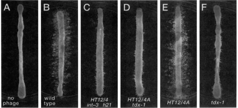
FIGURE
2.-Trails formed by the abortive transduction of Fli recipient strains. The recipient is strain lTl864.5. Phage donors were as follows: none (A), MS57 (B), MS607 (C), NBP.52 (D), NBP.51 ( E ) , KBP40 ( G ) . Phage genotypes are shown at the bottom of each panel. The experimental protocol is described in \~.\TF.RIAI.S ASD VI:.'rHODS.formed in parallel with the experiments shows in Figures 2 and 4 to verify the accuracy of the Fli"complete transductant titers ( i e . , the same mixture that was plated on the swarm agar w a s plated on Bochner medium).
Complementation of fdx-f mutants by wild-type P22: The FliD'-abortive transduction assay was used to detect comple- mentation of the abortive transduction phenotype of tdx-Z
mutants. Recipient cells were grown as described for the quan- titative transduction assay. Wild-type helper phage was grown on strain TT17615 that cannot provide the Fli' function. The M.O.I. of helper phage was 3.5, while that of phage NBP40 was 1.6. Phage were premixed, added to cells, and allowed to adsorb at room temperature for 20 min; the mixture was then deposited on motility agar containing tetracycline and the plates incubated at 37" for 24 hr.
RESULTS
Assays for abortive transduction: We employ hvo assays for abortive transduction. Our qualitative assay is based on the motility phenotype of abortively trans- duced fli mutants (described in MATERIALS AND METH-
ODS). Transduction of a nonmotile (Fli-) strain withy?
DNA generates complete transductants (which multiply and form confluent "swarms" throughout the motility plate) and abortive transductants (which form discrete colony trails traversing the motility plate; STOCKER 1956;
LEDERBERG 1956). In the experiment shown in Figure 2, complete transductants are prevented from growing and only the nonmotile recipient and abortive trans- ductants may grow. The presence of trails on a motility plate in this experiment is a qualitative indication of the occurrence of abortive transduction.
Our quantitative assay for abortive transduction is based on selection for inheritance of tetracycline resis- tance. For this assay the recipient carries a large dele- tion of the chromosomal region homologous to the transduced fragment, which carries the defective trans- poson T n l O n e t . Under the conditions of the experi- ment no complete transductants are observed because
the recipient's deletion is too large to be repaired by a single transduced fragment. The elimination of com- plete transductants greatly facilitates the scoring of abortive transductants.
The high-transducing phage
HT12/4
is impaired for the formation of abortive transductants: The original P22 mutant HT12/4 is one of several phage mutants isolated by virtue of its increased frequency of complete transduction (SCHMIECER 1972). While using this phage in standard transductional crosses we observed that abortive transductants were not as readily apparent as in crosses mediated by a different high transducing phage (HT105/1) or by wild-type P22. This defect in abortive transduction is seen in the motility assay depicted in Figure 2.Figure 2 shows trails formed by abortive transduction of a recipient carrylng the mutation fliD5055::TnlOd- Tet. In Figure 2B the transducing phage is wild-type P22; numerous trails (evidence of abortive transduc- tion) are evident. In Figure 2C the donor lysate was made with phage MS607, a derivative of the original phage HT12/4 of SCHMIEGER. It is apparent that this lysate generates fewer abortive transductants (deduced by the lack of trails). For all panels in this figure, a p
proximately equal numbers of FliD' complete transduc- tants have been deposited on each plate for each cross (see MATERIALS AND METHODS). If the ratio of complete
to abortive transductants is the same for each phage, we would expect equal numbers of abortive trails in each panel. Since there are virtually no trails evident for phage HT12/4, we conclude that this phage is defec- tive for the ability to specify abortive transductants. When a higher concentration of HT12/Merived lysate
is used, some trails do appear; thus the ability of phage HT12/4 to perform abortive transduction is impaired but not completely lost (data not shown).
22 N. R. Benson and J. Roth
no
phage
wild type
HT 12/4 HT 12/4A
int-3 tdx-
1
h2
1
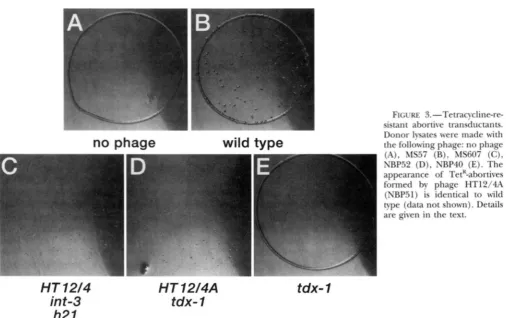
on inheritance of TetK. Figure 3B shows wild-type, TetK- abortive transductant colonies. Figure 3C shows abor- tive transductants made by phage HT12/4 ( M S 6 0 7 ) . In the TetK-based assay, phage HT12/4 still forms abortive transductants but the abortive colonies are smaller and less distinct than wild-type-TetK abortives. The reason for the difference between the Fli and Tet assays is unclear, but the results of the Tet assay seem significant and will be discussed later. TetR-abortive and complete transductants made by wild-type and HTI 2/4 phage are quantified in Table 3.
The genetic basis for the HT12/4transducing pheno- type: The HT12/4 phage used in the above experi- ments contained the 1121 and the int-3 mutations in addition to the HT12/4 mutation. To examine the con- tribution of these mutations to the HT phenotype, we performed phage crosses to replace these alleles with their wild-type counterparts (described in MATERIALS
AND METHODS). These crosses generated recombinant phages that displayed transducing phenotypes not seen for either of the parent phages.
Transduction characteristics of these novel transduc- ing phage are shown in Table 3 (see also Figures 2 and
3). One class of phage, assigned the genotype tdx-I, shows slightly increased complete transduction (two-
fold, compared to wild-type) and a deficiency in the formation of abortive transductants (Figure 2F; Figure
3E; Table 3). The second class of transducing phage, assigned the genotype HT12/4A, is 10-fold elevated for
FIGURE 3.-Tetracycline-re- sistant abortive transductants.
Donor lysates were made with the following phage: no phage (A), MS57 (B), MS607 (C), NBP52
(D),
NBP40 (E). The appearance of TetR-abortives formed by phage HT12/4A (NBP51) is identical to wild type (data not shown). Details are given in the text.tdx- 1
the formation of complete transductants and makes an excess of abortive transductants compared to wild-type phage (Table 3; Figure 2). Since the above phages lack the intand h21 alleles, we conclude that these mutations have no role in the transducing phenotype of phage HT12/4.
We interpret these data to indicate that the HT12/ 4 phenotype is due to both the tdx-1 and HT12/4A mutations. This hypothesis was confirmed by the follow- ing cross. Phage NBP52 (HTI2/4A tdx-1) was crossed with phage NBP63 ( c l
-
7 5-urn). Recombinant c l - 7 5’ phages were isolated and their transducing phenotypes analvzed. Again, recombinant phages with either thetdx-I or the HT12/4A genotypes were recovered (data
not shown). Thus, we conclude that the HT12/4 pheno- type reflects the combined effects of at least two separa- ble mutations that we designate ldx-1 and HT12/4A.
CASJENS et nl. (1992) have cloned gene 3 of phage HTl2/4 and demonstrated that the single base-pair mu- tation present in gene 3 of phage HT12/4 is necessary and sufficient to endow P22 with the HT phenotype and altered pnc-site specificity characteristic of phage
These authors remarked in their work that their recon- structed HT12/4A phage showed altered transduction properties compared to the original HT12/4 phage. We have compared the quantitative transducing phenotype and pnc-site specificity of CAS~ENS et al.’s reconstructed
HT12/4A phage with the phage we have designated
P22 Abortive Transduction Mutant 23
TABLE
4Efficiency of plating P22 strains harboring different combinations of erjand t& alleles on Salmonella strains with different red and supE alleles
~
Efficiency of plating on different hosts"
~~
Phage Phage genotype Wild type supE r e d I recAl supE
NBP85 cI-7; tdx-1 0.95 1 0.88 0.83
NBP183 cl-7; e$(am) 0.29 1 1.47 X 10⁻⁷ 0.83
NBPl79 cl-7; &(am); tdx-1 1.41 X 1 1.1 x 10⁻⁶ 0.76
10" (NBP179).'
HT12/4A (NBP51) and found them to be identical
(data not shown).
The tdjol mutation is lethal in an erfam background
Figure 1 describes a model for the transduction process in which an abortive transductant is formed by a pro- tein-mediated, noncovalent circularization of the trans- duced DNA (SANDRI and BERGER 1980b). If this model is correct, then P22 DNA might also assume such a structure as an intermediate during the essential circu- larization of the phage genome. Since the P22-encoded Erf protein is known to catalyze the circularization step (BOTSTEIN and MATZ 1970; POTEETE 1988), we con- structed an erf-am tdx-1 double mutant anticipating that such a phage might show a revealing phenotype.
Phages carrying null
erf
alleles cannot circularize their genome by the Erf-mediated pathway and are de- pendent on the host's RecA protein for circularization. Thus E r f phages make plaques on wild-type hosts but not on recA hosts. Table 4 shows the efficiency of plating tdx-1 phages, erf-am phages and erf-am tdx-l phages on various hosts. The erf-am tdx-l double mutant does not plaque on our wild-type host (line 3) , while phages carrying either the tdx-1 or the erf-am mutation plaque efficiently on the wild-type host (lines 1 and2).
From these data we conclude that an erf-am tdx-l phage can- not use the host's RecA pathway of recombination, sug- gesting that the tdx-l mutation blocks the RecAdepen- dent pathway of circularization.Genetic mapping of the &I allele: The erf-um tdx-
1 "lethal phenotype allowed us to map the tdx-l muta- tion using amber mutations in a variety of essential genes in combination with an @-am mutation. Prelimi- nary cross data suggested that the tdx-1 mutation re- sided near genes 7, 20 and 16 (data not shown). Table 5 shows the results of crosses between erf-am tdx-1 phages with erf-am 2@am phages, or erf-am 16-am phages. The genetic distance between the 16-am allele and the tdx-1 mutation (line 5) is approximately threefold smaller than the distances separating the 2@am and tdx-
1 alleles (line 4) or the 16-am and 2@am alleles (line 6). This data localizes the tdx-1 mutation in or near gene 16.
, I
The wild-type strain is DB7000. The supE strain is MS1363. The r e d 1 strain is 'IT18642. The r e d 1 supE strain is TT18640. The plating efficiencies are normalized to the titer of phage on strain MS1363. All lysates were prepared on strain MS1363 at 25" as described in MATERIALS AND METHODS. The data presented is the plaquing efficiency relative to the supE strain. Data is shown for one Darticular lvsate. for which the value of 1 corresponds to 2.35 X 10" (NBP85), 3.89 X 10" (NBP183), 1.46 X
The abortive transduction phenotype of &I mutant phage can be corrected by &+-helper particles: When
16-am or 16-ts phage are grown under nonpermissive
conditions they form progeny particles that lack the product of gene 16 (gp16; HOFFMAN and LEVINE 1975a). These gpl6defective particles are noninfec- tious but can be rendered infectious by coinfection with phage particles that contain wild-type gp16 (HOFFMAN and LEVINE 1975a,b). HOFFMAN and LEVINE showed that the DNA of the coinfecting, gp16+ helper phage need not be expressed and concluded that the correcting activity must be effected by gp16 present in the helper- phage particle. If the tdx-1 mutation affects gene 16, as
suggested by the mapping data in Table 5, then the abortive-transduction defect of tdx-1 phages might be corrected by coinfecting, wild-type phage.
Figure 4 shows a complementation experiment utiliz- ing a j i D mutant recipient. Figure 4D clearly shows that wild-type, coinfecting phage can complement the tdx-1 donor phage for the formation of abortive transduc- tants (compare C and D). In this experiment the helper
TABLE 5
Genetic mapping of the tdro-1 mutation
Relevant genotypes of recombining phages"
% tdx+ am+
Phage 1 Phage 2 recombinant phage*
tdx-1
-
20-am
-
4 0 - 5 ; 4 0 - 51 &am -
tdx-1 2@am 0.29; 0.25
tdx-1 16-am 0.09; 0.08
1 6 a m 20-am 0.27; 0.28
In these crosses both phages carry the e$am(HI 173) and cl-7 mutations. The tdx-1 phage is NBPl79, the P@am phage is NBP187, and the 16-am phage is NBP188.
24 N. R. Rcnson and J , Roth
FIGLXK 4.-Correcting the abor- tive transduction phenotype of tdx-I phages with wild-type helper phage. T h e wild-type, helper phage
(NRPJI) has been grown on a Fli- strain (TT1761.5) and cannot pro- vide Fli function (B). The tdx-I phage was NBP40; the recipient in this experiment was NBPIIPI. A complete discussion can be found in the text.
no
phage
wild-type
helper
tdx-
1
tdx-
1
+
wild-type
helper
phage was grown on a host containing the same Jli
mutation as the recipient and therefore cannot possibly contribute j / i + sequences (Figure
4R).
Furthermore, since the recipient is a lysogen, the helper phage cannot express its late genes suggesting that the Tdx' function is provided by the helper-phage capsid. Particles defec- tive in gp16 d o n o t provide this help (data not shown). This result is consistent with the tdx-1 mutation residing in gene 16. However, we have recently determined that gp2Odefective particles are also helped by wild-type helper phage (and also fail to help tdx-1; data not shown); therefore, the outcome of the tdx-I correction experiment is consistent with either a mutation in gene 20 or gene 16.The tdx-2 mutation affects an early step in the matura- tion of injected e f u m tdx-2 phage DNA: The growth defect of rrjkm /&-I phages could be early or late in development. If the growth defect of q f u m tdx-I phages reflects a failure to circularize the injected genome (as
the involvement of the qpum allele implies), we would
expect that this phage would be defective in both Ivsog- eny and lysis. Alternatively, if the doubly mutant phage is defective in a later stage of lytic growth, we might expect it to lysogenize a wild-type host with wild-type efficiency.
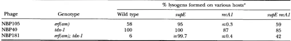
Table 6 shows the ability of phages carrying various combinations of nfand tdx alleles to lysogenize various hosts. The data show that singly mutant q j h n o r tdx-I
phages can lysogenize a wild-type host with high effi- ciency; however, the doubly-mutant Prporn tdx-I phages lysogenize a wild-type host
-
IO-fold less frequently than the n - a m phage. The ohsenation that an +[m tdx-Iphage retains some ability to lysogenize a rwAA. cell (6%
of infected cells) compared to a rprA- cell (50.4%)
indicates that the tdx-I mutation reduces but does not eliminate circularization by the RecA pathway.
Once circularized, erf-am tdx-1 phage exhibit wild-type lytic growth: The data in Table 6 tlocs not rule out the possibility that @-urn tdx-I phages have both pre- and postcircularization defects. To determine if nj-am tdx-l phages have a defect in development subsequent to circu- larization of the phage genome, we assessed the ability
of such phage to develop when the phage DNA was circu- larized by an Erf/RecA-independent mechanism.
An alternative means of achieving circularization is by use of the site-specific recombination system of the phage. Excision of a prophage from the host chromo- some requires the P22-encoded Int and Xis proteins and the &acting sites that flank the prophage. This site-specific recombination system does not require Erf o r RecA function ( POTEETE 1988). If "am tdx-1 phages are defective only in circularization (or an earlier meta- bolic step) we would predict that induction of an @-
elm tdx-I prophage would yield an amount of phage
comparable to that released by inducing a wild-type prophage.
The results presented in Table
7
clearly demonstrate that the lysates produced by mitomvcin C induction ofwild-type, tdx-I, qpam or @am tdx-I prophages are nearly equal in titer, whether the induction was carried out in a st(@ or sufi" genetic background. Because an qpum, tdx-I phage has a wild-type burst size once it has been circularized, we conclude that n - a m tdx-1 mutant phages are not defective for any developmental lytic step after circularization of the phage genome.
DISCUSSION
P22 Abortive Transduction Mutant 25
This mutation, tdx-I, was initially discovered as one of TABLE 7
two mutations contributing to the high-transducing Titers of lysates made by inducing lysogens of varying phenotype of the phage HT12/4. We have demon- prophage genotype
strated the following.
1.
2.
3.
4.
5 .
-
The tdx-1 mutation reduces the ability to form abor- tive transductants and slightly increases the forma- tion of complete transductants.
The tdx-1 mutation appears to map in or near P22- gene 16.
The abortive transduction defect of tdx-l-transduc- ing particles can be corrected by wild-type, coinfect- ing phage, a fact that is consistent with known phe- notypes of gplfj-defective particles (HOFFMAN and LEVINE 1975a,b) and gp20-defective particles (N. BENSON, unpublished data).
An
@-am tdx-1 mutant phage cannot grow on a wild- type host; from which we conclude that the tdx-lmutation affects the Red-catalyzed pathway of phage chromosome circularization.
The erf-am-tdx-l lethal phenotype is circumvented when the phage genome is circularized by the phage’s site-specific recombination system; from this observation we conclude that the defect in @-am tdx-I phage development is at or before the critical circularization stage of infection.
Our data also suggest a more complex involvement of gp3 in the formation and/or stability of abortive transductants (in addition to the packaging of DNA). First, the TetR-abortive transduction data presented in Figure 3 and Table 3 show that phages carrying the tdx-
I allele (in a wild-type gene 3 background) are severely deficient for the formation of TetR-abortive transduc- tants (Figure 3E). However, when the tdx-1 mutation is accompanied by an HT12/4A mutation (a mutant gene 3 background), TetR-abortive transductants are “resurrected” (albeit weakly; compare D and E in Fig- ure 3). Second, the ratio of abortive to complete trans- ductants for phage HT12/4A is 120, while the ratio of complete to abortive transductants for wild-type P22 is 11.8 (Table 3 ) . This suggests that the mutant gene 3
allele in an HT12/4A phage affects the formation of abortive transductants. Both these observations are consistent with the mutant gp3 (specified by the HT12/ 4A mutation) being packaged in the phage capsid. This
Titer of lysates made by induction of wild-type and
supE lysogenic hosts”
Prophage Wild-type (su ) supE host
Phage genotype host (X10” )
(xlo”o)
NBP31 Wild type 9.7 3.4 NBP40 tdx-1 8.2 3.8
NBP105 @-am 18 4.5
NBP181 tdx-I; @-am 10 2.4
Lysogenic strains are as follows: wild type, TT18650, 1187, 1195 and 1193 (top to bottom); supE, TT18646, 1186, 1194 and 1185. Numbers shown are the average of two indepen- dent induction experiments. The titer of the lysates was deter- mined on MS1363 (sup@.
conclusion is surprising since gp3 has not been found as a component of the capsid. However, it is possible that a few molecules of gp3 might be present in the capsid and escape detection (S. CASJENS, personal com- munication).
The tdx-1 mutation maps to a region of the P22 ge- nome that encodes proteins carried in the mature viral particle. We have attempted to assign the tdx-1 mutation to a precise locus with several experiments. When @-
am tdx-1 phages are crossed with a plasmid containing the P22 EcoRI-B fragment (9.28 kb;JAcmoN et al. 1978),
@-am tdx+ progeny phages are recovered. The EcoRI-B fragment extends from the middle of gene 8 through gene 16 (the EcoRI site is 40 nucleotides past the end of gene 16). However, when @-am tdx-l phages are crossed with a minimal clone of gene 16 (pBU5; UM- LAW and DREISEIKELMANN 1992) no @-am tdx” phages are recovered, nor are such recombinants seen with crosses using clones that include genes 16 and 20 (pro- vided by PETER BERGET; data not shown). Furthermore, all spontaneous reverent phages (those plating on a wild-type host) are genotypically erf’ tdx-1 and never
@-am tdx’ (data not shown). These experiments, taken in consideration with the mapping data in Table 5 and the complementation data in Figure 4, suggest that a locus in addition to genes 16 or 20 may be involved.
TABLE 6
The ability of phage carrying different erfand tdx alleles to lysogenize red+ and r e d l hosts
% lysogens formed on various hosts“
Phage Genotype Wild type supE recA I supE recAl
NBP105 erf(am) 58 95 50.3 59
NBP40 tdx-1 100 100 87 85
NBP181 @(am); tdx-1 6 299.7 50.4 42
26 N. R. Benson and J. Roth
We have also attempted to complement the Tdx-1 phenotype with the plasmid pBU5, which produces gp16 under the control of the Lac repressor. Strains harboring pBU5 cannot rescue @-am tdx-1 phage al- though these strains can rescue 16-am phage (in a plat- ing assay; data not shown). Furthermore, growing tdx- 1 mutant phage (NBP40) through a gp16 (pBU5) pro- ducing strain does not enable the progeny phage to specify abortive transductants nor does plating tdx-l-
transducing particles on a host expressing gp16 effect an observable increase in abortive transductants (data not shown). We conclude that gp16 alone cannot res- cue the Tdx-1 mutant phenotypes. These observations reinforce our belief that the Tdx-1 phenotypes are due to the combined effects of mutations in genes 16 or 20
and an additional, unidentified locus.
We have been unable to unambiguously assign the
tdx-1 mutations to known P22 loci, nonetheless, several conclusions can be drawn. The fact that the tdx-1 muta- tions affect generalized transducing particles and can be corrected by coinfecting phage (Figure 4) suggests that the Tdx+ proteins are carried within the viral parti- cle and are injected with DNA into the new host. Thus, the identity of Tdx+ must be restricted to the injected components of the mature P22. This narrows Tdx+ to the products of genes
7,
16, 20 or 26; all of which encode proteins believed to be injected with the DNA(ISRAEL 1977).
In considering the role of injected phage proteins, it is useful to review early events in phage infection. In- jected phage DNA must cross the outer membrane, the
periplasmic space and the inner membrane to reach the cytoplasm. Unlike some T-phages, the architecture of the P22 particle does not seem to allow for direct phage penetration of the membranes and injection into the cytoplasm (DREISEIKELMANN 1994). Rather, it seems likely that phage (and transducing) DNA must first en- ter the periplasmic space and then traverse the inner membrane.
Once inside the cytoplasm, the double-stranded DNA must evade the RecBCD nuclease that can degrade the phage nucleic acid. During the later stages of infection the P22 genome avoids RecBCD-catalyzed degradation by circularizing and then by guiding synthesis of the phage Abcl and 2 proteins. These proteins physically inhibit the RecBCD-catalyzed degradation of the linear, concatemeric DNA from which infectious particles are made (POTEETE 1988). It is not known how P22 DNA escapes RecBCD-catalyzed degradation of DNA before circularization; sequestration of the blunt ends by in- jected phage proteins is a likely possibility. We propose that injected phage proteins protect DNA ends and promote genome circularization (and may also help in transport across the membrane as discussed below). Specifically we suggest that formation of abortively transduced fragments requires the presence of these proteins at DNA ends. Normally these proteins assist in
the circularization of a phage genome that possesses homologous, direct repeats at each end. Since trans- duced fragments do not possess such repeats, the circu- larization process is aborted leaving a stable, protein- held DNA circle (an abortively transduced fragment). The proposal that an abortive transductant is formed by circularization of a transduced DNA fragment was suggested for the transducing phage P1 by SANDRI and
BERGER (1980). These authors showed that the mobility of P1-abortively transduced DNA in native gels or neu- tral sucrose gradients was characteristic of a circular molecule. When this suspected circular DNA was puri- fied and subjected to denaturing conditions, or ex- posed to Pronase, the DNA assumed the mobility char- acteristic of full-sized, linear P1-particle DNA. Given the many parallels between P1 and P22 transduction (OZEKI
and IKEDA 1968), it is likely that these phages share a similar mechanism for abortive transduction. The tdx-
1 mutant characterized here is an excellent candidate for a defective P22 analogue of the Plcircularization apparatus.
Our model for P22 transduction (Figure 1) incorpo- rates the hypothesis of SANDRI and BERGER (1980) that abortive transduction is due to protein mediated, non- covalent circularization of the transduced DNA. Figure
1 shows a complex of gp7/20/16 at one end of the DNA fragment (it is possible that both ends are protected; see BENSON and ROTH 1994). We suggest that the eventual fate of the transducing fragment is determined by a competition for the unprotected end by the protected end (resulting in a circle and abortive transduction) and RecBCD enzyme (resulting in degradation and re- combination). Circle closure is usually the winner in this competition, a fate that makes sense for the phage genome because this protects against RecBCD degrada- tion. Thus, we visualize abortive transductants as a trapped intermediate of a circle-closure reaction; phage DNA transiently assumes this form before progressing to a recombinant, closed circle.
There are genetic and physical data relevant to this model. HOFFMAN and LEVINE (1975a,b) have conducted an extensive analysis of gene 16 mutations and their effect on the fate of injected DNA. These authors con- cluded that gp16 did not protect injected DNA from host degradation but might be involved in some aspect of the translocation of DNA across the cell membrane into the intracellular space. UMLAUF and DREISEIKEG MANN (1992) have cloned gene 16 and shown that (1)
gp16 expressed in E. coli cannot rescue gene 2 mutants of bacteriophage T4 ( i e . , cannot protect naked T 4
P22 Abortive Transduction Mutant 27
tect blunt, double-stranded DNA ends; a conclusion consistent with our data.
The data discussed above lead us to hypothesize that translocation and protection of injected DNA are ef- fected by the coordinated actions of at least gp7/20/
16, probably in a complex. The data presented here, in addition to our unpublished data, suggests that the Tdx-1 phenotype is effected by a minimum of two muta- tions, one of which we believe to lie in either gene 16 or 20, and another located within the boundaries of the EcoRI-B fragment. We suggest that the tdx-1 muta- tions affect the wild-type proteins in such a way that noncovalent circle closure (abortive transduction) is
impaired but that DNA transport and protection activi- ties are conserved.
In summary, we have described a mutant phage that is defective in both the phenomenon of abortive trans- duction and the RecAdependent circularization of the
P22
genome. We suggest that the phenomenon of abor- tive transduction is best described as a futile attempt by injectedP22
proteins to circularize transduced DNA.We extend our thanks to MIRIAM SUSSKIND and TONY POTEETE for supplying phage P22 strains and plasmids, B. UMLAUF and B. DREISEIKELMANN for plasmid pBU5, PETER BERGET for various P22 clones, and SHERWOOD CASJENS for communicating unpublished data on the contents of the phage particle, supplying P22 plasmids and for helpful comments on the manuscript. This work was supported in its initial stages by a postdoctoral research fellowship awarded to N.R.B. by the National Institute of Allergy and Infectious Diseases (5-
F32-AI08250-03).
LITERATURE CITED
BENSON, N. R., and B. S. GOLDMAN, 1992 Rapid mapping in Salmo- nella typhimurium with MudP22 prophages. J. Bact&ol. 174:
1673-1681.
BENSON, N. R., and J. ROTH, 1994 Suppressors of recB mutations in
Salmonella typhimurium. Genetics 138: 11-29.
BOCHNER, B., H. C. HUANG, G. L. SCHREVIN and B. N. AMES, 1980 A positive selection for loss of tetracycline resistance. J. Bacteriol.
BOTSTEIN, D., and M. J. MATZ, 1970 A recombination function es- sential to the growth of bacteriophage P22. J. Mol. Biol. 5 4
CASJENS, S., and M. HAYDEN, 1988 Analysis in vivo of the bacterio- phage P22 head-full nuclease. J. Mol. Biol. 1 9 9 467-474. ~ J E N S , S . , S. L. AMPS SON, S. RANDALL, IC EPPLER, H. Wu et al., 1992
Molecular genetic analysis of bacteriophage P22 gene 3 product, a protein involved in the initiation of head-full DNA packaging.
CHELALA, C., and P. MARGOLIN, 1974 Effect of deletions on co- transduction linkage in Salmonella typhimunum: evidence that bac- terial chromosomal deletions affect the formation of transducing DNA fragments. Mol. Gen. Genet. 131: 97-106.
DAVIS, R. W., D. BOTSTEIN and J. R. ROTH, 1980 Advanced Bacte-
143: 629-933.
417-440.
J. Mol. BIOI. 227: 1086-1099.
rial Genetics. Cold Spring Harbor Laboratory, Cold Spring Harbor,
NY.
DREISEIKELMANN, B., 1994 Translocation of DNA across bacterial membranes. Microbiol. Rev. 5 8 293-316.
EBEL-TSIPIS, J., D. BOTSTEIN and M. S. Fox, 1972a Generalized trans- duction by bacteriophage P22 in Salmonelkz typhimurium. I. Molec- ular origin of transducing DNA. J. Mol. Biol. 71: 433-448. EBEL-TSIPIS, J., M. S. Fox and D. BOTSTEIN, 1972b Generalized trans
duction by bacteriophage P22 in Salmonella typhimunum. 11.
Mechanism of integration of transducing DNA. J. Mol. Biol. 71:
HARTMAN, P. E., Z. HARTMAN and D. SERMAN, 1960 Complementa- tion mapping by abortive transduction of histidine requiring
Salmonella mutants. J. Gen. Microbiol. 2 2 354-368.
HOFFMAN, B., and M. LEVINE, 1975a Bacteriophage P22 virion p r e tein which performs an essential early function. I. Analysis of 16(ts) mutants. J. Virol. 16: 1536-1546.
HOFFMAN, B., and M. LEVINE, 1975b Bacteriophage P22 virion pro- tein which performs an essential early function. 11. Characteriza- tion of the gene 16 function. J. Virol. 16: 1547-1559.
ISRAEL, J. V., T. F. ANDERSON and M. LEVINE, 1967 In uitro morph- genesis of phage P22 from heads and base-plate parts. Proc. Natl. Acad. Sci. USA 57: 284-291.
ISRAEL, V., 1977 I. Identification and ejection from wild-type and defective particles. J. Virol. 2 3 91-97.
JACKSON, E. N., H. I. MILLER and M. L. A D A M S , 1978 EcoRl restric- tion endonuclease cleavage site map of bacteriophage P22 DNA. J. Mol. Biol. 118: 347-363.
JACKSON, E. N., F. LASKI and C. ANDRES, 1982 Bacteriophage P22 mutants that alter the specificity of DNA packaging. J. Mol. Biol.
LEDERBERG, J., 1956 Linear inheritance in transductional clones. Genetics 41: 845-871.
LEDERBERG, J. E., E. M. LEDERBERG, N. P. ZINDER and E. R. LIVELY, 1951 Recombination analysis of bacterial heredity. Cold Spring Harbor Symp. Quant. Biol. 1 6 413-441.
MIESEL, L., and J. R. ROTH, 1994 Salmonella r e d mutations increase recombination in a short sequence transduction assay. J. Bacte-
OZEKI, H., 1956 Abortive transduction in purine-requiring mutants of Salmonella typhimunum. Carnegie Institution of Washington, Genetic Studies of Bacteria, Publication 612: 97-106.
OZEKI, H., and H. IKEDA, 1968 Transduction mechanisms, pp. 245- 278 in Annual Reukw of Genetics, edited by H. L. ROMAN. Annual Reviews, Inc. Palo Alto, CA.
POTEETE, A. R., 1988 Bacteriophage P22, pp. 647-682 in The Bacte- riophages, Vol. 2, edited by RICHARD CALENDER. Plenum Press, New York.
SANDN, R. M., and BERGER, H., 1980a Bacteriophage-PI-mediated generalized transduction in Eschachia coli: structure of abortively transduced DNA. Virology 106: 30-40.
SCHMIEGER, H., 1972 Phage-P22 mutants with increased or de- creased transduction abilities. Mol. Gen. Genet. 154: 75-88. STOCKER, B. A. D., 1956 Abortive transduction of motility in Salmo-
nella; a non-replicated gene transmitted through many genera- tions to a single descendant. J. Gen. Microbiol. 15: 575-598. STOCKER, B. A. D., N. D. ZINDER and J. LEDERBERG, 1953 Transduc-
tion of flagella characteristics in Salmonella. J. Gen. Microbiol.
SUSSKIND, M. M., and D. BOTSTEIN, 1978 Molecular genetics of bac- teriophage P22. Microbiol. Rev. 42: 385-413.
UMLAUF, B., and B. DREISEIKELMANN, 1992 Cloning, sequencing and overexpression of gene 16 of Salmonella bacteriophage P22. Virology 188: 495-501.
449-469.
154 551-563.
ri01. 176: 4092-4103.
9 410-433.