NIGHOT, MEGHALI PRASHANT. Role of ClC-2 and MAPK in Gastric Barrier Function. (Under the direction of Anthony Blikslager).
The gastric barrier is composed of a single layer of columnar epithelium, mucus and bicarbonate layer, and the intraepithelial tight junctions residing at the apical-most region of the paracellular space. The gastric barrier serves as the first line of defense against a hostile
luminal environment. Once the epithelial barrier is disrupted, epithelial repair mechanisms must rapidly re-form a continuous epithelial monolayer and seal paracellular tight junctions in order to prevent entry of protons and bacteria. We studied the role of chloride channel ClC-2 and MAPK proteins in the gastric barrier function. We designed, developed, and optimized in vivo acid-injury and in vitro NSAID-injury model for our studies. We found that
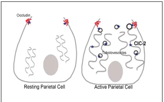
ClC-2 activation protects gastric mucosal barrier dysfunction by preventing disruption of tight junction protein occludin in both in vivo acid injury in pig gastric mucosa as well as in vitro NSAID injury in gastric epithelial cells. In parallel studies based on genetically modified mice, we found altered acid secretion, reduced H+K+ATPase expression, reduced parietal cell population, and disorganization of gastric glands in the ClC-2 knockout mice. Thus ClC-2 appears to have a critical role in the proper differentiation and organization of gastric glands.
In another piece of study we investigated the role of MAPK (Mitogen Activated
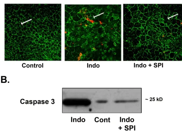
expression of occludin. Indomethacin also induced apoptosis via activation of caspase-3 and inhibition of p38 MAPK prevented indomethacin-induced activation of caspase-3. Collectively this data suggests that NSAID indomethacin induces gastric epithelial barrier dysfunction via changes in expression and localization of occludin and induction of apoptosis in a p38 MAPK-dependent mechanism.
© Copyright 2011 by Meghali P. Nighot
by
Meghali Prashant Nighot
A dissertation submitted to the Graduate Faculty of North Carolina State University
in partial fulfillment of the requirements for the degree of
Doctor of Philosophy
Comparative Biomedical Science
Raleigh, North Carolina 2012
APPROVED BY:
_______________________________ ______________________________ Dr. Anthony T. Blikslager Dr. Sam L. Jones Chair of Advisory Committee
BIOGRAPHY
Meghali Thakre Nighot was born and brought up in Nagpur, a city in the central part of India. She completed her Bachelor’s degree in Veterinary Sciences and Master’s in veterinary Parasitology from Nagpur Veterinary College. She worked in Poultry industry and Government of India for few years before joining North Carolina State University for PhD study in August 2008.
ACKNOWLEDGMENTS
I would like to acknowledge and appreciate the enormous support that I received throughout my PhD studies at North Carolina State University. I sincerely thank Dr. Anthony Blikslager for providing me this wonderful opportunity of graduate research in his laboratory. I have always found an accomplished mentor and an amazing person in him. I would also like to thank Dr. Sam Jones, Dr. Jody Gookin, and Dr. Adam Moeser for agreeing to be member of my Graduate committee and their valuable advice throughout my degree.
I would like to express my deep gratitude to my fellow laboratory colleagues Tracy Hill, Karen Young, Liara Gonzalez, Katie Tolbert, Liz Lenon, Amelia Gibson, Beth Overman, Eue Jae Sung, Raphael, and Katie Sheats for their support and help with my research studies. I thank the staff at the Central Procedures Laboratory and the Laboratory of Animal Resources for their excellent technical advice and support. The services of Laboratory for Advanced Electron and Light Optical Methods, CVM are greatly appreciated. Dr. Troy Ghashghaei helped us with confocal microscopy.
This is also a time to say thanks to colleagues and friends including Susan, Mihika, Arun, Aparna, Sudhakar, Shraddha and others for their help throughout this period.
TABLE OF CONTENTS
LIST OF TABLES... vi
LIST OF FIGURES...vii
CHAPTER I. ROLE OF ClC-2 CHLORIDE CHANNEL IN GASTRIC MUCOSA... 1
Introduction... 2
ClC-2 structure and organization... 2
ClC-2 regulation…... 5
ClC-2 function…... 7
Gastric barrier function...10
Measurement of gastric barrier function... 10
ClC-2 chloride channel in gastric mucosa... 11
Role of ClC-2 chloride channel in gastric barrier function... 12
Conclusion………... 14
References... 16
CHAPETR II. GASTROINTESTINAL TIGHT JUNCTIONS AND MAPK SIGNALING PATHWAYS... 28
Introduction... 29
Structure and composition of epithelial tight junctions... 30
Signaling to tight junction...….. 31
Mitogen Activated Protein Kinases (MAPK) signaling pathways ... 32
ERK1/2MAPK... 34
JNK MAPK... 35
p38 MAPK... 36
Effect of MAPK Kinases on tight junctions…... 38
Conclusion... 40
References…... 42
CHAPETR III. GASTROPROTECTIVE PROPERTIES OF THE NOVEL PROSTONE SPI-0811 AGAINST ACID-INJURED PORCINE MUCOSA……….. 50
Abstract...51
Introduction……... 52
Materials and Methods………... 54
Results... 59
Discussion...63
Figure legends... 67
CHAPETR IV. THE PROTECTIVE EFFECT OF CLC-2 AGONIST SPI-0811 ON INDOMETHACIN-INDUCED EPITHELIAL BARRIER DYSFUNCTION IN
HUMAN GASTRIC EPITHELIAL CELLS ……... 83
Abstract…... 84
Introduction... 85
Methods... 88
Results…... 93
Discussion... 96
Figure legends... 101
References... 109
CHAPETR V. INDOMETHACIN INDUCES GASTRIC EPITHELIAL BARRIER DYSFUNCTION AND APOPTOSIS VIA P38 AND JNK MAPK IN MKN28 CELLS... 115
Abstract... 116
Introduction…... 117
Methods... 119
Results... 123
Discussion... 128
Figure legends…... 132
LIST OF TABLES
LIST OF FIGURES
CHAPTER I
Figure 1. Localization of ClC-2 in Parietal cells………... 15 CHAPETR II
Figure 1. Mitogen Activated Protein Kinases Family………….……….. 33 CHAPTER III
Figure 1. Electrical responses and 3H-mannitol fluxes of acid-injured porcine gastric mucosa………... 67 Figure 2. Histological and Immunofluorescence findings in acid-injured gastric mucosal tissues……….…... 69 Figure 3. Western analysis for Expression of TJ proteins in membrane fractions in acid injured porcine gastric mucosa……….….…... 71 Figure 4. Electrical responses and 3H-mannitol fluxes of acid-injured porcine gastric mucosa with inhibitors……….……….……. 72 Figure 5. Western analysis for Expression of ClC-2 protein in porcine
mucosa………... 74
CHAPTER IV
Figure 1. Dose curve for indomethacin-induced loss of TER in MKN-28 cell line... 101 Figure 2. Effect of Cobiprostone (SPI-0811) on indomethacin-induced decrease in TER and increase in epithelial permeability in MKN-28 cells……… 102 Figure 3. Western expression of TJ proteins………103 .
Figure 6. Western analysis for Expression of ClC-2 in MKN-28 cells…………106
Figure 7. Effect of Cobiprostone (SPI-0811) on indomethacin-induced decrease in the transepithelial resistance in stomach mucosa…………...………..107
CHAPTER V Figure 1. Dose curve for indomethacin-induced loss of TER in MKN-28 cell line……… 132
Figure 2. Effect of MAPK inhibition on indomethacin-induced changes in the epithelial barrier in MKN-28 cells……….………... 133
Figure 3. Western analysis of Tight Junction proteins……….... 134
Figure 4. Western analysis of Phosphorylation of MAPK…….………... 135
Figure 5. Immunolocalization of TJ protein occludin ……… 136
Figure 6. Expression of activated caspase-3………..…... 137
CHAPTER I
LITERATURE REVIEW
Introduction
The ClC super family of voltage-gated Cl- channels consists of nine isoforms that are expressed in a wide variety of tissues and organs. The mutation/disruption of some of the ClC isoforms is associated with a variety of human or animal diseases. One of these isoforms, ClC-2, has been shown to have a critical functional role in tissues such as brain, testis, retina, and intestine. The physiological function and regulation of ClC-2 channels in the intestine has been intensely researched as a substitute for dysfunctional CFTR in the fatal condition of cystic fibrosis. The work in the last two decades has demonstrated a variety of tissue specific functions of ClC-2 besides chloride transport activity. This paper will review the latest knowledge about function and regulation of ClC-2 in general, and specifically its role in the gastric mucosa.
ClC-2: Structure and distribution
ClC members in tissue homeostasis has been demonstrated by, mouse models, experimental expression systems and human diseases associated with either mutations or functional defects in ClC family members e.g. myotonia (ClC-1) (41), epilepsy (ClC-2) (35), Bartter III- renal salt loss (ClC-Kb) (25), Dent’s disease- proteinuria (ClC-5) (63), and osteopetrosis (ClC-7) (42). The crystal structure of the ClC identified by Dutzler et al. (23) in Salmonella enterica and Escherichia coli aided greatly in defining its function and regulation. The membrane spanning 18 helices form two halves of the double barreled structure and two identical pores. A glutamate residue that protrudes into the pore is proposed to be a binding site for chloride (36). The gating mechanism of ClC-2 is thought to be influenced by intracellular Cl -concentrations. According to this hypothesis, E217, the conserved glutamate residue, acts as a hyper polarization-dependent protopore gate in ClC-2 and that access of intracellular Cl- to this site stabilizes the open state of ClC-2 channel (53). Two large highly conserved CBS (cystathionine beta synthase) domains in the C terminus of mammalian but not bacterial ClC-2 have been a subject of interest to researchers. In vitro studies have shown that the CBS domains from several proteins, including ClC-2, can bind to nucleotides like ATP suggesting that they may act as energy sensors (66).
dendritic spines, cell bodies and axon initial segments of neurons, and in the end feet of astrocytes. The presence of ClC-2 in axonal membranes with intense γ-aminobutyric acid (GABA) responsive synaptic transmission activity has been demonstrated to be consistent with chloride transport that leads to efficient GABA (A) receptor-mediated inhibition of synaptic transmission (68, 72).
ClC-2 regulation
ClC-2 on the cell surface and increased sensitivity of ClC-2 gating towards intracellular chloride concentrations (31). Importantly, the partnership of Hsp90 with ClC-2 would also explain varying expression and activity of ClC-2 in the different expression systems and tissues (16).
ClC-2 functions
Conventionally, ClC-2 has been considered to be a ubiquitous, inwardly rectifying plasma membrane Cl- channel that is activated by membrane hyperpolarization, cell swelling, and mildly acidic extracellular pH (28, 35). However, many of the physiological functions of ClC-2 were derived from knock out (KO) studies or studies in heterogeneous expression systems, and the results from genetic knock studies were not always consistent. Recent studies using newer research tools have been useful in defining the functional role of ClC-2. For instance, contrary to the previous notion that ClC-2 helps maintain synaptic inhibition by lowering intracellular Cl- concentrations ([Cl(-)](i)), new simulation studies show that ClC-2 mediates chloride influx instead of efflux (64). Similarly, regarding the fundamental function of a chloride channel, although ClC-2 has been shown to contribute to native chloride secretion and antisense treatment reduced Cl- secretion in the human intestinal Caco-2 cells (49), additional disruption of ClC-Caco-2 in CFTR KO mice did not exacerbate the phenotype (75).
homeostasis. ClC-2 knockout mice suffer from severe retinal degeneration and testicular degeneration; both of these tissues have a blood barrier and depend on the supporting tissue for nutrition (retinal pigment epithelial cells and Sertoli cells, respectively) (8, 35, 69). Overall, most of the examples from KO studies suggest an important role of ClC-2 in cell-to-cell contact, communication, and homeostasis aside from chloride transport. ClC-2 was believed to contribute to the regulatory volume decrease in human glioma cells and inhibition of ClC-2 with siRNA was shown to inhibit cell proliferation of a human glioma cell line U-87 (24, 74). Another member of the ClC family, ClC-3, has been shown to mediate swelling-activated Cl- current and cell volume regulation in human gastric epithelial cells (38). The role of ClC family members in tissue homeostasis is also evident from a report where expression of chloride intracellular channel 1 (CLIC1) was found to be significantly (1.95-fold) up-regulated in 67.9% of gastric patients and was well correlated with lymph node metastasis, lymphatic invasion, perineural invasion, pathological staging, and a poor prognosis (14).
Gastric barrier function
Measurement of gastric epithelial barrier function in the in vitro and in vivo injury models
In order to study mechanisms of ClC-2 mediated gastric epithelial barrier function, it is necessary to be able to sensitively and dynamically measure gastric barrier function. The most sensitive measure of mucosal barrier function is transepithelial resistance (TER), since this measurement reflects the degree to which ions traverse tissue. There are two routes ions may traverse epithelium: transcellular and paracellular. The properties of the paracellular space differ dramatically between distinct tissues based on differences in tight junction structure. The measurements of TER largely reflect the tight junction resistance. Alternatively, the paracellular pathway can be assessed using a variety of probes that selectively traverse epithelium via the paracellular space. An example of such a probe is mannitol, which has been used in permeability studies as mucosal-to-serosal fluxes in tissues placed in Ussing chambers. This approach is also extended to study in vivo responses of modulators of intestinal secretion. In the in vitro studies, we have also used FITC-Dextran to measure permeability in the MKN-28 cells.
ClC-2 chloride channel in the gastric mucosa
that may be involved in gastric acid secretion based on the following results: (1) ClC-2 was cloned from rabbit gastric mucosa and its electrophysiological properties including anion selectivity (I− < Cl−) were similar to those of the native channel in H+,K+-ATPase-containing gastric vesicles (18, 19); (2) ClC-2 in rabbit (45, 46) and human gastric mucosa (19) is activated by cAMP-dependent phosphorylation, and this mechanism is consistent with an essential role of intracellular cAMP in the HCl secretory mechanism; and (3) anti-ClC-2 antibody showed signals in the rabbit gastric parietal cells (67). In contrast, other groups have suggested that ClC-2 is not associated with gastric acid secretion based on the following results: (1) histamine-stimulated gastric acidification in ClC-2-deficient mice was not significantly different from that in wild-type mice (33); (2) anion selectivity of ClC-2 cloned from rats and rabbits is Cl− < I− (11, 39); (3) ClC-2 in rats (33, 67) and rabbits (67) cannot be activated by cAMP-dependent phosphorylation. Clearly, further research is required to identify the precise function of ClC-2 in Cl- secretion in gastric HCl secretion.
Role of ClC-2 chloride channel in gastric epithelial barrier function
known as the mucosal barrier. Failure of this defense leads to lesions ranging from the single mucosal cell wound to the progression of ulceration and penetration into the mucosa.
In our experimental studies we have found that ClC-2 agonist (Cobiprostone, Sucampo pharmaceuticals Inc.) protected gastric injury, both, ex-vivo and in vitro. Our data demonstrate that ClC-2 agonist prevented gastric epithelial barrier dysfunction in porcine model of acid-injury and NSAID-induced injury model in the gastric cell line. Furthermore, in our studies ClC-2 activation protected gastric mucosal barrier dysfunction via preservation of expression and localization of tight junction protein occludin. In the human gastric cell line, indomethacin-induced gastric epithelial barrier dysfunction was prevented by ClC-2 agonist by maintaining TJ barrier via preserving expression of occludin as well via preventing indomethacin-induced apoptosis. The molecular mechanism of protective role of ClC-2 against gastric epithelial barrier dysfunction and apoptosis needs to further investigated.
organization of gastric glands (Figure 1). Further studies are needed to elucidate the role of ClC-2 in the development and organization of gastric mucosa.
Conclusion
REFERENCES
1. Ahmed N, Ramjeesingh M, Wong S, Varga A, Garami E and Bear CE. Chloride channel activity of ClC-2 is modified by the actin cytoskeleton. Biochem.J. 352 Pt 3: 789-794, 2000.
2. Allen A and Flemstrom G. Gastroduodenal mucus bicarbonate barrier: protection against acid and pepsin. Am.J.Physiol.Cell.Physiol. 288: 1: C1-19, 2005.
3. Allen A, Flemstrom G, Garner A and Kivilaakso E. Gastroduodenal mucosal protection. Physiol.Rev. 73: 4: 823-857, 1993.
4. Ambizas EM and Ginzburg R. Lubiprostone: a chloride channel activator for treatment of chronic constipation. Ann.Pharmacother. 41: 6: 957-964, 2007.
5. Bali M, Lipecka J, Edelman A and Fritsch J. Regulation of ClC-2 chloride channels in T84 cells by TGF-alpha. Am.J.Physiol.Cell.Physiol. 280: 6: C1588-98, 2001.
6. Bassil AK, Borman RA, Jarvie EM, McArthur-Wilson RJ, Thangiah R, Sung EZ, Lee K and Sanger GJ. Activation of prostaglandin EP receptors by lubiprostone in rat and human stomach and colon. Br.J.Pharmacol. 154: 1: 126-135, 2008.
8. Bosl MR, Stein V, Hubner C, Zdebik AA, Jordt SE, Mukhopadhyay AK, Davidoff MS, Holstein AF and Jentsch TJ. Male germ cells and photoreceptors, both dependent on close cell-cell interactions, degenerate upon ClC-2 Cl(-) channel disruption. EMBO J. 20: 6: 1289-1299, 2001.
9. Britton FC, Hatton WJ, Rossow CF, Duan D, Hume JR and Horowitz B. Molecular distribution of volume-regulated chloride channels (ClC-2 and ClC-3) in cardiac tissues. Am.J.Physiol.Heart Circ.Physiol. 279: 5: H2225-33, 2000.
10. Camilleri M, Bharucha AE, Ueno R, Burton D, Thomforde GM, Baxter K, McKinzie S and Zinsmeister AR. Effect of a selective chloride channel activator, lubiprostone, on gastrointestinal transit, gastric sensory, and motor functions in healthy volunteers. Am.J.Physiol.Gastrointest.Liver Physiol. 290: 5: G942-7, 2006.
11. Canfield V, West AB, Goldenring JR and Levenson R. Genetic ablation of parietal cells in transgenic mice: a new model for analyzing cell lineage relationships in the gastric mucosa. Proc.Natl.Acad.Sci.U.S.A. 93: 6: 2431-2435, 1996.
12. Catalan M, Niemeyer MI, Cid LP and Sepulveda FV. Basolateral ClC-2 chloride channels in surface colon epithelium: regulation by a direct effect of intracellular chloride. Gastroenterology 126: 4: 1104-1114, 2004.
14. Chen CD, Wang CS, Huang YH, Chien KY, Liang Y, Chen WJ and Lin KH. Overexpression of CLIC1 in human gastric carcinoma and its clinicopathological significance. Proteomics 7: 1: 155-167, 2007.
15. Chu S, Blaisdell CJ, Bamford P and Ferro TJ. Interferon-gamma regulates ClC-2 chloride channel in lung epithelial cells. Biochem.Biophys.Res.Commun. 324: 1: 31-39, 2004.
16. Cid LP, Niemeyer MI and Sepulveda FV. ClC-2 channels get new partners. Focus on "association between Hsp90 and the ClC-2 chloride channel upregulates channel function". Am.J.Physiol.Cell.Physiol. 290: 1: C42-4, 2006.
17. Cornejo I, Niemeyer MI, Zuniga L, Yusef YR, Sepulveda FV and Cid LP. Rapid recycling of ClC-2 chloride channels between plasma membrane and endosomes: role of a tyrosine endocytosis motif in surface retrieval. J.Cell.Physiol. 221: 3: 650-657, 2009.
18. Cuppoletti J, Baker AM and Malinowska DH. Cl- channels of the gastric parietal cell that are active at low pH. Am.J.Physiol. 264: 6 Pt 1: C1609-18, 1993.
19. Cuppoletti J, Malinowska DH, Tewari KP, Li QJ, Sherry AM, Patchen ML and Ueno R. SPI-0211 activates T84 cell chloride transport and recombinant human ClC-2 chloride currents. Am.J.Physiol.Cell.Physiol. 287: 5: C1173-83, 2004.
21. Dhani SU, Kim Chiaw P, Huan LJ and Bear CE. ATP depletion inhibits the endocytosis of ClC-2. J.Cell.Physiol. 214: 1: 273-280, 2008.
22. Dhani SU, Mohammad-Panah R, Ahmed N, Ackerley C, Ramjeesingh M and Bear CE. Evidence for a functional interaction between the ClC-2 chloride channel and the retrograde motor dynein complex. J.Biol.Chem. 278: 18: 16262-16270, 2003.
23. Dutzler R, Campbell EB, Cadene M, Chait BT and MacKinnon R. X-ray structure of a ClC chloride channel at 3.0 A reveals the molecular basis of anion selectivity. Nature 415: 6869: 287-294, 2002.
24. Ernest NJ, Weaver AK, Van Duyn LB and Sontheimer HW. Relative contribution of chloride channels and transporters to regulatory volume decrease in human glioma cells. Am.J.Physiol.Cell.Physiol. 288: 6: C1451-60, 2005.
25. Estevez R and Jentsch TJ. CLC chloride channels: correlating structure with function. Curr.Opin.Struct.Biol. 12: 4: 531-539, 2002.
26. Furukawa T, Ogura T, Zheng YJ, Tsuchiya H, Nakaya H, Katayama Y and Inagaki N. Phosphorylation and functional regulation of ClC-2 chloride channels expressed in Xenopus oocytes by M cyclin-dependent protein kinase. J.Physiol. 540: Pt 3: 883-893, 2002.
28. Grunder S, Thiemann A, Pusch M and Jentsch TJ. Regions involved in the opening of CIC-2 chloride channel by voltage and cell volume. Nature 360: 6406: 759-762, 1992.
29. Gyomorey K, Yeger H, Ackerley C, Garami E and Bear CE. Expression of the chloride channel ClC-2 in the murine small intestine epithelium. Am.J.Physiol.Cell.Physiol. 279: 6: C1787-94, 2000.
30. Hinzpeter A, Fritsch J, Borot F, Trudel S, Vieu DL, Brouillard F, Baudouin-Legros M, Clain J, Edelman A and Ollero M. Membrane cholesterol content modulates ClC-2 gating and sensitivity to oxidative stress. J.Biol.Chem. 282: 4: 2423-2432, 2007.
31. Hinzpeter A, Lipecka J, Brouillard F, Baudoin-Legros M, Dadlez M, Edelman A and Fritsch J. Association between Hsp90 and the ClC-2 chloride channel upregulates channel function. Am.J.Physiol.Cell.Physiol. 290: 1: C45-56, 2006.
32. Holmes KW, Hales R, Chu S, Maxwell MJ, Mogayzel PJ,Jr and Zeitlin PL. Modulation of Sp1 and Sp3 in lung epithelial cells regulates ClC-2 chloride channel expression. Am.J.Respir.Cell Mol.Biol. 29: 4: 499-505, 2003.
33. Hori K, Takahashi Y, Horikawa N, Furukawa T, Tsukada K, Takeguchi N and Sakai H. Is the ClC-2 chloride channel involved in the Cl- secretory mechanism of gastric parietal cells? FEBS Lett. 575: 1-3: 105-108, 2004.
35. Jentsch TJ. CLC chloride channels and transporters: from genes to protein structure, pathology and physiology. Crit.Rev.Biochem.Mol.Biol. 43: 1: 3-36, 2008.
36. Jentsch TJ. Chloride channels are different. Nature 415: 6869: 276-277, 2002.
37. Jiang B, Hattori N, Liu B, Kitagawa K and Inagaki C. Expression of swelling- and/or pH-regulated chloride channels (ClC-2, 3, 4 and 5) in human leukemic and normal immune cells. Life Sci. 70: 12: 1383-1394, 2002.
38. Jin NG, Kim JK, Yang DK, Cho SJ, Kim JM, Koh EJ, Jung HC, So I and Kim KW. Fundamental role of ClC-3 in volume-sensitive Cl- channel function and cell volume regulation in AGS cells. Am.J.Physiol.Gastrointest.Liver Physiol. 285: 5: G938-48, 2003.
39. Karam SM. A focus on parietal cells as a renewing cell population. World J.Gastroenterol. 16: 5: 538-546, 2010.
40. Kirk KL. Chloride channels and tight junctions. Focus on "Expression of the chloride channel ClC-2 in the murine small intestine epithelium". Am.J.Physiol.Cell.Physiol. 279: 6: C1675-6, 2000.
42. Kornak U, Kasper D, Bosl MR, Kaiser E, Schweizer M, Schulz A, Friedrich W, Delling G and Jentsch TJ. Loss of the ClC-7 chloride channel leads to osteopetrosis in mice and man. Cell 104: 2: 205-215, 2001.
43. Lamb FS, Clayton GH, Liu BX, Smith RL, Barna TJ and Schutte BC. Expression of CLCN voltage-gated chloride channel genes in human blood vessels. J.Mol.Cell.Cardiol. 31: 3: 657-666, 1999.
44. Lipecka J, Bali M, Thomas A, Fanen P, Edelman A and Fritsch J. Distribution of ClC-2 chloride channel in rat and human epithelial tissues. Am.J.Physiol.Cell.Physiol. 282: 4: C805-16, 2002.
45. Malinowska DH. Cl- channel blockers inhibit acid secretion in rabbit parietal cells. Am.J.Physiol. 259: 4 Pt 1: G536-43, 1990.
46. Malinowska DH, Kupert EY, Bahinski A, Sherry AM and Cuppoletti J. Cloning, functional expression, and characterization of a PKA-activated gastric Cl- channel. Am.J.Physiol. 268: 1 Pt 1: C191-200, 1995.
47. Moeser AJ, Haskell MM, Shifflett DE, Little D, Schultz BD and Blikslager AT. ClC-2 chloride secretion mediates prostaglandin-induced recovery of barrier function in ischemia-injured porcine ileum. Gastroenterology 127: 3: 802-815, 2004.
ClC-2 chloride channel, lubiprostone. Am.J.Physiol.Gastrointest.Liver Physiol. 292: 2: G647-56, 2007.
49. Mohammad-Panah R, Gyomorey K, Rommens J, Choudhury M, Li C, Wang Y and Bear CE. ClC-2 contributes to native chloride secretion by a human intestinal cell line, Caco-2. J.Biol.Chem. 276: 11: 8306-8313, 2001.
50. Murray CB, Chu S and Zeitlin PL. Gestational and tissue-specific regulation of C1C-2 chloride channel expression. Am.J.Physiol. 271: 5 Pt 1: L829-37, 1996.
51. Nascimento DS, Reis CU, Goldenberg RC, Ortiga-Carvalho TM, Pazos-Moura CC, Guggino SE, Guggino WB and Morales MM. Estrogen modulates ClC-2 chloride channel gene expression in rat kidney. Pflugers Arch. 446: 5: 593-599, 2003.
52. Nehrke K, Arreola J, Nguyen HV, Pilato J, Richardson L, Okunade G, Baggs R, Shull GE and Melvin JE. Loss of hyperpolarization-activated Cl(-) current in salivary acinar cells from Clcn2 knockout mice. J.Biol.Chem. 277: 26: 23604-23611, 2002.
53. Niemeyer MI, Cid LP, Zuniga L, Catalan M and Sepulveda FV. A conserved pore-lining glutamate as a voltage- and chloride-dependent gate in the ClC-2 chloride channel. J.Physiol. 553: Pt 3: 873-879, 2003.
55. Nighot PK and Blikslager AT. ClC-2 regulates mucosal barrier function associated with structural changes to the villus and epithelial tight junction. Am.J.Physiol.Gastrointest.Liver Physiol. 299: 2: G449-56, 2010.
56. Nighot PK, Moeser AJ, Ryan KA, Ghashghaei T and Blikslager AT. ClC-2 is required for rapid restoration of epithelial tight junctions in ischemic-injured murine jejunum. Exp.Cell Res. 315: 1: 110-118, 2009.
57. Nilius B and Droogmans G. Amazing chloride channels: an overview. Acta Physiol.Scand. 177: 2: 119-147, 2003.
58. Ornellas DS, Nascimento DS, Christoph DH, Guggino WB and Morales MM. Aldosterone and high-NaCl diet modulate ClC-2 chloride channel gene expression in rat kidney. Pflugers Arch. 444: 1-2: 193-201, 2002.
59. Palmada M, Dieter M, Boehmer C, Waldegger S and Lang F. Serum and glucocorticoid inducible kinases functionally regulate ClC-2 channels. Biochem.Biophys.Res.Commun. 321: 4: 1001-1006, 2004.
60. Park K, Begenisich T and Melvin JE. Protein kinase A activation phosphorylates the rat ClC-2 Cl- channel but does not change activity. J.Membr.Biol. 182: 1: 31-37, 2001.
62. Pena-Munzenmayer G, Catalan M, Cornejo I, Figueroa CD, Melvin JE, Niemeyer MI, Cid LP and Sepulveda FV. Basolateral localization of native ClC-2 chloride channels in absorptive intestinal epithelial cells and basolateral sorting encoded by a CBS-2 domain di-leucine motif. J.Cell.Sci. 118: Pt 18: 4243-4252, 2005.
63. Piwon N, Gunther W, Schwake M, Bosl MR and Jentsch TJ. ClC-5 Cl- -channel disruption impairs endocytosis in a mouse model for Dent's disease. Nature 408: 6810: 369-373, 2000.
64. Ratte S and Prescott SA. ClC-2 channels regulate neuronal excitability, not intracellular chloride levels. J.Neurosci. 31: 44: 15838-15843, 2011.
65. Santos Ornellas D, Grozovsky R, Goldenberg RC, Carvalho DP, Fong P, Guggino WB and Morales M. Thyroid hormone modulates ClC-2 chloride channel gene expression in rat renal proximal tubules. J.Endocrinol. 178: 3: 503-511, 2003.
66. Scott JW, Hawley SA, Green KA, Anis M, Stewart G, Scullion GA, Norman DG and Hardie DG. CBS domains form energy-sensing modules whose binding of adenosine ligands is disrupted by disease mutations. J.Clin.Invest. 113: 2: 274-284, 2004.
68. Sik A, Smith RL and Freund TF. Distribution of chloride channel-2-immunoreactive neuronal and astrocytic processes in the hippocampus. Neuroscience 101: 1: 51-65, 2000.
69. Strange K. Of mice and worms: novel insights into ClC-2 anion channel physiology. News Physiol.Sci. 17: 11-16, 2002.
70. Thiemann A, Grunder S, Pusch M and Jentsch TJ. A chloride channel widely expressed in epithelial and non-epithelial cells. Nature 356: 6364: 57-60, 1992.
71. Vij N and Zeitlin PL. Regulation of the ClC-2 lung epithelial chloride channel by glycosylation of SP1. Am.J.Respir.Cell Mol.Biol. 34: 6: 754-759, 2006.
72. Walz W. Chloride/anion channels in glial cell membranes. Glia 40: 1: 1-10, 2002.
73. Wroblewski LE, Shen L, Ogden S, Romero-Gallo J, Lapierre LA, Israel DA, Turner JR and Peek RM,Jr. Helicobacter pylori dysregulation of gastric epithelial tight junctions by urease-mediated myosin II activation. Gastroenterology 136: 1: 236-246, 2009.
74. Yang XY, Lai XG, Zhang Y, Pei JM, Yang AG and Zhou SS. siRNA-mediated silencing of ClC-2 gene inhibits proliferation of human U-87 glioma cells. Ai Zheng 25: 7: 805-810, 2006.
CHAPTER II
LITERATURE REVIEW
Introduction:
Tight junctions (TJs) form a barrier to the paracellular diffusion of ions and solutes across the epithelia. The role of signaling pathways that regulate organization of TJs and permeability has been a focus of intense research efforts. Recent research indicates that different types of signaling proteins and signaling pathways are associated with tight junction function. Specifically, MAPK which are among the best characterized signaling pathways in terms of the control of cell growth and differentiation, have recently been shown to play an important role in the regulation of TJ and paracellular permeability.
Structure and composition of epithelial tight junctions
Signaling to tight junction
The intracellular signaling pathways that regulate TJ assembly and function have received a lot of interest recently, but it is often difficult to differentiate between the pathways that regulate tight junctions and those that alter paracellular permeability. In other words, regulation of TJ assembly and paracellular permeability are to some extent different domains where signaling pathways can act selectively or have overlapping effects. Several different types of signaling pathways and proteins have been shown to participate in the stimulation of TJ assembly (5, 8). It has been shown that PKA (Protein Kinase A) activation prevents the disassembly of tight junctions in a calcium switch model (11, 14, 31); however its role in TJ permeability is controversial. Also, G-coupled Proteins have been shown to regulate TJ assembly (19, 30). The G protein-mediated TJ regulation is explained by increased paracellular permeability caused by the action of Gα12 on ZO-1 (20, 36). Similarly, it has also been demonstrated that Protein Kinase C is
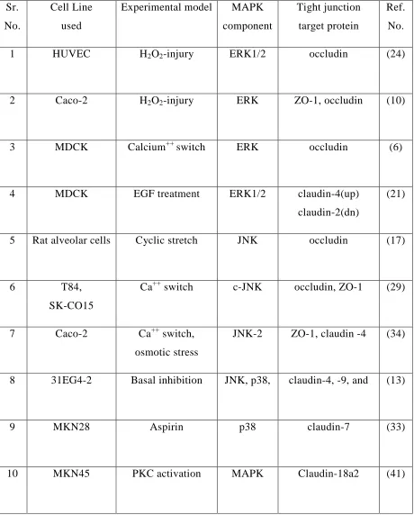
important in neoplastic processes involving gastric mucosa. For instance, claudin-18a2 is a splice variant of claudin-18 gene that is involved in cell adhesion as well as the paracellular barrier for H+ in the gastric mucosa (22). In gastric cancers of intestinal phenotype claudin-18a2 is down regulated and activation of PKC with phorbol 12-myristate 13-acetate (PMA) increases claudin-18a2 expression in MKN45 human gastric cancer cell line (41). Furthermore, the expression of claudin-18a2 for which two activator protein (AP)-1 promoter binding sites were identified to be critical, was suppressed by the use of PKC and MAPK inhibitors, indicating importance of PKC/MAPK/AP-1 signaling pathway in the regulation of TJ protein claudin-18 (41). Recent evidence indicates that besides the well-established role of MAPK signaling in cell differentiation and cell proliferation, MAPK are also involved in the regulation of epithelial barrier dysfunction via TJ permeability (1, 18, 21, 33, 34).
Mitogen Activated Protein Kinase (MAPK) signaling
(14, 18). Extracellular kinases that have been added more recently included ERK3, ERK4, ERK5, ERK 7, and ERK 8 (7, 25). The most well characterized groups of MAPK are ERK1/2, p38, and JNK (Figure 1) which are activated by either extracellular signals or by various stress factors.
Figure 1: Mitogen-activated protein kinases (MAPK) are a family of Ser/Thr protein kinases widely conserved among eukaryotes and are involved in many cellular programs such as cell proliferation, cell differentiation, cell movement and cell death. MAPK signaling cascades are organized hierarchically into three-tiered modules. MAPKs are phosphorylated and activated by MAPK-kinases (MAPKKs), which in turn are phosphorylated and activated by MAPKK-kinases (MAPKKKs). The MAPKKKs are in turn activated by interaction with the family of small GTPases and/or other protein kinases, connecting the MAPK module to cell
surface receptors or external stimuli (adapted from
ERK1/2
has been shown to increase gastric cancer risk via activation of the ERK signaling pathway and ERK single-nucleotide polymorphism (SNP) rs5999749 was found to be one of the important genetic determinants for gastric carcinogenesis (40). Although the mechanism by which CagA-mediated ERK signaling affects TJ barrier in the stomach mucosa is not clear, intracellular CagA is associated with the epithelial TJ intracytoplasmic protein ZO-1 (38). Furthermore, the interaction of CagA with PAR1b/MARK2 polarity-regulating kinase has been shown to disrupt the tight junctions (3).
JNK-c-Jun-NH2 terminal activated Protein Kinases (SAPK- Stress activated kinases)
regulated by different MAPK components. Similar recent studies by Samak et al. (34) used calcium switch models in Caco-2 cells and osmotic stress in JNK1 and JNK 2 knock out mouse studies to demonstrate that JNK-2 and not JNK-1 is involved in inducing epithelial barrier dysfunction via down regulation of the TJ protein ZO-1 and up regulation of claudin-4 following osmotic stress-induced TJ disruption in intestinal epithelium. Further in vivo studies are needed to elucidate the exact roles of distinct isoforms of JNK and the mechanism involved in TJ disassembly.
p38 Mitogen Activated Protein Kinases
The p38 mitogen activated proteins kinases (p38) are stress-activated kinases that, together with ERK1/2, and JNK form the critical components of the mitogen activated protein kinase (MAPK) cascades. Four p38 isoforms (p38α, p38β, p38γ, and p38δ) have
effects of p38 on claudin expression. Recently Chuenkitivanon et al. (15) showed that H2O2 induced hyper permeability in ECV304 endothelial cells originating from human umbilical vein involved phosphorylation of p38 MAPK and down regulation of ZO-1 and occludin. It is presumed that p38 MAPK-mediated disruption of tight junctions and adherens junctions contributes to acquisition of malignant properties noted in poorly differentiated gastric phenotypes. In one study, expression of E-cadherin, ZO-1, occludin and claudin-4 was found to be reduced in poorly differentiated gastric cancer cell lines KATO-III and MKN45 cells as compared to well differentiated MKN7 and MKN28 gastric cells (4).
Effect of MAPK on Tight Junctions
Table 1: MAPK signaling at Tight Junction Sr.
No.
Cell Line used
Experimental model MAPK component
Tight junction target protein
Ref. No.
1 HUVEC H2O2-injury ERK1/2 occludin (24)
2 Caco-2 H2O2-injury ERK ZO-1, occludin (10)
3 MDCK Calcium++ switch ERK occludin (6)
4 MDCK EGF treatment ERK1/2 claudin-4(up) claudin-2(dn)
(21)
5 Rat alveolar cells Cyclic stretch JNK occludin (17)
6 T84,
SK-CO15
Ca++ switch c-JNK occludin, ZO-1 (29)
7 Caco-2 Ca++ switch, osmotic stress
JNK-2 ZO-1, claudin -4 (34)
8 31EG4-2 Basal inhibition JNK, p38, claudin-4, -9, and (13)
9 MKN28 Aspirin p38 claudin-7 (33)
Conclusions
proliferation, cell differentiation, apoptosis, and epithelial barrier dysfunction. Therefore, it will be important to study cross talk between TJs and other well established regulatory systems such as cadherin–based adhesion signaling and signaling from the extracellular matrix (7, 42). It seems that activation or phosphorylation of MAPK is associated with epithelial barrier dysfunction and thus is of immense therapeutic potential in important gastrointestinal disorders/diseases such as Inflammatory Bowel Syndrome (IBS), Inflammatory Bowel Disease, and NASID induced gastro-enteropathy.
REFERENCES
1. Aggarwal S, Suzuki T, Taylor WL, Bhargava A and Rao R. Contrasting Effects of ERK on Tight Junction Integrity in Differentiated and Under-Differentiated Caco-2 Cell Monolayers. Biochem.J. 2010.
2. Aijaz S, Balda MS and Matter K. Tight junctions: molecular architecture and function. Int.Rev.Cytol. 248: 261-298, 2006.
3. Amieva MR, Vogelmann R, Covacci A, Tompkins LS, Nelson WJ and Falkow S. Disruption of the epithelial apical-junctional complex by Helicobacter pylori CagA. Science 300: 5624: 1430-1434, 2003.
4. Atsumi T, Kato K, Uno K, Iijima K, Koike T, Imatani A, Ohara S and Shimosegawa T. Pathophysiological role of the activation of p38 mitogen-activated protein kinases in poorly differentiated gastric cancer. Pathol.Int. 57: 10: 635-644, 2007.
6. Balda MS, Gonzalez-Mariscal L, Matter K, Cereijido M and Anderson JM. Assembly of the tight junction: the role of diacylglycerol. J.Cell Biol. 123: 2: 293-302, 1993.
7. Balda MS and Matter K. Epithelial cell adhesion and the regulation of gene expression. Trends Cell Biol. 13: 6: 310-318, 2003.
8. Balda MS and Matter K. Transmembrane proteins of tight junctions. Semin.Cell Dev.Biol. 11: 4: 281-289, 2000.
9. Basuroy S, Dunagan M, Sheth P, Seth A and Rao RK. Hydrogen peroxide activates focal adhesion kinase and c-Src by a phosphatidylinositol 3 kinase-dependent mechanism and promotes cell migration in Caco-2 cell monolayers. Am.J.Physiol.Gastrointest.Liver Physiol. 299: 1: G186-95, 2010.
10. Basuroy S, Seth A, Elias B, Naren AP and Rao R. MAPK interacts with occludin and mediates EGF-induced prevention of tight junction disruption by hydrogen peroxide. Biochem.J. 393: Pt 1: 69-77, 2006.
12. Bruewer M, Hopkins AM, Hobert ME, Nusrat A and Madara JL. RhoA, Rac1, and Cdc42 exert distinct effects on epithelial barrier via selective structural and biochemical modulation of junctional proteins and F-actin. Am.J.Physiol.Cell.Physiol. 287: 2: C327-35, 2004.
13. Carrozzino F, Pugnale P, Feraille E and Montesano R. Inhibition of basal p38 or JNK activity enhances epithelial barrier function through differential modulation of claudin expression. Am.J.Physiol.Cell.Physiol. 297: 3: C775-87, 2009.
14. Chen Y, Lu Q, Schneeberger EE and Goodenough DA. Restoration of tight junction structure and barrier function by down-regulation of the mitogen-activated protein kinase pathway in ras-transformed Madin-Darby canine kidney cells. Mol.Biol.Cell 11: 3: 849-862, 2000.
17. Cohen TS, Gray Lawrence G, Khasgiwala A and Margulies SS. MAPK activation modulates permeability of isolated rat alveolar epithelial cell monolayers following cyclic stretch. PLoS One 5: 4: e10385, 2010.
18. Davis RJ. Signal transduction by the JNK group of MAP kinases. Cell 103: 2: 239-252, 2000.
19. de Almeida JB, Holtzman EJ, Peters P, Ercolani L, Ausiello DA and Stow JL. Targeting of chimeric G alpha i proteins to specific membrane domains. J.Cell.Sci. 107 ( Pt 3): Pt 3: 507-515, 1994.
20. Denker BM, Saha C, Khawaja S and Nigam SK. Involvement of a heterotrimeric G protein alpha subunit in tight junction biogenesis. J.Biol.Chem. 271: 42: 25750-25753, 1996.
21. Flores-Benitez D, Rincon-Heredia R, Razgado LF, Larre I, Cereijido M and Contreras RG. Control of tight junctional sealing: roles of epidermal growth factor and prostaglandin E2. Am.J.Physiol.Cell.Physiol. 297: 3: C611-20, 2009.
23. Hewitt KJ, Agarwal R and Morin PJ. The claudin gene family: expression in normal and neoplastic tissues. BMC Cancer 6: 186, 2006.
24. Kevil CG, Oshima T, Alexander B, Coe LL and Alexander JS. H(2)O(2)-mediated permeability: role of MAPK and occludin. Am.J.Physiol.Cell.Physiol. 279: 1: C21-30, 2000.
25. Kyriakis JM and Avruch J. Mammalian mitogen-activated protein kinase signal transduction pathways activated by stress and inflammation. Physiol.Rev. 81: 2: 807-869, 2001.
26. Leone V, di Palma A, Ricchi P, Acquaviva F, Giannouli M, Di Prisco AM, Iuliano F and Acquaviva AM. PGE2 inhibits apoptosis in human adenocarcinoma Caco-2 cell line through Ras-PI3K association and cAMP-dependent kinase A activation. Am.J.Physiol.Gastrointest.Liver Physiol. 293: 4: G673-81, 2007.
27. Li D and Mrsny RJ. Oncogenic Raf-1 disrupts epithelial tight junctions via downregulation of occludin. J.Cell Biol. 148: 4: 791-800, 2000.
29. Naydenov NG, Hopkins AM and Ivanov AI. c-Jun N-terminal kinase mediates disassembly of apical junctions in model intestinal epithelia. Cell.Cycle 8: 13: 2110-2121, 2009.
30. Nigam SK, Denisenko N, Rodriguez-Boulan E and Citi S. The role of phosphorylation in development of tight junctions in cultured renal epithelial (MDCK) cells. Biochem.Biophys.Res.Commun. 181: 2: 548-553, 1991.
31. Nilsson M, Fagman H and Ericson LE. Ca2+-dependent and Ca2+-independent regulation of the thyroid epithelial junction complex by protein kinases. Exp.Cell Res. 225: 1: 1-11, 1996.
32. Nusrat A, Giry M, Turner JR, Colgan SP, Parkos CA, Carnes D, Lemichez E, Boquet P and Madara JL. Rho protein regulates tight junctions and perijunctional actin organization in polarized epithelia. Proc.Natl.Acad.Sci.U.S.A. 92: 23: 10629-10633, 1995.
33. Oshima T, Miwa H and Joh T. Aspirin induces gastric epithelial barrier dysfunction by activating p38 MAPK via claudin-7. Am.J.Physiol.Cell.Physiol. 295: 3: C800-6, 2008.
35. Steed E, Balda MS and Matter K. Dynamics and functions of tight junctions. Trends Cell Biol. 20: 3: 142-149, 2010.
36. Takaishi K, Sasaki T, Kotani H, Nishioka H and Takai Y. Regulation of cell-cell adhesion by rac and rho small G proteins in MDCK cells. J.Cell Biol. 139: 4: 1047-1059, 1997.
37. Tsukita S, Furuse M and Itoh M. Multifunctional strands in tight junctions. Nat.Rev.Mol.Cell Biol. 2: 4: 285-293, 2001.
38. Tummala S, Keates S and Kelly CP. Update on the immunologic basis of Helicobacter pylori gastritis. Curr.Opin.Gastroenterol. 20: 6: 592-597, 2004.
39. Wang Y, Zhang J, Yi XJ and Yu FS. Activation of ERK1/2 MAP kinase pathway induces tight junction disruption in human corneal epithelial cells. Exp.Eye Res. 78: 1: 125-136, 2004.
41. Yano K, Imaeda T and Niimi T. Transcriptional activation of the human claudin-18 gene promoter through two AP-1 motifs in PMA-stimulated MKN45 gastric cancer cells. Am.J.Physiol.Gastrointest.Liver Physiol. 294: 1: G336-43, 2008.
42. Yu QH and Yang Q. Diversity of tight junctions (TJs) between gastrointestinal epithelial cells and their function in maintaining the mucosal barrier. Cell Biol.Int. 33: 1: 78-82, 2009.
Chapter III
GASTRO-PROTECTIVE PROPERTIES OF THE NOVEL PROSTONE SPI-0811 IN ACID-INJURED PORCINE GASTRIC MUCOSA
ABSTRACT
Aim: To evaluate the protective properties of novel prostone ClC-2 agonist SPI-0811 in porcine model of gastric acid injury.
Method: Porcine gastric mucosa was mounted in Ussing chambers and injured by bathing mucosal tissues in an HCl Ringer’s solution (pH 1.5) with or without SP1-0811(1µM), CFTR inhibitor (inhibitor 172, 10µM, apical) and ClC-2 inhibitor ZnCl2, 300µM, apical), on the apical surface of tissues. Transepithelial resistance and mucosal-to-serosal 3H-mannitol fluxes were measured over a 90-minute period. Tissues were analyzed by morph metric techniques, immunofluorescence and by western blots.
Results: Compared with control tissues, acid exposure decreased TER and increased 3 H-mannitol flux. Pretreatment of gastric mucosa with SPI-0811 was protective against acid-induced decreases in TER (TER Ω.cm2 = 50 vs. 100) and abolished increases in flux (3 H-mannitol flux 0.10 vs. 0.04 µM.cm2). Evidence of histological damage in the presence of acid was markedly attenuated by SPI-0811. Immunofluroscence and western analysis for occludin revealed enhanced localization to the region of the tight junction after treatment with SPI-0811. Pretreatment with the ClC-2 inhibitor ZnCl2, but not the selective CFTR inhibitor 172, attenuated SPI-0811-mediated mucosal protection, suggesting a role for ClC-2. Prostone may serve both protective and reparative roles in injured tissues.
Conclusion: ClC-2 agonist SPI-0811 stimulated enhancement of mucosal barrier function by protecting tight junction protein occludin in porcine gastric mucosa and thus protected the gastric acid injury in porcine stomach.
INTRODUCTION It is becoming increasingly evident that many patients suffer from gastric ulcers, particularly in groups of patients such as those in intensive care facilities [1-4]. This suggests that medications that provide gastro protection have the potential to reduce morbidity associated with gastric ulceration. For decades, agents that suppress acid secretion [5-7] have been widely used for treatment of gastric ulcers[1,7-10].Gastric ulcer disease and repair is complex, involving inflammation, cell proliferation, formation of granulation tissue, and angiogenesis[3,4,11] .However, gastro protection has also been studied in depth. For example, studies with rebamipide or misoprostol, geranylgeranyl hydrochloride (HSP70) have revealed that these compounds have gastro protective properties as well enhancing ulcer healing [2,7,12]
.Nonetheless, our understanding of the mechanisms of gastro protection is incomplete and there is the prospect of novel pharmacological agents, aside from antacids, proton pump inhibitors, and prostanoid activators or analogs, which might protect the stomach[2,13].
that ensure rapid repair of defects in the epithelial monolayer [3,4,15-17]. The next line of mucosal defense is formed by a continuous layer of surface epithelial cells which secrete mucus and bicarbonate and generate PGs, heat shock proteins, TFFs, and cathelicidins. Because of the presence of phospholipids on their surfaces, these cells are hydrophobic, repelling acid- and water-soluble damaging agents. Interconnected by tight junctions, the surface epithelial cells form a “barrier” preventing back diffusion of acid and pepsin. Once the epithelial barrier is disrupted, epithelial repair mechanisms must rapidly re-form a continuous epithelial monolayer in order to prevent entry of protons and bacteria [3,4,14,15,18]. The remarkable phenomenon of epithelial restitution during which epithelium re-seals mucosal defects in the presence of acidic environment has been a subject of several studies.
fluxes of 3H-mannitol in ischemic-injured intestinal mucus [20, 22-24]. Given the previous findings that activation of ClC-2 by the prostone Lubiprostone [22, 25 ] enhanced recovery of barrier function in ischemic-injured porcine intestine, the present study was performed to evaluate the ability of the novel ClC-2 agonist SPI-0811 to provide gastro protection in acid-injured porcine gastric mucosa.
MATERIALS AND METHODS
Chemicals
ZnCl2, Bumetanide, 3H-mannitol, and CFTR inhibitor 172 were purchased from Sigma-Aldrich (St. Louis, MO). SPI-0811 was provided by Sucampo Pharmaceuticals Inc., Bethesda, MD.
Experimental Animals
Ussing chamber studies
Experimental Treatments
Once tissues were mounted on Ussing chambers, treatments aimed at inhibiting ClC-2 (ZnCl2, 300µM, apical), CFTR (inhibitor 172, 10µM, apical), or NKCC1 (bumetanide, 100µM, basolateral) were added. Alternatively, SPI-0811 was administered to the apical surface of tissues (1µM) to enhance epithelial Cl- secretion via ClC-2. Following a 30 min equilibration period, HCl (1N in Ringer’s) was added to the mucosal surface of the tissue to induce acid injury. The pH was monitored with a pH meter (Hanna Instruments Inc., Ann Arbor, MI, USA) and maintained at 1.5 by adding 1N HCl as needed during the experiment.
Mucosal-to-serosal fluxes of 3H-mannitol
To assess mucosal permeability, 0.2µCi/ml 3H-mannitol was placed on the mucosal side of tissues after experimental treatments. After a 15 min equilibration period, standards were taken from the mucosal side of each chamber and a 30 min flux period was established by taking 0.5ml samples from the serosal compartments. The presence of 3H was established by measuring emission in a liquid scintillation counter (Rack Beta, Perkin Elmer Life and Analytical Sciences, Boston MA, USA). Unidirectional mucosal-to-serosal 3H-mannitol fluxes were calculated using a previously established spreadsheet [17].
Histological analyses
(5µm) and stained with hematoxylin and eosin and PAS. For each tissue, mucosal epithelial lining and gastric pits (crypts) were identified to assess the damage caused by acid (pH 1.5).
Immunofluorescence labeling of occludin
For this procedure, tissues were embedded in optimal cutting temperature (OCT) medium, frozen, and sectioned at 5µm. Tissue sections were blocked with 2% BSA followed by incubation with rabbit anti-occludin polyclonal antibody (1:150, Zymed, San Francisco, CA, USA) overnight at 40C. Sections were washed with PBS and incubated for 45 min with FITC-conjugated anti-rabbit secondary antibody. Sections were mounted in fluorescent mounting medium, and well-orientated gastric pits were examined with a photomicroscope linked to a digital camera.
Gel electrophoresis and western blotting
Following Ussing chamber experiments, gastric mucosal samples were snap frozen and stored at -70°C prior to performing SDS-PAGE. Tissue aliquots were thawed at 4°C and added to chilled lysis buffer, including protease inhibitors (0.5mM Pefabloc, 0.1mM 4-nitrophenyl phosphate, 0.04 mM glycerophosphate, 0.1mM Na3VO4, 40μg/ml bestatin, 2μg/ml aprotinin, 0.54μg/ml eupeptic, and 0.7μg/ml pepstatin A) (Sigma-Aldrich Inc., St.
and boiled for 4 min. Lysates were loaded on an SDS polyacrylamide gradient gel, and electrophoresis was carried out according to standard protocols. Proteins were transferred to a PVDF membrane (Immobilon®, Millipore, Billerica, MA, USA) by using an electro blotting minitransfer apparatus. Membranes were blocked at room temperature for 2 h in Tris-buffered saline plus 0.05% Tween 20 (TBST) and 5% dry powdered milk, and then incubated overnight in primary antibody at 4°C in rabbit anti-rat ClC-2 (Alpha Diagnostics, San Antonio, TX, USA), rabbit anti-CFTR (Santa Cruz Biotech Inc., CA, USA) or rabbit anti-occludin (Zymed Laboratories Inc., San Francisco, CA, USA). After multiple washings in TBST, membranes were incubated with horseradish peroxidase conjugated secondary antibody, and developed for visualization of protein with luminol enhancer solution (Pierce, Rockford, IL, USA).
For preparation of detergent soluble and detergent insoluble fractions of gastric mucosa, the tissue samples were extracted in lysis buffer (20mM Tris, 5mM MgCl2, 0.3mM EGTA, 210μg/ml sodium fluoride, 18.5μg/ml sodium orthovanadate, 30mM sodium
Statistical analyses
Data were reported as mean ±SE. For Ussing chamber experiments, an experimental number of 6 was used (n=6). Data was analyzed by using an analysis of variance (ANOVA) and t-test using a commercial statistical package (Sigmastat, Systat Software, San Jose, CA, USA). The significance level was set at p<0.05.
RESULTS
Acid injury model
Effects of the ClC-2 agonist SPI-0811 on TER and mucosal-to-serosal 3H-mannitol fluxes in acid-injured porcine gastric mucosa
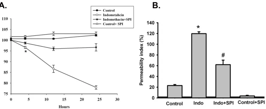
As previously described, porcine gastric mucosa was mounted on Ussing chambers and the mucosal surface was exposed to acid Ringer’s (pH 1.5) for 90 minutes. Mucosa subjected to acid injury had significantly lower TER values when compared to the gastric mucosa bathed in normal Ringer’s, indicating electrophysiological evidence of disruption of epithelial barrier function. Alternatively, pretreatment of the apical side of gastric mucosa with the ClC-2 agonist SPI-0811 (1µM) nullified decreases in TER due to acid injury (fig 1A). Epithelial permeability was assessed by mucosal-to- serosal fluxes of 3H-mannitol in control and acid-injured tissues in the presence or absence of apical SPI-0811. In agreement with TER responses, acid-injured tissues exhibited increases in permeability to 3H- mannitol as compared to control uninjured tissue. Pretreatment of the apical surface of mucosal tissues SPI-0811 ablated changes in permeability noted in untreated acid-injured tissues (fig 1B).
Histological and Immunofluorescence microscopic findings in acid-injured gastric mucosa
acid, although the epithelium appeared to have flattened to maintain the barrier and the gastric pits appeared dilated (fig 2A).
Using PAS staining for mucus we observed that the loss of mucus by acid injury was seen restored by pretreatment of ClC-2 agonist (fig 2B). In further experiments, we performed Immunofluorescence of occludin to assess the gastric mucosal tight junctions (TJ). As compared to apical immunolocalization of occludin in the control uninjured gastric mucosal lining epithelium, acid-injured tissues revealed loss of occludin Immunofluorescence (fig 2C). In contrast, gastric mucosa pretreated with SPI-0811 and subsequently exposed to acid for 90 minutes had evidence of localization of occludin to the TJ.
Expression of occludin in gastric mucosal tissues
acid injury was severe enough to cause surface epithelium and inter epithelial tight junctions. On the other hand, occludin expression in the detergent insoluble fraction of tissues treated with SPI-0811 appeared similar to tissues treated with Ringer’s solution alone, suggesting this prostone prevented loss of occludin. Cellular expression of occludin as a whole was also seen markedly reduced compared to control tissue and this loss was prevented with pretreatment with SPI-0811(fig. 3A and 3B).
Role of Cl- channels in SPI-0811-mediated protection of TER and barrier permeability in acid-injured gastric mucosa
To explore the potential role of Cl- secretion in SPI-0811-associated gastro protection, mucosa was treated with the NKCC1 inhibitor bumetanide (100µM, basolateral) and SPI-0811 (1µM, apical), which blocked the ability of SPI-0811 to reduce acid injury (data not shown). In more targeted studies in tissues pretreated with SPI-0811, tissues were pretreated with pharmacological inhibitors of the apical chloride channels ClC-2 and CFTR. Pretreatment of acid-injured mucosa with the ClC-2 inhibitor ZnCl2 (300 µM, apical) abolished the gastro protective properties of SPI-0811 as determined by change in TER and mannitol fluxes. On the other hand, pretreatment with CFTR inhibitor 172 (10µM, apical) had no effect (fig 4A, B)
Expression of ClC-2 and CFTR in porcine gastric mucosa
CFTR in porcine gastric mucosa. As shown in figure 5, ClC-2 was expressed ~98 kDA in porcine gastric homogenates. ClC-2 expression was observed in porcine gastric mucosa which confirmed the expression of ClC-2 in porcine stomach. Western analyses showed no evidence of CFTR in gastric mucosa, whereas CFTR was clearly present in porcine jejunum as a positive control (data not shown).
Discussion
protection and acute gastric injury are the direct application of acid or ethanol to gastric mucosa [8, 31].
explanation would be that the extensive reduction in pH resulted in loss of tight junction proteins into the lumen, followed by erosion of epithelium and ultimately ulceration. Tissues pretreated with SPI-0811 evaluated by immunofluorescence were protected from this effect, with localization of occludin to the apical epithelium similar to that of control tissues. Additionally, occludin remained in the detergent insoluble fraction in western analyses under the influence of SPI-0811 indicating localization to the TJs.
There are opposing views regarding the role of ClC-2 in gastric chloride secretion; some investigators have suggested involvement of ClC-2 in gastric acid secretion [29, 36, 37] while another group has indicated that ClC-2 chloride secretion plays no role in production of HCl [38]. It is also noteworthy that the apical ClC-2 channel has been suggested to serve as a route for both bicarbonate and Cl- exit into the lumen [29, 39].This raises the possibility that ClC-2-mediated bicarbonate secretion attenuates acid injury to some extent by raising the pH. However in our studies, constant monitoring and adjustment of pH to a level of 1.5 would nullify this possibility. We also found that SPI-0811 produces a gastro protective effect not only via increasing gastric pH, but also by protecting and restoring the gastric
mucus level (fig 2B). The importance of gastro protection via mucus protection has been
previously shown [7, 40, 34].
Figure 1: Barrier function of acid injured porcine gastric mucosa to pretreatment of SPI-0811 (1µM). (A) Porcine gastric mucosa mounted on Ussing chambers challenged with mucosal acid (HCl, pH 1.5) over a 90 minute period had significantly lower TER when compared with control tissues bathed in normal Ringer’s (NR) (TER Ω.cm2
=100 for acid-treated tissues versus 50 for control tissues). Mucosal application of the ClC-2 agonist SPI- (Fig1. continued from page 68)
Figure 2. Histological findings in acid-injured gastric mucosal tissues. (A) Tissues bathed in normal Ringer’s appeared uninjured after 90 minutes in Ussing chambers, whereas those exposed to acid had substantial evidence of epithelial injury. Pretreatment with SPI-0811 ameliorated injury evident in acid-injured tissues. Bar = 100µm. Each panel is representative of at least 3 separate animals. (B). PAS-Alcian blue staining findings in Control tissues (Figure 2.continued on page 70)
REFERENCES
1. Montrose, MH. Choosing sides in the battle against gastric acid. J Clin Invest 2001; 108: 1743-1744 [PMID: 11748256 DOI: 10.1172/JCI14652].
2. Park, SH, Cho, CS, Lee, OY, Jun, JB, Lin, SR, Zhou, LY, Yuan, YZ, Li, ZS, Hou, XH, Zhao, HC, Kachintorn, U, Kositchaiwat, C, Lertkupinit, C . Comparison of Prevention of NSAID-Induced Gastrointestinal Complications by Rebamipide and Misoprostol: A Randomized, Multicenter, Controlled Trial-STORM STUDY. J Clin Biochem Nutr 2007; 40: 148-155 [PMID: 18188417 DOI:10.3164/jcbn.40.148].
3. Tarnawski, A . Molecular mechanisms of ulcer healing. Drug News Perspect 2000; 13: 158-168 [PMID: 12937606].
4. Tarnawski, AS. Cellular and molecular mechanisms of gastrointestinal ulcer healing. Dig Dis Sci 2005; 50 Suppl 1: S24-33 [PMID: 16184417 DOI:10.1007/s10620-005-2803-6].
6. Romano, M, Razandi, M, Ivey, KJ . Role of sulphydryl compounds in the defense of rat gastric epithelial cells against oxygen reactive metabolite-induced damage. Ital J Gastroenterol 1991; 23: 55-59 [PMID: 1747503].
7. Romano, M, Razandi, M, Ivey, KJ. Effect of cimetidine and ranitidine on drug induced damage to gastric epithelial cell monolayers in vitro. Gut 1989; 30: 1313-1322 [PMID: 2583559].
8. Okabe, S, Amagase, K . An overview of acetic acid ulcer models--the history and state of the art of peptic ulcer research. Biol Pharm Bull 2005; 28: 1321-1341 [PMID:
16079471].
9. Okabe, S, Amagase, K . An overview of acetic acid ulcer models and their utility for drug screening. Nippon Yakurigaku Zasshi 2003; 122: 73-92 [PMID: 12843575].
10. Suzuki, T, Yoshida, N, Nakabe, N, Isozaki, Y, Kajikawa, H, Takagi, T, Handa, O, Kokura, S, Ichikawa, H, Naito, Y, Matsui, H, Yoshikawa, T . Prophylactic effect of rebamipide on aspirin-induced gastric lesions and disruption of tight junctional protein zonula occludens-1 distribution. J Pharmacol Sci 2008; 106: 469-477 [PMID:
11. Wallace, JL, Granger, DN . The cellular and molecular basis of gastric mucosal defense. FASEB J 1996; 10: 731-740 [PMID: 8635690].
12. Aihara, E, Hayashi, S, Amagase, K, Kato, S, Takeuchi, K . Prophylactic effect of rebamipide against the irritative and healing impairment actions of alendronate in rat stomachs. Inflammopharmacology 2007; 15: 196-202 [PMID: 17943251
DOI:10.1007/s10787-007-1592-2].
13. Arakawa, T, Kobayashi, K, Yoshikawa, T, Tarnawski, A . Rebamipide: overview of its mechanisms of action and efficacy in mucosal protection and ulcer healing. Dig Dis Sci 1998; 43: 5S-13S [PMID: 9753220].
14. Allen, A, Flemstrom, G, Garner, A, Kivilaakso, E . Gastroduodenal mucosal protection. Physiol Rev 1993; 73: 823-857 [PMID: 8415927].
15. Allen, A, Flemstrom, G . Gastroduodenal mucus bicarbonate barrier: protection against acid and pepsin. Am J Physiol Cell Physiol 2005; 288: C1-19 [PMID: 15591243
DOI:10.1152/ajpcell.00102.2004].
17. Rainsford, KD. Mechanisms of gastrointestinal damage by NSAIDS. Agents Actions Suppl 1993; 44: 59-64 [PMID: 8372736].
18. Ito, S, Lacy, ER, Rutten, MJ, Critchlow, J, Silen, W . Rapid repair of injured gastric mucosa. Scand J Gastroenterol Suppl 1984; 101: 87-95 [PMID: 6336239].
19. Matter, K, Balda, MS . Occludin and the functions of tight junctions. Int Rev Cytol 1999; 186: 117-146 [PMID: 9770298].
20. Blikslager, AT, Moeser, AJ, Gookin, JL, Jones, SL, Odle, J . Restoration of barrier function in injured intestinal mucosa. Physiol Rev 2007; 87: 545-564 [PMID: 17429041 DOI:10.1152/physrev.00012.2006].
21. Moeser, AJ, Haskell, MM, Shifflett, DE, Little, D, Schultz, BD, Blikslager, AT . ClC-2 chloride secretion mediates prostaglandin-induced recovery of barrier function in ischemia-injured porcine ileum. Gastroenterology 2004; 127: 802-815 [PMID:
15362036].
23. Blikslager, AT, Roberts, MC, Rhoads, JM, Argenzio, RA. Prostaglandins I2 and E2 have a synergistic role in rescuing epithelial barrier function in porcine ileum. J Clin Invest 1997; 100: 1928-1933 [PMID: 9329955 DOI: 10.1172/JCI119723].
24. Takezono, Y, Joh, T, Oshima, T, Suzuki, H, Seno, K, Yokoyama, Y, Alexander, JS, Itoh, M . Role of prostaglandins in maintaining gastric mucus-cell permeability against acid exposure. J Lab Clin Med 2004; 143: 52-58 [PMID: 14749685 DOI:
10.1016/S0022214303001896].
25. Bao, HF, Liu, L, Self, J, Duke, BJ, Ueno, R, Eaton, DC . A synthetic prostone
activates apical chloride channels in A6 epithelial cells. Am J Physiol Gastrointest Liver Physiol 2008; 295: G234-51 [PMID: 18511742 DOI:10.1152/ajpgi.00366.2007].
26. Wong, V . Phosphorylation of occludin correlates with occludin localization and function at the tight junction. Am J Physiol 1997; 273: C1859-67 [PMID: 9435490].
27. Nighot, PK, Blikslager, AT . ClC-2 regulates mucosal barrier function associated with structural changes to the villus and epithelial tight junction. Am J Physiol Gastrointest Liver Physiol 2010; 299: G449-56 [PMID: 20489043 DOI:10.1152/ajpgi.00520.2009].
jejunum. Exp Cell Res 2009; 315: 110-118 [PMID: 18976652 DOI:10.1016/j.yexcr.2008.10.001].
29. Malinowska, DH . Cl- channel blockers inhibit acid secretion in rabbit parietal cells. Am J Physiol 1990; 259: G536-43 [PMID: 1699427]
.
30. Cuppoletti, J, Malinowska, DH, Tewari, KP, Li, QJ, Sherry, AM, Patchen, ML, Ueno, R . SPI-0211 activates T84 cell chloride transport and recombinant human ClC-2 chloride currents. Am J Physiol Cell Physiol 2004; 287: C1173-83 [PMID: 15213059 DOI:10.1152/ajpcell.00528.2003].
31. Morris, GP, Wallace, JL. The roles of ethanol and of acid in the production of gastric mucosal erosions in rats. Virchows Arch B cell Pathol Incl Mol Pathol 1981; 38: 23-38 [PMID: 6119834].
32. Akiba, Y, Furukawa, O, Guth, PH, Engel, E, Nastaskin, I, Sassani, P, Dukkipatis, R, Pushkin, A, Kurtz, I, Kaunitz, JD. Cellular bicarbonate protects rat duodenal
mucosa from acid-induced injury. J Clin Invest 2001; 108: 1807-1816 [PMID: 11748264 DOI: 10.1172/JCI12218].
34. Romano, M, Razandi, M, Ivey, KJ . Effect of ranitidine on taurocholate-, ethanol-, and indomethacin-induced damage to gastric epithelial cells in vitro. Digestion 1989; 43: 212-221 [PMID: 2612744].
35. Romano, M, Razandi, M, Sekhon, S, Krause, WJ, Ivey, KJ . Human cell line for study of damage to gastric epithelial cells in vitro. J Lab Clin Med 1988; 111: 430-440 [PMID: 3127509].
36. Sherry, AM, Malinowska, DH, Morris, RE, Ciraolo, GM, Cuppoletti, J.
Localization of ClC-2 Cl- channels in rabbit gastric mucosa. Am J Physiol Cell Physiol 2001; 280: C1599-606 [PMID: 11350755].
37. Kosiek, O, Busque, SM, Foller, M, Shcheynikov, N, Kirchhoff, P, Bleich, M, Muallem, S, Geibel, JP . SLC26A7 can function as a chloride-loading mechanism in parietal cells. Pflugers Arch 2007; 454: 989-998 [PMID: 17404755 DOI:
10.1007/s00424-007-0254-y].
38. Hori, K, Takahashi, Y, Horikawa, N, Furukawa, T, Tsukada, K, Takeguchi, N, Sakai, H . Is the ClC-2 chloride channel involved in the Cl- secretory mechanism of gastric parietal cells? FEBS Lett 2004; 575: 105-108 [PMID: 15388342
39. Hogan, DL, Crombie, DL, Isenberg, JI, Svendsen, P, Schaffalitzky de Muckadell, OB, Ainsworth, MA. Acid-stimulated duodenal bicarbonate secretion involves a CFTR-mediated transport pathway in mice. Gastroenterology 1997; 113: 533-541 [PMID: 9247473].
40. Rainsford, KD, Willis, C . Relationship of gastric mucosal damage induced in pigs by antiinflammatory drugs to their effects on prostaglandin production. Dig Dis Sci 1982; 27: 624-635 [PMID: 6953009].
CHAPTER IV
THE PROTECTIVE EFFECT OF ClC-2 AGONIST SPI-0811 ON INDOMETHACIN –INDUCED EPITHELIAL BARRIER DYSFUNCTION IN