GENERAL ABSTRACT
SHIM, MINSUB. Proteasomal Regulation of CCAAT/Enhancer Binding Protein alpha (C/EBPα) and Diminished Expression of C/EBPα in Squamous Cell Carcinomas
The CCAAT/enhancer binding proteins (C/EBPs) are members of the basic leucine zipper (bZIP) class of transcription factors. C/EBPα is involved in mitotic
growth arrest/differentiation and has been implicated as a human tumor suppressor in acute myeloid leukemia. C/EBPα is abundantly expressed in mouse keratinocytes. The purpose of this study was to examine the regulation of the C/EBPα protein and to determine if C/EBPα expression/function is altered in skin squamous cell carcinomas.
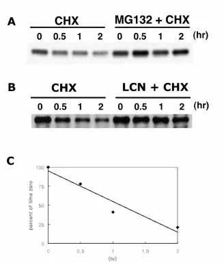
We found that C/EBPα is a short-lived protein with a half-life of ~ 1 hour and treatment with proteasome inhibitors blocked the degradation of C/EBPα protein. Poly-ubiquitinated C/EBPα were detected in BALB/MK2 and C/EBPα was degraded
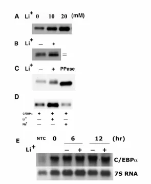
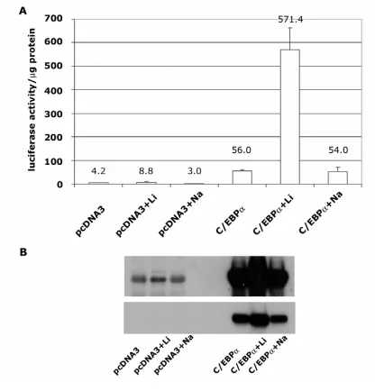
by the proteasome in an ATP- and ubiquitin-dependent manner. GSK3 is a known C/EBPα kinase and treatment of keratinocytes with LiCl, an inhibitor of GSK3 resulted in; i) an increase in C/EBPα protein levels, ii) increased electrophoretic mobility of C/EBPα protein and iii) no increase in C/EBPα mRNA levels suggesting that GSK3-mediated phosphorylation of C/EBPα may target it for proteasomal degradation. However, a mutant C/EBPα containing mutations in the GSK3 phosphorylation sites
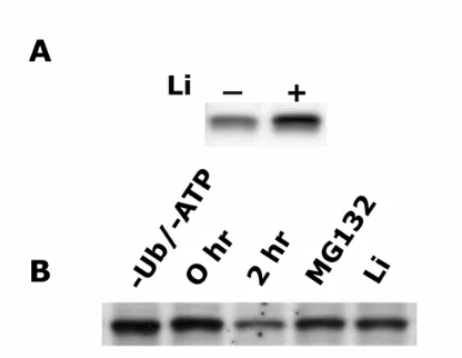
produced a six-fold increase in the half-life of C/EBPα protein. In vitro studies revealed that LiCl inhibited proteasomal degradation of C/EBPα. These results demonstrate C/EBPα is degraded via a proteasomal pathway and LiCl stabilizes C/EBPα through a GSK3 independent pathway involving inhibition of proteasome
activity.
The expression of C/EBPα was evaluated in mouse skin SCC lines. C/EBPα
mRNA and protein levels were greatly diminished or undetectable in all seven SCC cell lines compared to normal keratinocytes. Forced expression of C/EBPα resulted in the inhibition in SCC cell proliferation. Expression of C/EBPα also resulted in the
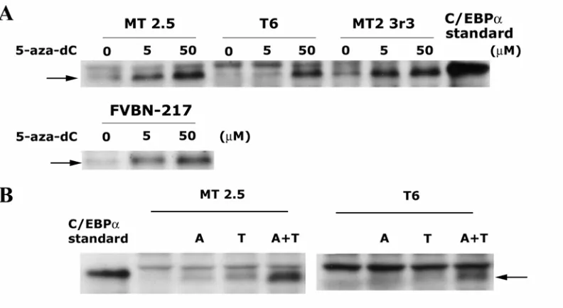
expression of loricrin, a late stage marker of squamous differentiation. Treatment with 5’-aza-deoxycytidine increased C/EBPα expression in some SCC cell lines suggesting the C/EBPα promoter region may be transcriptionally silenced by hypermethylation. C/EBPα expression was negligible in all 14/14 SCC examined compared to normal epidermis. These results suggest the loss of C/EBPα expression may contribute to the
PROTEASOMAL REGULATION OF CCAAT/ENHANCER BINDING
PROTEIN ALPHA (C/EBPα) AND DIMINISHED EXPRESSION OF C/EBPα IN
SQUAMOUS CELL CARCINOMAS
by
MINSUB SHIM
A dissertation submitted to the graduate faculty of North Carolina State University
in partial fulfillment of the requirements for the degree of Doctor of Philosophy
TOXICOLOGY
Raleigh
APPROVED BY
BIOGRAPHY
Minsub Shim was born on January 21, 1967 in Seoul, The Republic of Korea. He
enrolled Yonsei University, Seoul, The Republic of Korea in 1986 where he earned a
Bachelor of Science degree in Biochemistry in 1990. He attended the Graduate School
of Yonsei University in 1991 and got a Master of Science degree in Biochemistry in
1993. He then began working as a researcher in LG R&D Institute, Daejeon, The
Republic of Korea until 1998. And then he started working toward a Ph.D. in
Toxicology at North Carolina State University, Raleigh, North Carolina under the
ACKNOWLEDGMENTS
I extend my most sincere gratitude to my adivisor, Dr. Robert C. Smart for training me
to think and to do research as a scientist. I would like to thank Dr. Jonathan M.
Horowitz, Dr. B. Alex Merrick, and Dr. Yoshiaki Tsuji for serving on my advisory
committee. I would also like to thank Dr. Jun Ninomiya-Tsuji and colleagues in the
same laboratory, Dr. Songyun Zhu, Tina powers, Sarah Fry, and Kyungsil Yoon. My
utmost thanks go to my family, especially my wife, for endless support and for always
TABLE OF CONTENTS
page
LIST OF TABLES ……… vi
LIST OF FIGURES ………. vii
GENERAL INTRODUCTION ………... 1
MANUSCRIPT I Lithium Stabilizes C/EBPα Protein through a GSK3 Independent Pathway Involving Direct Inhibition of Proteasomal Activity ……….. 27
Abstract ……….. 28
Introduction ………. 29
Materials and Methods ………... 32
Results ……… 38
Discussion ………... 45
Acknowledgments ………... 49
References ………. 50
MANUSCRIPT II C/EBPα Expression is Diminished in Skin Squamous Cell Carcinomas (SCCs) and Re-expression in SCC Cell Lines Inhibits Proliferation ………. 65
Abstract ………. 66
Introduction ……….. 67
Materials and Methods ……… 69
Results and Discussion………. 74
Acknowledgments ……… 78
References ……….. 79
GENERAL DISCUSSION ……….. 90
page
APPENDIX C/EBPβ Modulates the Early Events
of Keratinocyte Differentiation Involving Growth Arrest,
LIST OF TABLES
page
APPENDIX
LIST OF FIGURES
page
MANUSCRIPT I
1. C/EBPα is degraded via protesome ……….. 54
2. C/EBPα is ubiquitinated and is a proteasome substrate ……….. 55
3. Effect of lithium on C/EBPα protein ……….. 57
4. Lithium-induced increase in C/EBPα levels is accompanied by an increase
in C/EBPα transactivation activity ……… 59
5. Effect of lithium on C/EBPα protein level is GSK3 independent ……….... 61
6. Lithium stabilizes C/EBPα protein by interfering with proteasomal activity ……. 62
7. Lithium treatment increases p53 levels in BALB/MK2 cells
and blocks p53 degradation by HeLa S-100 fraction……… 64
MANUSCRIPT II
1. C/EBPα protein and mRNA levels are greatly diminished in SCC cell lines ……. 84
2. Treatment with methylation inhibitor induces C/EBPα expression
in SCC cell lines ………... 85
3. Forced expression of C/EBPα induces growth arrest in SCC cell lines ……. 86
4. Forced expression of C/EBPα induces expression of loricrin
page
APPENDIX
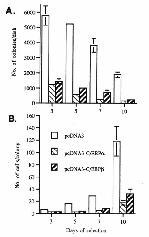
1. Transactivationpotential of endogenous C/EBPs in primarykeratinocytes…… 160
2. Forced expression of C/EBPβ and C/EBPα inhibits
BALB/MK2 keratinocyte growth………. 161
3. Forced expression of C/EBPβ alters BALB/MK2 keratinocyte morphology… 162
4. Transient transfection of BALB/MK2 keratinocytes
with pcDNA3-C/EBPβ inhibits growth……… 163
5. Transient transfection of BALB/MK2 keratinocytes with pcDNA3-C/EBPβ
increases K1 and K10 expression ………. 164
6. Immunofluorescence detection of the coexpression of C/EBPβ and K1
in pcDNA3-C/EBPβ-transfected BALB/MK2 keratinocytes……… 165
7. C/EBPα and C/EBPβ expression in the epidermis of wild-type,
C/EBPβ-heterozygous, and C/EBPβ-deficient adult mice ………. 166
8. C/EBPβ-deficient mice demonstrate alteration in epidermal K1
and K10 expression……….. 167
9. Altered expression of K1 and K10 in attached, spontaneously detached,
and suspension cultured primary keratinocytes from C/EBPβ-deficient
mice……… 168
10. C/EBPβ-deficient epidermal keratinocytes are resistant
GENERAL INTRODUCTION
The regulation of gene expression is complex and is subject to multiple levels
of control including the regulation of the rate of mRNA synthesis, the stability of the
mRNA, the rate at which the mRNA is translated, as well as the stability of the
translated product. The most common type of regulation is at the transcriptional level
which controls the rate of mRNA synthesis. Every gene has sequences upstream of its
coding sequence to control the rate of transcription. These sequences can be divided
into two general classes; i) sequences which control basal transcription and ii)
sequences which control the rate of transcription in response to cellular signals. At the
transcriptional starting point, there is a tendency for the first base of mRNA to be
adenine, flanked on either side by pyrimidines. This region is called the initiator (Inr)
and most promoters have a sequence called the TATA box, usually located ~ 25 bp
upstream of starting point. These sequences provide binding sites for general
transcription machinery which includes RNA polymerase II, TATA binding proteins
(TFIID, TFIIA, TFIIB, TFIIF, TFIIE, THIIH, and TFIIJ), and activators. While
general transcription machinery initiates and maintains RNA transcription, specific
transcription factors are responsible for the control of transcription in response to
various stimuli. The sequences that these factors bind to are called response elements.
There are ~2000 to 3000 transcription factors in mammals and it is these site-specific
transcription factors that mediate the final steps of cell signaling from cell surface to
nucleus. Transcription factors contain at least two domains ; i) a DNA binding domain
functions to activate or suppress transcription.
CCAAT/ENHANCER BINDING PROTEIN (C/EBP)
CCAAT/Enhancer Binding Proteins are members of bZIP transcription factors
CCAAT/enhancer binding proteins (C/EBPs) are members of the basic leucine
zipper (bZIP) class of transcription factors. This class of transcription factor also
includes Fos and Jun proteins which bind to AP-1 site, CREB/ATF (cAMP responsive
element binding protein/activation transcription factor), PAR-domain proteins, and
yeast/plant derived bZIP proteins (1). bZIP transcription factors are characterized by a
basic DNA binding domain and a leucine-zipper region. The leucine zipper region is a
heptad repeat of leucines within a 35 amino acid sequence which forms an amphipatic
α-helix with the leucines being located on one side of the helix per every two turns (2).
These leucines allow the two α-helices of separate monomers to dimerize by
hydrophobic interactions (zipping). The first C/EBP protein, C/EBPα was identified
as a heat-stable factor in rat liver nuclei and since it bound to the CCAAT box present in
several promoters as well as to the core homology sequence
[TGTGG(A/T)(A/T)(A/T)G] of certain viral enhancers, it was termed CCAAT/enhancer
binding protein (3). Later, other members of C/EBP family were identified and all of
C/EBP family members have conserved C-terminal parts, which is a basic amino acid
rich DNA binding domain and a leucine zipper domain (4-10).
C/EBP Family Members and Tissue Expression Profile
C/EBPα (also known as C/EBP, RcC/EBP-1) (2, 8, 9, 11-14), C/EBPβ (also known as
NF-IL6, IL-6DBP, LAP, CRP2, NF-M, AGP/EBP, ApC/EBP) (4-6, 8, 9, 14-18),
C/EBPδ (also known as NF-IL6β, CRP3, CELF, RcC/EBP2) (8, 9, 13, 14, 19-21),
C/EBPε (also known as CRP-1) (9, 12, 18, 22-24), C/EBPγ (also known as Ig/EBP-1) (7,
14) and C/EBPζ (also known as CHOP-10, GADD153) (10, 25, 26). The expression
profile of each C/EBP member has been described at the mRNA and protein level in
various tissues (4-11, 14, 23, 24, 27, 28). There are some discrepancies between the
levels of C/EBP mRNA and protein (6, 9), suggesting the existence of
post-transcriptional regulatory mechanisms. C/EBPα is highly expressed in liver, adipose,
lung, peripheral leukocytes, epidermis, intestine and skeletal muscle (8, 9, 11, 28, 29).
In liver, adipose, and epidermis, the highest level of C/EBPα mRNA expression was
observed in terminally differentiated cells. C/EBPβ expression is highest in liver,
intestine, lung, adipose, spleen, kidney, epidermis, and myelomonocytic cells (4, 8, 9,
15, 16, 27), while C/EBPδ is expressed in adipose, lung, and intestine (8, 9, 19).
C/EBPγ and C/EBPζ are expressed ubiquitously (7, 10) and C/EBPε is highly expressed
in myeloid and lymphoid cells (22-24, 30).
Structure and Properties of C/EBPs
As mentioned previously, all C/EBP family transcription factors have a highly
conserved C-terminal bZIP motif, which includes a DNA binding domain that is rich in
basic amino acids and a leucine zipper domain that mediates dimerization between
C/EBP isoforms. C/EBP family members can either homodimerize or heterodimerize
with other members of the C/EBP family through their dimerization domain (leucine
Dimerization between different isoforms has the potential to increase the repertoire of
transcriptional responses resulting from various stimuli. For example, a tissue that
expresses the α and β isoforms will contain three species of C/EBPs : the α/α
homodimer, the α/β heterodimer, and the β/β homodimer. In contrast to the C-temini,
the N-termini of C/EBPs are less conserved (less than 20 % sequence homology). The
N-termini of C/EBP family (except for C/EBPγ and C/EBPζ) contains three activation
domains which are required for binding to general transcription factors and activating
transcription (31-36). In addition, N-termini of C/EBPα, C/EBPβ, and C/EBPε
contain negative regulatory domains which silence the function of activation domains
(31, 33-35, 37, 38). While most of C/EBP transcription factors function as
transcriptional activators, some C/EBP transcription factors function as negative
regulators of transcription. For example, the N-terminally truncated form of
C/EBPβ, LIP (liver inhibitory protein), lacks a transactivation domain and can bind to
full-length C/EBPβ or other C/EBP transcription factors resulting in non-functional
C/EBP homo- or heterodimers (39). C/EBPγ and C/EBPζ can also function as
dominant negative inhibitors (10, 36). Recently, it has been shown that mutations
occur in C/EBPα in certain forms of acute myeloid leukemia and these mutations result
in a truncated form of C/EBPα that functions as a dominant-negative inhibitor of
full-length C/EBPα (40). Protein-protein interactions between C/EBP transcription factors
and other classes of protein molecules have been reported, such as C/EBPα-NFκB (41),
C/EBPβ-NFκB (42-45), C/EBPδ-NFκB (46, 47), C/EBPβ-glucocorticoid receptor (48),
C/EBPβ-Sp1 (49), C/EBPβ-Myb (50), and C/EBPα-AML1 (51) interactions. For
C/EBPs interaction with the NFκB or glucocorticoid receptor, it has been shown that the
direct protein-protein interaction, the functional interaction of C/EBPs with p300 (52),
Rb (53, 54), and p21 (55, 56) has been reported.
C/EBP family members have simple gene structures. C/EBPα, C/EBPβ, and
C/EBPδ are intronless genes (6, 8, 9, 57) while the mouse C/EBPγ gene consists of two
exons (58). C/EBPε gene has three exons and C/EBPζ has four exons (23, 25). The
promoter of C/EBPα gene is subject to autoregulation which occurs in human and
mouse by different mechanisms (59, 60). While C/EBPα binds to the murine C/EBPα
promoter and results in an increased C/EBPα expression, human C/EBPα induces USF,
another transcription factor, which induces C/EBPα expression. C/EBPα promoter
can be repressed by nuclear factor C/EBP undifferentiated protein (CUP), an isoform of
activator protein-2α (AP-2α), which binds to repressive elements in the C/EBPα gene
promoter and silence the gene until late in the differentiation program (61). There are
two isoforms of C/EBPα (42 kD and 30 kD) and the smaller isoform of C/EBPα (30
kD) can be generated by initiating translation at an alternative in-frame AUG codon (62,
63). This smaller isoforms still has an intact DNA binding domain and leucine-zipper
domain however, its transactivating activity is low as it also has an altered
transactivation domain. C/EBPβ mRNA can give rise to three C/EBPβ isoforms (38
kD, 35 kD, and 20 kD) among which 35 kD (LAP-liver activating protein) and 20 kD
(LIP-liver inhibitory protein) are the major isoforms found in cells (39). While these
forms of C/EBPβ are considered to result from alternative translation start sites, Baer et
al showed that 20 kD C/EBPβ is generated predominantly by in vitro proteolytic
cleavage during its isolation from cells (64). Four forms of C/EBPε (32 kD, 30 kD, 27
Like LIP, C/EBPγ and C/EBPζ can function as negative regulators of C/EBP activity
although their inhibitory mechanisms are different. While C/EBPγ lacks
transactivation domain (36), C/EBPζ contains two proline residues in the basic region
resulting in disruption of α-helical structure (10). C/EBPζ can heterodimerize with
other C/EBP members however, such heterodimers are not able to bind to target
sequences because of the altered DNA binding domain.
Biological Function of C/EBPs
The function of C/EBP family members has been investigated using target gene
promoter analysis, overexpression/inhibition of C/EBPs, and gene-knockout animals.
These analyses revealed roles of C/EBP family members in a number of cellular
processes, including growth arrest/differentiation, inflammation, liver regeneration,
metabolism, and other cellular signaling events.
(I) Role of C/EBPs in Cell Differentiation
It has been shown that C/EBP transcription factors have an important role in
preadipocyte differentiation (65-67). The promoter region of many adipocyte-specific
genes contains C/EBP binding sites and C/EBP transcription factors have a critical role
in preadipocyte differentiation. C/EBPα expression is highest in terminally
differentiated adipocytes (28) and the induction of C/EBPβ and C/EBPδ mRNA
precedes C/EBPα mRNA expression during preadipocyte differentiation (8, 59, 67).
Since C/EBPα promoter is activated by all three C/EBP isoforms (C/EBPα, C/EBPβ,
and C/EBPδ), C/EBPβ and C/EBPδ have been proposed to function in early
transcription of adipocyte-specific genes. Overexpression of C/EBPα or C/EBPβ
induced preadipocyte differentiation while inhibition of C/EBPα by antisense RNA
blocked preadipocyte differentiation (68-71). Furthermore, embryonic fibroblasts
from C/EBPβ and C/EBPδ deficient mice were unable to differentiate in response to
hormonal stimulus and C/EBPα deficient mice had dramatically reduced lipid
accumulation in adipose tissue (72, 73). C/EBP transcription factors also have
important roles in differentiation of myeloid cells (74). The promoter region of many
genes expressed in myeloid cells contains C/EBP binding sites and ectopic expression
of C/EBPs activates a number of target genes in myeloid cells. Studies from C/EBPα
knockout mice showed that these mice lack mature neutrophils (75). This defect
correlates with a lack of receptors for granulocyte colony stimulating factor (G-CSF) or
interleukin-6 (IL-6) and expression of these receptors restores granulopoiesis in
C/EBPα deficient mice (75, 76). C/EBPε deficient mice generate atypical
granulocytes that are functionally defective, lacking an oxidative burst (24). Lastly,
C/EBPβ deficient mice show defects in macrophage activation, suggesting a potential
role of C/EBPβ in macrophage activation or differentiation (77-79). In addition to
adipocytes and myeloid cells, C/EBPs are implicated in differentiation of other type of
cells including hepatocytes, mammary epithelial cells, keratinocytes, intestinal epithelial
cells, and neuronal cells (28, 80-84). Studies with C/EBPβ deficient mice show that
C/EBPβ is important for functional differentiation of mammary epithelial cells and
expression of milk proteins. Female C/EBPβ deficient mice are sterile due to defects
in differentiation of periovulatory granulosa cells in response to luteinizing hormone
(II) Role of C/EBPs in Energy Metabolism
C/EBP transcription factors are also important in the control of energy
metabolism. C/EBPα deficient mice die within a few hours due to hypoglycemia.
This hypoglycemia results from two metabolic defects in liver. i) the level of liver
glycogen is undetectable because of the reduced expression of glycogen synthase. ii)
the expression of glucose-6-phosphatase, phosphoenolpyruvate carboxykinase, and
tyrosine aminotransferase, which are required to synthesize glucose, are decreased (73).
In initial studies on C/EBPβ deficient mice, it was observed that C/EBPβ deficient mice
were generally healthy (78, 79), however, a second study by Croniger et al (86) showed
that mice with A-phenotype survived, whereas mice with B-phenotype died soon after
birth due to hypoglycemia, similar to C/EBPα deficient mice. While C/EBPα
deficient mice lack liver glycogen, C/EBPβ deficient mice with B-phenotype are able to
synthesize/store glycogen but unable to mobilize hepatic glycogen (87). The basis for
the different phenotypes of the C/EBPβ deficient mice has not been described, however,
some evidence suggests that the expression of non-inbred modifier genes are involved
(87).
(III) Role of C/EBPs in Inflammation
The activity and expression levels of C/EBP transcription factors are regulated
by inflammatory stimuli and different cytokines. For example, C/EBPβ and C/EBPδ
mRNA are highly upregulated by lipopolysaccharide (LPS) and cytokines such as
interleukin-1 (IL-1), interleukin-6 (IL-6), and tumor necrosis factor-α (TNF-α) (77,
88-90). C/EBPβ was originally identified as an inducible protein by IL-6 or IL-1 (4, 5).
plasma proteins in hepatocytes, and components of inflammatory signaling pathways
have binding sites for C/EBPs and gene knockout studies have provided further
evidence that C/EBP transcription factors are important in regulation of the
inflammatory responses (74, 77). Acute phase proteins are plasma proteins whose
levels are regulated during the acute phase of inflammation (91). These liver-specific
or liver-enriched proteins can be divided into two classes. Class I genes require both
interleukin-1 (IL-1) and IL-6 for maximal induction, whereas class II genes are
responsive to IL-6. It has been shown that the promoter regions of most class I genes
(hemopexin, haptoglobin, α1-acid glycoprotein, serum amyloid A1, A2, and A3,
complement C3, C-reactive protein) contain C/EBP binding motifs (77). C/EBPβ
deficient mice show impaired expression of some acute phase genes (serum amyloid A
and P proteins, C3) and TNF-α while the expression of other acute phase genes
(hemopexin, haptoglobin) are not affected, possibly due to compensation by other
C/EBP members (77). C/EBP binding motifs are also present in regulatory region of
genes encoding several cytokines such as IL-6, IL-1β, TNF-α, IL-8, IL-12 and proteins
which are important in macrophage/granulocyte functions such as nitirc oxide synthase,
lysozyme, myeloperoxidase, granulocyte colony stimulating factor (74). Serum IL-6
levels are not changed in C/EBPβ deficient mice however serum IL-6 levels are
elevated in aging C/EBPβ deficient mice (79, 92), suggesting C/EBPβ is dispensable for
IL-6 gene expression. However, C/EBPβ regulates TNF-α and serum TNF-α
induction is impaired in C/EBPβ deficient mice (79). In addition, deletion of C/EBPε
gene, a C/EBP family member expressed in cells of the myelomonocytic lineage,
resulted in decreased expression of several cytokines such as interferon-γ (IFN-γ),
(IV) Role of C/EBPs in Cell Proliferation
C/EBPα negatively regulates cell proliferation. C/EBPα expression is highest
in terminally differentiated adipocytes/hepatocytes/keratinocytes while C/EBPα
expression is down-regulated after partial hepatectomy (proliferation) (28).
Overexpression of C/EBPα results in strong growth arrest in cultured cells (93) and
abnormal cell proliferation is observed in liver and lung of C/EBPα deficient mice (94).
Furthermore, the expression levels of C/EBPα are reduced in some tumors/tumor cell
lines (95, 96). However, the importance of C/EBPα in the regulation of growth arrest
and differentiation is exemplified by recent studies in which dominant-negative
mutations in C/EBPα were found in some patients with human acute myeloid leukemia
(AML) (40). Such mutations in C/EBPα are thought to result in a differentiation block
of the granulocytic blasts and have implicated C/EBPα as a tumor suppressor gene. In
addition, it has been suggested that AML1-ETO, a fusion protein resulting from t(8;21)
translocation in AML, suppresses C/EBPα expression indirectly by inhibiting positive
autoregulation of the C/EBPα promoter (97). The growth and differentiation
regulatory functions of C/EBPα are complex and multifaceted. For example, C/EBPα
has been proposed to regulate p21 expression (56) and interact with Rb family proteins
(98). C/EBPα also has been shown to directly repress E2F function through its
physical associations with E2F and this repression is necessary for growth arrest and
adipocyte and granulocyte differentiation (99). However, recent studies indicate that
C/EBPα can block growth independent of its DNA binding and transcriptional activity
by forming a complex with cdk2 (cyclin-dependent kinase 2) and cdk4 and thereby
et al showed that C/EBPα cooperates with p21 to inhibit cdk2 activity (101). Thus, in
addition to its DNA binding/transcription factor activity, C/EBPα can modulate growth
arrest and differentiation by protein/protein interactions with cell cycle regulatory
proteins independent of its transcription activity.
Regulation of C/EBPs
(I) Transcriptional/Translational Control
The levels and activity of C/EBPs are modulated by various stimuli, such as
cytokines, hormones, nutrients, and cellular stresses. The levels and activity of
C/EBPs are subjected to regulation at the transcriptional, translational, and
post-translational level. In addition, the degradation of C/EBPs can affect the levels of
C/EBPs in the cell. A number of signals can negatively or positively affect C/EBP
mRNA levels (74) and the promoter regions of C/EBPα, β, δ, ε, ζ have been
investigated. The C/EBPα promoter region has binding sites for C/EBP, Sp1, nuclear
factor (NF)-1, NF-Y, c-myc, and upstream stimulating factor (USF) and is activated by
overexpression of C/EBPα or C/EBPβ (59, 102), indicating C/EBPα promoter can be
autoactivated. Autoactivation of C/EBPβ and C/EBPδ promoter also has been
observed in some species (21, 103). Interestingly, c-myc, a proto-oncogene, inhibits
transcription of C/EBPα by binding the core promoter region of C/EBPα (104, 105).
The promoter of C/EBPε is transcribed by two alternative promoters which contain no
TATAAA box but has a number of purine rich sequences for the Ets family of
transcription regulators (23, 24). The C/EBPε promoter also has a retinoic acid
damage/cellular stress (26) and the C/EBPζ promoter contains an AP-1 binding site
which is responsible for C/EBPζ induction by oxidative stress (107). The C/EBPζ
promoter has a conserved C/EBP site which has an important role in its induction during
acute phase response in hepatocytes (108). Production of different C/EBPα or
C/EBPβ isoforms by usage of alternative translation initiation codons is a major
mechanism of translational control (12, 39, 62, 63).
(II) Post-Translational Control
Transactivation, DNA binding activity, or localization of C/EBPs can be
modulated by post-translational modification. Phosphorylation is the most frequent
post-translational modification and several kinases that phosphorylate C/EBPs have
been identified. The transactivation activity of C/EBPβ can be induced by
phosphorylation of Thr235 by a Ras/mitogen-activated protein kinase (MAPK) pathway
(80, 109), Ser105 by protein kinase C (PKC) (110), and Ser 276 by Ca2+
/calmodulin-dependent protein kinase (111). TGF-α induces activation of the p90 ribosomal S
kinase (RSK), which results in the phosphorylation of rat C/EBPβ on Ser-105 and of
mouse C/EBPβ on Thr-217 (112). C/EBPζ is phosphorylated by p38 MAPK during
cellular stress and this phosphorylation results in increased transactivation activity (113).
PKC-mediated phosphoryation of C/EBPα at Ser248, Ser277, and Ser299 attenuates its
DNA binding activity (114) and insulin induced phosphorylation of C/EBPβ by PI3
kinase (phosphatidylinositol-3-kinase) suppresses transactivation activity of
C/EBPβ (115). DNA binding activity of C/EBPβ is negatively regulated via
phosphorylation of Ser240 by PKC and in vitro phosphorylation study showed that
inhibits DNA binding activity (116). On the other hand, forskolin-induced activation
of cAMP/PKA in rat PC12 cells results in the translocation of C/EBPβ into the nucleus,
where it activates c-fos gene transcription (117). Antioxidant-induced nuclear
translocation of C/EBPβ in colorectal cancer cell line is also regulated by PKA
mediated phosphorylation (118). Glycogen synthase kinase 3 (GSK3) has been
identified as a C/EBPα kinase (119). However, the functional significance of this
phosphorylation is not known. Transcriptional activity of C/EBPε as well as other
C/EBP family members (C/EBPα, C/EBPβ, and C/EBPδ) is positively controlled by
sumoylation at a conserved inhibitory domain (120). Subramanian et al showed that
C/EBPα interacts directly with the E2 SUMO-conjugating enzyme Ubc9 and can be
sumoylated in vitro (121). Recently, it has been shown that C/EBPγ and C/EBPζ are
ubiquitinated and subsequently degraded by the proteasome (122).
CCAAT ENHANCER BINDING PROTEINS IN EPIDERMIS
Epidermis
The epidermis is a stratified squamous epithelium composed mainly of
keratinocytes that form four different layers. In addition to retaining body fluids to
prevent dehydration, the epidermis provides an essential barrier function against
environmental insults such as physical/chemical trauma and microorganisms. Each
epidermal layer represents a different stage in epidermal differentiation beginning from
basal keratinocytes which become post-mitotic and move upwards through the spinous
epidermal cells is basal stem cells which generate transient amplifying cells that
undergo a limited number of divisions and withdraw from cell cycle. As these
post-mitotic cells move upward, epidermal differentiation begins and when they reach the
outer surface, they become enucleated, flattened, and dead cells that are subsequently
sloughed and replaced by inner cells that move upwards (123). Therefore, the
epidermis is a tissue which is in a constant state of dynamic equilibrium, replenishing
itself throughout life.
During epidermal differentiation, the expression and the repression of specific
genes are strictly regulated as a basal keratyinocyte transits through the different stages
of differentiation (124). For example, the transition of basal keratinocytes from the
basal layer to the spinous layer is accompanied by the repression of keratin 5 and
keratin 14 and upregulation of keratin 1 (K1) and keratin 10 (K10) which are early
differentiation markers. As a keratinocyte moves from spinous to granular layer, the
expression of K1 and K10 is suppressed while the expression of late differentiation
markers, involucrin and loricirn, are induced. Finally, epidermal transglutaminase
cross-links these structural proteins and forms a cornified envelope, resulting in the
mature squame following digestion of intracellular organelles.
Role of C/EBPα and C/EBPβ in epidermal differentiation
The expression of C/EBP family members in mouse and human epidermis
has been investigated and some of C/EBP family members are highly expressed in
epidermis (29, 125, 126). C/EBPα, C/EBPβ, and C/EBPδ mRNA are expressed
expression in the epidermis are higher in epidermis than in liver and in lung, tissues
known to express high levels of these C/EBP members (29). C/EBPα and C/EBPβ
proteins are expressed abundantly in mouse keratinocytes while C/EBPδ protein
expression is low. C/EBPα protein is expressed in the nuclei and cytoplasm of
suprabasal kertinocytes and is weakly expressed in a perinuclear manner in some basal
keratinocytes. C/EBPα expression is observed in non-proliferative compartments of
hyperplastic epidermis and papillomas however, C/EBPα expression is greatly
diminished in squamous cell carcinomas (SCC). C/EBPβ expression shows a highly
ordered and specific pattern in epidermis. C/EBPβ expression is exclusive to the
nuclei of three-cell clusters of suprabasal keratinocytes and these clusters are repeated at
regular intervals throughout the epidermis, which is consistent with the central
suprabasal column of the epidermal proliferative unit.
It has been shown that C/EBPα and C/EBPβ are associated with epidermal
differentiation. The terminal differentiation of keratinocytes can be induced by
increasing the level of extracellular calcium in the media (127). When terminal
differentiation is induced by calcium, expression of C/EBPα and C/EBPβ is increased
(29). In addition, forced expression of C/EBPα or C/EBPβ induces differentiation-like
morphological changes and growth arrest in BALB/MK2 keratinocytes. C/EBPβ has
been shown to modulate keratin 1 (K1) and keratin 10 (K10) expression, early
keratinocyte differentiation markers. Forced expression of C/EBPβ increases K1 and
K10 expression in BALB/MK2 keratinocytes (80) and the promoter region of the K10
gene contains three functional C/EBP binding sites (128). Primary keratinocytes
are resistant to calcium induced growth arrest (80). The phorbol ester
12-O-tetradecanoylphorbol-13-acetate (TPA) is a potent inducer of keratinocyte
differentiation and of involucrin gene expression (129), a late keratinocyte
differentiation marker. Agarwal et al showed that a C/EBP site in the proximal
regulatory region of the human involucrin gene is required for involucrin expression in
response to phorbol ester (130). The human involucrin reporter plasmid is activated
by C/EBPα whereas C/EBPβ and C/EBPδ inhibit both TPA- and C/EBPα-dependent
involucrin promoter activation. In addition, a C/EBP binding site has been identified
in the promoter region of α2 and α5 integrin genes in human keratinocytes and the
overexpression of C/EBPα or C/EBPβ inhibited the activity of these promoters (131).
Since detachment of keratinocytes from basement membrane triggers terminal
differentiation and integrin expression is down-regulated during keratinocyte terminal
differentiation, the up-regulation of C/EBPs may contribute to the inhibition of integrin
expression during keratinocyte differentiation.
Role of C/EBPs in Epidermal Proliferation and Cell Survival
C/EBPs also have an important role in epidermal proliferation and cell survival.
It has been shown recently that C/EBPβ deficient mice are completely refractory to a
variety of carcinogen-induced skin tumors and H-ras transgenic mice that carry the
C/EBPβ deletion mutation display a significant reduction in tumorigenesis (132). In
addition, oncogenic ras potently stimulated C/EBPβ transactivation activity and
C/EBPβ cooperated with ras to transform NIH 3T3 cells, suggesting C/EBPβ as a
downstream mediator of ras signaling. Ras-induced C/EBPβ transactivation activity is
phosphorylation site, is substituted with alanine. The positive role of C/EBPβ in cell
proliferation also has been described in other systems. For example, Greenbaum et al
showed that C/EBPβ mediates growth response pathways in the regenerating liver and
is required for a normal proliferative response (133) and Buck et al showed that p90
ribosomal S kinase (RSK) mediated phosphorylation of rat C/EBPβ on Ser-105 and of
mouse C/EBPβ on Thr-217 is critical for TGF-α induced proliferation of hepatocytes
(112). Recently, it was reported that a certain C/EBPβ isoform transforms normal
mammary epithelial cells and induces an epithelial to mesenchymal transition in culture
(134).
C/EBPβ also appears to be important in regulating cell survival. For example,
a seventeen-fold increase in the number of apoptotic keratinocytes is observed in
DMBA-treated, C/EBPβ deficient epidermis compared with wild-type epidermis (132).
The observed increase in the number of apoptotic cells in DMBA-treated, C/EBPβ
deficient epidermis is consistent with a survival/anti-apoptotic role for C/EBPβ. It is
possible that in normal keratinocytes C/EBPβ regulates epidermal differentiation as well
as survival, which is required to complete the differentiation program. However, in the
presence of oncogenic H-ras, the prosurvival function of C/EBPβ may dominate over
the differentiating function of C/EBPβ and C/EBPβ may contribute to clonal expansion
of oncogenic H-ras containing cells. It has been proposed that in C/EBPβ deficient
epidermis the prosurvival function of C/EBPβ is blocked and cells containing oncogenic
ras mutations undergo apoptosis, inhibiting tumor formation. In support of this, Buck
et al observed that hepatotoxin CCl4 activates RSK, resulting in the phosphorylation of
treatment induces apoptosis in C/EBPβ deficient mice (135). They showed that
C/EBPβ phosphorylation on Thr-217 by RSK creates a functional XEXD caspase
substrate/inhibitor box and inhibits activation of procaspase 1 and 8.
UBIQUITIN/PROTEASOME-MEDIATED PROTEOLYTIC SYSTEM
The cellular integrity is maintained by dynamic equilibrium between synthesis
and degradation of each protein. Cells must control the rates of both synthesis and
destruction of their proteins in order for proper cellular function. For proper
degradation of target proteins, cells contain two major degradation machineries: i)
proteases (cathepsin B, D, H, and L, calpain I and II) and ii) multicatalytic, multisubunit
ubquitin/proteasome in nucleus/cytoplasm. While proteases make several cuts in its
target protein without energy input resulting in several fragments of polypeptide,
ubiquitin/proteasome system requires energy expenditure and cleaves every peptide
bond in target protein. Recently, many cellular proteins involved in the critical cellular
processes, such as proliferation, differentiation, DNA repair, and apoptosis, have been
discovered to be degraded by the ubiquitin/proteasome pathway (136, 137).
Furthermore, deregulated proteasomal degradation has been found in a number of
human diseases, including cancer, neurodegenerative diseases, and myodegenerative
diseases and proteasome inhibitors are being developed as a therapeutic agent.
Structure of Proteasome
of the 20S proteasome is well conserved in all organisms from archaebacteria to yeast to
human (138-140). The 20S core unit forms the hollow barrel shape in which four
heptameric rings are stacked forming central chamber where proteolysis occurs. Two
outer rings are composed of seven α-subunits and two inner rings are composed of
seven β-subunits. Eukaryotic proteasomes have two additional 19S regulatory units
(141) which bind to the 20S proteasome forming a lid and a base in the 26S proteasome.
19S units contain binding sites for ubiquitinated proteins, enzymes that depolymerize
ubiquitin chain, and six distinct ATPases. The ATPase function of the 19S unit unfolds
target protein and facilitates the entry of target protein into the 20S proteasome (142).
To ensure that every peptide bond in target protein is susceptible for cleavage, 20S
proteasome has multiple proteolytic activities: i) chymotrypsin-like, ii) trypsin-like,
iii) caspase-like, iv) branched-amino acid preferring, and v) small neutral amino
acid-preferring (143-145). Based on mutational studies in yeast, these distinct proteolytic
activities are mapped to different subunits in 20S proteasome (146-150).
Ubiquitin/Proteasome System
In ubiquitin/proteasome system, the degradation of a protein begins with the
conjugation of multiple ubiquitins to the target protein (151) although certain proteins
do not require ubiquitination for proteasomal degradation (152). Ubiquitin is a
well-conserved, ubiquitously expressed small molecular weight (76 amino acids) protein.
Ubiquitin is conjugated to a target protein through a reversible isopeptide bond
catalyzed by a sequential action of E1, E2, and E3 ligase. The conjugation reaction is
initiated by formation of a high-energy thioester bond between the C-terminus of
ubiquitin is transferred to E2 ligase which transfers ubiquitin to E3 ligase. E3 ligase
catalyzes the last step of the ubiquitin conjugation reaction and ubiquitin is ligated to ε
-amino group of lysine residues in the target protein through an isopeptide bond.
Ubiquitin contains seven lysine residues (K6, K11, K27, K29, K33, K48, and K63) and
each of these lysine residues can form an isopeptide bond with C-terminal glycine of
next ubiquitin, resulting in polyubiquitination (153-155). However, the most common
type of ubiquitin chain is the K-48 linked type chain which signals the target protein for
proteasomal degradation (156). Most organisms contain one or two E1 ligases and it is
these E1 ligases that catalyze activation of ubiquitin. A single E1 ligase is known in
yeast (Uba1) and deletion of this gene is lethal in yeast (157). Many E2 ligases have
been identified in yeast (13 E2 ligases) and mammals (more than 20 E2 ligases) (158,
159). Some E2 ligases are involved in specific cellular processes as ubiquitin
conjugating enzymes (UBC). For example, yeast E2 ligase UBC2/RAD6 is involved
in the degradation of N-end rule substrates and in DNA repair (160). UBC3/CDC34 is
required for G1/S transition via the degradation of cell cycle regulators (160, 161).
However, it is believed that specificity of E2 ligases in cellular processes results from
their association with a distinct E3 ligase. E3 ligases are responsible for ligation of
ubiquitin to the target protein for proteasomal degradation. They recognize specific
motifs in their substrate and confer the substrate specificity. Four families of E3 ligase
have been identified. i) N-end rule family, ii) HECT (Homologous to E6-AP carboxy
terminus) domain family, iii) ring finger family, and iv) multisubunit complex family.
E3α ligase (UBR1-yeast homologue of E3α) is a large protein (~ 200 kD) and contains
three distinct functional domains for substrate recognition. A type-1 site recognizes
recognizes bulky hydrophobic residues (Leu, Ile, Phe, Trp, and Tyr) (162, 163). The
third substrate binding site recognizes a non-N-end rule substrate (164, 165). The
HECT family E3 ligase contains a 350 amino acid-long C-terminal sequence which is
homologous to E6-AP (human papilloma virus E6 oncoprotein associating protein) and
at least 20 members of this E3 ligase family have been identified (166, 167). E6-AP
mediates the ubiquitination of p53, resulting in proteasomal degradation of p53. The
ring finger motif was originally identified in human RING1 gene product and it contains
the Cys-X2-Cys-X(9-39)-Cys-X(2-3)-Y-X2-Cys-X(4-48)-Cys-X2-Cys sequence, where X can
be any amino acid and Y can be Cys or His (168). Proto-oncoprotein c-cbl is the
prototype of ring E3 ligase which ubiquitinates receptor protein tyrosine kinase and
targets it for proteasomal degradation (169-171). APC (anaphase-promoting complex)
and SCF complex (Skp1, cullin/cdc53, and F-box proteins) are the member of the
multisubunit E3 ligase complexes (172, 173). Known substrates for APC E3 ligase are
certain cyclins, anaphase inhibitors, and spindle-associated proteins (174). SCF E3
ligase complex recognizes a number of important cell cycle regulators such as G1
cyclins, CDK inhibitor, and other short-lived proteins (173). It has been proposed that
Skp1 binds to F-box proteins, a different component of the SCF complex, and the F-box
proteins recognize substrates in a phosphorylation dependent manner (175-177).
Isopeptide bond between ubiquitin and substrate can be cleaved by deubiquitinating
enzymes such as UCH (ubiquitin C-terminal hydrolases) and UBP (ubiquitin-specific
processing enzymes) (178). These enzymes are involved in i) regeneration of
C-terminus of ubiquitin after degradation of attached protein, ii) reversing the
ubiquitination from wrong target proteins, and iii) disassembly of polyubiquitin chains
The substrate proteins for ubiquitination are recognized by distinct ubiquitin
ligases via specific motifs. Several structural motifs for ubiquitination have been
identified including genetically programmed signal, phosphorylation, and damaged
protein. Certain proteins contain short N-terminal sequences of amino acids and the
nature of these N-terminal amino acids determines the rate of ubiquitination. This is
the first identified structural motif for ubiquitination and is referred to as the N-end rule.
Ubiquitination of target proteins can be positively or negatively regulated by
phosphorylation. These target proteins include cyclins/cyclin dependent kinase
inhibitors, checkpoint regulators, cellular signaling components (e.g. SMADs, TGF-β,
IκBα, β-catenin, c-Jun, and p53), and cell surface receptors. Phosphorylated target
proteins are recognized by F-box protein which is a component of SCF complex E3
ligase. Furthermore, the activity of ubiquitin ligase is regulated by phosphorylation.
The exposure of hydrophobic residues which are normally embedded in protein-protein
interface or inside the protein can function as a signal for ubiquitination. Exposure of
such residues can result from protein misfolding or inappropriate assembly of the
subunit into a multisubunit complex and proteasomal surveillance ensures the
degradation of these unwanted proteins.
Proteasomal Defects Associated with Human Diseases
Since the proteasome regulates the cellular levels of numerous proteins, defects
in the proteasomal system can result in human diseases. For example, a defect in the
gene encoding human E3 ligase (UBE3A) is identified in Angelman’s syndrome, a
movement or balance disorder (179, 180). Another type of inherited proteasomal
defect is Liddle syndrome which is characterized by abnormally high blood pressure
due to incapability to maintain the balance of salt and water. In these patients,
increased half-life of sodium channel due to deletion or mutation of ubiquitination site
in sodium channel protein allows excessive reabsorption of sodium and water (181).
Skeletal muscle wasting in some pathological states also results from accelerated
ubiquitin-mediated proteolysis. US2 and US11, the proteins encoded by human
cytomegalovirus (CMV), target MHC class I heavy chain molecule for degradation by
directing it from the ER to the cytoplasm, thereby evading immune system (182).
Accumulation of ubiquitin conjugates is observed in senile plaques, lysosomes,
endosomes, and intracellular inclusion bodies in neurological disorders such as
Alzheimer’s disease and Parkinson’s disease although it is not known whether this
accumulation is a direct result of defects in the proteasomal pathway (183).
Most cancers are caused by activating mutations in proto-oncogenes and/or
inactivating mutations in tumor suppressor genes and the proteasomal degradation
system regulates the levels of many of these gene products (136)). Certain
modifications during tumorigenesis can change the stability of these oncoproteins/tumor
suppressor proteins by affecting the proteasome-mediated degradation and some
proteins involved in tumorigenesis contain ubiquitin ligase activity. For example, p53
is targeted for proteasomal degradation by mdm2 which is a p53 target gene containing
E3 ubiquitin ligase activity. While mdm2 targets p53 for degradation, mdm2 is
self-ubiqutinated and degraded. Cyclin dependent kinase inhibitor p21waf/cip, another p53
mediated phosphorylation of p21waf/cip triggers its degradation (184). Rb
(Retinoblastoma) protein is a tumor suppressor and negatively regulates G1/S transition
by interacting with E2F transcription factor. Rb protein is degraded in ubiquitin
dependent manner (185). In addition, free E2F is degraded in ubiquitin dependent
manner by the 26S proteasome (186-188). Mutations in the tumor suppressor gene
APC is associated with the development of colon cancer. Mutations in the APC gene
leads to accumulation of β-catenin which is normally degraded via the proteasome and
increased level of β-catenin is related to carcinogenesis (189, 190). Abl is a cellular
homologue of the transforming gene of Abelson murine leukemia virus and activation
of abl occurs as a consequence of a chromosomal translocation which generates a
bcr-abl oncogenic fusion protein (191-194). It has been reported that bcr-bcr-abl fusion
protein targets abl-interactor proteins (ABI1 and ABI2), which are inhibitors of tyrosine
kinase activity of abl, for proteasomal degradation (195). Oncogenic protein myc has
a very short half-life (30 min) and is degraded by the proteasome. Sears et al showed
that ERK mediated phosphorylation of Ser62 is required for ras-induced stabilization of
myc while GSK3 mediated phosphorylation of Thr58, which is dependent on the prior
phosphorylation of Ser 62, is associated with the degradation of myc (196). Increased
activity of the NFκB transcription factor promotes cell survival in some tumors
(197-200) and NFκB is activated by the ubiquitin/proteasome pathway (201, 202). In
response to stimuli, IκBα which normally complexes with NFκB is phosphorylated by
THESIS RESEARCH FOCUS AND HYPOTHESES
In manuscript I, we hypothesized that C/EBPα is degraded via proteasomal
pathway and that GSK3 regulates this process by targeting C/EBPα for proteasomal
degradation. GSK3 is a ser/thr kinase that phosphorylates and inhibits the activity of
glycogen synthase and two highly homologous forms of mammalian GSK3, GSK3α
and GSK3β, have been identified. GSK3β is expressed ubiquitously in eukaryotes
including yeast, C. elegans, Drosophila, Xenopus, mice and humans and is expressed in
most tissues (203). Recent studies have indicated that GSK3β phosphorylates a
variety of cell cycle regulatory proteins including p21 (184), β-catenin (204), cyclin D
(205) and c-myc (196) and in doing so targets these molecules for proteasomal
degradation. We investigated the degradation mechanism of C/EBPα and
demonstrated that C/EBPα is degraded via the ubiquitin/ATP-dependent proteasomal
pathway and lithium, a known inhibitor of GSK3 (206, 207), inhibits C/EBPα
degradation by interfering with the proteasomal degradation of C/EBPα in a
GSK3-independent manner.
In manuscript II, we hypothesized that loss of C/EBPα is important in
development of SCC. The role of C/EBPα as a negative regulator of cell proliferation
has been suggested from the several studies and C/EBPα deficient mice display
abnormal proliferation of certain type of cells (94). In addition, it has been reported
that C/EBPα levels are reduced in hepatocellular carcinomas (208) and lung cancer cell
lines (96). Recently, dominant-negative mutation in C/EBPα has been found in human
gene (40). We demonstrated that C/EBPα expression levels are greatly diminished in
mouse SCC / SCC cell lines and forced expression of C/EBPα inhibits the proliferation
of SCC cell lines. In addition, we showed that C/EBPα is related to the differentiation
of keratinocytes and transcription of C/EBPα gene may be silenced by promoter
hypermethylation in SCC cell lines. Our results suggest a tumor suppressor function
MANUSCRIPT I
Lithium Stabilizes C/EBPα Protein through a GSK3-Independent Pathway
Involving Direct Inhibition of Proteasomal Activity
Minsub Shim and Robert C. Smart
Cell Signaling and Cancer Group, Department of Environmental and Molecular
Toxicology, North Carolina State University
CORRESPONDING AUTHOR:
Robert C. Smart, Ph. D
Department of Environmental and Molecular Toxicology
North Carolina State University
Raleigh, NC 27695-7633
Phone : (919) 515-7245, Fax : (919) 515-7169, email : [email protected]
Running Title; C/EBPα is degraded via a ubiquitin-proteasome pathway
in Press in Journal of Biological Chemistry
Abbreviations; C/EBP : CCAAT/enhancer binding protein, GSK3 : Glycogen synthase
ABSTRACT
C/EBPα, a basic leucine zipper transcription factor, is involved in mitotic growth arrest
and differentiation. Given that numerous proteins involved in cell cycle regulation are
degraded via the ubiquitin-proteasome system, we examined whether C/EBPα protein is
degraded via a proteasomal mechanism. In cycloheximide-treated BALB/MK2
keratinocytes we found that C/EBPα is a short-lived protein with a half-life of ~ 1 hour.
Treatment with proteasome inhibitors, MG-132 or lactacystin, blocked the degradation
of C/EBPα protein. Higher molecular weight species of ubiquitinated C/EBPα were
detected in BALB/MK2 and in vitro studies confirmed that C/EBPα is degraded by the
proteasome in an ATP- and ubiquitin-dependent manner. GSK3 is a known C/EBPα
kinase and treatment of keratinocytes with LiCl, an inhibitor of GSK3 resulted in; i) a
five-fold increase in C/EBPα protein levels, ii) increased electrophoretic mobility of
C/EBPα protein and iii) no increase in C/EBPα mRNA levels suggesting that
GSK3-mediated phosphorylation of C/EBPα may target it for proteasomal degradation.
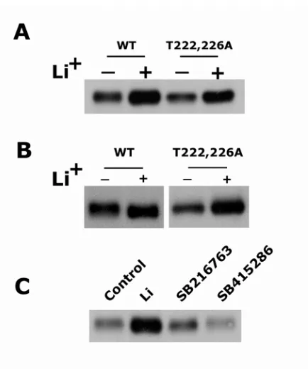
However, a mutant C/EBPα containing T to A mutations in the GSK3 phosphorylation
sites (T222A and T226A) retained its response to LiCl and additional pharmacological
inhibitors of GSK3 did not alter C/EBPα levels indicating the effects of LiCl on
C/EBPα are GSK3 independent. LiCl treatment of BALB/MK2 cells inhibited
C/EBPα degradation and produced a six-fold increase in the half-life of C/EBPα protein.
In vitro studies revealed that LiCl inhibited proteasome activity and the ensuing
degradation of C/EBPα. These results demonstrate C/EBPα is degraded via an
ubiquitin-dependent proteasomal pathway and LiCl stabilizes C/EBPα through a GSK3
INTRODUCTION
CCAAT/enhancer binding protein α (C/EBPα) is a member of the basic leucine zipper
(bZIP) class of transcription factors. There are six members of the C/EBP family
(C/EBPα, C/EBPβ, C/EBPδ, C/EBPε, C/EBPγ and C/EBPζ) (1-3). The C-terminal
region of C/EBP contains a basic domain that is responsible for binding specific DNA
sequences and a leucine zipper domain that functions in dimerization (4). C/EBPα can
form homodimers as well as heterodimers with other members of the C/EBP family.
The N-terminal region of C/EBPα contains three transactivation elements and an
additional highly conserved region (CR4) that is thought to have a regulatory function.
C/EBPα is highly expressed in liver, fat, lung, peripheral leukocytes, epidermis,
intestine and skeletal muscle (5-7). In a numerous cell types, including preadipocytes
(8,9), various myeloid cells (10,11), hepatocytes (12) and keratinocytes (6,13), C/EBPα
is involved in the regulation of mitotic growth arrest and/or differentiation. Consistent
with this, C/EBPα null mice display cell proliferation defects in liver and lung (14,15).
C/EBPα also plays a key role in energy homeostasis. C/EBPα null mice die shortly
after birth due to altered hepatic glucose and glycogen metabolism and also display
defects in white adipose tissue differentiation (14).
The importance of C/EBPα in the regulation of growth arrest and differentiation
is exemplified by recent studies in which dominant-negative mutations in C/EBPα were
found to be associated with human acute myeloid leukemia (16,17). Such mutations in
C/EBPα are thought to result in differentiation block of the granulocytic blasts and have
implicated C/EBPα as a tumor suppressor gene. The growth and differentiation
regulatory functions of C/EBPα are complex and multifaceted. For example, C/EBPα
proteins (20,21). C/EBPα has been shown to directly repress E2F function through its
physical associations with E2F and this repression is necessary for growth arrest and
adipocyte and granulocyte differentiation (22). However, recent studies indicate that
C/EBPα can block growth independent of its DNA binding and transcriptional activity
by forming a complex with cdk2 and cdk4 and thereby blocking cyclin-cdk interactions
and cell cycle progression (23). Thus, it appears that in addition to its DNA
binding/transcription factor activity, C/EBPα can modulate growth arrest and
differentiation by protein/protein interactions with cell cycle regulatory proteins
independent of its transcription activity. Therefore, the identification of cellular
processes that impinge on the regulation of C/EBPα protein stability may be critical in
understanding the regulation and/or deregulation of C/EBPα-induced growth arrest and
differentiation.
The ubiquitin-proteasome degradation plays an important role in the degradation
of cellular proteins which are involved in regulating various cellular process, including
cell cycle regulation, differentiation and apoptosis (24,25). Recent studies have
indicated that GSK3 phosphorylates a number of cell cycle regulatory proteins
including p21 (26), β-catenin (27), cyclin D (28) and c-myc (29) and in doing so targets
these molecules for proteasomal degradation. Lithium, an inhibitor of GSK3 (30,31),
blocked the proteasomal degradation of these proteins. C/EBPα can be
phosphorylated by GSK3 on Thr 222 and Thr 226 and this phosphorylation can be
blocked by lithium (32,33). However, the functional significance of GSK3-mediated
phosphorylation is not known nor is it known whether C/EBPα is degraded via a
proteasomal pathway or whether GSK3 or lithium treatment can alter C/EBPα protein
degradation of proteins that are GSK3 substrates has generally been attributed to its
inhibition of GSK3, lithium also inhibits a number of other cellular kinases and more
recently lithium has been shown to a be an inhibitor of chymotryptic activity of both the
20S and 26S proteasome (34). Therefore, we have examined whether C/EBPα is
degraded via a proteasomal mechanism and investigated the role of GSK3 and lithium
on C/EBPα protein stability. We demonstrate that C/EBPα is degraded via
ubiquitin-dependent proteasomal pathway and that lithium stabilizes C/EBPα protein through a
MATERIALS AND METHODS
Materials
Fetal bovine serum, trypsin, human recombinant epidermal growth factor (hEGF),
Lipofectin, and tris-glycine gels were purchased from Invitrogen. Ca2+-free Eagle’s
minimal essential medium (EMEM) was purchased from BioWhittaker. The enhanced
chemiluminescence (ECL) reagents were purchased from PerkinElmer Life Sciences.
HRP-linked donkey anti-rabbit IgG was purchased from Amersham. Ubiquitin, HeLa
S-100, energy regeneration system (ERS),ubiquitin-aldehyde, MG-132 and lactacystin
were purchased from Boston Biochem. Cycloheximide and monoclonal anti-Flag
antibody were purchased from Sigma. Bio-Rad protein assay reagent was purchased
from BioRad. Construction of pcDNA3-C/EBPα has been previously described (13).
λ-phosphatase was purchased from New England Biolabs. GSK3 inhibitor SB216763
and SB415286 were from GlaxoSmithKline. pCMV-Flag expression vector was a
kind gift from Dr. Jun-Tsuji (North Carolina State University, Raleigh). Rabbit
polyclonal anti-ubiquitin antibody, rabbit polyclonal anti-C/EBPα antibody, rabbit
polyclonal anti-p53 antibody, and protein A/G-plus agarose were purchased from
SantaCruz Biotechnology.
Transfection and lithium treatment
BALB/MK2 keratinocytes (a gift from Dr. Weissman, University of North Carolina,
Chapel Hill) were plated at 2.5 x 10⁵ cells/60 mm culture dish in Ca2+ free EMEM
supplemented with 8 % Chelex-treated fetal bovine serum, 4 ng of hEGF per ml, and
0.05 mM calcium chloride. Two days after plating, BALB/MK2 keratinocytes were
transfecetd with 2 µg of pcDNA3-C/EBPα and 12 µg of Lipofectin 2 ml serum free
manufacturer’s protocol. Twenty-four hours following transfection, cells were refed
with EMEM supplemented with 8 % Chelex-treated fetal bovine serum, 4 ng of hEGF
per ml, and 0.05 mM calcium chloride and incubated with 20 mM lithium chloride for
24 hr.
Preparation of nuclear and whole cell lysates
Nuclear extracts were prepared as previously described by Schreiber et al (35). For
the preparation of whole cell lysates, cells were washed with cold PBS, harvested by
scraping, collected by brief centrifugation. Cells were lysed in lysis buffer (10 mM
HEPES, pH 7.9, 10 mM KCl, 0.1 mM EDTA, 0.1 mM EGTA, 1 mM DTT, 1mM PMSF,
100 µg/ml aprotinin, 100 µg/ml leupeptin, 1mM sodium orthovanadate, 0.6 % NP-40)
by sonication and then one-tenth volume of 5 M NaCl was added. Incubation mixture
was vortexed, incubated for 15 min on ice, and centrifuged (14,000g, 10 min, 4 oC).
Supernatants were stored at –80 oC until use. Protein concentration was determined by
the Bio-Rad protein assay reagent.
Western blot analysis
Equal amounts of protein were precipitated by adding equal volume of 20%
trichloroacetic acid and washed with acetone (-20 oC). Protein samples were
solubilized and boiled in SDS sample buffer for 2 min, and then separated by
SDS-PAGE. The separated proteins were transferred to an Immobilon-P membrane
(Millipore). Following incubation in blocking buffer (PBS with 1 % bovine serum
albumin, 5 % non fat dry milk, and 0.1 % Tween-20) for 1 hr at room temperature, the
membranes were probed for 2 hr at room temperature with rabbit polyclonal IgG raised
against C/EBPα (SantaCruz Biotechnology). The membranes were washed and then
Detection was made with an enhanced chemiluminescence reagent followed by
exposure of the membrane to film.
Luciferase assay
BALB/MK2 keratinocytes were plated at 1 x 10⁵ cells/well in 12 well culture plate.
Two days after plating, BALB/MK2 keratinocytes were transfecetd in triplicate with
100 ng of pcDNA3-C/EBPα and 400 ng of the specified C/EBP-dependent
promoter/reporter plasmid as described in the text, and 3 µg of Lipofectin in 0.5 ml
serum free EMEM according to manufacture’s protocol. Forty-eight hours later, cells
were harvested and the luciferase activity was determined by using luciferase assay kit
(Promega). Protein concentration was determined with the Bio-Rad protein assay
reagent.
Dephosphorylation of C/EBPα by phosphatase treatment
BALB/MK2 cells were transfected with pcDNA3-C/EBPα and 48 hr following
transfection, nuclear extracts were isolated as described above, except that the
phosphatase inhibitor (sodium orthovanadate) was omitted. 50 µg of nuclear extracts
was incubated with 400 units of λ-phosphatase in 50 µl of phosphatase buffer (50 mM
Tris-HCl pH 7.5, 0.1 mM EDTA, 5 mM dithiothreitol, 0.01% Brij 35, and 2 mM
MnCl2) at 30 °C for 1 hr. The reaction was stopped by adding equal volume of 20%
trichloroacetic acid. Precipitated proteins were washed with acetone (-20 oC) and were
solubilized and boiled in SDS sample buffer for 2 min. Protein samples were
separated by SDS-PAGE and analyzed by western blotting.
Northern blot analysis
Total RNA was isolated from untransfected or pcDNA3-C/EBPα transfected
labeled with [α-32P] dCTP by using Ready-To-Go labeling beads (Amersham). RNA
was electrophoresed on agarose gel containing formaldehyde, transferred to zeta-probe
GT membrane (BioRad) and UV cross linked. Blots were incubated at 65 °C in
hybridization buffer (0.25 M Na2HPO4 pH 7.2, 7 % SDS) and sequentially washed with
washing buffer 1 (20 mM Na2HPO4 pH 7.2, 5 % SDS) and washing buffer 2 (20 mM
Na2HPO4, 1% SDS) at room temperature. Films were exposed to membrane at –80 °C
and developed.
Inhibition of C/EBPα degradation by proteasomal inhibitors
BALB/MK2 cells were transfected with pcDNA3-C/EBPα. Forty-six hours following
transfection, cells were incubated with either 25 µM MG-132 or 10 µM lactacystin for
30 min prior to the addition of 50 µg/ml cycloheximide. Lysates were prepared at the
indicated time points and subjected to western blot analysis. MG-132 was prepared as
a 25 mM stock solution in Me2SO, and lactacystin was prepared as a 5 mM stock
solution in Me2SO. Control experiments were carried out with Me2SO.
In vitro proteasomal degradation assay
Degradation reactions were carried out in a final volume of 10 µl and contained 30 µg
of S-100 fraction as the source of ubiquitin-proteasomal system components. The
reaction mixtures contained 50 mM Tris-HCl, pH 7.6, 5 mM MgCl2, 1 mM DTT, 1 mM
ATP along with nucler extracts from pcDNA3-C/EBPα transfected BALB/MK2 cells as
a proteasomal substrate (125 ng/reaction) supplemented with energy regeneration
system (ERS) and 1 mM ubiquitin. For the inhibition of proteasomal activity, 25 µM
of MG-132 or 10 mM LiCl was included. Reactions were incubated at 37 °C for 2 hr
and were terminated by boiling after the addition of an equal volume of 2× SDS sample
western blot analysis
Effect of Li+ on C/EBPα stability
BALB/MK2 cells were transfected with pcDNA3-C/EBPα and treated with 20 mM
lithium chloride as described above. Twenty-two hours after the lithium treatment,
cells were incubated with 50 µg/ml cycloheximide. Cells were harvested at various
time points and extracts were subjected to western blot analysis followed by
densitometric analysis.
Detection of ubiquitinated C/EBPα
For the detection of ubiquitinated C/EBPα in the cell, BALB/MK2 keratinocytes were
plated on 60 mm culture dish and transfected with pCMV-Flag-C/EBPα. Twenty-four
hours later transfected cells were treated with 20 mM LiCl for 24 hours. For MG-132
treatment, cells were incubated with 25 µM MG-132 for 2 hours. Forty-eight hours
following transfection, cells were washed with ice-cold PBS, lysed in ice-cold
immunoprecipitation buffer (20 mM HEPES pH 7.4, 150 mM NaCl, 12.5 mM β
-glycerophosphate, 1.5 mM MgCl2, 2 mM EGTA, 10 mM NaF, 2 mM DTT, 1 mM PMSF,
5 mM N-ethylmaleimide and Mini Complete protease inhibitor tablet from Roche
Molecular Biochemicals) and rocked for 20 min at 4 °C. N-ethylmaleimide was
included to inhibit isopeptidase activity. The lysed cells were scraped and centrifuged
at 14000g for 10 min at 4 °C. Anti-Flag monoclonal antibody and protein A/G-agarose
plus were added to cleared lysates and rocked overnight at 4 °C. Immunoprecipitates
were centrifuged at 2500g and washed three times with ice-cold lysis buffer.
Immunoprecipitated proteins were boiled in 2X SDS sample buffer, electrophoresed and
analyzed by western blot. In vitro ubiquitination reactions were carried out in a final