Scholarship@Western
Scholarship@Western
Electronic Thesis and Dissertation Repository
11-30-2015 12:00 AM
The Effects Of Perceived Predation Risk On The Avian Brain
The Effects Of Perceived Predation Risk On The Avian Brain
Emma C. Hobbs
The University of Western Ontario Supervisor
Dr. Liana Zanette
The University of Western Ontario Graduate Program in Biology
A thesis submitted in partial fulfillment of the requirements for the degree in Master of Science © Emma C. Hobbs 2015
Follow this and additional works at: https://ir.lib.uwo.ca/etd
Part of the Biology Commons
Recommended Citation Recommended Citation
Hobbs, Emma C., "The Effects Of Perceived Predation Risk On The Avian Brain" (2015). Electronic Thesis and Dissertation Repository. 3360.
https://ir.lib.uwo.ca/etd/3360
This Dissertation/Thesis is brought to you for free and open access by Scholarship@Western. It has been accepted for inclusion in Electronic Thesis and Dissertation Repository by an authorized administrator of
(Thesis format: Integrated Article)
Emma Caroline Hobbs
Graduate Program in Biology
A thesis submitted in partial fulfillment of the requirements for the degree of
Master of Science
The School of Graduate and Postdoctoral Studies The University of Western Ontario
London, Ontario, Canada
ii
Abstract
Predators do not affect prey solely through direct killing. The fear (i.e. the prospect of imminent, violent death) of predators shapes prey ecology– the mere presence of a predator leaves lasting effects. Current models of fear are based on post-traumatic stress disorder (PTSD) in humans. The scientific community has identified brain regions involved in
mammalian fear processing. The neurobiological effects of predator fear on wild animals are unknown. I exposed wild black-capped chickadees (Poecile atricapillus) to auditory
playbacks simulating acute and chronic predation risk and quantified the expression of short- and long-term immediate-early genes in brain regions implicated in the avian fear network: the nucleus taeniae of the amygdala (TnA), hippocampus (Hp), and caudal nidopallium (NC). The TnA and Hp showed short- and long-term changes in response to predation risk. NC results were ambiguous. I provide new information to be incorporated into the biomedical model of fear and the field of predator-prey ecology.
Keywords
iii
Co-Authorship Statement
Dr. Scott MacDougall-Shackleton will be the second co-author on the manuscripts to
be published from this thesis. Scott provided his expertise with regards to the capture of my
study species, surgeries, and particularly in the design and implementation of my
immunohistochemistry protocols and the microscopy required in order to obtain my results.
His animal use protocols also allowed me to carry out this research.
Dr. Michael Clinchy will be the third co-author on the manuscripts to be published
from this thesis. Mike provided his guidance in the design of my overall experiments,
particularly in relation to the auditory playback treatments and data analysis. He provided
valuable background knowledge about predator-prey ecology and the neurobiology of fear, in
addition to essential feedback on my research as a whole.
Dr. Liana Zanette will be the fourth co-author on the manuscripts to be published
from this thesis. Liana provided a great deal of knowledge that I required to design and carry
out my studies. She helped develop my experimental protocols, provided feedback on data
analysis and the development of my manuscripts. Her NSERC grants supported the studies
iv
Acknowledgements
I am so grateful for the incredible amount of support I have received from my family,
friends, and mentors throughout my time at Western. I could never have completed this
demanding project without their constant encouragement.
First, I have to thank my parents for their support through months of early mornings
of bird-catching and long nights of manuscript writing, as well as for supporting me in every
way for the past 24 years. I am so lucky that they have been there whenever I needed to think
out loud and always provided me with the words of encouragement I needed to keep going.
Without them, I would never have finished this research and I cannot thank them enough.
My supervisors, Drs. Liana Zanette and Michael Clinchy made this research possible,
and for that I am extremely grateful. Over the years, they have taught me how important it is
to do good science, and have constantly challenged me to think harder, write better, and to
express how exciting my research really is. I am thankful for their guidance and the lessons
they have taught me about how research should be done, not to mention the character and
research skills they have instilled in me over many months of hard work.
Dr. Scott MacDougall-Shackleton also deserves a great deal of credit for the guidance
he has provided me over the years, as both an advisory committee member and a mentor.
Without Scott’s expertise, hours of troubleshooting in the lab and many, many hours of
microscope work would have been in vain. He went above and beyond to help me with my
research and to make me feel welcome in his lab, and for that I am so grateful.
My hilarious and amazing lab-mates Natalie Cheng, Blair Dudeck, and Ben Walters
v
their support. I feel so lucky to have worked with such kind, intelligent, and ridiculous
people. You kept me laughing through thick and thin, and I know that these friendships will
last much longer than the two years we spent together. To the rest of the Zanchy lab, Marek
Allen, David Swan, Justin Suraci, Devin Roberts, Ashael Raveh, and Lauren Witterick, thank
you for your amazing feedback and unending support over many hours of lab lunches,
practice presentations, manuscript editing, and Grad Clubbing.
So many others have helped me along the way. Shannon Mischler, without your
company during months of early morning winter bird catching, I would probably still be
frozen somewhere in the forest. Thank you for your friendship and your help with my
research. Michela Rebuli and Andrew Gould provided a great deal of assistance during my
time at the Advanced Facility of Avian Research. My tireless volunteers Opal Sekler and
Maddison Wilson cared for my chickadees as if they were their own and never complained
about being dragged through feet of snow to catch more. My advisor David Sherry also
provided valuable input on my project and access to the facilities required to complete this
research. Dr. Raj Rajakumar was a great source of knowledge and assisted in the
development of this research.
To my best friend and best roommate Dawn Bannerman, thank you for supporting me
and keeping me laughing and well-fed over the course of this project. I am not sure either of
us would have completed our research alone, and I would not have wanted to try. To Graham
Bracken, your encouragement, insightful comments, hours of writing company, and endless
supplies of coffee and pizza made the completion of this research possible. Finally, I would
like to thank a long list of others who have made the last two years an amazing experience,
vi
Kayla Gradil, Tim Hain, Michael Hasstedt, Tosha Kelly, Malcolm Lau, Ricki Lovett, Zander
vii
Table of Contents
Abstract ... ii
Co-Authorship Statement ... iii
Table of Contents ... vii
List of Figures ... x
Chapter 1 ... 1
General Introduction ... 1
1.1 Perceived predation risk ... 1
1.2 Anti-predator responses ... 2
1.3 Fear and post-traumatic stress disorder (PTSD) ... 4
1.4 Effects of fear on the brains of wild animals ... 8
1.5 Measuring fear in the brain ... 11
1.6 The importance of wild animals ... 13
1.7 Research goals ... 15
1.8 Study species ... 16
1.9 References ... 18
Chapter 2 ... 30
2.1 Introduction ... 30
2.2 Methods ... 36
Overview ... 36
Perceived Predation Risk Manipulations ... 37
Brain Processing ... 41
Statistical Analyses ... 42
2.3 Results ... 43
Nucleus taeniae of the amygdala (TnA) ... 43
viii
Caudal nidopallium (NC) ... 45
2.4 Discussion ... 49
2.5 References ... 57
Chapter 3 ... 68
3.1 Introduction ... 68
3.2 Methods ... 71
Overview ... 71
Perceived Predation Risk Manipulation ... 71
Brain Processing ... 72
Statistical Analyses ... 73
3.3 Results ... 73
3.4 Discussion ... 79
3.5 References ... 84
Chapter 4 ... 91
General Discussion ... 91
4.1 Perceived predation threat results in short-term and lasting changes in the avian brain ... 92
4.2 Consequences for avian neurobiology ... 95
4.3 Consequences for biomedical fear research ... 96
4.4 Consequences for applied conservation ... 97
4.5 Future directions ... 99
4.6 References ... 103
Appendices ... 111
Appendix A: Immediate-early gene immunoreactivity in brain regions of interest .. 111
Appendix B: Playback information ... 113
ix
x
List of Figures
Figure 1.1. Regions proposed to play a role in the processing of fear in the avian brain: the nucleus taeniae of the amygdala (TnA), the hippocampus (Hp), and the caudal nidopallium (NC) as viewed in one hemisphere of a coronal brain slice. Locations of brain regions are indicated by labels and red outlines. ………...11
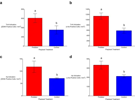
Figure 2.1. Auditory predator playbacks result in significantly higher numbers of ZENK (a) and c-fos (b) positive cells in the nucleus taeniae of the amygdala (TnA) in the brains of black-capped chickadees in comparison ton-predator (control) playbacks. The same effect can be seen in the hippocampus (Hp) of chickadees using ZENK (c) and c-fos (d). Means (±SE) represented by different letters are significantly different (p < 0.05). ... 46
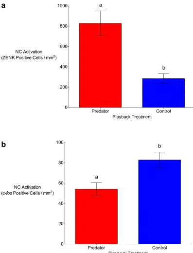
Figure 2.2 The caudal nidopallium (NC) of black-capped chickadees is differentially active in response to auditory predator and non-predator (control) playbacks depending on the immediate-early gene (IEG) used to measure this activation. Using ZENK, the predator treatment resulted in significantly higher NC immunoreactivity than the control treatment (a). The opposite pattern occurred when the IEG c-fos was used to quantify the immunoreactivity (b). Means (±SE) represented by different letters are significantly different (p < 0.05). ... 47
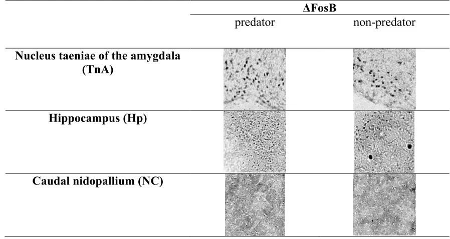
Figure 2.3. In the nucleus taeniae of the amygdala (TnA) (a) and hippocampus (Hp) (b) of black-capped chickadees, there was significantly greater ΔFosB immunoreactivity seven days post-stimulus in response to auditory predator playbacks in comparison to non-predator (control) playbacks. There was no significant difference in ΔFosB immunoreactivity between the two playback treatments in the caudal nidopallium (NC) (c). Means (±SE) represented by different letters are significantly different (p < 0.05). ... 48
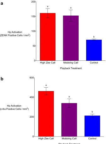
Figure 3.1. Auditory playbacks of chickadee high zee calls result in significantly higher numbers ZENK (a) and c-fos (b) positive cells in the nucleus taeniae of the amygdala (TnA) in the brains of black-capped chickadees in comparison to auditory chickadee mobbing call and non-predator (control) playbacks. Means (±SE) represented by different letters are significantly different from the control (p < 0.05). ... 76
Figure 3.2. Auditory playbacks of chickadee high zee and mobbing calls result in
xi
in the brains of black-capped chickadees in comparison to non-predator (control) playbacks. Means (±SE) represented by different letters are significantly different from the control (p < 0.05). ... 77
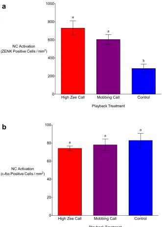
Figure 3.3. Auditory playbacks of chickadee high zee and mobbing calls result in
significantly higher numbers ZENK (a) positive cells in the caudal nidopallium (NC) in the brains of black-capped chickadees in comparison to non-predator (control) playbacks. Chickadee high zee and mobbing call playbacks do not result in significantly different c-fos NC activation from the controls (b). Means (±SE) represented by different letters are
Chapter 1 General Introduction
1.1Perceived predation risk
Predators have traditionally been seen as direct killers, reducing prey populations
through consumption alone (Abrams 1993; Preisser et al. 2005; Schmitz et al. 2008).
Models of predator-prey dynamics have most often focused on predators, while treating
prey as unresponsive victims of predation (Murdoch & Oaten 1975; Taylor 1984; Turchin
2003). We now know that predators impact prey not only by how many they kill, but also
by how many they threaten. The presence, and resulting perceived risk, of predators has
far greater impacts on prey and ecosystems than direct predation alone (Schmitz et al.
2008). These non-consumptive predator effects have profound effects on prey by altering
their physiology, behavior, and morphology.
The success rate of predators in the wild is not often considered in studies of
predator-prey dynamics, despite extremely low rates of prey capture by predators across
taxa (Vermeij 1982). As predators have limited success when catching prey, prey often
survive these ‘near-miss’ predator attacks (McLaughlin et al. 2000; Cresswell & Quinn
2010; Combes et al. 2013). For example, it is estimated that sharks fail to kill bottlenose
dolphins (Tursiops aduncus) at least 11% of the time after they have successfully bitten
the dolphin (Heithaus 2001). Very few species have capture rates that approach even
90%, and may not kill all prey they successfully capture (Vermeij 1982). The fact that
predators do not kill all prey they pursue is clarified further by the fact that up to 70% of
living individuals in some prey species have scars from non-lethal predator attacks
To illustrate, 13% of giraffes (Giraffa camelopardalis) over one year of age exhibited
claw marks on their hindquarters from non-lethal lion (Panthera leo) attacks (Strauss &
Packer 2013). We should expect that these life-threatening predator confrontations would
have substantial, lasting impacts on all aspects of prey ecology. The ability of animals to
recognize and respond to predation risk and avoid predator attack forms the foundation
on which non-consumptive predator effects change prey populations and communities
(Lima & Steury 2005). In order to act appropriately based on the risk of predation in their
environments, prey use anti-predator tactics to decrease their conspicuousness and to
improve their chances of escaping attack (Lima & Dill 1990; Brown et al. 1999; Nelson
et al. 2004; Caro 2005).
1.2 Anti-predator responses
Anti-predator responses can be behavioural, aimed at allowing prey to better
conceal themselves or to enable them to detect predators more successfully (Abrams
1986; Lima & Dill 1990; Schmitz et al. 2008). Prey may increase their use of refuges or
decrease their feeding, parenting, or mating activity, rendering them less conspicuous
(Lima & Dill 1990; Abrams 1993; Brown & Kotler 2004; Creel et al. 2005; Schmitz et
al. 2008). For example, when faced with increased densities of tiger sharks (Galeocerdo
cuvier), bottlenose dolphins abandoned highly profitable, yet risky, shallow foraging
habitats to forage in less profitable, but safe deep habitat (Heithaus & Dill 2006). In
addition to behavioural alteration, the physiology of prey including glucocorticoid levels,
metabolic rates, and oxidative stress can all be impacted by an increase in perceived
predation risk (Hik et al. 2001; Apfelbach et al. 2005; Slos & Stoks 2008; Sheriff et al.
levels can reduce the function of gonadotropins, affecting reproduction (Sheriff et al.
2009); increased metabolism can result in greater energy use and increased need for food
(Chabot et al. 1996); and oxidative stress can reduce body condition, immune function,
reproduction, and survival (Janssens & Stoks 2014). These changes in behaviour and
physiology were thought to be solely acute, occurring during and immediately after a
near-lethal encounter with a predator, and quickly dissipating, returning the individual to
its previous state. Recently, however, studies have shown that these anti-predator
responses can also endure, affecting reproduction and survival (Preisser & Bolnick 2008;
Anson et al. 2013). Elk (Cervus canadensis) in the Yellowstone National Park population
showed a significant decline in calf recruitment in the years following the reintroduction
of wolves (Canis lupus) to the park, despite the fact that wolves rarely kill calves,
suggesting that perceived predation risk alone resulted in these declines (Creel et al.
2007). Zanette et al. (2011) found a 40% decrease in song sparrow (Melospiza melodia)
offspring production in response to high perceived predation risk in the absence of direct
predation.
Increased predation risk can also result in enduring changes in prey morphology
through developmental phenotypic plasticity. In invertebrates, fish, and some
amphibians, these developmental changes take the form of inducible morphological
defences, alterations in body form that assist prey in surviving predator attacks (Preisser
et al. 2005; Preisser & Bolnick 2008). The classic example of an inducible morphological
defence is the spiny “helmet” that Daphniapulex develop in response to increased
predation risk (Krueger & Dodson 1981); other organisms develop spikes or tougher
external morphological defences in response to an increase in predation risk, but it has
been proposed that analogous defences are predator-induced changes in neurobiology
(Kavaliers & Choleris 2001; Apfelbach et al. 2005; Sheriff et al. 2009; Clinchy et al.
2010, 2013). These changes can affect neurotransmitters, neuroarchitecture, plasticity,
and gene expression, and can persist over the long term (Slos & Stoks 2008; Zoladz et al.
2008, 2012; Clinchy et al. 2010, 2013; Cohen et al. 2012). Most information about how
the brain responds to threat come from studies of post-traumatic stress disorder (PTSD)
in humans. Results of this research demonstrate that fear (i.e., the prospect of imminent,
violent death due to perceived predation risk) has profound impacts on brain structure
and function over the long-term.
1.3 Fear and post-traumatic stress disorder (PTSD)
Fear is a natural, evolutionary response to threats in the environment. It results in
physiological responses that support defensive behaviours (i.e., anti-predator behaviours;
fighting, fleeing, remaining motionless) (Schmitz et al. 1997; Nelson et al. 2004;
Macleod et al. 2014). These physiological and defensive responses are a means by which
an organism can increase its chances of survival in the face of a threat to its life. An
organism can respond innately to, or learn through experience about, a variety of possible
threats (i.e., predators, social behaviours, pain) and determine how best to respond to
these threats in order to survive. In this way, fear is beneficial, as it allows an individual
to respond to a threat to its survival (Boonstra 2013). However, responding to fear can be
detrimental to an organism’s other functions, like feeding or reproduction, if like in
with a previous life-threatening event or if the fear response far outlasts the actual threat
(Shiromani et al. 2009).
PTSD is a chronic, incapacitating disorder that results from a traumatic
experience in which one perceives a potential loss of life (Shiromani et al. 2009; Cohen
et al. 2012). PTSD is characterized by extreme fear caused by the initial traumatic event,
repeated re-experiencing of this event, avoidance of cues related to the trauma, and
hyperarousal and hypervigiliance for at least one month after the event– although most
patients experience these symptoms for much longer (Shiromani et al. 2009). These
behavioural and psychological symptoms occur in tandem with changes in physiology.
Patients with PTSD show increased sensitivity in the negative feedback system of the
hypothalamic-pituitary-adrenal (HPA) axis, resulting in abnormal glucocorticoid levels
(Yehuda 2002; Shiromani et al. 2009; Chattarji et al. 2015). Cortisol levels in PTSD
patients have been found to be below normal for decades after the traumatic event,
despite high levels of corticotropin-releasing factor in cerebrospinal fluid (Yehuda 2002).
Patients also have increased circulating norepinephrine and thyroid hormones, which
contribute to behavioural symptoms (e.g. hypervigilance) (Yehuda 2002).
Fear also has dramatic impacts on the function of the mammalian brain, and this
fear is something that can be measured. Different types of fear– fear of pain, fear of
aggressive conspecifics, and fear of predators– are processed in distinct pathways (Gross
& Canteras 2012). In the study of PTSD in humans and fear in other mammals, three
brain regions are commonly implicated in the processing of this fear: the amygdala, the
hippocampus, and the prefrontal cortex (Shin et al. 2006; Sotres-Bayon et al. 2006;
Although the amygdala is involved in many functions, its critical role in the
processing of fear is unambiguous and ubiquitous in mammalian species (Shiromani et
al. 2009). In the brains of normal humans, the amygdala is active in response to fearful
faces and aversive stimuli, providing a protective function by alerting individuals to
relevant threats or cues of threat in their environments, and by processing these threats
(Shiromani et al. 2009). In PTSD patients, however, the amygdala is hyperresponsive to
both trauma-related stimuli (e.g. combat sounds) (Liberzon et al. 1999; Protopopescu et
al. 2005; Shin et al. 2006) and innocuous stimuli associated with the event (e.g. locations,
people) (Yehuda 2002). Although few studies have investigated amygdala structure in
PTSD patients, some have found decreased amygdala volume in those exhibiting
symptoms (Rogers et al. 2009). The amygdala has projections to the hippocampus and
prefrontal cortex, and regulates the response to stress in each of these regions (Kilpatrick
& Cahill 2003; Akirav & Maroun 2007; Shiromani et al. 2009; Chattarji et al. 2015).
The role of the hippocampus is more diverse than that of the amygdala; it is
involved in the formation of memory, learning, and the processing of spatial information
(Shiromani et al. 2009). However, the hippocampus is also involved specifically in fear
processing, as evidence suggests a role for the hippocampus in the processing of spatial
information to do with fear, the formation of fear memories, and the extinction of fear
responses (Kim & Diamond 2002; Shin et al. 2006; Cornwell et al. 2012; Gross &
Canteras 2012; Wang et al. 2013; Wotjak & Pape 2013). The hippocampus
communicates with the amygdala and feedback moves bidirectionally between these
regions (Shiromani et al. 2009; Chattarji et al. 2015). Similarly to the amygdala, studies
Chattarji et al. 2015), and it has been suggested that these abnormalities may relate to
memory and cognitive deficits seen in these patients, and may in fact be a risk factor for
the disorder itself (Shiromani et al. 2009). Unlike the amygdala, which shows heightened
activity in PTSD, hippocampal activity has been found to be lower than normal in PTSD
patients (Schuff et al. 2001). The hippocampus plays an important regulatory role with
regards to the HPA axis; decreased hippocampal function results in less HPA axis
inhibition and consequently greater activation in this axis, leading to an increased stress
response (Shiromani et al. 2009; Chattarji et al. 2015).
The prefrontal cortex is the third brain region implicated in the processing of fear,
although, like the hippocampus, it has various functions (Gross & Canteras 2012;
Chattarji et al. 2015). The primary role of the prefrontal cortex is in executive control
including decision-making, as well as in the formation of fearful memories (Shin et al.
2006; Akirav & Maroun 2007; Maroun 2012). It has been shown to regulate the stress
response by providing “top-down” control to the amygdala, inhibiting the amygdala fear
response under normal circumstances (Quirk & Beer 2006; Sotres-Bayon et al. 2006;
Akirav & Maroun 2007; Shiromani et al. 2009; Chattarji et al. 2015). However, recent
studies suggest that in PTSD, the prefrontal cortex is hyporesponsive, leading to
uninhibited amygdala activity; this diminished activity is one of the most consistent
findings in the PTSD literature (Cerqueira et al. 2007; Shiromani et al. 2009; Chattarji et
al. 2015).
Taken together, these three brain regions and the lasting changes in their structure
and function following a traumatic experience are crucial to our understanding of the
other mammals. Using mammalian models in the lab, researchers have been able to delve
deeper into this brain network to gain further understanding of its function and response
to trauma, and to attempt to model a PTSD-like disorder (Adamec & Shallow 1993;
Wiedenmayer 2004; Cohen et al. 2012). Initially, researchers undertaking mammalian
laboratory studies of fear used aversive stimuli like restraint, foot shock, swimming
stress, or changes in social hierarchy to induce a stress response in the study organisms
(Clinchy et al. 2010). However, recent research has focused on the use of auditory and
olfactory predator cues. These are perceived by rats and mice in the lab as
life-threatening, but cause them no pain, best mimicking PTSD-eliciting stimuli (Adamec &
Shallow 1993; Staples et al. 2005, 2009; Mackenzie et al. 2010; Cohen et al. 2012). The
exposure of mammals in the lab to predator cues is a powerful means by which to
investigate the effects of fear on the brain and has also resulted in numerous sustained
behavioural, physiological, and neurobiological changes similar to symptoms of human
PTSD (Wiedenmayer 2004; Zoladz et al. 2008, 2012; Mitra et al. 2009; Clay et al. 2011).
These fear-induced changes include effects on anxiety, hormone levels, and gene
expression in the brain (Adamec & Shallow 1993; Adamec et al. 2004; Staples et al.
2005; Costantini et al. 2010; Clinchy et al. 2011). Like in PTSD, a single, traumatic
exposure to a threatening cue can result in lasting changes in lab mammals for weeks or
months (Adamec & Shallow 1993; Adamec et al. 2004; Clinchy et al. 2010; Wotjak &
Pape 2013).
1.4 Effects of fear on the brains of wild animals
It is evident that the brains of humans and other mammals in the laboratory are
living under natural conditions (Creel & Christianson 2008). Recent studies have shown
that captive raised animals may be less responsive to aversive stimuli than wild animals
(Wiedenmayer 2004). Little study has gone into what the neurobiological effects of
real-world predation threat are on wild, free living animals, especially non-mammalian
species. Very little is known about the avian brain regions and networks processing
predator fear, and no investigation into the long-term activation of the avian brain in
response to predator threat has occurred. It should be expected, however, that an
encounter with a predator (or a simulation of such an encounter with an auditory or
olfactory cue) would be perceived as life threatening by prey, and that this type of
traumatic event should result in lasting changes in the brain, as seen in humans with
PTSD and mammalian lab models (Clinchy et al. 2010; Cohen et al. 2012; Boonstra
2013). The life-long predator fear that wild animals experience is more intense than any
simulation that could be carried out in a laboratory setting, and therefore should result in
extreme impacts on their neurobiology.
The quantification of the impacts of predator-induced fear on wild animals is
additionally a more meaningful metric than measuring this same fear in lab mammals.
Because wild animals have almost certainly experienced predator threat in their
environment, they are likely functioning at a fear level higher than the baseline that
would be expected of a predator-naïve lab model. As a result, any significant increases in
any measure of fear (behavioural, physiological, or neurobiological) in a wild animal
represent a meaningful impact of fear (Creel & Christianson 2008; Clinchy et al. 2010,
2013; Cohen et al. 2012).
predator-induced fear in the avian brain: the nucleus taeniae of the amygdala (TnA), the
hippocampus (Hp), and the caudal nidopallium (NC). The nucleus taeniae of the
amygdala (TnA) is known to be the avian homologue of the medial amygdala, and is
proposed to be the avian fear centre (Cohen & Goff 1978; Charlier et al. 2005). It has
been proposed that this region, like its mammalian counterpart, acts as a switchboard,
gathering information about potential threats in the environment and routing this
information to other areas of the brain for processing. Previous studies have shown the
TnA and its mammalian homologue to be activated in response to aversive stimuli, such
as foot shock, as well as to unambiguous cues of predation threat including predator
mounts and, in the case of the mammalian amygdala, olfactory predator cues (Dielenberg
et al. 2001; Li et al. 2004; Brito et al. 2011; Marzluff et al. 2012; Cross et al. 2013).
The proposed role of the avian hippocampus (Hp), homologue of the mammalian
hippocampus, in the processing of predator-induced fear is more ambiguous than that of
the TnA; the Hp has been implicated in many processes, including several fear-related
functions such as the formation of memory of fearful stimuli and processing of spatial
and social information (Clayton & Lee 1998; Colombo & Broadbent 2000; Kim &
Diamond 2002; Mayer et al. 2010; Nishizawa et al. 2011; Cornwell et al. 2012; Cross et
al. 2013).
The role of the NC in the processing of predator-induced fear has thus far been
ambiguously described, as it is involved in many processes. The NC is analogous to the
mammalian prefrontal cortex, and it has been proposed that it is involved in executive
function and decision-making (Veit & Nieder 2013), and has previously been found to be
2013) (Figure 1.1.).
Figure 1.1. Regions proposed to play a role in the processing of fear in the avian brain: the nucleus taeniae of the amygdala (TnA), the hippocampus (Hp), and the caudal nidopallium (NC) as viewed in one hemisphere of a coronal brain slice. Locations of brain regions are indicated by labels and red outlines.
1.5 Measuring fear in the brain
One of the major obstacles to studying the effects of perceived predation risk on
the brain is how we measure this fear (Lima & Dill 1990). In order to measure changes in
activation in the brain, the protein products of immediate-early genes (IEGs) can be
previously to investigate activation in the avian and mammalian brains, ZENK (Kimpo &
Doupe 1997; Bailey & Wade 2003; Phillmore et al. 2003; Knapska & Kaczmarek 2004;
Charlier et al. 2005; Leitner et al. 2005; Avey et al. 2008; Mayer et al. 2010; Brito et al.
2011) and c-fos (Kimpo & Doupe 1997; Dielenberg et al. 2001; Wiedenmayer & Barr
2001; Charlier et al. 2005; Staples et al. 2005; Cunningham et al. 2008; Vanelzakker et
al. 2011). These IEGs are used as short-term markers of brain activation, as the protein
products of these genes are produced and degraded in active neurons within hours of a
stimulus exposure (Cole et al. 1989; Kimpo & Doupe 1997; Guzowski et al. 2001;
Thiriet et al. 2001; Mokin & Keifer 2005).
ZENK is a gene encoding a nuclear transcription factor protein, ZENK, which is
rapidly induced following exposure to an extracellular stimulus. ZENK protein binds to
DNA and activates transcription of target genes, protein products of which are required
for cell division and differentiation. ZENK is not produced in all neuron populations, but
cells expressing ZENK protein in their nuclei are considered activated (Cole et al. 1989;
Guzowski et al. 2001; Thiriet et al. 2001; Mokin & Keifer 2005). The immediate-early
gene c-fos encodes the c-fos protein, which is rapidly translated and acts as a
transcriptional regulator for several target genes. Like ZENK, c-fos is not expressed in all
neurons, but when it is, this is an indication that this cell has been activated by an
external stimulus (Guzowski et al. 2001; Thiriet et al. 2001; Mokin & Keifer 2005).
In order to measure long-lasting changes in activity in the mammalian brain,
researchers have used the IEG FosB. The protein product of this gene, FosB, is a
transcription factor that is induced by chronic external stimuli. A splice variant of the
acting as a transcriptional regulator, influencing plasticity and behaviour (McClung et al.
2004; Nestler 2008). ΔFosB is present in active mammalian neurons for at least a week
following chronic stimulus exposure. Labelling ΔFosB over the weeks post-stimulus
provides a picture of lasting activation as a result of the stimulus (McClung et al. 2004;
Nestler 2008). Despite its use in mammalian studies, ΔFosB has never before been
labelled in the avian brain. It is unknown whether it is produced or can be labelled in the
neurons of birds.
1.6 The importance of wild animals
The vast majority of research into the effects of fear on the brain has so far taken
place in a biomedical context with controlled stimuli and human patients or model
mammalian subjects. It is unknown whether predator-induced fear results in similar
changes in the brains of wild animals, especially non-mammals, exposed to constant and
unpredictable predation threat in their environments. Wild animals were not considered in
fear research until recently; they were previously thought to be unaffected by predator
fear in the long term, as it was thought to be maladaptive for a free-living animal to be
debilitated by stress (Sapolsky 2004). Fear of a predator was considered an acute stress
response immediately following a predator encounter and quickly dissipating (Krebs
2002). However, studies have shown that fear effects on free-living animals result in
lasting changes in behaviour and reproduction (Wiedenmayer 2004; Creel & Christianson
2008; Hawlena & Schmitz 2010); for example, by changing foraging activity (Brown &
Kotler 2004; Heithaus & Dill 2006; Zanette et al. 2013), habitat selection (Creel et al.
2005; Eggers et al. 2006), and reproduction (Eggers et al. 2006; Zanette et al. 2006,
cues may prove more advantageous for the study of PTSD than mammalian lab models
(Clinchy et al. 2010, 2013; Cohen et al. 2012). The threat of predation in the real world is
not an occasional or predictable one. Wild animals could have a life-threatening
encounter with a predator at any time, every day of their lives. Predator-induced changes
in behaviour, physiology, and neurobiology in response to persistent and unpredictable
predator-induced fear in the wild are easily related to PTSD. This fear is a valid,
ethologically relevant experience for wild animals, and the resulting effects may mimic
those seen in PTSD patients to an even greater degree than the dramatic effects already
demonstrated in the lab (Wiedenmayer 2004). Thus, the study of fear in wild animals is
crucial to further clarify how life-threatening experiences affect behaviour, physiology,
and neurobiology, and will provide biomedical researchers with new information about
the causes and symptoms of PTSD in humans.
In addition to the biomedical applications of the study of fear on the brains of
wild animals, this research is the first step to linking changes in the brains of wild
animals to changes in their behaviour and physiology, and the effects of these changes on
reproduction and population dynamics. We now know that the fear of predators has
impacts on foraging, reproduction, and parental care, with effects spanning generations
(Creel et al. 2005, 2007; Eggers et al. 2006; Zanette et al. 2006, 2011, 2012, 2013;
Travers et al. 2010), and results in altered physiology (Clinchy et al. 2004; Creel et al.
2007; Hawlena & Schmitz 2010; Newman et al. 2012; Zanette et al. 2012). These
changes in turn can lead to decreased survival and fecundity in prey species, having
population level effects and changing prey demography. However, no connection has
activity. We should expect, based on links between changes in the brain and changes in
behaviour seen in PTSD, that similar connections will be found in studies of wild
animals. The first step to making these important links is the investigation of how fear is
changing the brains of these animals, and later investigating parallel changes in behaviour
and physiology.
1.7 Research goals
We know that life-threatening events change the brains of humans, and that the
brains of laboratory mammals are affected similarly in response to simulations of
predator threat, both in the short- and long-term. It is unknown whether wild animals are
affected to the same degree as those raised in captivity, and the networks processing
predator fear in the brains of non-mammalian taxa are not yet well understood. No
experiment has tested the acute and lasting effects of predator stimuli on wild-caught
non-mammals.
In Chapter 2, I address three brain regions of interest in avian fear processing in
black-capped chickadees (Poecile atricapillus), and investigate acute and lasting
activation changes in these regions in response to short-term and chronic simulations of
predation risk. In Chapter 3, I investigate short-term activation changes in the same
chickadee brain regions in response to black-capped chickadee alarm calls, which act as
social cues of different degrees of predation threat. In Chapter 4, I discuss the broader
implications of my findings and their application to our biomedical understanding of how
fear impacts the brain and to prey ecology. I also suggest aspects of
1.8 Study species
Black-capped chickadees are one of the most recognizable birds in North
America. Their range covers almost all of Canada and most of the United States (all
general life history of chickadees reviewed here is from Smith 1991, unless noted
otherwise). Chickadees are resident year round, making them ideal research subjects, as
they are accessible in any season. Chickadees, ranging in mass from 10 to 14 g, are
characterized by their dark cap and bib, white cheeks, and dark back. They feed
frequently and cache food for later use in multiple locations.
Chickadees live in nonbreeding flocks in the fall and winter, and defend territories
in monogamous breeding pairs in the spring and summer. Both sexes excavate the
cavities in which they build and incubate their nests, but females alone construct the nest
and incubate the eggs. Common predators of chickadees include sharp-shinned (Accipter
striatus) and Cooper’s hawks (Accipter cooperii), and commonly in the area in which I
conducted my research, northern saw-whet owls (Aegolius acadicus).
Chickadees have a complex social system consisting of an extensive vocal
repertoire of at least 11 distinct vocalizations, encoding different messages. When
confronted with a perceived threat, chickadees use one of two vocalizations to alert
conspecifics. In the case of a moderately threatening predator, chickadees use the
mobbing call. This call is used by both sexes to alert other flock members to a potential
threat, calling the flock together to confront, or mob the threat. An example of a
moderately alarming predator is a stationary avian predator within view of at least one
member of the flock. In the case of extreme alarm, such as a flying avian predator or a
this high frequency call. The call alerts flock members to the threat and induces
immobility until the threat is no longer imminent.
The chickadees used in my study are resident to the area of London, ON around
the University of Western Ontario year round. Chickadees approach feeders easily,
making them easy to capture in Potter traps for study, although capture is easier in fall
and winter when chickadees flock together and food is scarce. Wild-caught chickadees
are known acclimatize well to captivity, and the chickadees I used did well in
1.9 References
Abrams, P.A. (1986). Adaptive responses of predators to prey and prey to predators: the failure of the arms-race analogy. Evolution, 40, 1229–1247.
Abrams, P.A. (1993). Why predation rate should not be proportional to predator density. Ecology, 74, 726–733.
Adamec, R., Walling, S. & Burton, P. (2004). Long-lasting, selective, anxiogenic effects of feline predator stress in mice. Physiol. Behav., 83, 401–410.
Adamec, R.E. & Shallow, T. (1993). Lasting effects on rodent anxiety of a single exposure to a cat. Physiol. Behav., 54, 101–109.
Akirav, I. & Maroun, M. (2007). The role of the medial prefrontal cortex-amygdala circuit in stress effects on the extinction of fear. Neural Plast., 2007, 1–11.
Anson, J.R., Dickman, C.R., Boonstra, R. & Jessop, T.S. (2013). Stress triangle: do introduced predators exert indirect costs on native predators and prey? PLoS One, 8, 1–10.
Apfelbach, R., Blanchard, C.D., Blanchard, R.J., Hayes, R.A. & McGregor, I.S. (2005). The effects of predator odors in mammalian prey species: A review of field and laboratory studies. Neurosci. Biobehav. Rev., 29, 1123–1144.
Avey, M.T., Kanyo, R. A., Irwin, E.L. & Sturdy, C.B. (2008). Differential effects of vocalization type, singer and listener on ZENK immediate early gene response in black-capped chickadees (Poecile atricapillus). Behav. Brain Res., 188, 201–208.
Bailey, D.J. & Wade, J. (2003). Differential expression of the immediate early genes FOS and ZENK following auditory stimulation in the juvenile male and female zebra finch. Mol. Brain Res., 116, 147–154.
Boonstra, R. (2013). Reality as the leading cause of stress: rethinking the impact of chronic stress in nature. Funct. Ecol., 27, 11–23.
Brito, I., Britto, L.R.G. & Ferrari, E.A.M. (2011). Induction of Zenk protein expression within the nucleus taeniae of the amygdala of pigeons following tone and shock stimulation. Brazilian J. Med. Biol. Res., 44, 762–766.
Brown, J.S. & Kotler, B.P. (2004). Hazardous duty pay and the foraging cost of predation. Ecol. Lett., 7, 999–1014.
Brown, J.S., Laundre, J.W., & Gurung, M. (1999). The ecology of fear: optimal foraging, game theory and trophic interactions. J. Mammal., 80, 385–399.
Cerqueira, J.J., Mailliet, F., Almeida, O.F.X., Jay, T.M. & Sousa, N. (2007). The prefrontal cortex as a key target of the maladaptive response to stress. J. Neurosci., 27, 2781–2787.
Chabot, D., Gagnon, P. & Dixon, E.A. (1996). Effect of predator odors on heart rate and metabolic rate of wapiti (Cervus elaphus canadensis). J. Chem. Ecol., 22, 839– 868.
Charlier, T.D., Ball, G.F. & Balthazart, J. (2005). Sexual behavior activates the expression of the immediate early genes c-fos and Zenk (egr-1) in
catecholaminergic neurons of male Japanese quail. Neuroscience, 131, 13–30.
Chattarji, S., Tomar, A., Suvrathan, A., Ghosh, S. & Rahman, M.M. (2015).
Neighborhood matters : divergent patterns of stress-induced plasticity across the brain. Nat. Neurosci., 18, 1364–1375.
Clay, R., Hebert, M., Gill, G., Stapleton, L.A., Pridham, A., Coady, M., et al. (2011). Glucocorticoids are required for extinction of predator stress-induced
hyperarousal. Neurobiol. Learn. Mem., 96, 367–377.
Clinchy, M., Schulkin, J., Zanette, L.Y., Sheriff, M.J., McGowan, P.O. & Boonstra, R. (2010). The neurological ecology of fear: insights neuroscientists and ecologists have to offer one another. Front. Behav. Neurosci., 5, 1–6.
Clinchy, M., Sheriff, M.J. & Zanette, L.Y. (2013). Predator-induced stress and the ecology of fear. Funct. Ecol., 27, 56–65.
Clinchy, M., Zanette, L., Boonstra, R., Wingfield, J.C. & Smith, J.N.M. (2004).
Balancing food and predator pressure induces chronic stress in songbirds. Proc. Biol. Sci., 271, 2473–2479.
Clinchy, M., Zanette, L., Charlier, T.D., Newman, A.E.M., Schmidt, K.L., Boonstra, R., et al. (2011). Multiple measures elucidate glucocorticoid responses to
environmental variation in predation threat. Oecologia, 166, 607–614.
Cohen, D.H. & Goff, D.M. (1978). Effect of avian basal forebrain lesions, including septum, on heart rate conditioning. Brain Res. Bull., 3, 311–318.
Cohen, H., Kozlovsky, N., Alona, C., Matar, M.A. & Joseph, Z. (2012). Animal model for PTSD: From clinical concept to translational research. Neuropharmacology, 62, 715–724.
Cole, A.J., Saffen, D.W., Baraban, J.M. & Worley, P.F. (1989). Rapid increase of an immediate early gene messenger RNA in hippocampal neurons by synaptic NMDA receptor activation. Nature, 340, 474–476.
Colombo, M. & Broadbent, N. (2000). Is the avian hippocampus a functional homologue of the mammalian hippocampus? Neurosci. Biobehav. Rev., 24, 465–484.
Cornwell, B.R., Arkin, N., Overstreet, C., Carver, F.W. & Grillon, C. (2012). Distinct contributions of human hippocampal theta to spatial cognition and anxiety. Hippocampus, 22, 1848–1859.
Costantini, D., Rowe, M., Butler, M.W. & McGraw, K.J. (2010). From molecules to living systems: historical and contemporary issues in oxidative stress and antioxidant ecology. Funct. Ecol., 24, 950–959.
Creel, S. & Christianson, D. (2008). Relationships between direct predation and risk effects. Trends Ecol. Evol., 23, 194–201.
Creel, S., Christianson, D., Liley, S. & Winnie, J.A. (2007). Predation risk affects reproductive physiology and demography of elk. Science, 315, 960.
Creel, S., Winnie, J., Maxwell, B., Hamlin, K. & Creel, M. (2005). Elk alter habitat selection as an antipredator response to wolves. Ecology, 86, 3387–3397.
Cresswell, W. & Quinn, J.L. (2010). Attack frequency, attack success and choice of prey group size for two predators with contrasting hunting strategies. Anim. Behav., 80, 643–648.
Cross, D.J., Marzluff, J.M., Palmquist, I., Minoshima, S., Shimizu, T. & Miyaoka, R. (2013). Distinct neural circuits underlie assessment of a diversity of natural dangers by American crows. Proc. Biol. Sci., 280, 20131046.
Cunningham, J.T., Mifflin, S.W., Gould, G.G. & Frazer, A. (2008). Induction of c-Fos and DeltaFosB immunoreactivity in rat brain by vagal nerve stimulation. Neuropsychopharmacology, 33, 1884–1895.
Eggers, S., Griesser, M., Nystrand, M. & Ekman, J. (2006). Predation risk induces changes in nest-site selection and clutch size in the Siberian jay. Proc. Biol. Sci., 273, 701–706.
Gross, C.T. & Canteras, N.S. (2012). The many paths to fear. Nat. Rev. Neurosci., 13, 651–658.
Guzowski, J.F., Setlow, B., Wagner, E.K. & McGaugh, J.L. (2001). Experience-dependent gene expression in the rat hippocampus after spatial learning: a
comparison of the immediate-early genes Arc, c-fos, and zif268. J. Neurosci., 21, 5089–5098.
Hawlena, D. & Schmitz, O.J. (2010). Physiological stress as a fundamental mechanism linking predation to ecosystem functioning. Am. Nat., 176, 537–556.
Heithaus, M.R. & Dill, L.M. (2006). Does tiger shark predation risk influence foraging habitat use by bottlenose dolphins at multiple spatial scales? Oikos, 114, 257–264.
Heithaus, R. (2001). Shark attacks on bottlenose dolphins (Tursiops aduncus) in Shark Bay, Western Australia: attack rate, bite scar frequencies, and attack seasonality. Mar. Mammal Sci., 17, 526–539.
Hik, D.S., McColl, C.J. & Boonstra, R. (2001). Why are Arctic ground squirrels more stressed in the boreal forest than in alpine meadows? Ecoscience, 8, 275–288.
Janssens, L. & Stoks, R. (2014). Chronic predation risk reduces escape speed by
increasing oxidative damage: a deadly cost of an adaptive antipredator response. PLoS One, 9, e101273.
Kilpatrick, L. & Cahill, L. (2003). Amygdala modulation of parahippocampal and frontal regions during emotionally influenced memory storage. Neuroimage, 20, 2091– 2099.
Kim, J.J. & Diamond, D.M. (2002). The stressed hippocampus, synaptic plasticity and lost memories. Nat. Rev. Neurosci., 3, 453–462.
Kimpo, R.R. & Doupe, A.J. (1997). FOS is induced by singing in distinct neuronal populations in a motor network. Neuron, 18, 315–325.
Knapska, E. & Kaczmarek, L. (2004). A gene for neuronal plasticity in the mammalian brain: Zif268/Egr-1/NGFI-A/Krox-24/TIS8/ZENK? Prog. Neurobiol., 74, 183– 211.
Krebs, C.J. (2002). Two complementary paradigms for analysing population dynamics. Philos. Trans. R. Soc. Lond. B. Biol. Sci., 357, 1211–1219.
Krueger, D.A. & Dodson, S.I. (1981). Embryological induction and predation ecology in Daphnia pulex. Limnol. Oceanogr., 26, 219–223.
Leitner, S., Voigt, C., Metzdorf, R. & Catchpole, C.K. (2005). Immediate early gene (ZENK, Arc) expression in the auditory forebrain of female canaries varies in response to male song quality. J. Neurobiol., 64, 275–284.
Li, C.-I., Maglinao, T.L. & Takahashi, L.K. (2004). Medial amygdala modulation of predator odor-induced unconditioned fear in the rat. Behav. Neurosci., 118, 324– 332.
Liberzon, I., Taylor, S.F., Amdur, R., Jung, T.D., Chamberlain, K.R., Minoshima, S., et al. (1999). Brain activation in PTSD in response to trauma-related stimuli. Biol. Psychiatry, 45, 817–826.
Lima, S.L. & Steury, T.D. (2005). Perception of predation risk: the foundation of nonlethal predator-prey interactions. In: Ecol. predator-prey Interact. (eds. Barbosa, P. & Castellanos, I.). Oxford University Press, Oxford, UK, pp. 166– 188.
Mackenzie, L., Nalivaiko, E., Beig, M.I., Day, T.A. & Walker, F.R. (2010). Ability of predator odour exposure to elicit conditioned versus sensitised post traumatic stress disorder-like behaviours, and forebrain δFosB expression, in rats. Neuroscience, 169, 733–742.
Macleod, C.D., Macleod, R., Learmonth, J.A., Cresswell, W. & Pierce, G.J. (2014). Predicting population-level risk effects of predation from the responses of individuals. Ecology, 95, 2006–2015.
Maroun, M. (2012). Medial prefrontal cortex: multiple roles in fear and extinction. Neurosci., 19, 370–383.
Marzluff, J.M., Miyaoka, R., Minoshima, S. & Cross, D.J. (2012). Brain imaging reveals neuronal circuitry underlying the crow’s perception of human faces. Proc. Natl. Acad. Sci., 109, 15912–15917.
Mayer, U., Watanabe, S. & Bischof, H.J. (2010). Hippocampal activation of immediate early genes Zenk and c-Fos in zebra finches (Taeniopygia guttata) during learning and recall of a spatial memory task. Neurobiol. Learn. Mem., 93, 322–329.
McClung, C.A., Ulery, P.G., Perrotti, L.I., Zachariou, V., Berton, O. & Nestler, E.J. (2004). ΔfosB: a molecular switch for long-term adaptation in the brain. Mol. Brain Res., 132, 146–154.
McLaughlin, R.L., Grant, J.W.A. & Noakes, D.L.G. (2000). Living with failure: the prey capture success of young brook charr in streams. Ecol. Freshw. Fish, 9, 81–89.
Mokin, M. & Keifer, J. (2005). Expression of the immediate-early gene – encoded protein Egr-1 ( zif268 ) during in vitro classical conditioning. Learn. Mem., 12, 144–149.
Murdoch, W.W. & Oaten, A. (1975). Predation and population stability. Adv. Ecol. Res., 9, 1–131.
Nelson, E.H., Matthews, C.E. & Rosenheim, J.A. (2004). Predators reduce prey population growth by inducing changes in prey behaviour. Ecology, 85, 1853– 1858.
Nestler, E.J. (2008). Review. Transcriptional mechanisms of addiction: role of DeltaFosB. Philos. Trans. R. Soc. Lond. B. Biol. Sci., 363, 3245–3255.
Newman, A.E.M., Zanette, L.Y., Clinchy, M., Goodenough, N. & Soma, K.K. (2012). Stress in the wild: chronic predator pressure and acute restraint affect plasma DHEA and corticosterone levels in a songbird. Stress, 16, 363–367.
Nishizawa, K., Izawa, E.I. & Watanabe, S. (2011). Neural-activity mapping of memory-based dominance in the crow: Neural networks integrating individual
discrimination and social behaviour control. Neuroscience, 197, 307–319.
Orr, M. V, Hittel, K. & Lukowiak, K. (2010). Predator detection enables juvenile Lymnaea to form long-term memory. J. Exp. Biol., 213, 301–307.
Phillmore, L.S., Bloomfield, L.L. & Weisman, R.G. (2003). Effects of songs and calls on ZENK expression in the auditory telencephalon of field- and isolate-reared black capped chickadees. Behav. Brain Res., 147, 125–134.
Preisser, E.L. & Bolnick, D.I. (2008). The many faces of fear: comparing the pathways and impacts of nonconsumptive predator effects on prey populations, 3, 5–8.
Protopopescu, X., Pan, H., Tuescher, O., Cloitre, M., Goldstein, M., Engelien, W., et al. (2005). Differential time courses and specificity of amygdala activity in
posttraumatic stress disorder subjects and normal control subjects. Biol. Psychiatry, 57, 464–473.
Quirk, G.J. & Beer, J.S. (2006). Prefrontal involvement in the regulation of emotion: convergence of rat and human studies. Curr. Opin. Neurobiol., 16, 723–727.
Rogers, M.A., Yamasue, H., Abe, O., Yamada, H., Ohtani, T., Iwanami, A., et al. (2009). Smaller amygdala volume and reduced anterior cingulate gray matter density associated with history of post-traumatic stress disorder. Psychiatry Res. Neuroimaging, 174, 210–216.
Rose, J. & Colombo, M. (2005). Neural correlates of executive control in the avian brain. PLoS Biol., 3, e190.
Santa Cruz Biotechnology (2014). c-fos (4): sc-52 information. Santa Cruz Biotechnology Inc., Dallas, TX, USA.
Santa Cruz Biotechnology (2014). Egr-1 (C-19): sc-189 information. Santa Cruz Biotechnology Inc., Dallas, TX, USA.
Schmitz, O.J., Beckerman, A.P. & Brien, K.M.O. (1997). Behaviorally mediated trophic cascades: effects of predation risk on food web interactions. Ecology, 78, 1388– 1399.
Schmitz, O.J., Grabowski, J.H., Peckarsky, B.L., Preisser, E.L., Trussell, G.C., Vonesh, J.R., et al. (2008). From individuals to ecosystem function: toward an integration of evolutionary and ecosystem ecology. Ecology, 89, 2436–2445.
Sheriff, M.J., Krebs, C.J. & Boonstra, R. (2009). The sensitive hare: sublethal effects of predator stress on reproduction in snowshoe hares. J. Anim. Ecol., 78, 1249–1258.
Shin, L.M., Rauch, S.L. & Pitman, R.K. (2006). Amygdala, medial prefrontal cortex, and hippocampal function in PTSD. Ann. N. Y. Acad. Sci., 1071, 67–79.
Shiromani, P.J., Keane, T.M. & LeDoux, J.E. (Eds.). (2009). Post-Traumatic Stress Disorder: Basic Science and Clinical Practice. Humana Press, New York NY, USA.
Slos, S. & Stoks, R. (2008). Predation risk induces stress proteins and reduces antioxidant defense. Funct. Ecol., 22, 637–642.
Smith, S.M. (1991). The Black-Capped Chickadee: Behavioral Ecology and Natural History. Cornell University Press, Ithaca, NY, USA.
Sotres-Bayon, F., Cain, C.K. & LeDoux, J.E. (2006). Brain mechanisms of fear
extinction: historical perspectives on the contribution of prefrontal cortex. Biol. Psychiatry, 60, 329–336.
Staples, L.G., Hunt, G.E., Cornish, J.L. & McGregor, I.S. (2005). Neural activation during cat odor-induced conditioned fear and “trial 2” fear in rats. Neurosci. Biobehav. Rev., 29, 1265–1277.
Staples, L.G., McGregor, I.S. & Hunt, G.E. (2009). Long-lasting FosB/ΔFosB
immunoreactivity in the rat brain after repeated cat odor exposure. Neurosci. Lett., 462, 157–161.
Strauss, M.K.L. & Packer, C. (2013). Using claw marks to study lion predation on giraffes of the Serengeti. J. Zool., 289, 134–142.
Taylor, R.J. (1984). Predation. 1st edn. Chapman and Hall Ltd., NY, NY, USA.
Travers, M., Clinchy, M., Zanette, L., Boonstra, R. & Williams, T.D. (2010). Indirect predator effects on clutch size and the cost of egg production. Ecol. Lett., 13, 980–988.
Turchin, P. (2003). Complex population dynamics: a theoretical/empirical synthesis. Princeton University Press, Princeton, NJ, USA.
Vanelzakker, M.B., Zoladz, P.R., Thompson, V.M., Park, C.R., Halonen, J.D., Spencer, R.L., et al. (2011). Influence of pre-training predator stress on the expression of c-fos mRNA in the hippocampus, amygdala, and striatum following long-term spatial memory retrieval. Front. Behav. Neurosci., 5, 1–30.
Veit, L. & Nieder, A. (2013). Abstract rule neurons in the endbrain support intelligent behaviour in corvid songbirds. Nat. Commun., 4, 1–11.
Vermeij, G.J. (1982). Unsuccessful predation and evolution. Am. Nat., 120, 701–720.
Wang, M.E., Fraize, N.P., Yin, L., Yuan, R.K., Petsagourakis, D., Wann, E.G., et al. (2013). Differential roles of the dorsal and ventral hippocampus in predator odor contextual fear conditioning. Hippocampus, 23, 451–466.
Wiedenmayer, C.P. (2004). Adaptations or pathologies? Long-term changes in brain and behavior after a single exposure to severe threat. Neurosci. Biobehav. Rev., 28, 1– 12.
Wiedenmayer, C.P. & Barr, G.A. (2001). Developmental changes in c-fos expression to an age- specific social stressor in infant rats. Behav. Brain Res., 126, 147–157.
Wotjak, C.T. & Pape, H.-C. (2013). Neuronal circuits of fear extinction. Eur. J. Neurosci., 31, 599–612.
Zanette, L., Clinchy, M. & Smith, J.N.M. (2006). Food and predators affect egg production in song sparrows. Ecology, 87, 2459–2467.
Zanette, L.Y., Clinchy, M., Leonard, M.L., Horn, A.G., Haydon, D.T. & Hampson, E. (2012). Brood-parasite-induced female-biased mortality affects songbird demography: negative implications for conservation. Oikos, 121, 1493–1500.
Zanette, L.Y., Hobson, K.A., Clinchy, M., Travers, M. & Williams, T.D. (2013). Food use is affected by the experience of nest predation: implications for indirect predator effects on clutch size. Oecologia, 172, 1031–1039.
Zanette, L.Y., White, A.F., Allen, M.C. & Clinchy, M. (2011). Perceived predation risk reduces the number of offspring songbirds produce per year. Science, 334, 1398– 1401.
Zoladz, P.R., Conrad, C.D., Fleshner, M. & Diamond, D.M. (2008). Acute episodes of predator exposure in conjunction with chronic social instability as an animal model of post-traumatic stress disorder. Stress, 11, 259–281.
Chapter 2
2.1 Introduction
All organisms face the threat of predator attack. How individuals respond to these
threats impacts all aspects of ecology, from individual physiology to population
dynamics. Understanding the mechanisms by which prey process these life-threatening
encounters is essential to our knowledge of predator-prey ecology and has broad
implications for the study of human anxiety disorders.
Research regarding the effects of predators on their prey has traditionally focused
on direct predation (i.e., predators killing prey for food) and, more recently, on the
short-term effects of non-lethal predation events (i.e., perceived predation risk). For example,
increased predation risk can cause prey to intensify anti-predator behaviours in order to
become less conspicuous in the presence of predators (Lima 1998; Schmitz et al. 2008).
Perceived predation risk and predator encounters can also impact physiology (Clinchy et
al. 2004) including glucocorticoids (Hik et al. 2001; Creel et al. 2007; Newman et al.
2012) and measures of oxidative stress (Slos & Stoks 2008; Travers et al. 2010).
However, we now know that these acute changes in behaviour and physiology occur in
tandem with lasting indirect predator effects– persistent changes in behaviour,
physiology, and morphology resulting from a perceived predation threat or an actual
predation attempt (Preisser et al. 2005; Creel & Christianson 2008; Preisser & Bolnick
2008; Ferrari 2014). In fish, amphibians, and invertebrates, increased threat of predation
can cause organisms to induce morphological defences in order to protect against
‘helmet’ that Daphnia pulex develop in response to increased predation pressure
(Krueger & Dodson 1981).
In terrestrial vertebrates, which seldom develop induced external morphological
defences in response to increased predation risk, predator-induced defences may take the
form of changes in the morphology and activity of neurons in the brain, most examples of
which come from studies of post-traumatic stress disorder (PTSD) in humans (Shiromani
et al. 2009; Orr et al. 2010; Cohen et al. 2012; Clinchy et al. 2013). As a result of
research into the causes and symptoms of PTSD, it is now possible to measure some
impacts of fear (i.e., the prospect of imminent, violent death due to perceived predation
risk) on the brains of prey, as well as to demonstrate that the neurobiological effects of
predator-induced fear in prey persist over time.
Humans exposed to a fearful, seemingly inescapable, and life-threatening
stimulus are at risk of developing PTSD (Shiromani et al. 2009; Zoladz et al. 2012;
Chattarji et al. 2015). After as little as one brief, traumatic experience, the resulting
changes in behaviour, physiology, and neurobiology are immediate and can be life-long
(Yehuda 2002; Wiedenmayer 2004; Shiromani et al. 2009; Chattarji et al. 2015) (Chapter
1). Until recently, most research into fear and its effects has been biomedical, stemming
from our interest in PTSD and other fear-related disorders. The aim of this biomedical
research has traditionally been to investigate the effects of chronic psychological stress in
humans, whereas ecological research has traditionally focused on the acute physiological
crisis of prey surviving a predator attack (Clinchy et al. 2013). Recently, however, small
mammalian models have been used to further investigate the causes and symptoms of
al. 2012). Predator cues are reliable indicators of a possible attack and thus are perceived
as life-threatening– yet are painless– fitting the aetiology of PTSD better than any stimuli
previously used (Adamec & Shallow 1993; Staples et al. 2005, 2009; Mackenzie et al.
2010; Cohen et al. 2012). Exposure to these predator cues could result in “psychological
stress” or “fear” similar to PTSD-inducing fear in humans (Adamec et al. 2004; Clinchy
et al. 2013) and has resulted in numerous sustained changes in behaviour (Adamec et al.
2004; Mackenzie et al. 2010; Staples 2010), physiology (Clay et al. 2011), and
neurobiology (Mitra et al. 2009) (Chapter 1). In studying predator-induced fear and the
similar fear of losing one’s life leading to PTSD, researchers have identified which parts
of the mammalian brain are active in response to fearful stimuli (including fear of
predators), and often focus on three main regions: the medial amygdala, the
hippocampus, and the prefrontal cortex (Rosen & Schulkin 1998; Kilpatrick & Cahill
2003; Phelps 2004; Shin et al. 2006; Shiromani et al. 2009; Clinchy et al. 2013; Chattarji
et al. 2015).
The amygdala is the region most unequivocally implicated in the processing of all
fearful stimuli and is thought to be central to the network processing fear in the
mammalian brain, acting as a switchboard that collects information about environmental
threats and directs them into the complex and distinct efferent pathways for the fear of
pain, conspecifics, or predators (Cohen & Goff 1978; Shin et al. 2006; Gross & Canteras
2012; Chattarji et al. 2015). The hippocampus is a crucial component of the
predator-induced fear pathway, collecting spatial and contextual information from the environment
(Cornwell et al. 2012) and forming memories of fearful experiences (Kim & Diamond
functions, some of which are related to the processing of fearful stimuli. It gathers
information about perceived threats and allows individuals to make decisions and
respond appropriately to fearful stimuli (Shin et al. 2006; Akirav & Maroun 2007;
Shiromani et al. 2009; Gross & Canteras 2012; Maroun 2012). All of these regions have
been shown to be active over the short- and long-term in response to life-threatening fear
or predation threat in humans and mammalian models (Shiromani et al. 2009; Mackenzie
et al. 2010; Martinez et al. 2011; Wang et al. 2013; Chattarji et al. 2015). They are
connected in a feedback network with bidirectional communication among the regions
(Chattarji et al. 2015) (Chapter 1).
Predator-induced fear was not thought to dramatically affect animals in the wild
until recently, when studies showed that fear effects on free-living animals result in
lasting changes in behaviour and reproduction (Kavaliers & Choleris 2001; Wiedenmayer
2004; Creel & Christianson 2008; Hawlena & Schmitz 2010; Zanette et al. 2011). It is
unknown whether the brains of other taxa are altered by the experience of fear in the
wild, which brain regions are impacted, and if these changes are quantifiable. The
dramatic behavioural effects of predator-induced fear in wild mammals have led
researchers to suggest that wild animals exposed to predator cues will be more
advantageous for the study of PTSD than lab models (Clinchy et al. 2010, 2013; Cohen et
al. 2012). The ability to measure the effects of fear in wild animals previously living in
their natural environment with their predators is powerful (Creel & Christianson 2008),
as these wild animals– constantly vulnerable to the possibility of predator attack– already
function above the physiological and behavioural baseline of predator-naïve, lab-raised