1071-412X/05/$08.00⫹0 doi:10.1128/CDLI.12.3.453–464.2005
Copyright © 2005, American Society for Microbiology. All Rights Reserved.
Impaired Accessory Cell Function in a Human Dendritic Cell Line
after Human Immunodeficiency Virus Infection
Prarthana Beuria, Houchu Chen, Michael Timoney, and Kirk Sperber*
Division of Clinical Immunology, Mount Sinai School of Medicine, New York, New York
Received 20 July 2004/Returned for modification 13 October 2004/Accepted 28 December 2004
We generated human dendritic cell (DC) hybridoma cell lines by fusing HGPRT-deficient promonocytic U937 cells with immature DCs obtained by culturing peripheral blood monocytes with interleukin-4 (IL-4; 1,000 U/ml) and granulocyte-macrophage colony-stimulating factor (100 U/ml) for 7 days and mature DCs by
treatment with tumor necrosis factor alpha (12.5g/ml) for 3 days. Only one fusion with immature DCs was
successful and yielded four cell lines—HB-1, HB-2, HB-3, and HB-9—with an overall fusion efficiency of 0.0015%. The cell lines were stable in long-term culture, displayed morphological features typical of DCs, and expressed distinct class I and class II molecules not present on U937 (A*031012, B*51011, Cw*0701, DRB3*01011 52, and DR5*01011). A representative cell line, HB-2, that expressed DC markers including CD83, CD80 and CD86 could be induced to produce IL-12 through CD40 stimulation. After human immuno-deficiency virus (HIV) infection, there was impairment of antigen-presenting cell (APC) function, which was manifested by an inability to stimulate allogeneic T-cell responses. There was no change in expression of major histocompatibility complex class I and class II antigens, CD83, CD40, CD4, CD11c, CD80, CD86, CD54, and CD58, or IL-12 production in the HIV-infected HB-2 cells. The HIV-infected HB-2 cells induced T-cell apoptosis in the cocultures. T-cell proliferation could be partially restored by using ddI, indinivir, and blocking anti-gp120 and anti-IL-10 antibodies. Our data suggest that there are multiple mechanisms that DCs use to inhibit T-cell responses in HIV-infected patients. The HB-2 cell line could be a useful model system to study APC function in HIV-infected DCs.
Dendritic cells (DCs) are potent antigen-presenting cells (APCs) that have the unique property of being able to gener-ate primary T-cell responses (3, 4, 60). The roles that DCs play in the immunopathogenesis of human immunodeficiency virus (HIV) disease have been extensively studied, but some ques-tions still remain unanswered. It was not clear whether DCs can be directly infected with HIV or if the virus is only present on the cell surface or if both processes occur simultaneously (37, 46, 47). However, recent work has demonstrated that DCs can take up HIV into endosome-like compartments, where it can be transferred to T cells (35). Turnville et al. have dem-onstrated that DCs transfer HIV type 1 (HIV-1) to CD4⫹T cells in two distinct phases (63). In the first phase DCs divert the virus from the endolysosomal pathway to the DC–T-cell synapse, while in the second phase the transmission occurs by de novo infection of the DCs. Study of the immunologic func-tion of infected DCs, as well as DCs isolated from HIV-infected individuals, has yielded ambiguous results (7, 8, 10, 11, 12, 13, 19, 24, 25, 44, 44, 45, 53). Some reports have identified APC defects with DCs, whereas others have not. This could be partially explained by different effects of HIV infection on DC subpopulations, the purity of the DC population studied, and different percentages of infected DCs. A cloned stable cell line that can be infected with HIV may be useful for studying the effect of HIV infection on DC APC function, although it may not be reflective of the different types of DCs, including Lang-erhans cells, monocyte-derived, immature, mature, and
my-eloid and plasmacytoid DCs. There are currently no available cloned and stable human DC lines. However, it is possible to fuse DCs with tumor cells by using either either polyethylene glycol or electrofusion to create a tumor-DC hybrid (54).
We have previously demonstrated that both human periph-eral blood monocytes and macrophages are able to fuse with the U937 promonocytic cell line to form stable hybrids (56). Using these cell lines, we demonstrated different monocyte defects after HIV infection (57, 58, 65). These studies have been extended here; we have made human DC hybridomas by fusing immature DCs obtained from peripheral blood mono-cytes with HGPRT-deficient U937 cells, followed by aminop-terin selection. The hybridomas express DC markers and do-nor class I and II antigens and have functional properties not found in the U937 fusion partner, including interleukin-12 (IL-12) production and the ability to stimulate allogeneic T-cell responses. In addition, these hybridomas can be uniformly infected with HIV. In the present study we have used one of these cell lines (HB-2) to investigate DC function after HIV infection and have found that infection leads to defective ac-cessory cell function manifested by an inability to stimulate allogeneic T-cell proliferative responses. This is due to multi-ple factors including gp120, IL-10, and the induction of apo-ptosis.
MATERIALS AND METHODS
DC generation.Peripheral blood mononuclear cells were isolated from buffy coats obtained from healthy blood donors by using Ficoll-Paque (Amersham Pharmacia Biotech, Piscataway, N.J.) density gradient centrifugation (65). Cells were washed three times with sterile phosphate-buffered saline (PBS) and re-suspended in RPMI 1640 (Life Technologies, Grand Island, N.Y.) supplemented with 10% male AB human serum (Life Technologies) and 1%
penicillin-strep-* Corresponding author. Mailing address: Division of Clinical Im-munology, Box 1089, 1 Gustave Levy Place, Mount Sinai School of Medicine, New York, NY 10029. Phone: (212) 659-9265. Fax: (212) 987-5593. E-mail: [email protected].
453
on August 17, 2020 by guest
http://cvi.asm.org/
tomycin-glutamine (Life Technologies). The freshly isolated peripheral blood mononuclear cells were incubated at 37°C in 5% CO2in culture flasks and
allowed to adhere for 45 min. The nonadherent cells were removed by several washes with sterile PBS. The adherent monocytes were then cultured in RPMI 1640 (Life Technologies) supplemented with 1% male AB human serum (Life Technologies) and 1% penicillin-streptomycin-glutamine (Life Technologies) and 100 U of granulocyte-macrophage colony-stimulating factor (BD Pharmin-gen, San Diego, Calif.)/ml and 1,000 U of IL-4 (BD Pharmingen)/ml. The medium was replaced at day 3 and day 7, yielding immature DCs. On day 7, the medium was replaced and supplemented with tumor necrosis factor alpha (TNF-␣; BD Pharmingen) at a final concentration of 5 ng/ml. Cells were cultured in TNF-␣-supplemented medium for 3 days to yield mature DCs at day 10.
Fusion and selection of DC hybridomas.Our previously generated HGPRT-deficient U937 promonocytic cell line was fused with DCs in the presence of 35% polyethylene glycol (pH 8.2) (aldehyde free, MW1500; Sigma Chemicals, St. Louis, Mo.) for 8 min (55). A ratio of 10:1 DCs to U937 cells was used to maximize fusion efficiency and to increase chances of DC-U937 fusions. After fusion, the cells were cultured in complete medium (CM), i.e., RPMI 1640 (Life Technologies) supplemented with 10% fetal calf serum (Life Technologies) and 1% penicillin-streptomycin-glutamine (Life Technologies), with the addition of aminopterin at 10⫺5M (Sigma), hypoxanthine at 10⫺4M (Sigma), and thymidine
at 10⫺5
M (Sigma) (HAT medium) for 12 weeks in 96-well plates to select products. Cells were fed three times a week with HAT medium. After 10 to 12 weeks, the viable hybridomas were removed from HAT medium and cultured in CM.
HLA typing.The tumor-DC hybridomas (HB), along with the parent U937 cell line, were typed for class I and class II antigens at the Mount Sinai Blood Bank Tissue Typing facility by using PCR sequence-specific primers with class I and class II primers supplied by One Lambda, Inc. (Los Angeles, Calif.). HLA allele designations follow the guidelines of the WHO Nomenclature Committee.
Flow cytometry analysis.The HB-2 cell line was characterized by immunoflu-orescence staining (41, 56). Immature DCs, the U937 cell line, and the DC hybridomas were stained with fluorescein isothiocyanate (FITC)-conjugated an-tibodies directed against different cell surface markers, including HLA class I (W6/32 obtained from the American Type Culture Collection, Manassas, Va.), CD40 (626; a gift from Shu Man Fu, University of Virginia), and HLA-DR, CD83, CD14, CD4, CD54, CD58, CD80, CD86, and CD11c (Pharmingen). Cells were analyzed by flow cytometry (FACSCalibur and Cellquest software), with gating on live cells (56). For intracytoplasmic staining, cells were fixed and permeabilized with 70% ethanol for 30 min at 4°C. The cells were then washed three times with PBS, and the 5145 polyclonal human anti-HIV-1 antibodies (17) or control human antibodies were added for 30 min at 4°C, followed by affinity-purified FITC-conjugated goat anti-human immunoglobulin antibodies (Tago, Burlingame, Calif.), and then analyzed as described above. Our laboratory has routinely used 70% ethanol to permeabilize cells for intracytoplasmic staining (17, 39, 54).
Isolation of purified CD3ⴙand CD3ⴙCD45RAⴙT cells.We isolated purified CD3⫹T cells and CD3⫹CD45RA⫹T-cell populations by RosetteSep (Stem Cell Technologies, Vancouver, British Columbia, Canada) (59). RosetteSep is a rapid, easy cell separation kit for the isolation of highly purified CD3 CD45RA⫹ T cells from whole blood. The cells were obtained by negative selection. Whole blood was added to a RosetteSep cocktail, and cells are cross-linked with tet-ramer complexes and then incubated at room temperature, layered over Ficoll-Hypaque, and centrifuged for 20 min. The enriched CD3⫹CD45RA⫹T cells are then isolated.
HIV-1 infection of HB-2 cells.The HB-2 cells were infected with the HIV-1IIIB
(50) T-cell-tropic virus and the HIV-1BaL(29) monocytotropic virus. These
HIV-1 strains were obtained through the AIDS Research and Reference Re-agent Program, Division of AIDS Research, National Institutes of Allergy and Infectious Diseases (Germantown, Md.). A multiplicity of infection of 1 was used to infect the HB-2 cells with both HIVIIIBand HIVBaLfor 90 min at 37°C, followed
by three PBS washes and culture in CM (57). The cells were stained intracytoplas-mically to determine the presence of HIV-1 proteins as described above (17). The p24 antigen levels in the culture supernatants were measured at day 5 according to manufacturers’ protocol by using a commercial antigen capture enzyme-linked immunosorbent assay (ELISA; Dupont, Wilmington, Del.) (58).
MLR.The HIV-infected and uninfected HB-2 cells were used as stimulators in unidirectional mixed lymphocyte reactions (MLRs). The stimulator population was irradiated at a dose of 6,000 rads (cesium source). One hundred thousand CD3⫹T cells or CD3⫹CD45RA⫹T cells were cocultured with various concen-trations of irradiated human DC hybridomas (103to 105) in 0.2 ml of CM in
triplicate in round-bottom plates (Linbro, Oxnard, Calif.) at 37°C in a 5% CO2
incubator for 5 days. One hundred thousand T cells in 100l were used as the
responder cells in all of the experiments. The total cells in the coculture exper-iments varied from 101,000 (103
HB-2 or 103
HB-2HIV) in 101l to 110,000 (10 4
HB-2 or 104HB-2
HIV) in 110l and 200,000 (105HB-2 or 105HB-2HIV) in 200
l. At 16 h before harvesting, 1Ci of [3
H]thymidine (ICN Pharmaceuticals, Aurora, Ohio) was added to each well. The cells were harvested onto glass fiber filters, and incorporated radiolabel was measured by scintillation counting (39). In some experiments, 10M didanosine (ddI; Sigma) and 10g of indinivir (IDV; Merck, Readingtown, N.J.), as well as the 5145 anti-gp120 (17) and anti-IL-10 antibodies alone or in combination, were added to the MLR. The results are presented for these experiments are reported as the means plus or minus the standard deviations.
IL-12 and IL-10 production.IL-12 p70 and IL-10 levels were measured in the culture supernatants from unstimulated and anti-CD40 MAb-stimulated (10 g/ml) uninfected and HIV-infected HB-2 cells. After 48 h, the IL-12 and IL-10 levels in the culture supernatant were measured by using commercially available antigen capture ELISA kits (R&D Systems, Minneapolis, Minn.) (65).
Apoptosis assay.FITC-labeled Annexin V, a phospholipid-binding protein of the Annexin family, was used to measure apoptosis by using a commercially available kit (Coulter, Hialeah, Fla.). After incubation of the HIV-infected and uninfected HB-2 cells with allogeneic T cells, the cells were washed with ice-cold PBS, followed by centrifugation at 500⫻gat 4°C. The cells were incubated with Annexin V FITC at room temperature for 10 min in the dark, followed by staining with phycoerythrin-labeled anti-CD3 antibodies. The cells were then analyzed by flow cytometry to measure the CD3⫹and Annexin V⫹T cells (59).
Statistical analysis. Statistically significant differences in proliferative re-sponses, cytokine production, p24 levels, and mean fluorescence intensity (MFI) values were determined by using the SAS statistical program (Cary, N.C.).P values ofⱕ0.05 were considered statistically significant.
RESULTS
In this study we generated stable clonal human DC hybrid-oma cell lines that we used to study DC function after HIV infection. The human DC hybridomas were obtained by allow-ing peripheral blood monocytes to become immature DCs by treatment with IL-4 and granulocyte-macrophage colony-stim-ulating factor for 7 days, followed by TNF-␣ for 3 days to obtain mature DCs. Both immature and mature DCs were then fused with a mutagenized HAT-sensitive U937 promono-cytic cell line. Only the immature DCs were capable of forming stable hybrids. Interestingly, the phenotype of the DC hybrid-omas (CD80⫹and CD86⫹) represents a mature phenotype. However, this does not necessarily mean that the mature DCs were the fusion partner for the human DC hybridomas. Im-mature DCs without maturation stimuli (TNF-␣, CpG, etc.) undergo apoptosis, making it unlikely that any of the immature DCs could have matured before fusion with the U937 cell line (4). What more likely happened was that the fusion process itself may have caused some maturation in the immature DCs. Mature DCs were capable of fusion, but such hybrids were unstable in culture. It is possible that the terminally differen-tiated state of the mature DCs may have precluded the gen-eration of stable cell lines.
Fusion and fusion efficiency.Although the fusion efficiency
to make DC-tumor cell hybridomas has been reported to be between 15 and 20% (54), the fusion of the immature DCs with U937 to form stable hybridomas cell lines was an inefficient process. Although conditions were optimized to enhance the possibility of a DC-U937 fusion (10/1 ratio of DCs to U937), only a total of four cell lines were generated. Of the five fusions performed, only one was successful, yielding an overall effi-ciency of 0.0015%. Even in the successful fusion, the effieffi-ciency was only 0.08%. Growth positive wells were noted 10 to 12 weeks after the fusion and were expanded for further analysis. The immature DC hybridomas were relatively stable in
on August 17, 2020 by guest
http://cvi.asm.org/
tinuous culture for more than 6 months, and the doubling time of the DCs hybridomas was ca. 24 h.
Presence of donor class I antigens on human DC hybrid-omas, morphological appearance, and adherent growth
pat-tern. In order to document that the growth positive wells
represent true hybridomas, we analyzed the expression of class I and class II antigens on these cells. All of the human DC hybridomas came from one fusion. As illustrated in Table 1, a representative human hybridoma cell line, HB-2, from the successful fusion has the A*031012 locus, the B*51011 locus, the C*0701 locus, the DRB3*01011 locus, and the DRB5* 01011 locus that were not present on the U937 parent line. Although HLA typing of the donor DCs was not available,
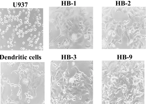
the presence of three donor-derived class I antigens and two class II antigens on the HB-2 cells is consistent with fusion and provides proof that the cell lines are true hybridomas. In conjunction with the expression of donor class I antigens, other changes were noted as well. U937 is an oval non-descript cell without any distinguishing features (Fig. 1). In contrast, the four hybridomas have a distinct stellate mor-phology with dendritic processes similar to primary DCs (Fig. 1). All of the hybridomas grew in an adherent pattern, unlike U937 that grows unattached to plastic sur-faces.
Expression of normal dendritic cell antigens and IL-12
pro-duction. The human DC hybridomas are representative of
FIG. 1. Morphological appearance of the human DC hybridomas. Photograph (magnification,⫻360) of immature DCs, U937, HB-1, HB-2, HB-3, and HB-9. The human DC hybridomas are larger and have a stellate appearance with dendritic processes similar to the immature DCs but are unlike U937, which is a nondescript cell line that has no remarkable features.
TABLE 1. HLA typinga
Class I
HLA type
Class II
HLA type
HB-2 U937 HB-2 U937
A A*0301, A*031012 A*0301 DRB1 DRB1*1401, DRB1*1605 DRB1*1605
B B*1801, B*51011 B*5106, B*1801, Bw 51011(5) DRB3 DRB3*01011
C Cw*0102, Cw*0701 Cw*0102, Cw*02021 DRB5 DRB5*01011
aHLA typing was performed on the HB-2 human DC hybridoma line HB-2 by using polymerase chain-specific primers for class I and class II.
on August 17, 2020 by guest
http://cvi.asm.org/
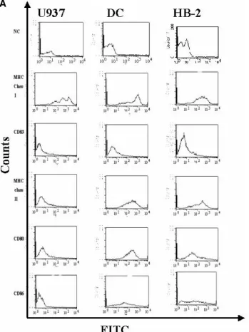
myeloid DCs since they were derived from CD14⫹monocytes (40, 51). We will present the results from one representative human DC hybridoma line, HB-2, chosen for its stability and ability to grow in long-term culture. We first determined whether the HB-2 cell line expressed common DCs markers not found on the U937 cells. We stained U937, primary DC and HB-2 cells with antibodies directed against major
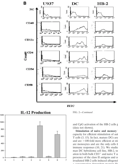
histo-compatibility complex (MHC) class I, MHC class II, CD80, CD86, and CD83 and demonstrated that HB-2 expresses all of these cell surface proteins like primary DCs but unlike U937, which only expresses MHC class I (Fig. 2A). DCs also express CD4, CD11c, and CD40 and have a high surface density of adhesion molecules, including ICAM-1 (CD54) and LFA-3 (CD58), that facilitates interactions with lymphocytes (3, 4,
FIG. 2. Surface expression of DC markers on HB-2 cells. (A) HB-2 cells, U937 cells, and the primary DCs were stained by direct immuno-fluorescence with FITC-labeled anti-HLA-class I, HLA-DR, CD83, CD80, and CD86 antibodies, along with isotype-matched controls, and then analyzed by flow cytometry, with gating on live cells. These results are representative of an experiment repeated three times. (B) The HB-2 cells, U937 cells, and DCs were directly stained with FITC-labeled anti-CD11c, CD4, CD58, CD54, and CD40 antibodies, along with isotype control antibodies and then analyzed by flow cytometry, gating on live cells. These findings are representative of an experiment repeated three times. (C) Induction of IL-12 production. The HB-2 cells, U937 cells, and DCs were stimulated with anti-CD40 MAbs (10g/ml) for 48 h. IL-12 production was determined by using an IL-12 p70 antigen capture ELISA. These findings are representative of an experiment repeated three times.
Pvalues (comparing IL-12 production in unstimulated cells and in antibody-stimulated DCs and HB-2 cells) ofⱕ0.05 are considered statistically significant.
on August 17, 2020 by guest
http://cvi.asm.org/
60). HB-2 cells also expressed CD4, CD11c, CD40, CD54, and CD58, again unlike U937 cells, which did not (Fig. 2B). Anal-ysis of IL-12 secretion by the U937 cells, primary DCs, and HB2 cells revealed no IL-12 production above background in the unstimulated cells, in contrast to CD40-stimulated HB2 cells and primary DCs that secreted significantly large amounts of IL-12 (P⫽0.01 andP⫽0.02, respectively) (14), unlike the U937 cells, in which IL-12 was not detectable (Fig. 2C). TNF-␣
and CpG activation of the HB-2 cells produced similar results (data not shown).
Stimulation of naive and memory T cells. DCs have the
capacity for efficient stimulation of autologous and allogeneic T cells (3, 15). In fact, mature DCs are the most potent APCs and are⬎100-fold more efficient at stimulating an MLR than are monocytes and are the only cells that can induce primary immune responses (10, 21). We studied the ability of the hu-man DC hybridoma cell line, HB-2, to induce T-cell prolifer-ation in both bulk CD3⫹and naive T cells. Consistent with the presence of the class II antigens and costimulatory molecules, irradiated HB-2 cells induced allogeneic T-cell proliferation in a concentration-dependent manner like the primary DCs but unlike U937 (Fig. 3). As further evidence that the HB-2 cells were true human DC hybridomas, we investigated whether they could stimulate CD45RA⫹-T-cell proliferation. A dose-dependent increase in T-cell proliferation similar to that seen with the unsorted T-cell populations was observed in both HB-2 and primary DCs but not in U937 (Fig. 3).
HIV-1 infection of DCs.DCs are commonly the first immune
cells to encounter foreign organisms such as HIV (5, 7, 28, 47). DCs play an important role in the generation of protective immunity but may also be subverted as part of the life cycle of
FIG. 2—Continued
on August 17, 2020 by guest
http://cvi.asm.org/
different pathogens (22, 23). HIV-1 uses receptors expressed by DCs, including DC-SIGN, CD4, CCR5, and CXCR4 for infection (30, 44, 43). HIV-1 can remain dormant in DCs and exploits DC trafficking to lymphoid tissue to infect permissive CD4⫹T cells (31, 33, 46). To investigate whether HIV has any effect on DC accessory cell function, we first infected HB-2 cells with two different strains of HIV: HIV-1IIIB, a
T-cell-tropic strain, and HIV-1BaL, a monocytotropic strain. As noted
in the introduction, one problem in studying APC function in HIV-infected DCs is that the percentage of infected cells is small and uninfected cells may give false-negative results in functional assays. A false-negative result means that there were no defects in antigen presentation under experimental condi-tions since there were sufficient numbers of uninfected DCs to restore normal APC function. As illustrated in Fig. 4, HB-2 cells could be infected with HIV-1IIIBand HIVBaL, as
demon-strated by peak shifts corresponding to intracellular virus after staining with the 5145 anti-HIV-1 antibodies.
Effect of HIV infection on stimulation of an MLR. HIV
causes many deleterious effects on APCs resulting in defective function. Since HB-2 cells can stimulate both naive and mem-ory T cells, we infected the HB-2 cells with HIVBaLfor 10 days
to determine whether its accessory cell function was impaired. We then cocultured the HIV-infected HB-2 cells with unsorted alloreactive T cells and with sorted CD45RA⫹T cells from two different donors (T-1 and T-2) and assessed proliferation by measuring thymidine incorporation. Unsorted alloreactive (P
⫽0.04 andP⫽0.05) and CD45RA⫹T cells (P⫽0.01 andP
⫽0.02) cocultured with HB-2HIVcells infected with HIVBaL
failed to proliferate (Fig. 5). There are several possibilities that could explain the lack of T-cell proliferation, including loss of MHC class II and costimulatory and adhesion molecules, al-tered cytokine production in the HB-2 cells, transmission of HIV to the cocultured T cells, the effect of an HIV protein, or the induction of apoptosis.
Expression of cell surface markers and IL-10 and IL-12
production in HB-2HIVcells. Loss of MHC class II antigens
(HLA-DR) and costimulatory molecules (CD80 and CD86) has been reported after HIV infection in monocytic cells, which could explain the inability of the HB-2HIVcells to induce
T-cell stimulation in our system (38, 49, 52). Furthermore, decreased expression of both CD54 and CD58 has been re-ported in monocytes of HIV-infected patients (61). This would
FIG. 3. Stimulation of an MLR. (Top) Negatively selected Ro-setteSep isolated T cells (105) were cocultured with various
concen-trations (103to 105) of irradiated (6,000 rads) HB-2 cells for 5 days. A
total of 1Ci of thymidine was added during the last 16 h of culture. Baseline stimulation was determined by culturing T cells in medium alone. The data represent the mean values of three separate experi-ments. (Bottom) Stimulation of naive T cells. Various concentrations (103 to 105) of immature and mature DCs, HB-2, and U937 were
irradiated (6,000 rads) and cocultured with negatively selected Ro-setteSep CD3⫹ CD45RA⫹ T cells for 5 days. A total of 1Ci of thymidine was added during the last 16 h of culture. Baseline stimu-lation was determined by culturing T cells in media alone. These findings are representative of an experiment repeated three times. Columns: 1, T cells alone (T); 2, T⫹U937 (103); 3, T⫹U937 (104);
4, T⫹U937 (105); 5, T⫹DCs (103); 6, T⫹DC (104); 7, T⫹DCs
(105); 8, T⫹HB-2 (103); 9, T⫹HB-2 (104); 10, T⫹HB-2 (105).
FIG. 4. 1 infection. The HB-2 cells were infected with HIV-1IIIBand HIV-1BaLfor 5 days, followed by intracytoplasmic staining
with the human anti-HIV antibody 5145 (5g/ml) and negative control human antibodies, followed by affinity-purified FITC-conjugated goat anti-human antibodies, and then analyzed by flow cytometry, with gating on live cells. These findings are representative of an experiment repeated three times.
on August 17, 2020 by guest
http://cvi.asm.org/
also contribute to defective HB-2HIV–T-cell interactions. To
address the effect of HIV infection on different regulatory surface proteins, staining with specific MAbs was performed. Uninfected HB-2 cells express MHC class I, MHC class II, CD83, CD86, CD80, CD54 and CD58, CD11c, and CD4 (Fig. 2A and B). As illustrated in Table 2, we compared the MFIs for HLA class I, HLA class II, CD11c, CD40, CD4, CD80, CD86, CD83, CD58, and CD54 on uninfected HB-2 cells to the expression antigens on HB2 cells infected with either HIVIIIB
or HIVBaL. This was determined by dividing the MFI staining
for HLA class I, HLA class II, CD11c, CD40, CD4, CD80, CD86, CD83, CD58, and CD54 on uninfected HB-2 cells by the MFIs for the same antigens on the HB-2 cells infected with HIVIIIBand HIVBaLand multiplying that value by 100. There
was no significant change in the MFIs of class I, class II, CD11c, CD40 CD4, CD80, CD86, CD83, CD58, and CD54 in HB-2 cells infected with HIVIIIBand HIVBaLcompared to the
uninfected controls. It has been demonstrated in HIV-infected monocytes that there is loss of IL-12 production and induction of IL-10 that has been associated with impaired immune
re-sponses (65). It has also been demonstrated that IL-10 can cause DC apoptosis (42). The lack of myeloid DC activation by HIV is also consistent with the findings of Fonteneau et al., who reported that HIV activates plasmacytoid DCs and in-duces bystander maturation of myeloid DCs (27). We stimu-lated the HB-2HIVcells through CD40 for 48 h with anti-CD40
antibodies to investigate whether there were any changes in IL-12 or IL-10 production. There was no significant difference in IL-10 and IL-12p70 production between the uninfected HB-2 cells and HB-2 cells infected with HIVBal(Fig. 6).
Effect of antiretroviral therapy and anti-gp120 antibodies
on T cells cocultured with HB-2HIVcells.DC cells can transmit
HIV to T cells that could result in the diminished capacity of T cells to be stimulated by the HB-2HIV cells (33, 48). As
shown in Fig. 5, HIV infection of HB-2 cells significantly re-duced T-cell proliferation in the MLR (P ⫽ 0.02). We next determined the effect of HIV infection of the HB-2 cells on allogeneic T-cell proliferation by adding the antiretroviral drugs ddI and IDV to our cocultures. IDV (P ⫽ 0.01) was more efficient in suppressing the inhibition of the proliferative
FIG. 5. Stimulation of an MLR. Uninfected HB-2 cells or HB-2 cells infected with HIVBaLwere cocultured with purified bulk populations of
T cells (105) or purified CD3⫹CD45RA⫹T cells (105) from two different donors (T-1 and T-2) for 5 days. Cells were pulsed with [3H]thymidine
for the last 16 h of culture and then counted in a scintillation counter.Pvalues comparing T-cell stimulation induced by uninfected HB-2 cells to T-cell stimulation by HB-2HIVcells ofⱕ0.05 are statistically significant. Columns: 1, T-1 cells alone (T-1); 2, T-1⫹HB-2; 3, T-1⫹HB-2HIV; 4,
T-2 cells alone (T-2); 5, T-2⫹HB-2; 6, T-2⫹HB-2HIV.
on August 17, 2020 by guest
http://cvi.asm.org/
responses than ddI as expected (Fig. 7A). The combination of ddI and IDV (P⫽0.01) was no more effective than either drug alone and significantly blocked p24 production in the cocul-tures as noted in Fig. 7A but did not completely restore reac-tivity in the MLR (26). Another possibility is that HIV viral proteins, especially gp120, produced by HIV are well known to cause T-cell dysfunction (18, 62). IDV and other protease inhibitors block HIV proteases that prevent replication of ma-ture virions but do not affect the release of HIV gp120 secreted by immature defective virions (20). In addition, it has been reported that gp120 produced by HIV-infected primary DCs can suppress T-cell proliferation in cocultures without inducing apoptosis (37). To determine whether this was happening in our system, we treated HIV-infected HB-2 cells with the
poly-FIG. 6. IL-12 and IL-10 production after HIV infection. Superna-tants from HB-2 and HB-2HIVcells stimulated with CD40
anti-bodies (10g/ml) for 48 h were analyzed for IL-12 and IL-10 produc-tion by using a direct binding ELISA (R&D). NS, not statistically significant. These findings are representative of an experiment re-peated three times. Columns 1 to 3 show IL-12 production; columns 4 to 6 show IL-10 production. Columns: 1, HB-2; 2, HB-2⫹anti-CD40; 3, HB-2HIV⫹anti-CD40; 4, HB-2; 5, HB-2⫹anti-CD40; 6, HB-2HIV
⫹anti-CD40.
FIG. 7. Effect of antiretroviral therapy and gp120 and anti-IL-10 antibodies on altered APC function of HIV-infected HB-2 cells. (A) Antiretroviral therapy with anti-IL-10 and anti-gp120 antibodies. A 10M concentration of ddI and 10g of IDV/ml were added to cocultures of HB-2 and HB-2HIVcells and T cells. Thymidine
incor-poration was measured by scintillation counting, and p24 antigen levels measured by antigen capture ELISA were assessed simultaneously at day 5 in the cocultures of HB-2, HB-2HIV, and T cells. Then, 10g of
anti-gp120, 5145 and anti-IL-10 antibodies/ml were added alone or in combination with or without ddI and IDV to cocultures of HB-2 cells, HB-2HIVcells, and T cells.Pvalues comparing either thymidine
in-corporation or p24 levels between uninfected HB-2 T-cell cocultures and T-cell HB-2HIVcocultures, as well as between HB-2 T-cell
cocul-tures and HB-2HIVT-cell cocultures treated with IDV alone, ddI⫹
IDV, and ddI⫹IDV⫹anti-IL-10⫹5145 ofⱕ0.05 are statistically significant. These findings are representative of an experiment re-peated three times.Columns: 1, T cells alone (T); 2, T⫹HB-2; 3, T ⫹HB-2HIV; 4, T⫹HB-2⫹ddI; 5, T⫹HB-2HIV⫹ddI; 6, T⫹HB-2
⫹IDV; 7, T⫹HB-2HIV⫹IDV; 8, T⫹HB-2⫹IDV⫹ddI; 9, T⫹
HB-2HIV⫹IDV⫹ddI; 10, T⫹HB-2⫹5145; 11, T⫹HB-2HIV⫹
5145; 12, T⫹HB-2⫹anti-IL-10; 13, T⫹HB-2HIV⫹anti-IL-10; 14,
T⫹HB-2⫹anti-IL-10⫹5145; 15, T⫹HB-2⫹anti-IL-10⫹5145; 16, T⫹HB-2⫹ddI, IDV, anti-IL-10, and 5145; 17, T⫹HB-2HIV⫹
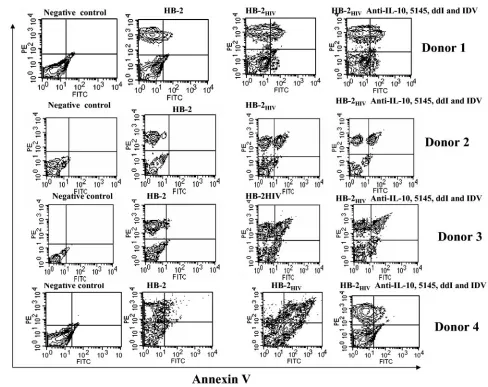
ddI, IDV, anti-IL-10, and 5145. (B) Induction of apoptosis in T cells by HB-2HIVcells. T cells were cocultured with HB-2, HB-2HIV, and
HB-2HIVcells treated with ddI (10M), IDV (10g/ml), anti-IL-10 (10
g/ml), and 5145 (10g/ml) antibodies for 3 days, and apoptosis was determined by staining with FITC-labeled Annexin V and phyco-erythrin-labeled anti-CD3 MAbs. Apoptosis was determined by dual staining in the upper right quadrant. These are experiments with four different T-cell donors. Columns: 1, T cells alone (T); 2, T⫹HB-2; 3, T⫹HB-2HIV; 4, T⫹HB-2HIV⫹ddI; 5, T⫹HB-2HIV⫹IDV; 6, T
⫹HB-2HIV⫹IDV⫹ddI; 7, T⫹HB-2HIV⫹5145; 8, T⫹HB-2HIV⫹
anti-IL-10; 9, T⫹HB-2HIV⫹anti-IL-10⫹5145; 10, T⫹HB-2HIV⫹
ddI, IDV, anti-IL-10, and 5145. TABLE 2. Expression of cell surface antigens on HB-2 cells
infected with HIVIIIBand HIVBaLa
Cell type
% Modulation of MFI⫾SD in cells infected with:
HIVBaL HIVIIIB
MHC class I 91⫾25 89⫾13
MHC class II 87⫾19 93⫾20
CD11c 84⫾27 91⫾18
CD40 118⫾36 102⫾25
CD4 99⫾34 116⫾30
CD80 90⫾13 95⫾27
CD86 86⫾25 96⫾28
CD83 93⫾17 87⫾24
CD58 94⫾21 100⫾25
CD54 107⫾31 110⫾25
aValues are expressed as the percent modulation of the control MFI on HB-2
cells infected with either HIVIIIBor HIVBaL. In these experiments, the MFI for
HLA class I, HLA class II, CD11c, CD40, CD4, CD1a, CD80, CD86, CD83, CD58, or CD54 antigens on uninfected HB-2 cells was divided by the MFI staining of the same antigens on HB-2 cells infected with either HIVIIIBor
HIVBaL. This number was multiplied by 100 to determine the percent
modula-tion of the control antigens after HIVIIIBand HIVBaLinfection. These results
represent the findings from three different experiments.
on August 17, 2020 by guest
http://cvi.asm.org/
clonal anti-gp120 antibody 5145 (10g/ml) and repeated the MLR (17). Compared to the uninfected HB-2 cells, the 5145 antibodies partially restored T-cell proliferation in the HB-2HIV cocultures (Fig. 7A). We also treated uninfected HB-2
cells with ddI and IDV at the same concentrations that we used with the HB-2HIV cells to ensure that the effects on T-cell
proliferation that were observed were not due to the toxicity of the drugs (Fig. 7A).
Effect of anti-IL-10 antibodies on T-cell proliferation and
the induction of apoptosis.Although there was no
upregula-tion of IL-10 after HIV infecupregula-tion, Granelli-Piperno et al. (33) have reported that cocultures of HIV-infected DCs and T cells suppress allogeneic proliferative responses through the gener-ation of a populgener-ation of IL-10-producing T cells. To test whether this was occurring in our system, we added anti-IL-10 antibody to the MLRs and determined cellular proliferation by measuring thymidine incorporation. Similar to the results ob-tained with the 5145 antibody, the addition of IL-10 anti-body (10 g/ml) to the HB-2HIV T-cell cocultures partially
restored T-cell proliferation (Fig. 7A). The combination of the 5145 and anti-IL-10 antibodies restored the reactivity in the MLR but was still less than the response observed in the
uninfected HB-2 cells. Even the combination of ddI, IDV, anti-IL-10, and 5145 antibodies that suppressed HIV replica-tion (P ⫽ 0.01) and blocked IL-10 and gp120 still did not restore complete reactivity (Fig. 7A). Similar to the experi-ments performed with ddI and IDV, we used uninfected HB-2 in MLRs with 5145 and anti-IL-10 antibodies to ensure that the effects on T-cell proliferation were not due to the toxicities of the antibodies. Another possibility to explain the lack of T-cell proliferation in the HB-2HIVcocultures is the induction
of apoptosis.
In support of this, Lichtner et al. have reported that HIV-infected DCs induce apoptotic death in CD4⫹T cells through the induction of TNF-␣-related death factors, including FasL, TRAIL, TNF-␣, and TWEAK (39). We determined whether the HB-2HIV-infected cells were inducing apoptosis by
mea-suring T-cell apoptosis assay with annexin V and anti-CD3 antibodies. T cells were cocultured with uninfected 2, HB-2HIV, and HB-2HIVcells treated with ddI, IDV, and the 5145
anti-gp120 and anti-IL-10 antibodies. Compared to the unin-fected HB-2 cells, T cells cocultured with the HB-2HIVcells
underwent more apoptosis than T cells cocultured with HB-2 cells, as determined by the presence of a CD3⫹ Annexin V
FIG. 7—Continued.
on August 17, 2020 by guest
http://cvi.asm.org/
costaining population with four different donors (Fig. 7B). T cells cocultured with HB-2HIV, along with ddI, IDV, 5145, and
anti-IL-10 still underwent apoptosis.
DISCUSSION
We have previously reported that macrophages can be fused with an appropriate fusion partner for the generation of im-mortalized cloned human macrophage hybridomas (55). We have extended these studies to DCs to show that, like macro-phages, they can also be fused and immortalized to make hybridomas. Our cell lines were proven to be true hybridomas by the acquisition of donor class I antigens present in the donor cells but not on the U937 parent line (Table 1). In addition, the hybridomas displayed distinctive stellate DC mor-phology, since they were larger than U937 and grew in an adherent pattern (Fig. 1). Further evidence that our cell lines were true hybridomas came from cell surface staining experi-ments that demonstrated the presence of DC specific markers (Fig. 2A and B), the capacity to produce IL-12 (Fig. 2C) and induce primary T-cell responses unlike the U937 parent line (Fig. 3). As noted in the Introduction, DCs have been previ-ously shown to be capable of cell fusion forming hybrid cell lines with tumor cells to make cancer vaccines, but these were not clonal or stable in long-term culture (20).
There were no differences between the four human DC cell lines. When we generated the human macrophage hybridomas (55), different populations of macrophages were identified based on staining with Vmaxantibodies (55) that identify
mac-rophages at different anatomical sites (55). The antibodies that were used in these experiments to characterize the DC lines may not completely identify subpopulations of myeloid DCs, so it is conceivable that HB-1, HB-2, HB-3, and HB-9 may still represent different DC subpopulations. Furthermore, culturing DCs from peripheral blood monocytes either represents a lim-ited subpopulation of DCs or, alternatively, only limlim-ited num-bers of subpopulations of DCs may be capable of fusion to form stable hybridomas. Thus, it is also possible that selection bias may have occurred during the generation of HB-1, HB-2, HB-3, and HB-9, and the experiments presented here may represent events occurring only in a selected DC subpopula-tion. We are currently performing experiments with HIV-in-fected primary DCs to compare to the results obtained with the human DC hybridomas. This must be kept in mind since the data from functional studies with different HIV-infected sub-types of DCs, Langerhans cells, monocyte-derived, immature, mature, and myeloid and plasmacytoid DCs have given vari-able results (7, 8, 9, 11, 12, 19, 24, 25, 43, 44, 45, 53).
Nevertheless, the HB-2 hybridoma cells have many charac-teristics of normal DCs and were used to assess DC function after HIV infection (Fig. 4). Using this cell line we demon-strated defects in APC function (Fig. 3). The HB-2HIVcells did
not stimulate primary immune responses in an MLR (Fig. 5) despite the fact that there were no alterations in MHC class I, class II, CD11c, CD40, CD80, CD86, CD83, CD58, and CD54 expression (Table 2) or IL-12 and IL-10 production (Fig. 6). These results differ from our previous studies with HIV-in-fected human macrophage hybridomas, where there was loss of MHC class II expression, downregulation of IL-12, and induc-tion of IL-10 (49, 65). Differences in APC funcinduc-tion between
DCs and monocytes have been observed in parasitic and my-cobacterial infections in studies using murine model systems (34, 64).
HIV infection did not activate the DC hybridomas and re-duces their intrinsic allostimulatory capacity. As noted above, the lack of myeloid DC activation by HIV is consistent with recent work by Granelli-Piperno et al. (33) and Fonteneau et al. (27). The impaired stimulatory capacity of the HB-2HIV
cells was caused by multiple mechanisms, including gp120, IL-10, and induction of apoptosis, unlike the results of other groups that have identified single pathways for the inhibition of T-cell activity (Fig. 7). Kawamura et al. (36), using HIV-in-fected primary human DCs, have demonstrated that gp120 blocks HIV proliferation in an allo-MLR and that T-cell pro-liferation was completely restored with sCD4. Granelli-Pip-erno et al. have shown that HIV-infected DCs induce a T-cell population that produces IL-10 and that proliferation could be restored by using anti-IL-10 antibodies (32). Lichtner et al. demonstrated that HIV-infected DCs induced apoptosis in CD4⫹T cells by TNF-␣-related death factors, including FasL, TRAIL, TNF-␣, and TWEAK (39). Experiments are currently under way to block induced apoptosis by using FasL, anti-TRAIL, anti-TNF-␣, and anti-Tweak antibodies. It has also been reported that thymic cytotoxic factors are released from HIV-infected thymic DCs after HIV infection (6). An inability to induce naive and memory T-cell responses by HIV-infected DCs early in the course of HIV infection either through gp120 and IL-10 or the induction of apoptosis could delay immune responses, allowing for increased dissemination of the virus. Induction of apoptosis by HIV-infected DCs could also con-tribute to naive and memory T-cell depletion seen in AIDS patients.
In conclusion, DCs can be fused with the U937 cell line to create stable hybridomas, thereby yielding immortalized cell lines. The representative human DC hybridoma cell, HB-2, expresses DC surface molecules found on monocyte-derived DCs, including CD83, and possesses functional characteristics of monocyte-derived DCs, including stimulation of naive T cells and IL-12 production. The HB-2 cell line can be infected with HIV-1 and used as a model to investigate DC T-cell interactions after HIV infection. Using this model system, we have demonstrated impaired APC function manifested by an inability to stimulate an MLR that was caused by multiple mechanisms, including IL-10 and gp120-induced apoptosis. The HB-2 cell line could be a useful model system to study APC function in HIV-infected DCs.
ACKNOWLEDGMENT
This study was supported by National Institutes of Health grants AI-44236 and AI-45343.
REFERENCES
1.Andreesen, R., K. J. Bross, J. Sterols, and F. Emmrich. 1986. Human macrophage maturation and heterogeneity: analysis with a newly generated set of late differentiation antigens in situ. Blood67:1257–1264.
2.Andreesen, R., S. Gadd, U. Costabel, H. G. Leser, V. Speth, B. Cesnik, and R. C. Atkins.1988. Human macrophage maturation and heterogeneity: re-stricted expression of late differentiation antigens in situ. Cell Tissue Res.
253:271–279.
3.Banchereau, J., and R. Steinman.1998. Dendritic cells and the control of immunity. Nature392:245–252.
4.Banchereau, J., F. Briere, C. Caux, J. Davoust, S. Lebecque, Y. J. Liu, B. Pulendran, and K. Palucka.2000. Immunobiology of dendritic cells. Annu. Rev. Immunol.18:76–881.
on August 17, 2020 by guest
http://cvi.asm.org/
5.Baribaud, F., S. Pohlmann, and R. Doms.W.2001. The role of DC-SIGN and DC-signr in HIV and SIV attachment, infection, and transmission. Virology
20:1–6.
6.Beaulieu, S., A. Kessons, D. Landry, S. Montplaisir, D. Bergeron, and E. A. Cohen.2001. In vitro characterization of purified human thymic dendritic cells infected with human immunodeficiency virus type 1. Virology222:214– 226.
7.Blauvelt, A.1997. The role of skin dendritic cells in the initiation of human immunodeficiency virus infection. Am. J. Med.102:16–20.
8.Blauvelt, A., H. Asada, M. W. Saville, V. Klaus-Kovtun, D. J. Altman, R. Yarchoan, and S. I. Katz.1997. Productive infection of dendritic cells by HIV-1 and their ability to capture virus are mediated through separate pathways. J. Clin. Investig.100:2043.
9.Blauvelt, A., C. Chougnet, G. M. Shearer, and S. I. Katz.1996. Modulation of T cell responses to recall antigens presented by Langerhans cells in HIV-discordant identical twins by anti-interleukin (IL)-10 antibodies and IL-12. J. Clin. Investig.97:1550–1555.
10.Blauvelt, A., M. Clerici, D. R. Lucey, S. M. Steinberg, R. Yarchoan, R. Walker, G. M. Shearer, and S. I. Katz.1995. Functional studies of epidermal Langerhans cells and blood monocytes in HIV-infected persons. J. Immunol.
154:3506–3515.
11.Cameron, P. U., U. Forsum, H. Teppler, A. Granelli-Piperno, and R. M. Steinman.1992. During HIV-1 infection most blood dendritic cells are not productively infected and can induce allogeneic CD4⫹T cells clonal expan-sion. Clin. Exp. Immunol.88:226–236.
12.Cameron, P. U., P. S. Freudenthal, J. M. Barker, S. Gezelter, K. Inaba, and R. M. Steinman.1992. Dendritic cells exposed to human immunodeficiency virus type-1 transmit a vigorous cytopathic infection to CD4⫹cells. Science
257:383–387.
13.Canque, B., M. Rosenzwaig, S. Camus, M. Yagello, Bonne, M. M. L. Guigon, and J. C. Gluckman.1996. The effect of in vitro human immunodeficiency virus infection on dendritic cell differentiation and function. Blood88:4215– 4228.
14.Cella, M., D. Scheidegger, K. Palmer-Lehman, P. Lane, A. Lanzavecchia, and G. Alber.1996. Ligation of CD40 on dendritic cells triggers production of IL-12 and enhances T cell stimulatory capacity: T-T help via APC acti-vation. J. Exp. Med.184:747–752.
15.Cella, M., F. Sallusto, and A. Lanzavecchia.1997. Origin, maturation and antigen presenting function of dendritic cells. Curr. Opin. Immunol.9:10–16. 16.Chapuis, F., M. Rosenwajg, M. Yagello, M. Ekman, P. Biberfeld, and J. C. Gluckman.1997. Differentiation of human dendritic cells from monocytes in vitro. Eur. J. Immunol.27:431–441.
17.Chen, H., Y. K. Yip, I. George, M. Tyorkin, E. Salik, and K. Sperber.1998. Chronically HIV-1 infected monocytic cells induce apoptosis in cocultured T cells. J. Immunol.161:4257–4267.
18.Chirmule, N., and S. Pahwa.1996. Envelope glycoproteins of human immu-nodeficiency virus type 1: profound influences on immune functions. Micro-biol. Rev.60:386–406.
19.Chougnet, C., S. S. Cohen, T. Kawamura, A. L. Landay, H. A. Kessler, E. Thomas, A. Blauvelt, and G. M. Shearer.1999. Normal immune function of monocyte-derived dendritic cells from HIV-infected individuals: implica-tions for immunotherapy. J. Immunol.163:1666–1672.
20.Corelier, P., and D. S. Strayer.2003. Mechanisms of alpha1-antitrypsin inhibition of cellular serine proteases and HIV-1 protease that are essential for HIV-1 morphogenesis. Biochem. Biophys. Acta30;1638:197–207. 21.Croft, M., D. D. Duncan, and S. L. Swain.1992. Response of naive
antigen-specific CD4⫹T cells in vitro: characteristics and antigen presenting require-ments. J. Exp. Med.176:1431–1437.
22.Dietz, A. B., P. A. Bulur, C. A. Brown, V. S. Pankratz, and S. Vuk-Pavlovic.
2001. Maturation of dendritic cells infected by recombinant adenovirus can be delayed without impact on transgene expression. Gene Ther.8:419–423. 23.Engelmayer, J., M. Larsson, M. Sublewew, A. Chahroundi, W. I. Cox, R. M. Steinman, and N. Bhardwaj.1999. Vaccinia virus inhibits the maturation of human dendritic cells: a novel mechanism of immune evasion. J. Immunol.
163:6762–6768.
24.Fan, Z., X. L. Huang, L. Zheng, C. Wilson, L. Borowski, J. Liebmann, P. Gupta, J. Margolick, and C. Rinaldo.1997. Cultured blood dendritic cells retain HIV-1 antigen-presenting capacity for memory CTL during progres-sive HIV-1 infection. J. Immunol.159:4973–4982.
25.Feldman, S., D. Stein, S. Amrute, T. Denny, Z. Garcia, P. Kloser, Y. Sun, N. Megjugorac, and P. Fitzgerald-Bocarsly.2001. Decreased interferon-␣ pro-duction in HIV-infected patients correlates with numerical and functional deficiencies in circulating type 2 dendritic precursors. Clin. Immunol.101:
201–210.
26.Fan, Z., X. L. Huang, L. Borowski, J. W. Mellors, and C. R. Rinaldo, Jr.
2001. Restoration of anti-human immunodeficiency virus type 1 (HIV-1) responses in CD8⫹T cells from late-stage patients on prolonged antiretro-viral therapy by stimulation in vitro with HIV-1 protein-loaded dendritic cells. J. Virol.75:4413–4419.
27.Fonteau, J. F., M. Larsson, A. S. Beignon, K. McKenna, I. Dasilva, A. Amara, Y. J. Liu, J. D. Lifson, D. R. Littman, and N. Bhardwaj.2004. Human if immunodeficiency virus type 1 activates plasmacytoid dendritic
cells and concomitantly induces the bystander maturation of myeloid den-dritic cells. J. Virol.78:5223–5232.
28.Frankel, S. S., B. M. Wenig, A. P. Burke, P. Mannan, S. L. Thompson, S. L. Abbondanzo, A. M. Nelson, M. Pope, and R. M. Steinman.1999. Replication of HIV-1 in dendritic cell-derived syncytia at the mucosal surface of the adenoid. Science272:115–117.
29.Gartner, S., P. Markovits, D. M. Markovitz, R. Kaplan, R. C. Gallo, and M. Popovic.1986. The role of mononuclear cells phagocytes in HTLV-1II/LAV infection. Science233:215–219.
30.Geijtenbeek, T. B., D. S. Kwon, R. Torensman, S. J. van Vliet, G. C. van Diujnhoven, J. Middel, I. L. Cornelissen, H. S. Nottet, V. N. KewalRamani, D. R. Littman, C. G. Figdor, and Y. van Kooyk.2000. DC-SIGN, a dendritic cell-specific HIV-1-binding protein that enhances trans-infection of T cells. Cell100:587–597.
31.Granelli-Piperno, A., E. Delgado, V. Finkel, W. Paxton, and R. M. Steinman.
1998. Immature dendritic cells selectively replicate macrophage tropic (M-tropic) human immunodeficiency virus type 1, while mature cells efficiently transmit both M and T-tropic virus to T cells. J. Viol.72:2733–2737. 32.Granelli-Piperno, A., A. Golebiowska, C. Trumpfheller, F. Siegel, and R. M.
Steinman.2004. HIV-1-infected monocyte-derived dendritic cells do not undergo maturation but can elicit IL-10 production and T-cell regulation. Proc. Natl. Acad. Sci. USA101:7669–7674.
33.Granelli-Piperno, A., V. Finkel, E. Delgado, and R. M. Steinman.1988. Virus replication begins in dendritic cells during transmission of HIV-1 from ma-ture dendritic cells to T cells. Curr. Biol.14:21–29.
34.Hickman, S. P., J. Chan, and P. Salgame.2002. Mycotuberculosis induces differential cytokine production from dendritic cells and macrophages with divergent effects on naive T-cell polarization. J. Immunol.168:4636–4642. 35.Hope, T. J., D. McDonald, L. Wu, S. M. Bohks, V. N. KewalRamani, and D.
Unutmaz.2003. Recruitment of HIV and its receptors to dendritic cell-T cell junctions. Science300:1295–1297.
36.Kawamura, T., H. Gatanaga, D. L. Borris, M. Connors, H. Mitsuya, and A. Blauvelt.2003. Decreased stimulation of CD4⫹T cell proliferation and IL-2 production by highly enriched populations of HIV-infected dendritic cells. J. Immunol.170:4260–4266.
37.Knight, S. C.2001. Dendritic cells and HIV infection: immunity with viral transmission versus compromised cellular immunity. Immunobiology204:
614–620.
38.Le Naour, R., C. Lussoiz, H. Raoul, A. Mabondzo, and D. Dormont.1997. Expression of cell adhesion molecules at the surface of in vitro human immunodeficiency virus type-1 human monocytes: relationships with tumor necrosis factor alpha, interleukin 1beta, and interleukin 6 syntheses. AIDS Res. Hum. Retrovir.13:841–855.
39.Lichtner, M., C. Maranon, V. Pierre-Oliver, O. Azocar, D. Hanau, P. Lebon, M. Burgard, C. Rouziuoux, V. Vullo, H. Yagita, C. Rabourdin-Combe, C. Servet, and A. Hosmalin.2004. HIV type-1 infected dendritic cells induce apoptotic death in infected and uninfected primary CD4⫹lymphocytes. AIDS Res. Hum. Retrovir.20:175–182.
40.Liu, Y. J., H. Kanzler, V. Soumelis, and M. Gilliet.2001. Dendritic cell lineage, plasticity, and cross regulation. Nat. Immunol.2:585–589. 41.Louie, M., J. Yoo, T. Moran, L. Mayer, and K. Sperber.1995. Impairment of
monocytic function after influenza virus infection. Clin. Diagn. Lab. Immu-nol.2:426–433.
42.Ludewig, B., D. Graf, H. R. Gelderblom, Y. Becker, R. A. Kroczek, and G. Pauli.1995. Spontaneous apoptosis of dendritic cells is efficiently inhibited by TRAP (CD-40 ligand) and TNF-alpha, but strongly enhanced by inter-leukin-10. Eur. J. Immunol.25:1943–1950.
43.Macatonia, S. E., M. Gompels, A. J. Pinching, S. Patterson, and S. C. Knight.1992. Antigen presentation by macrophages but not by dendritic cells in human immunodeficiency virus (HIV) infection. Immunology75:
576–581.
44.Macatonia, S. E., R. Lau, S. Patterson, A. J. Pinching, and S. C. Knight.
1989. Suppression of immune responses by dendritic cells infected with HIV. Immunology67:285–289.
45.Macatonia, S. E., R. Lau, S. Patterson, A. J. Pinching, and S. C. Knight.
1990. Dendritic cell infection, depletion, and dysfunction in HIV-infected individuals. Immunology71:38–45.
46.Masurier, C., B. Salomon, Guettari, C. N. Pioche, F. Lachapelle, M. Guigon, and D. Klazmann.1998. Dendritic cells route immunodeficiency to lymph nodes after vaginal or intravenous administration to mice J. Virol.72:7822– 7829.
47.Piguet, V., and A. Blauvelt.2002. Essential roles for dendritic cells in the pathogenesis and potential treatment of HIV disease. J. Investig. Dermatol.
119:365–369.
48.Pohlmann, S., F. Baribaud, and R. W. Doms.2001. SIGN and DC-SIGNR: helping hands for HIV. Trends Immunol.22:643–649.
49.Polyak, S., H. Chen, D. Hirsch, I. George, R. Hirschberg, and K. Sperber.
1997. Impaired class II expression and impaired antigen uptake in monocytic cells after HIV-1 infection. J. Immunol.159:2177–2188.
50.Popovic, M., G. Sarnagaradharian, E. Read, and R. C. Gallo.1984. Detec-tion, isolaDetec-tion, and continuous production of cytopathic retroviruses (HTLV-III) from patients with AIDS and pre-AIDS. Science244:497–500.
on August 17, 2020 by guest
http://cvi.asm.org/
51.Puckl, W. F., O. Majdic, P. Kohl, J. Stockl, E. Riedl, C. Schneinecker, C. Bello-Fernandez, and W. Knopp.1996. Molecular and functional character-istics of dendritic cells generated from highly purified CD14⫹peripheral blood monocytes. J. Immunol.157:3850–3859.
52.Rakoff-Nahoum, S., H. Chen, T. Kraus, I. George, E. Oei, and K. Sperber.
2001. Regulation of class II expression in monocytic cells after HIV-1 infec-tion. J. Immun.167:2331–2342.
53.Sapp, M., J. Engelmayer, M. Larsson, A. Granelli-Piperno, R. Steinman, and N. Bhardwaj.1999. Dendritic cells generated from blood monocytes of HIV-1 patients are not infected and act as competent antigen presenting cells eliciting potent T-cell responses. Immunol. Lett.66:121–128. 54.Scott-Taylor, T. H., R. Pettengell, I. Clarke, G. Stuhler, M. C. La Barthe, P.
Walden, and A. G. Dalgleish.2000. Human tumor and dendritic cell hybrids generated by electrofusion: potential for cancer vaccines. Biochim. Biophys. Acta1500:265–279.
55.Sperber, K., A. Pizzimenti, V. Najfeld, and L. Mayer.1990. Identification of subpopulations of human macrophages through the generation of human macrophage hybridomas. J. Immunol. Methods129:31–40.
56.Sperber, K., G. Hamrang, M. J. Louie, T. Kalb, R. Banerjee, H. S. Choi, F. Paronetto, and L. Mayer.1993. Progressive impairment of monocytic func-tion in HIV-1-infected human macrophage hybridomas. AIDS Res. Hum. Retrovir.9:657–667.
57.Sperber, K., T. Kalb, V. Stecher, and L. Mayer.1993. Inhibition of human immunodeficiency virus type 1 replication by hydroxychloroquine in T cells and monocytes. AIDS Res. Hum. Retrovir.9:9–98.
58.Sperber, K., M. Louie, J. Proner, E. Sapira, S. Lin, and L. Mayer.1995. Hydroxychloroquine treatment of patients with human immunodeficiency virus type 1. Clin. Ther.17:622–636.
59.Sperber, K., P. Beuria, N. Singha, I. Gelman, P. Cortes, H. Chen, and T.
Kraus. 2003. Induction of apoptosis by HIV-1-infected monocytic cells. J. Immunol.170:1566–1578.
60.Stockwin, L. H., D. McGonagle, I. G. Martin, and G. E. Blair.2000. Den-dritic cells: immunological sentinels in health and disease. Immunol. Cell Biol.78:91–102.
61.Trial, J., H. H. Birdsall, J. A. Hallum, M. L. Crane, M. C. Rodriquez-Barradas, A. L. de Jong, B. Krishnan, C. E. Lacke, C. G. Figdor, and R. D. Rossen.1995. Phenotypic and functional changes in peripheral blood mono-cytes during progression of human immunodeficiency virus infection: effects of soluble immune complexes, cytokines, subcellular particulars from apo-ptotic cells, and HIV-1 encoded proteins on monocytes phagocytic function, oxidative burst, transendothelial migration and cell surface phenotype. J. Clin. Investig.95:1690–1701.
62.Turville, S. G., P. U. Cameron, J. Arthos, K. MacDonald, G. Clark, D. Hart, and A. L. Cunningham.2001. Bitter-sweet symphony: defining the role of dendritic cell gp120 receptors in HIV infection. J. Clin. Virol.22:229–239. 63.Turnville, S. G., J. J. Santos, P. U. Frank, P. U. Cameron, J. Wilkinson, M.
Miranda-Saksena, J. Dable, H. Stossel, N. Romani, M. Piatek, Jr., J. D. Lifson, M. Pope, and A. L. Cunningham.2004. Immunodeficiency virus uptake, turnover, and 2-phase transfer in human dendritic cells. Immunobi-ology103:2170–2179.
64.Von Stebut, E., Y. Belkaid, D. Jakob, D. L. Sacks, and M. C. Udey.1998. Uptake ofLeishmania majoramastigotes results in activation and interleukin 12 release from murine skin-derived dendritic cells: implications for the initiation of anti-Leishmaniaimmunity. J. Exp. Med.188:1547–1552. 65.Yoo, J., H. Chen, T. Kraus, D. Hirsch, S. Poylak, I. George, and K. Sperber.
1996. Altered cytokine and accessory cell function after HIV-1 infection. J. Immunol.156:1313–1320.