Scholarship@Western
Scholarship@Western
Electronic Thesis and Dissertation Repository
6-4-2012 12:00 AM
Role of ERK5 in Diabetic Vascular Complications
Role of ERK5 in Diabetic Vascular Complications
Yuexiu Wu
The University of Western Ontario Supervisor
Dr. Subrata Chakrabarti
The University of Western Ontario
Graduate Program in Pathology
A thesis submitted in partial fulfillment of the requirements for the degree in Doctor of Philosophy
© Yuexiu Wu 2012
Follow this and additional works at: https://ir.lib.uwo.ca/etd Part of the Medicine and Health Sciences Commons
Recommended Citation Recommended Citation
Wu, Yuexiu, "Role of ERK5 in Diabetic Vascular Complications" (2012). Electronic Thesis and Dissertation Repository. 586.
https://ir.lib.uwo.ca/etd/586
This Dissertation/Thesis is brought to you for free and open access by Scholarship@Western. It has been accepted for inclusion in Electronic Thesis and Dissertation Repository by an authorized administrator of
Role of ERK5 in Diabetic Vascular Complications
(Spine title: ERK5 in Diabetic Angiopathy)
(Thesis format: Integrated-Article)
by
Yuexiu Wu
Graduate Program in Pathology
A thesis submitted in partial fulfillment of the requirements for the degree of
Doctor of Philosophy
The School of Graduate and Postdoctoral Studies The University of Western Ontario
London, Ontario, Canada
ii
SCHOOL OF GRADUATE AND POSTDOCTORAL STUDIES
CERTIFICATE OF EXAMINATION
Supervisor
______________________________ Dr. Subrata Chakrabarti
Supervisory Committee
______________________________ Dr. Chandan Chakraborty
______________________________ Dr. Martin Sandig
______________________________ Dr. Zia A. Khan
Examiners
______________________________ Dr. Pedro Miguel Geraldes
______________________________ Dr. Qingping Feng
______________________________ Dr. Tianqing Peng
______________________________ Dr. Xiufen Zheng
The thesis by
Yuexiu Wu
entitled:
Role of ERK5 in Diabetic Vascular Complications
is accepted in partial fulfillment of the requirements for the degree of
Doctor of Philosophy
iii
ABSTRACT
Chronic complications are the leading cause of the mortality and morbidity in
diabetic patients. Endothelial cell dysfunction plays an important role in the pathogenesis
of chronic diabetic complications. Extracellular signal-regulated kinase 5 (ERK5) is
essential for maintaining normal endothelial function and vascular integrity. This project
investigated the role of ERK5 in the pathogenesis of diabetic retinopathy. Since increased
endothelin-1 (ET-1), vascular endothelial growth factor (VEGF) and fibronectin (FN) are
important features of diabetic complications, we examined the regulatory role of ERK5
on ET-1, VEGF and FN production in diabetes. We also studied the mechanism of ERK5
activation in this context.
We examined expression of ERK5, ET-1, VEGF and FN in endothelial cells as
well as in retinal tissues of diabetic rats. Results showed that ET-1, VEGF and FN
expression were increased in endothelial cells treated with high levels of glucose as well
as in retinal tissues of diabetic rats. These changes were associated with decreased ERK5
activation. We used constitutively active MEK5 (CAMEK5) recombinant adenovirus to
upregulate ERK5 signaling and showed that ET-1, VEGF and FN expression were
significantly inhibited in endothelial cells in both basal and high glucose conditions. In
contrast, ERK5 gene silencing stimulates ET-1, VEGF and FN expression. Dominant
negative MEK5 (DNMEK5) transduction resulted in increase of glucose-induced ET-1,
VEGF and FN synthesis. In vitro angiogenesis assay showed a markedly increased tube
formation angiogenesis after ERK5 gene knockdown, indicating that elevated VEGF by
iv regulated by neurotrophins.
Taken together, this study shows that ERK5 signaling may be involved in the
pathogenesis of diabetic vasculopathy. ERK5 may lend itself as a potential target for the
treatment of diabetic microangiopathy.
KEYWORDS: Diabetic Complications, extracellular signal-regulated kinase 5,
endothelin-1, vascular endothelial growth factor, fibronectin, transforming growth factor
v
Manuscript: Wu, Y., B.Feng, S.Chen, Y.Zuo, and S.Chakrabarti. 2010. Glucose-induced
endothelin-1 expression is regulated by ERK5 in the endothelial cells and retina of
diabetic rats. Can. J. Physiol Pharmacol. 88:607-615.
Yuexiu Wu Designed and conducted all experiments, performed data
analysis and drafted the manuscript
Biao Feng Assisted with animal experiment
Shali Chen Provided technical support
Yufeng Zuo ERK5 and MEK5 primer design for real-time PCR
Subrata Chakrabarti Supervisor, conceptual design of the study, edited manuscript
Manuscript: Wu, Y., Y.Zuo, R.Chakrabarti, B.Feng, S.Chen, and S.Chakrabarti. 2010.
ERK5 Contributes to VEGF Alteration in Diabetic Retinopathy. J. Ophthalmol.
2010:465824.
Yuexiu Wu Designed and conducted most of the experiments, performed
data analysis and draft the manuscript
Yufeng Zuo ERK5 siRNA transfection and ERK5 primer design
Rana Chakrabarti Assisted with animal studies
Biao Feng Performed animal studies
Shali Chen Provided technical support
vi
Manuscript: Wu Y, Feng B, Chen S & Chakrabarti S. ERK5 suppresses glucose-induced
extracellular matrix deposition via inhibiting TGFβ1 signaling in endothelial cells. (In
preparation)
Yuexiu Wu Designed and conducted all the experiments, performed data
analysis and draft the manuscript
Biao Feng Assisted with animal experiment
Shali Chen Provided technical support
Subrata Chakrabarti Supervisor, conceptual design of the study, edited manuscript
Manuscript: Wu Y & Chakrabarti S. Role of ERK5 in Chronic Diabetic Complications.
(In preparation)
Yuexiu Wu Drafted the paper
vii
This thesis is dedicated to the diabetic patients who are suffering from chronic
vascular complications, and also to clinicians and scientists who work to improve the
quality of life and find a cure of the disease.
viii
First of all, I thank my supervisor, Dr. Subrata Chakrabarti, for your wisdom,
guidance and support. Without your help and encouragement, I would not be able to go
through the journey. I have learned a lot from you, not only about diabetic research, but
also about dedication, professionalism and cooperation.
I would like to extend my gratitude to the members of my advisory committee:
Drs. Martin Sandig, Chandan Chakraborty and Zia Khan. Your challenges and advices
helped my research. I am forever thankful for your time, support and consideration.
My thanks also go to our lab members. Charlie and Francis, you are the wheel of
the lab. I have learned so much from you. Biju, Subro, Jane, Kara, Roksarna, Chunyan,
Linbo your enthusiasm to science inspires me. Also thanks for your friendship, I am so
glad to work with you guys.
I am so grateful to Pathology staff: Winnie, Mair, Tracey, Cheryl, Linda, Kathilyn,
Gail Than s for your help and suggestion for me and little Grace. ou ma e the
Pathology program great.
Last, but not least, I would like to thank my family. Mom and Dad, receive my
deepest gratitude. You have always been so supportive through every stage of my life,
thanks for everything you have done for me. I am grateful to my daughter Grace, thank
ix
and love during the past years. Your support and encouragement was in the end what
x
Page
CERTIFICATION OF EXAMINATION ii
ABSTRACT iii
CO-AUTHORSHIPS v
DEDICATION vii
ACKNOWLEDGEMENTS viii
TABLE OF CONTENTS x
LIST OF TABLES xvi
LIST OF FIGURES xvii
LIST OF APPENDICES xix
LIST OF ABBREVIATIONS xx
CHATPER 1: INTRODUCTION 1
1.1 DIABETES 1
1.2 Diabetic retinopathy 3
1.3 Mechanism of vascular endothelial injury in diabetic complications 5
1.3.1 Normal endothelium 5
1.3.2 Evidence of endothelial dysfunction in diabetes 6
1.3.3 Hyperglycemia is directly related to endothelial dysfunction 6
1.3.4 Characteristic factors involved in diabetic retinopathy 7
xi
1.5.1 Introduction of ERK5 14
1.5.2 Structure of ERK5 14
1.5.3 Mechanisms of ERK5 activation 18
1.5.3.1 Kinase activation of ERK5 18
1.5.3.2 Transcriptional activation of ERK5 22
1.5.4 Regulators of ERK5 signaling 23
1.5.5 Substrates of ERK5 signaling 24
1.6 Potential role of ERK5 in endothelial dysfunction in diabetes 25
1.6.1 Role of ERK5 in endothelial cells 25
1.6.2 Role of ERK5 in diabetic macroangiopathy 30
1.6.3 Role of ERK5 in diabetic microangiopathy 31
1.7 RATIONALE, HYPOTHESIS AND SPECIFIC AIMS 33
1.8 REFERENCES 34
CHAPTER 2: Glucose-induced endothelin-1 expression is regulated by
ERK5 in the endothelial cells and retina of diabetic rats
59
2.1 INTRODUCTION 60
2.2 MATERIALS AND METHODS 62
2.2.1 Cell culture 62
2.2.2 Gain and loss of function studies 63
2.2.3 Western blot analysis 63
2.2.4 RNA isolation and cDNA synthesis 64
xii
2.2.6 Animal experiments 67
2.2.7 Statistical analysis 67
2.3 RESULTS 68
2.3.1 Glucose caused ERK5 activation and upregulation of its
downstream molecules MEF2 and KLF2 in endothelial cells
68
2.3.2 ERK5 upregulation inhibited ET-1 expression in endothelial
cells
71
2.3.3 ERK5 downregulation increased ET-1 expression in
endothelial cells
77
2.3.4 Upregulated ERK5 signaling by CAMEK5 suppressed ET-1
expression in both basal and high glucose conditions
80
2.3.5 Similar alterations of ERK5 and ET-1 were observed in the
retina of diabetic rats
83
2.4 DISCUSSION 87
2.5 REFERENCES 90
CHAPTER 3: ERK5 contributes to VEGF alteration in diabetic
retinopathy
98
3.1 INTRODUCTION 99
3.2 MATERIALS AND METHODS 102
3.2.1
Cell culture 1023.2.2
Transfection of endothelial cells with siRNAs 102xiii
3.2.5
Real time reverse transcriptase polymerase chain reaction(RT-PCR)
104
3.2.6
Protein extraction and western blot analysis 1063.2.7
In vitro angiogenesis assay 1073.2.8
Animal experiments 1073.2.9
Immunohistochemistry 1083.2.10
Statistical analysis 1093.3 RESULTS 110
3.3.1 Glucose caused ERK5 alteration and VEGF upregulation 110
3.3.2 ERK5 downregulation led to increased VEGF expression 114
3.3.3 Functional significance of glucose-induced and
ERK5-mediated VEGF upregulation
117
3.3.4 ERK5 upregulation inhibited VEGF expression in
endothelial cells
120
3.3.5 Reduced ERK5 activation was associated with increased
VEGF mRNA expression in retinas of diabetic rats
123
3.4 DISCUSSION 126
3.5 REFERENCES 131
CHAPTER 4: ERK5 suppresses glucose-induced extracellular matrix
production via inhibiting TGFβ1 signaling in endothelial
cells
140
xiv
4.2.1 Cell culture 143
4.2.2 Viral gene transfer 144
4.2.3 Transfection of siRNA 144
4.2.4 RNA isolation and cDNA synthesis 145
4.2.5 Real time RT-PCR 145
4.2.6 Protein extraction 147
4.2.7 Western blot analysis 147
4.2.8 Enzyme-linked immunosorbent assay (ELISA) for FN 148
4.2.9 Animal experiments 149
4.2.10 Statistical analysis 149
4.3 RESULTS 150
4.3.1 Glucose-induced FN production was regulated by ERK5 150
4.3.2 Inhibition of ERK5 caused FN upregulation 156
4.3.3 ERK5 regulated FN production through TGFβ1 signaling 159
4.3.4 NGF regulated ERK5 signaling under high glucose
conditions
162
4.4 DISCUSSION 167
4.5 REFERENCES 173
CHAPTER 5 GENERAL DISCUSSION, CONCLUSION AND
FUTURE DIRECTIONS
183
5.1 GENERAL DISCUSSION AND CONCLUSION 183
xv
APPENDICES 204
xvi
Table
Description
Page
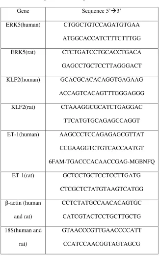
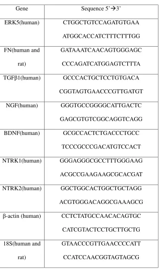
Table 2.1 Oligonucleotide sequences for real time PCR 66
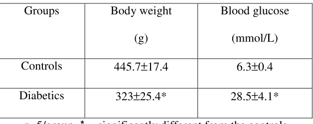
Table 2.2 Animal monitoring 84
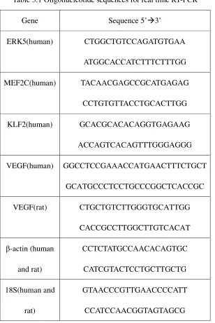
Table 3.1 Oligonucleotide sequences for real time PCR 105
xvii
Figure
PageFigure 1.1 Molecular mechanisms of high glucose-induced endothelial
dysfunction
12
Figure 1.2 Structure and activation of ERK5 16
Figure 1.3 Activators of ERK5 pathway 20
Figure 1.4 Potential role of ERK5 in chronic diabetic complications 28
Figure 2.1 Alteration of ERK5 signaling and ET-1 expression after high glucose
treatment
69
Figure 2.2 CAMEK5 constitutively activates ERK5 signaling and inhibits ET-1
expression in HUVECs
73
Figure 2.3 CAMEK5 constitutively activates ERK5 signaling and inhibits ET-1
expression in HMVECs
75
Figure 2.4 ERK5 siRNA transfection elevated ET-1 mRNA 78
Figure 2.5 ERK5 signaling upregulation by CAMEK5 inhibited ET-1 expression
in both basal and high glucose conditions
81
Figure 2.6 ERK5 and ET-1 mRNA expression in retinal tissues of diabetic rats 76
Figure 3.1 VEGF mRNA was inversely related to ERK5 signaling pathway in
endothelial cells treated with high glucose
112
Figure 3.2 ERK5 siRNA abolished ERK5 activation induced by high glucose and
promoted the increase in VEGF in endothelial cells
115
Figure 3.3 ERK5 siRNA enhanced tube formation in ECs under normal and high
glucose conditions
xviii expression in basal as well as high glucose conditions
Figure 3.5 Decreased ERK5 activation was associated with increased VEGF
mRNA expression in the retina of diabetic rats
124
Figure 4.1 FN expression was increased after high glucose treatment in HMVECs
as well as in retinal tissues of diabetic rats
152
Figure 4.2 ERK5 overexpression by CAMEK5 inhibited FN level with and
without glucose treatment
154
Figure 4.3 ERK5 downregulation by siERK5 and DNMEK5 increased FN levels 157
Figure 4.4 TGFβ1 signaling mediates the effect of ERK5 on FN 160
Figure 4.5 Alterations of neurotrophins and their receptors parallel ERK5 after
high glucose treatment
163
Figure 4.6 NGF treatment increased ERK5 phosphorylation and ERK5 expression
in HMVECs
165
Figure 5.1 A diagrammatic representation of the key findings of this study,
outlining possible role of ERK5 in diabetic retinopathy.
191
Figure App-1 pERK5 expression within 1 hour glucose treatment 206
Figure App-2 DNMEK5 transduction enhanced glucose-induced ET-1 and VEGF
mRNA in HMVECs
xix
APPENDICES Page
Appendix 1 Supplementary data 1 205
Appendix 2 Supplementary data 2 208
Appendix 3 Copyright agreement 1 211
Appendix 4 Copyright agreement 2 214
xx
AGE advanced glycation end products
ANOVA one way analysis of variance
AP-1 activating protein-1
BBE bovine brain extract
BCA bicinchoninic acid
BDNF brain-derived neurotrophic factor
BMK1 big mitogen-activated protein kinase 1
CAMEK5 constitutively active MEK5
CREB cAMP response element binding protein
Cx43 connexin 43
DCCT Diabetes Control and Complications Trial
DNERK5 dominant negative ERK5
DNMEK5 dominant negative MEK5
DR diabetic retinopathy
EC endothelial cell
ECM extracellular matrix
EGF epidermal growth factor
ELISA enzyme-linked immunosorbent assay
ERK1/2 extracellular signal-regulated kinase 1 and 2
ERK5 extracellular signal-regulated kinase 5
ET-1 endothelin-1
xxi
FN fibronectin
GPCR G protein-coupled receptors
hEGF human epidermal growth factor
hFGF-B human fibroblast growth factor - basic
HG high glucose (25mmol/L)
HIF1α hypoxia inducible factor 1 α
HMVEC human microvascular endothelial cell
HUVEC human umbilical vein endothelial cells
IL-6 interleukin 6
JNKs c-Jun NH2-terminal protein kinases
kDa kilodalton
KLF2 krueppel-like factor 2
LIF leukaemia inhibitory factor
LG low glucose (5mmol/L)
MAPK mitogen-activated protein kinase
MCA melting curve analysis
MEF2 myocyte enhancer factor 2
MEK mitogen-activated protein kinase kinase
MEKK mitogen-activated protein kinase kinase kinase
MOI multiplicity of infection
mRNA messenger RNA
xxii
NGF nerve growth factor
NLS nuclear localization signal
NO nitric oxide
NOS nitric oxide synthase
NPDR non-proliferative diabetic retinopathy
NTRK1 neurotrophic tyrosine kinase, receptor, type 1
NTRK2 neurotrophic tyrosine kinase, receptor, type 2
OC osmotic control
PBS phosphate-buffered saline
PDR proliferative diabetic retinopathy
pERK5 phospho-ERK5
PI3K phosphatidylinositol 3-kinase
PKC protein kinase C
AR peroxisome proliferator-activated receptor gamma
PVDF polyvinyl difluoride
R3-IGF-1 R3 insulin-like growth factor
ROS reactive oxygen species
RT-PCR reverse transcriptase polymerase chain reaction
SDS-PAGE sodium dodecyl sulfate-polyacrylamide gel
electrophoresis
SGK serum- and glucocorticoid-inducible kinase
xxiii
siRNA Small interfering RNA
STZ streptozotocin
SUMO small ubiquitin-like modifier
TEY Thr Glu Tyr
TGFβ1 transforming growth factor β 1
Tm melting temperature
tPA tissue plasminogen activator
UKPDS United Kingdom Prospective Diabetes Study
CHAPTER 1: INTRODUCTION
1.1Diabetes
Diabetes mellitus is characterized by high blood glucose levels, which produce
the classical symptoms of polyuria (frequent urination), polydipsia (increased thirst) and
polyphagia (increased hunger). Diabetes is usually diagnosed based on the fasting plasma
glucose levels of ≥7.0 mmol/L (≥126 mg/dL), or with nonfasting glucose levels of >11.1
mmol/L (>200 mg/dL) in the presence of classic symptoms (Vijan, 2010). There are two
main types of diabetes: type 1 and type 2. Symptoms are obvious and develop rapidly in
type 1 diabetes, while in type 2 diabetes symptoms may be subtle or absent and usually
develop slowly.
Type 1 diabetes is characterized by loss of the insulin-producing β cells of the
pancreas leading to an absolute insulin deficiency. It accounts for approximately 10% of
diabetes mellitus cases, mostly in children or in young adults. Wide variation in incidence
of type 1 diabetes in children has been well characterized worldwide. Incidence varies
from 0.1 per 100,000/year in China and Venezuela to 40.9 per 100,000/year in Finland
(DIAMOND Project Group, 2006). The incidence of type 1 diabetes has increased
rapidly over recent decades in young children (Patterson et al., 2009), which was thought
to be associated with changes in environment or lifestyle leading to the alteration of
humoral autoimmune response to islet antigens (Long et al., 2012). The presence of islet
tissue-specific autoantibodies in sera from patients with type 1 diabetes is in supportive
in NOD mice, that T-cell-mediated autoimmunity play a major role in the disease
development (Bluestone et al., 2010). Since pancreatic β cell loss and insulin deficency
are the charactistic features of type 1 diabetes, insulin supplementation is required for the
treatment.
Type 2 diabetes makes up about 90% of cases of diabetes mellitus. It is
characterized by insulin resistance with a relative insulin deficiency. In the early stage of
type 2 diabetes, insulin production may be increased due to the peripheral resistance. An
impairment of insulin secretion by pancreatic β cells may exist at later stage.
Development of type 2 diabetes is a multifactorial process involving genetics factors and
lifestyle (Ripsin et al., 2009). Most cases of type 2 diabetes involve many genes, each
being a small contributor. Lifestyle factors, important in the development of type 2
diabetes including: obesity (body mass index greater than thirty), lack of physical
activity, poor diet and stress. Most persons with type 2 diabetes are overweight and
obesity is thought to be the primary cause of type 2 diabetes in persons who are
genetically predisposed to the disease. Lifestyle modification through diet and exercise
can help reduce the incidence of type 2 diabetes in patients with “prediabetes” (Norris et
al., 2005). Interestingly, exercise significantly improved glycemic control, reduced
visceral adipose tissue, and reduce plasma triglyceride levels in patients with type 2
diabetes even without weight loss (Thomas et al., 2006).Therefore, lifestyle changes are
considered first-line therapy for the management of type 2 diabetes. If such measures are
inadequate, pharmacological interventions are initiated (Vijan, 2010;Rodbard et al.,
Both type 1 and type 2 diabetes can cause a number of complications affecting
multiple organs. Acute complications include hypoglycemia, diabetic ketoacidosis and
nonketotic hyperosmolar coma. Chronic complications typically develop after 10 to 20
years, and include macroangiopathy and microangiopathy in selected organ systems.
Chronic diabetic complications are the leading cause of mortality and morbidity in all
types of diabetes (Laight et al., 1999;Panus et al., 2003). Macroangiopathy in diabetes is
mainly due to an accelerated form of atherosclerosis, a pathological process initiated by
injury of endothelial cells. Atherosclerosis affects all clinically important sites i.e., the
coronary, the carotid and the peripheral arteries, thus increasing the risk of myocardial
infarction, stroke, intermittent claudication and ischaemic gangrene (Guerci et al.,
2001a). Diabetes also causes microvascular complications. These include diabetic
retinopathy and diabetic nephropathy (Klein, 1995). Diabetic nephropathy is a
progressive kidney disease caused by microangiopathy in the kidney glomeruli. It is
characterized by nephrotic syndrome and diffuse glomerulosclerosis (Tervaert et al.,
2010), and is a common cause of dialysis in Western countries. The present study is
focused on diabetic retinopathy (detailed below). However, it should be noted that most
of the pathological mechanisms may be similar in nephropathy and retinopathy.
1.2 Diabetic retinopathy
Diabetic retinopathy (DR) is a severe complication of diabetes, manifesting
vision loss and it is the most common cause of blindness in North America in the 25-74
years age group (Aiello et al., 1998).
DR has two phases, non-proliferative diabetic retinopathy (NPDR) and
proliferative diabetic retinopathy (PDR) (Hudson, 1996;Khan and Chakrabarti, 2007). In
NPDR phase, the vessels in the retina are weakened and leak, forming microaneurysms
and retinal hemorrhages, which leads to decreased vision. PDR is an advanced stage in
which progressive microvascular abnormalities cause areas of the retina to become
oxygen-deprived. New, but fragile, blood vessels develop on the surface of the retina or
the optic disc as the organ system attempts to maintain adequate oxygen levels within the
retina. This is called neovascularization, a characteristic feature of PDR. Unfortunately,
these delicate vessels hemorrhage easily. In the later phases of the disease, continued
abnormal vessel growth and scar tissue may cause tractional retinal detachment and
blindness (Mohamed et al., 2007). Several growth factors and vasoactive factors are
implicated in the development of PDR (Khan and Chakrabarti, 2003).
Laser photocoagulation is the standard treatment for proliferative retinopathy. The
Early Treatment Diabetic Retinopathy Study (ETDRS) results demonstrate that
photocoagulation is effective in reducing the risk of visual loss and increases the chance
of visual improvement (ETDRS, 1991). A vitrectomy is performed when there is vitreous
hemorrhage, or retinal detachment with or without vitreous hemorrhage (Flynn, Jr. et al.,
1992). VEGF plays an important role in mediating intraocular neovascularization in
intravitreal injection of anti-VEGF drug has emerged as a promising treatment for PDR
(Abdallah and Fawzi, 2009;Jardeleza and Miller, 2009). Recently, intravitreal anti-VEGF
agents have been used in combination with standard therapeutic regimens in the treatment
of proliferative diabetic retinopathy (Jardeleza and Miller, 2009;Montero et al., 2011).
1.3 Mechanisms of vascular endothelial injury in diabetic complications
Vascular endothelium is a major organ affected in chronic diabetic complications.
It is viewed as both the target organ and potential mediator of diabetic vascular
complications (Cosentino and Luscher, 1998;Laight et al., 1999).
1.3.1 Normal endothelium
The endothelium is the cell layer that lines blood vessels. It consists of a
metabolically active monolayer of cells that perform a variety of roles in vascular
homeostasis. Endothelium is a semipermeable barrier between the blood and the
interstitium, facilitating the exchange of water and small molecules. The endothelium
also participates in vasodilation. Furthermore, a healthy endothelium is able to inhibit
platelet aggregation and adhesion, smooth muscle cell proliferation and leucocyte
adhesion (Guerci et al., 2001b;Laight et al., 1999). The normal endothelial function is
achieved through the regulation of a number of endothelium-derived signaling and
adhesion molecules. Furchgott and Zawadzki (Furchgott and Zawadzki, 1980) discovered
by releasing nitric oxide (NO). Reactive oxygen species (ROS) are also produced by the
endothelium in response to shear stress and endothelial agonists. Superoxide anion
inactivates NO resulting in vasoconstriction in arteries (Mombouli and Vanhoutte, 1999).
1.3.2 Evidence of endothelial dysfunction in diabetes
Endothelial dysfunction has been observed to precede the onset of angiographic
lesions (Cosentino and Luscher, 1998). Vasodilation of coronary arteries in response to
pharmacological stimuli is reduced in diabetic rats (Sjogren and Edvinsson, 1988) and in
humans (Nitenberg et al., 1998), indicating an impaired endothelium. Alterations in
microcirculation consist of increased capillary pressure, blood flow and endothelial
permeability, which are reversible at an early stage in diabetes. Irreversible structural
modifications of the microvascular wall, such as thickening of the basement membrane
due to the extracellular accumulation of proteins, take place at later stages. These
alterations further affect microcirculation in diabetes by decreasing autoregulatory
capacity and blood flow reserve (Cosentino and Luscher, 1998).
1.3.3 Hyperglycemia is directly related to endothelial dysfunction
Diabetes-associated conditions such as hypertension, dyslipidemia and insulin
resistance are related to impaired endothelial function (Guerci et al., 2001a;Laight et al.,
1999;Panus et al., 2003). However, hyperglycemia is causally associated with endothelial
endothelial vasodilator function during either acute or chronic hyperglycemia in both
human (Akbari et al., 1998;Kim et al., 2003;Williams et al., 1998) and animal diabetes
(Pieper et al., 1995;Tesfamariam and Cohen, 1992). In addition, hyperglycemia is known
to increase endothelial permeability to macromolecules, delay cell replication, increase
the secretion of sclerotic matrix proteins, increase adhesive properties for leukocytes and
decrease the secretion of the pro-fibrinolytic agents, such as tissue plasminogen activator
(tPA)(Laight et al., 1999). Furthermore, both the Diabetes Control and Complications
Trial (DCCT) and the United Kingdom Prospective Diabetes Study (UKPDS) have
demonstrated correlation of poor glycemic control with increased incidences of
microvascular complications in patients with diabetes (DCCT, 1993;UKPDS, 1998).
Clinical trials have also shown that macrovascular complications such as coronary artery
disease (Kuusisto et al., 1994) and peripheral artery disease (Beks et al., 1995) is related
to glycemic levels.
1.3.4 Characteristic factors involved in diabetic retinopathy
The biochemical mechanisms of hyperglycemia-induced endothelial dysfunction
are under investigated at several laboratories. Potential mechanisms include
hyperglycemia-mediated formation of oxygen-derived free radicals, advanced glycation
end (AGE) products, protein kinase C(PKC) activation, and alteration of hexosamine
pathway (Brownlee, 2005). Hyperglycemia also causes an alteration in vasoactive factors
(Williams et al., 1998). It has been shown that hyperglycemia-induced endothelial
NO by oxygen-derived free radicals, and increased production of endothelium-derived
vasoconstrictors (Cosentino and Luscher, 1998;Giugliano et al., 1997;Williams et al.,
1998). Three characteristic factors involved in early and late stages of diabetic
retinopathy have been demonstrated including endothelin-1 (ET-1, involved in early and
late stages), vascular endothelial growth factor (VEGF, involved in early and late stages)
and fibronectin (FN, involved in late stage) (Chen et al., 2003b;Cukiernik et al.,
2004;Evans et al., 2000;Kaur et al., 2006;Roy et al., 1996;Takahashi et al., 1990).
ET-1, produced by endothelial cells, is a potent vasoconstrictive peptide (Masaki,
1989;Masaki, 1995;Yanagisawa et al., 1988). The activation of ET-1 also triggers the
release of vasodilator (Guerci et al., 2001b;Mombouli and Vanhoutte, 1999). ET-1 is
present in healthy subjects at low concentrations, and is involved in the maintenance of
blood pressure (Masaki, 2000). Increased plasma ET-1 levels have been demonstrated in
diabetics (Haak et al., 1992;Takahashi et al., 1990). In addition, circulating ET-1 levels
correlate with a number of vascular complications in diabetic patients with clinical
angiopathy (Laight et al., 1999). We have previously demonstrated that
hyperglycemia-induced augmented ET-1 production causes increased extracellular matrix (ECM) protein
deposition (Figure 1.1) (Chen et al., 2002;Chen et al., 2003a;Evans et al., 2000).
Furthermore, ET antagonist increases blood flow and prevents DR in diabetic animal
models (Deng et al., 1999;Shaw et al., 2006;Takagi et al., 1996).
FN is a multifunctional glycoprotein that plays important roles in various cellular
and ECs (Yamada and Olden, 1978;Yamada, 2000). Vascular basement membrane
thickening is a histological hallmark of diabetic retinopathy (Ljubimov et al., 1996;Roy et
al., 2010). We and others have shown that FN are overexpressed in retina vessels in
animal and human diabetes (Evans et al., 2000;Roy et al., 1996), as well as in ECs
cultured in high levels of glucose (Chen et al. 2003b; Chen et al. 2003a). It has been
shown that TGFβ1 and ET-1 mediate FN accumulation in target organs, including retina
in chronic diabetes (Evans et al., 2000;Khan et al., 2006;Sharma et al., 1996). FN may
provide outside-in signaling for angiogenesis by increased production of important
angiogenic factors like vascular endothelial growth factor (VEGF) (Astrof and Hynes,
2009;Khan et al., 2005).
VEGF is a key vasoactive factor implicated in the pathogenesis of DR. It is a
major angiogenic factor in proliferative DR (Pe'er et al., 1995;Ray et al., 2004). VEGF
also mediates the increased vascular permeability in the early stage of DR (Cukiernik et
al., 2004;Hammes et al., 1998). Elevated VEGF mRNA and protein expression in the
retina has been demonstrated in patients with DR (Boulton et al., 1998;Lutty et al.,
1996;Malecaze et al., 1994) and in the retina of streptozotocin-induced diabetic rats
(Cukiernik et al., 2004;Hammes et al., 1998). Inhibition of VEGF has been shown to
suppress retinal neovascularization in a murine model of ischemic retinopathy (Aiello et
al., 1995). Consistent with in vivo observations, in vitro studies show that high glucose
levels induce increased VEGF expression in ECs (Chen et al., 2000;Gao et al., 2008).
mediating glucose-induced increased permeability in ECs (Chen et al., 2000;Cukiernik et
al., 2004).
1.4 Signaling pathways implicated in glucose-induced endothelial dysfunction
Molecular mechanisms underlying the alteration of ET-1, VEGF, FN and
endothelial dysfunction remain to be fully elucidated. Hyperglycemia-induced activation
of protein kinase C (PKC) has been postulated to contribute to the vascular dysfunction in
diabetes (Bursell et al., 1997;Ishii et al., 1996;Koya et al., 1997). Glucose-induced PKC
activation increases mRNA expression of TGFβ1 and extracellular matrix components
such as fibronectin and alpha1 (type IV) collagen in the glomeruli of diabetic rats (Koya
et al., 1997). Phosphatidylinositol 3-kinases (PI3 kinase) is also considered to be involved
in the endothelial dysfunction in diabetes (Panus et al., 2003). We and others have shown
that both PKC and PI3 kinase pathways may converge on mitogen-activated protein
kinase (MAPK) (Figure 1.1), which may represent a common signal transduction of
glucose-induced endothelial dysfunction (Haneda et al., 1995;Haneda et al., 1997;Xin et
al., 2004). Extracellular signal-regulated kinases 1 and 2 (ERK1/2) has been
demonstrated to mediate high glucose-induced endothelial injury by increasing ET-1, FN
and VEGF (Xin et al., 2004;Xin et al., 2005). ET-1 mediates downstream effect of
glucose-induced ERK1/2 activation and stimulates FN accumulation in endothelial cells
(Xin et al., 2004). FN is involved in angiogenesis by increased production of important
angiogenic factors like vascular endothelial growth factor (VEGF) (Astrof and Hynes,
with activation of Akt/PKB, which is modulated by MAPK, PI3K and PKC (Xin et al.,
2005). Extracellular signal-regulated kinase 5 (ERK5) is the most recently identified
member of MAPK (Lee et al., 1995;Zhou et al., 1995). However the exact role of ERK5
Figure 1.1: Molecular mechanism of high glucose-induced endothelial dysfunction
A schematic outline of possible hyperglycemia-mediated alterations of signaling
mechanisms converging on MAPK, leading to increased ET-1, FN and VEGF production
in diabetic microangiopathy. PKC=protein kinase C, MAPK=mitogen-activated protein
1.5 Extracellular signal-regulated kinase 5 (ERK5)
1.5.1 Introduction of ERK5
MAPK plays a crucial role in regulating many cell processes including cell
survival, proliferation and differentiation (Chang and Karin, 2001;Pearson et al.,
2001;Widmann et al., 1999). There are four distinct subfamilies of MAPKs: extracellular
signal regulated kinase (ERK)1/2, ERK5, c-Jun NH2-terminal protein kinases (JNKs),
and p38 MAPKs (Chang and Karin, 2001;Pearson et al., 2001;Widmann et al., 1999).
ERK5, also termed big MAP kinase 1 (BMK1), is the most recently discovered member
of the MAPK family. It was cloned by two independent groups in 1995 (Lee et al.,
1995;Zhou et al., 1995). ERK5 transcript is abundant in heart, placenta, lung, kidney and
skeletal muscle (Lee et al., 1995;Zhou et al., 1995). ERK5 is highly expressed in
endothelial cells (Yan et al., 1999). A recent study has shown that ERK5 is expressed in
virtually all cells and is localized in nucleus as well as in the cytoplasm (Buschbeck and
Ullrich, 2005). Studies in knockout mice have shown that the ERK5 pathway is critical
for endothelial function and for the maintenance of vascular integrity (Hayashi et al.,
2001).
1.5.2 Structure of ERK5
Human ERK5 is a protein of 816 amino acids with a predicted molecular mass of
mammals (sharing 80-98% homology). Rat ERK5 is a protein of 812 amino acids that
share 97% homology with human ERK5. SMA-5 is a homolog of ERK5 in C. elegans
that is 60% similar to human ERK5 (Watanabe et al., 2005). Slt2p (Mpk1p) is an ERK5
ortholog in Saccharomyces cerevisiae (Truman et al., 2006).
ERK5 is more than twice the size of the other MAPKs due to its unique
C-terminal. The N-terminal of conserved MAPK catalytic domain share greater than 50%
homology with ERK1/2, which contains the Thr–Glu–Tyr (TEY) dual phosphorylation
motif in the activation loop (Figure 1.2) (Zhou et al., 1995). The C-terminal of ERK5
contains a nuclear localization signal (NLS) and two proline-rich regions (Figure 1.2).
The NLS is crucial for the nuclear localization of ERK5 upon stimulation. The
proline-rich regions may serve as binding sites for Src homology 3 (SH3) domain containing
Figure 1.2: Structure and activation of ERK5.
ERK5 is a protein of 816 amino acids. The N-terminal contains the TEY dual
phosphorylation motif in the activation loop. The C-terminal of ERK5 contains a nuclear
localization signal (NLS) and two proline-rich regions. ERK5 has two mechanisms for
signaling; kinase activation and transcriptional activation of targets. Kinase activation is
similar to other members of MAPKs. Transcriptional activation is unique for ERK5.
ERK5 is activated by dual phosphorylation at Thr218/Tyr220 by the upstream kinase
MEK5. Active ERK5 is able to undergo autophosphorylation on the C-terminal, leading
to an enhancement of ERK5 transcriptional activity. Diagram is modified from Drew et
NH2
COOH
substrate
Phosphorylation
activation
NH2
COOH
substrate
Phosphorylation
activation
1.5.3 Mechanisms of ERK5 activation
ERK5 has two mechanisms for signaling; kinase activation and transcriptional
activation of targets (Figure 1.2) (Nishimoto and Nishida, 2006;Roberts et al., 2009).
Kinase activation is similar to other members of MAPKs, However, the transcriptional
activation is unique for ERK5. These modes of activation are discussed below.
1.5.3.1 Kinase activation of ERK5
MAPK signalling cascades consists of three sequentially activated kinases: a
MAP kinase kinase kinase (MEKK); a MAP kinase kinase (MEK); and a MAP kinase
(MAPK). These kinase modules relay signals from extracellular agonists to cellular
targets. The signaling modules in the ERK5 pathway are composed of MEKK2/MEKK3,
MEK5, and ERK5 (Figure 1.3) (Chao et al., 1999;Lee et al., 1995;Sun et al., 2001;Zhou
et al., 1995). MEKK2/MEKK3 phosphorylate MEK5 on Ser311 and Thr315, resulting in
an increase in MEK5 activity (Chao et al., 1999). The MEK5 cDNA encodes a
444-amino acid protein, which displays 40% identity to other MEKs (English et al.,
1995;Zhou et al., 1995). MEK5 has multiple splice variants including 50 kDa MEK5α
and 40 kDa MEK5β isoforms (English et al., 1995;Zhou et al., 1995). MEK5β is
ubiquitously distributed, MEK5α is expressed most highly in liver and brain (English et
al., 1995). MEK5α is also found in endothelial cells (Hayashi et al., 2005). The
N-terminal of MEK5α contains a phox and Bem1p (PB1) domain, which mediates the
the PB1-dependent formation of the MEKK/MEK5 complex has been shown to inhibit
activation of MEK5α (Nakamura and Johnson, 2003;Seyfried et al., 2005). MEK5α is a
stronger activator of ERK5, compared to MEK5β, since MEK5β lacks an extended
N-terminus containing PB1 domain (Seyfried et al., 2005). Similarly, Cameron et al’ study
has shown that MEK5α, but not MEK5β, activates ERK5 (Cameron et al., 2004).
Mutation of dual phosphorylation MEK5α sites (Ser311Asp and Thr315Asp; MEK5α
(S311D/T315D)) activated ERK5, but similar mutation of MEK5β was unable to cause
ERK5 kinase activation or nuclear translocation (Cameron et al., 2004). It was also
shown that MEK5β is possibly a naturally occurring dominant-negative splice variant
protein that prevents excessive BMK1 activation and aberrant cell growth (Cameron et
Figure 1.3: Activators of ERK5 pathway.
The signaling modules in the ERK5 pathway are composed of MEKK2/MEKK3, MEK5,
and ERK5. ERK5 is activated by a variety of stimuli. It can be activated by serum and a
range of growth factors including EGF, FGF2, VEGF and NGF. It can also be activated
by cytokines such as LIF and IL-6. Additionally, ERK5 is activated by a range of stress
stresses (osmotic, shear, oxidative stress, hypoxia, ischemia)
Growth factors
(EGF, FGF2,
VEGF, NGF)
cytokines
(LIF, IL-6)
MEKK2/3
MEK5
ERK5
ERK5 is activated by dual phosphorylation at Thr218/Tyr220 by the upstream
kinase MEK5 (English et al., 1995;Lee et al., 1995;Zhou et al., 1995). MEK5
preferentially phosphorylates ERK5 on Thr218, which might induce a conformational
change and subsequent phosphorylation of Tyr220 (Mody et al., 2003). Active ERK5 is
able to undergo autophosphorylation on the C-terminal at a number of residues including
Thr28, Ser421, Ser433, Ser496, Ser731 and Thr733, leading to an enhancement of ERK5
transcriptional activity as described above. Activated ERK5 also phosphorylates MEK5
at residues 129, 137, 142 and 149 which are located in the region that is thought to
interact with ERK5 (Mody et al., 2003). PKCζ, an atypical protein kinase C, has been
reported to interact with MEK5 through their respective PB1 domains in EGF-induced
activation of ERK5 (Diaz-Meco and Moscat, 2001;Sumimoto et al., 2007). Interestingly,
a recent study demonstrated that PKCζ is directly associated with ERK5. PKCζ mediates
inhibitory phosphorylation of ERK5 by binding and phosphorylating serine 486, thus
suppressing ERK5 function in TNFα-mediated inflammatory process (Nigro et al., 2010).
1.5.3.2 Transcriptional activation of ERK5
The C-terminal region of ERK5 contains a transcriptional activation domain,
which is required for maximal transcriptional activity of target molecules (Buschbeck and
Ullrich, 2005;Kasler et al., 2000;Terasawa et al., 2003). Activated ERK5 phosphorylates
itself at the C-terminal at a number of residues (Mody et al., 2003) and
activity (Kasler et al., 2000;Morimoto et al., 2007). Once stimulated, phosphorylation of
ERK5 results in the activation of the kinase activity. ERK5 phosphorylates both
downstream target molecules and its carboxy-terminal region (Figure 1.2). Thus,
autophosphorylation of the C-terminal leads to a further increase in the transcription
activity of target molecules (Morimoto et al., 2007). In addition, Morimoto et al. showed
that the activated kinase activity of ERK5 is required for the C-terminal mediated
transcriptional activation of downstream targets. Mutation of phosphorylatable threonine
and serine residues to unphosphorylatable alanines significantly reduces the
transcriptional activation effect of ERK5 (Morimoto et al., 2007). Interestingly,
C-terminal also regulates the kinase activation of N-C-terminal. Deletion of C-C-terminal results
in dramatic increase of kinase activation of N-terminal (Buschbeck and Ullrich, 2005).
1.5.4 Regulators of ERK5 signaling
Similar to other MAPKs, ERK5 is activated by a variety of stimuli (Figure 1.3).
Studies have revealed that it is activated by serum (Kato et al., 1997), a range of growth
factors including epidermal growth factor (EGF) (Kato et al., 1998), fibroblast growth
factor-2 (FGF-2) (Kesavan et al., 2004), VEGF (Hayashi et al., 2001), and by cytokines
such as leukaemia inhibitory factor (LIF) (Nicol et al., 2001) and interleukin 6 (IL-6)
(Carvajal-Vergara et al., 2005). Additionally, neurotrophins, such as brain-derived
neurotrophic factor (BDNF), nerve growth factor (NGF), use the ERK5 pathway to
stress stimuli such as osmotic and oxidative stresses (Abe et al., 1996), fluid shear stress
(Yan et al., 1999), hypoxia (Sohn et al., 2002) and ischemia (Takeishi et al., 1999).
G-proteins are involved in the activation of ERK5 by growth factors (Obara and Nakahata,
2010). In addition, studies have shown that PKCζ mediates ERK5 activation by G
protein-coupled receptors (GPCR) (Diaz-Meco and Moscat, 2001;Garcia-Hoz et al.,
2010;Nigro et al., 2010). It has also been reported that G protein acts as an adaptor
protein in PKCζ-mediated ERK5 activation by GPCR (Garcia-Hoz et al., 2010).
1.5.5 Substrates of ERK5 signaling
A number of molecules have been identified as substrates of the ERK5 pathway.
The transcription factors of the myocyte enhancer factor 2 (MEF2) family are
best-characterized substrates of ERK5 (Kato et al., 1997;Kato et al., 2000;Yang et al., 1998).
MEF2 is a four-membered family of transcription factors including MEF2A, MEF2B,
MEF2C and MEF2D. ERK5 phosphorates and activates MEF2A, MEF2C and MEF2D,
but not MEF2B (Kato et al., 1997;Kato et al., 2000). The C-terminal tail of ERK5
contains a MEF2-interacting region and a transcriptional activation domain essential for
coactivation of MEF2 (Kasler et al., 2000). Activation of the MEF2 by the ERK5 is
essential for endothelial cell survival and proliferation (Kato et al., 1997;Olson, 2004). In
addition, Krueppel-like factor 2 (KLF2) is identified as an ERK5 responsive gene and
ERK5 drives KLF2 transcription by activating MEF2 (Sohn et al., 2005). KLF2 plays an
quiescence (Boon and Horrevoets, 2009;Dekker et al., 2006;Senbanerjee et al.,
2004;Sohn et al., 2005;Suzuki et al., 2005).
Ets-domain transcription factor (Sap1a) and serum- and glucocorticoid-inducible
kinase (SGK) have also been identified as the downstream targets of ERK5 and plays an
important role in cell proliferation induced by growth factors (Hayashi et al.,
2001;Kamakura et al., 1999). Moreover, the ERK5 signaling pathway stimulates the
transcriptional activity of c-Fos and Fra-1 (fos-related antigen 1), and members of the
AP-1 (activator protein 1) family (Terasawa et al., 2003). Other downstream substrates of
ERK5 include Cx43 (connexin 43; a gap junction protein) (Cameron et al., 2003), BAD
(a pro-apoptotic member of Bcl-2 family) (Pi et al., 2004), C-Myc proto-oncogene
(English et al., 1998) and CREB (cAMP response element binding protein) (Watson et
al., 2001).
1.6 Potential role of ERK5 in endothelial dysfunction in diabetes
1.6.1 Role of ERK5 in endothelial cell
ERK5 is highly expressed in the endothelial cells and is essential for maintaining
endothelial function and blood vessel integrity (Hayashi et al., 2001). ERK5-deletion is
lethal. These mice die around embryonic day 10 (E10) due to cardiovascular defects
are observed in the mekk3−/−, mek5−/− and mef2−/− embryos, suggesting that the
MEKK3/MEK5/ERK5/MEF2 cascade is critical to the cardiovascular development (Lin
et al., 1997;Wang et al., 2005;Yang et al., 2000). Additional studies by targeted deletion
of ERK5 gene in mice have shown that ERK5 is essential in endothelial cell physiology,
but not in the cardiac development (Hayashi et al., 2004). Endothelial cells specific
ERK5 ablation generates the same heart defects as those observed in global ERK5
knockout mutants, whereas cardiomyocyte specific ERK5 deletion mice are normal
(Hayashi et al., 2004). These results indicate that ERK5 is critical for endothelial cell
function and that the abnormal heart development in the mice lacking ERK5 is a
consequence of endothelial cell dysfunction (Hayashi et al., 2004). Additionally, ERK5 is
required to maintain vascular integrity in adult mice. Adult mice display hemorrhages in
multiple organs and die within 2–4 weeks after deletion of ERK5 (Hayashi et al., 2004).
In addition to these in vivo studies, ERK5 has been shown to be essential for endothelial
cells survival in vitro (Hayashi et al., 2004;Pi et al., 2004). Deletion of ERK5 induces
profound endothelial cell apoptosis. Introduction of exogenous ERK5 can prevent
endothelial cells from cell death (Hayashi et al., 2004). Similarly, activation of ERK5 by
constitutively active MEK5 (CAMEK5) significantly improved cell viability and
decreased apoptosis induced by growth factor deprivation (Pi et al., 2004). In addition,
CAMEK5 inhibited growth factor deprivation-induced apoptosis, whereas dominant
negative ERK5 (DNERK5) stimulated apoptosis in endothelial cells (Pi et al., 2004).
ERK5 pathway also mediates the shear stress-induced antiapoptotic effect in endothelial
cells (Pi et al., 2004;Yan et al., 1999). Inhibition of ERK5 activity by overexpression of
2004). Analysis of antiapoptotic mechanisms of ERK5 showed that MEF2C, a direct
substrate of ERK5 mediates endothelial cell survival signal (Hayashi et al., 2004).
Studies on the role of ERK5 in diabetic macrovascular and microvascular diseased are
Figure 1.4: Potential role of ERK5 in chronic diabetic complications.
hyperglycaemia
macroangiopathy
microangiopathy
ERK5
SUMOylation
Diabetic retinopathy
Diabetic nephropathy
myocardial infarction, stroke, intermittent claudication and ischemic gangrene
arthrosclerosis
ERK5
activation
1.6.2 Role of ERK5 in diabetic macrovascular diseases
It has been shown that ERK5 is involved in diabetic macroangiopathy.
Macroangiopathy in diabetes is mainly due to an accelerated form of atherosclerosis
(Guerci et al., 2001a). Steady and laminar blood flow is known to be atheroprotective,
and has been shown to be a strong activator of ERK5 (Yan et al., 1999). Also, ERK5
activation has been demonstrated to be atheroprotective (Pi et al., 2004). Inhibition of
ERK5 activity by dominant negative ERK5 (DNERK5) reduces the endothelial
cell-protective effect of shear stress (Pi et al., 2004), indicating that the ERK5 mediates the
shear stress-induced antiapoptotic effect in endothelial cells. This may be mediated by
phosphorylation of BAD (Pi et al., 2004). Sohn et al. revealed that KLF2 mediates
endothelial-protective effect of ERK5 (Sohn et al., 2005). KLF2 is a critical
transcriptional regulator for vasoprotective effect of shear stress (Boon and Horrevoets,
2009;Parmar et al., 2006). In addition, laminar flow-induced ERK5 activation has been
shown to confer an atheroprotective effect by inducing peroxisome proliferator-activated
receptor gamma (PPARγ) (Akaike et al., 2004) and inhibiting tumor necrosis factor α
(TNFα) mediated adhesion molecule expression in endothelial cells (Li et al., 2008).
However, protective effect of ERK5 is inhibited by SUMOylation in diabetes (Woo et al.,
2008). Small ubiquitin-like modifier (SUMO) covalently attaches to certain residues of
specific target proteins and negatively regulates transcription factors (Verger et al.,
2003;Wang and Tournier, 2006). Increased ERK5 SUMOylation was observed in the
al., 2008). ROS and advanced AGE are two well-known mediators of diabetic vascular
disease. H2O2 and AGE induce ERK5 SUMOylation and inhibit shear stress-mediated
ERK5’s transcription activity. Subsequently decreased KLF2 and eNOS expression lead
to endothelial dysfunction and accelerate atherosclerosis in diabetes (Woo et al., 2008).
1.6.3 Role of ERK5 in diabetic microangiopathy
A number of studies have been performed to study the role of ERK5 on diabetic
nephropathy. Mesangial expansion is an important structural change in diabetic
nephropathy (Dalla et al., 2001). Glucose activates ERK5 and promotes rat mesangial
cells proliferation (Suzaki et al., 2004). ERK5 also mediates human mesangial cell
activation by glucose as well as other agonists causing mesangial cell proliferation and
extracellular matrix accumulation (Dorado et al., 2008). Moreover, Suzaki et al has
demonstrated that ERK5 is activated in glomerular mesangial cells of diabetic rat and
concluded that ERK5-mediated mesangial cell growth may be involved in the
pathogenesis of diabetic nephropathy (Suzaki et al., 2004). Similarly, ERK5 increases
mesangial cell viability and collagen matrix accumulation in glomerulonephritis
(Urushihara et al., 2010). Furthermore, it has been shown that SGK1 is upregulated in
diabetic nephropathy and mediates extracellular matrix deposition in human mesangial
cells exposed to high glucose (Feng et al., 2005). SGK1 is a downstream target of ERK5
signaling (Hayashi et al., 2001), therefore, it is possible that ERK5 is involved in diabetic
nephropathy by activating SGK1. These results show that ERK5 activation contributes to
However, a recent report showed a different effect of ERK5 on renal disease.
ERK5 mediates renal protection against ischemia reperfusion injury (Kawakami et al.,
2012). It was shown that there is a kidney-specific ERK5 with a smaller molecular mass,
which may be partially functional. Overexpression of full length ERK5 in the mouse
kidneys provided protection against renal ischemia reperfusion injury (Kawakami et al.,
2012). It is possible that ERK5 overexpression may also protect patients with diabetes
from diabetic nephropathy.
Although ERK5 is potentially important in diabetic microangiopathy, the in-depth
investigation of glucose-induced ERK5 signaling has not been carried out. Furthermore
no studies have been performed with respect to diabetic retinopathy, a characteristic
1.7 Rationale, hypothesis and specific aims
As outlined above, ERK5 plays an important role in maintaining normal
endothelial function. Endothelial dysfunction is a key mechanism in the pathogenesis of
chronic diabetic complications, which leads to increase of endothelium derived factors
such as ET-1, VEGF and FN. It is possible that alteration of ERK5 may regulate these
endothelium derived factors.
Therefore, we hypothesize that alteration of ERK5 signaling plays a
pathogenesis role in diabetic vascular complication, specifically in the development
of diabetic retinopathy via increasing vasoactive factors and extracellular matrix
proteins.
Following are the specific aims of the study:
i. To study the role of ERK5 signaling in ET-1 expression in endothelial cells and in
the retina of diabetic animals.
ii. To study the role of ERK5 signaling in glucose-induced VEGF expression and
angiogenesis in endothelial cells.
iii. To study the role of ERK5 signaling in glucose-induced FN production and to
1.8References
1991. Early photocoagulation for diabetic retinopathy. ETDRS report number 9. Early
Treatment Diabetic Retinopathy Study Research Group. Ophthalmology 98:766-785.
1993. The effect of intensive treatment of diabetes on the development and progression
of long-term complications in insulin-dependent diabetes mellitus. The Diabetes Control
and Complications Trial Research Group. N. Engl. J. Med. 329:977-986.
1998. Intensive blood-glucose control with sulphonylureas or insulin compared with
conventional treatment and risk of complications in patients with type 2 diabetes
(UKPDS 33). UK Prospective Diabetes Study (UKPDS) Group. Lancet 352:837-853.
2006. Incidence and trends of childhood Type 1 diabetes worldwide 1990-1999. Diabet.
Med. 23:857-866.
Abdallah, W., and A.A.Fawzi. 2009. Anti-VEGF therapy in proliferative diabetic
retinopathy. Int. Ophthalmol. Clin. 49:95-107.
Abe, J., M.Kusuhara, R.J.Ulevitch, B.C.Berk, and J.D.Lee. 1996. Big mitogen-activated
Aiello, L.P., R.L.Avery, P.G.Arrigg, B.A.Keyt, H.D.Jampel, S.T.Shah, L.R.Pasquale,
H.Thieme, M.A.Iwamoto, J.E.Park, and . 1994. Vascular endothelial growth factor in
ocular fluid of patients with diabetic retinopathy and other retinal disorders. N. Engl. J.
Med. 331:1480-1487.
Aiello, L.P., T.W.Gardner, G.L.King, G.Blankenship, J.D.Cavallerano, F.L.Ferris, III,
and R.Klein. 1998. Diabetic retinopathy. Diabetes Care 21:143-156.
Aiello, L.P., E.A.Pierce, E.D.Foley, H.Takagi, H.Chen, L.Riddle, N.Ferrara, G.L.King,
and L.E.Smith. 1995. Suppression of retinal neovascularization in vivo by inhibition of
vascular endothelial growth factor (VEGF) using soluble VEGF-receptor chimeric
proteins. Proc Natl Acad Sci U. S. A 92:10457-10461.
Akaike, M., W.Che, N.L.Marmarosh, S.Ohta, M.Osawa, B.Ding, B.C.Berk, C.Yan, and
J.Abe. 2004. The hinge-helix 1 region of peroxisome proliferator-activated receptor
gamma1 (PPARgamma1) mediates interaction with extracellular signal-regulated kinase
5 and PPARgamma1 transcriptional activation: involvement in flow-induced
PPARgamma activation in endothelial cells. Mol. Cell Biol. 24:8691-8704.
Akbari, C.M., R.Saouaf, D.F.Barnhill, P.A.Newman, F.W.LoGerfo, and A.Veves. 1998.
Endothelium-dependent vasodilatation is impaired in both microcirculation and
Arosio, E., P.Minuz, and M.Prior. 1999. [Endothelial function and the microcirculation in
diabetes mellitus]. Ann. Ital. Med. Int. 14:106-113.
Astrof, S., and R.O.Hynes. 2009. Fibronectins in vascular morphogenesis. Angiogenesis.
12:165-175.
Beks, P.J., A.J.Mackaay, J.N.de Neeling, H.de Vries, L.M.Bouter, and R.J.Heine. 1995.
Peripheral arterial disease in relation to glycaemic level in an elderly Caucasian
population: the Hoorn study. Diabetologia 38:86-96.
Bluestone, J.A., K.Herold, and G.Eisenbarth. 2010. Genetics, pathogenesis and clinical
interventions in type 1 diabetes. Nature 464:1293-1300.
Boon, R.A., and A.J.Horrevoets. 2009. Key transcriptional regulators of the
vasoprotective effects of shear stress. Hamostaseologie. 29:39-3.
Boulton, M., D.Foreman, G.Williams, and D.McLeod. 1998. VEGF localisation in
diabetic retinopathy. Br. J Ophthalmol. 82:561-568.
Brownlee, M. 2005. The pathobiology of diabetic complications: a unifying mechanism.
Diabetes 54:1615-1625.
Bursell, S.E., C.Takagi, A.C.Clermont, H.Takagi, F.Mori, H.Ishii, and G.L.King. 1997.
abnormal retinal hemodynamics in diabetic rats. Invest Ophthalmol. Vis. Sci.
38:2711-2720.
Buschbeck, M., and A.Ullrich. 2005. The unique C-terminal tail of the mitogen-activated
protein kinase ERK5 regulates its activation and nuclear shuttling. J. Biol. Chem.
280:2659-2667.
Cameron, S.J., J.Abe, S.Malik, W.Che, and J.Yang. 2004. Differential role of
MEK5alpha and MEK5beta in BMK1/ERK5 activation. J. Biol. Chem. 279:1506-1512.
Cameron, S.J., S.Malik, M.Akaike, N.Lerner-Marmarosh, C.Yan, J.D.Lee, J.Abe, and
J.Yang. 2003. Regulation of epidermal growth factor-induced connexin 43 gap junction
communication by big mitogen-activated protein kinase1/ERK5 but not ERK1/2 kinase
activation. J. Biol. Chem. 278:18682-18688.
Carvajal-Vergara, X., S.Tabera, J.C.Montero, A.Esparis-Ogando, R.Lopez-Perez,
G.Mateo, N.Gutierrez, M.Parmo-Cabanas, J.Teixido, J.F.San Miguel, and A.Pandiella.
2005. Multifunctional role of Erk5 in multiple myeloma. Blood 105:4492-4499.
Chang, L., and M.Karin. 2001. Mammalian MAP kinase signalling cascades. Nature
Chao, T.H., M.Hayashi, R.I.Tapping, Y.Kato, and J.D.Lee. 1999. MEKK3 directly
regulates MEK5 activity as part of the big mitogen-activated protein kinase 1 (BMK1)
signaling pathway. J. Biol. Chem. 274:36035-36038.
Chen, S., M.D.Apostolova, M.G.Cherian, and S.Chakrabarti. 2000. Interaction of
endothelin-1 with vasoactive factors in mediating glucose-induced increased permeability
in endothelial cells. Lab Invest 80:1311-1321.
Chen, S., T.Evans, D.Deng, M.Cukiernik, and S.Chakrabarti. 2002. Hyperhexosemia
induced functional and structural changes in the kidneys: role of endothelins. Nephron
90:86-94.
Chen, S., Z.A.Khan, M.Cukiernik, and S.Chakrabarti. 2003a. Differential activation of
NF-kappa B and AP-1 in increased fibronectin synthesis in target organs of diabetic
complications. Am. J. Physiol Endocrinol. Metab 284:E1089-E1097.
Chen, S., S.Mukherjee, C.Chakraborty, and S.Chakrabarti. 2003b. High glucose-induced,
endothelin-dependent fibronectin synthesis is mediated via NF-kappa B and AP-1. Am. J.
Physiol Cell Physiol 284:C263-C272.
Cosentino, F., and T.F.Luscher. 1998. Endothelial dysfunction in diabetes mellitus. J.
Cukiernik, M., D.Hileeto, T.Evans, S.Mukherjee, D.Downey, and S.Chakrabarti. 2004.
Vascular endothelial growth factor in diabetes induced early retinal abnormalities.
Diabetes Res Clin. Pract. 65:197-208.
Dalla, V.M., A.Saller, M.Mauer, and P.Fioretto. 2001. Role of mesangial expansion in
the pathogenesis of diabetic nephropathy. J. Nephrol. 14 Suppl 4:S51-S57.
Dekker, R.J., R.A.Boon, M.G.Rondaij, A.Kragt, O.L.Volger, Y.W.Elderkamp,
J.C.Meijers, J.Voorberg, H.Pannekoek, and A.J.Horrevoets. 2006. KLF2 provokes a gene
expression pattern that establishes functional quiescent differentiation of the endothelium.
Blood 107:4354-4363.
Deng, D., T.Evans, K.Mukherjee, D.Downey, and S.Chakrabarti. 1999. Diabetes-induced
vascular dysfunction in the retina: role of endothelins. Diabetologia 42:1228-1234.
Diaz-Meco, M.T., and J.Moscat. 2001. MEK5, a new target of the atypical protein kinase
C isoforms in mitogenic signaling. Mol. Cell Biol. 21:1218-1227.
Dorado, F., S.Velasco, A.Esparis-Ogando, M.Pericacho, A.Pandiella, J.Silva,
J.M.Lopez-Novoa, and A.Rodriguez-Barbero. 2008. The mitogen-activated protein kinase Erk5
English, J.M., G.Pearson, R.Baer, and M.H.Cobb. 1998. Identification of substrates and
regulators of the mitogen-activated protein kinase ERK5 using chimeric protein kinases.
J. Biol. Chem. 273:3854-3860.
English, J.M., C.A.Vanderbilt, S.Xu, S.Marcus, and M.H.Cobb. 1995. Isolation of MEK5
and differential expression of alternatively spliced forms. J. Biol. Chem.
270:28897-28902.
Evans, T., D.X.Deng, S.Chen, and S.Chakrabarti. 2000. Endothelin receptor blockade
prevents augmented extracellular matrix component mRNA expression and capillary
basement membrane thickening in the retina of diabetic and galactose-fed rats. Diabetes
49:662-666.
Feng, Y., Q.Wang, Y.Wang, B.Yard, and F.Lang. 2005. SGK1-mediated fibronectin
formation in diabetic nephropathy. Cell Physiol Biochem. 16:237-244.
Flynn, H.W., Jr., E.Y.Chew, B.D.Simons, F.B.Barton, N.A.Remaley, and F.L.Ferris, III.
1992. Pars plana vitrectomy in the Early Treatment Diabetic Retinopathy Study. ETDRS
report number 17. The Early Treatment Diabetic Retinopathy Study Research Group.
Ophthalmology 99:1351-1357.
Furchgott, R.F., and J.V.Zawadzki. 1980. The obligatory role of endothelial cells in the