Pearls & Oy-sters:
False positives in short-segment nerve
conduction studies due to ulnar nerve
dislocation
B.J. Kim, MD, PhD
S.B. Koh, MD, PhD
K.W. Park, MD, PhD
S.J. Kim, MD, PhD
J.S. Yoon, MD, PhD
ABSTRACT
The possibility that a technical error may occur during nerve conduction studies due to ulnar nerve dislocation when the elbow is flexed has recently been suggested. We investigated normal volun-teers using ultrasonography to observe the effects of ulnar nerve dislocation during elbow flexion on short-segment nerve conduction studies. We found significant conduction block in all of the subjects with ulnar nerve dislocation, and the finding was defined as a technical error caused by volume conduction. The results of the present study suggest that caution should be exercised when interpreting the results of short-segment nerve conduction studies at the across-elbow segment due to the possibility of technical error induced by ulnar nerve dislocation.GLOSSARY
NCS⫽nerve conduction study; S-NCS⫽short-segment NCS; UNE⫽ulnar neuropathy at the elbow.Neurology®2008;70:e9–e13
A nerve conduction study (NCS) is the most valuable diagnostic tool in patients with
ulnar neuropathy at the elbow (UNE). However, the possibility that a technical error
induced by ulnar nerve dislocation may occur when the elbow is flexed has recently been
suggested.
1Indeed, the position of the elbow may have a significant influence on the
calculated ulnar nerve conduction velocity across the elbow,
2and when the elbow is
extended, it is thought that the ulnar nerve may become loose or redoubled in the ulnar
groove and that surface measurements do not accurately reflect the true distance of the
underlying nerve.
2,3Elbow flexion may stretch the nerve; therefore, the measurement of
the ulnar groove distance is a more accurate reflection of the distance along the nerve in
the ulnar groove. The recommended position of the elbow during ulnar NCS is flexion at
90 or 135 degrees in order to straighten the ulnar nerve.
4However, significant technical
error may be caused by ulnar nerve displacement when the elbow is flexed.
1NCS with short-segment interval stimulations has been commonly used to increase the
sensitivity for very focal lesions, because the effect of the abnormally slow segment is
progressively diluted as more and more normal segments are included in the
examina-tion.
5A short-segment NCS (S-NCS) can be performed by stimulating the nerve at 2-cm
intervals across the elbow in the flexed position.
6A significant conduction delay or focal
change in amplitude or waveform morphology across a segment is generally considered
abnormal; however, ulnar nerve displacement by elbow flexion may cause a more
signif-icant technical error in S-NCS than in routine NCS at the across-elbow segment.
In the present study, we investigated the effects of ulnar nerve displacement on S-NCS
at the across-elbow segment. We also measured the angle of the elbow when the ulnar
nerve was dislocated by elbow flexion.
METHODSParticipants and evaluation.We studied 234 elbows from 117 healthy volunteers (52 men, 65 women), ages 20 to 50 years (mean age, 30.4 y), who had normal NCS results in their upper extremities. Elbow flexion at approximately 135° was used for ulnar NCS in all subjects. None of the participants had any history or symptoms of elbow pain or trauma,
From the Departments of Neurology (B.J.K., S.B.K., K.W.P.) and Rehabilitation Medicine (S.J.K., J.S.Y.), College of Medicine, Korea University, Seoul.
Disclosure:The authors report no conflicts of interest. Address correspondence and
reprint requests to Dr. Joon Shik Yoon, Department of Rehabilitation Medicine, Korea University Guro Hospital, Korea University College of Medicine, 80, Guro-dong, Guro-gu, Seoul, 152-703, Korea [email protected]
RESIDENT & FELLOW SECTION
entrapment neuropathy, or systemic disease. Approval from the hospital institutional review board was obtained prior to conducting the study. Written informed consent, in which the potential risks of ultrasonography were outlined, was obtained from the participants before the procedure.
Real-time ultrasonographic study. A 7.5 to 12 MHz linear transducer (5000 System HDI; Philips Medical Sys-tems NA, WA) was applied on the line between the medial epicondyle and olecranon of subjects lying supine on an ex-amining table. We used the B mode to view the ulnar nerve in the postcondylar groove between the medial epicondyle and olecranon and used the color Doppler mode to differentiate arteries. A baseline nerve location was determined at zero degrees of elbow flexion, and we observed whether the ulnar nerve was displaced at any angle during elbow flexion using real-time ultrasonography. We classified the position of the ulnar nerve (during elbow flexion) into one of three types according to the classification system of a previous report.7 Type N (no dislocation) classification was assigned when the nerve moved anteriorly, but not to the tip of the epicondyle, during elbow flexion. The ulnar nerve position was classified as type S (subluxation) when the nerve moved onto the tip of the epicondyle. The position of the ulnar nerve was classified as type D (dislocation) when the nerve moved anteriorly be-yond the tip of the epicondyle. In cases of type D, we mea-sured the angle of the elbow using a goniometer when the ulnar nerve was dislocated (figure 1). To measure the angle, an axis of the goniometer was located on the skin of the medial epicondyle tip, and a proximal fixed arm of the goni-ometer was aligned parallel to the long axis of the upper arm. A distal movable arm was then attached to the medial surface of the forearm long axis. The elbow angle was de-fined as the angle made by the movement of the distal mov-able arm with forearm flexion from the extended position.
Short-segment nerve conduction study. S-NCSs for the ulnar nerve were performed in subjects with type S and D classifications by an electromyographer who was blind to the classification type. The S-NCSs were performed at an elbow position of approximately 135°. Prior to the S-NCS, we confirmed that none of the subjects among those with type S and D classifications had weakness in the ulnar inner-vated muscles or sensory changes in the fourth and fifth fin-gers. To achieve approximately 135° of elbow flexion in the
most comfortable position possible, subjects were positioned supine on an examining table and asked to abduct their up-per arm to 90° and to rest it on a pillow such that the hand approached the ipsilateral ear, resulting in elbow flexion. The midpoint of the ulnar groove between the tip of the me-dial epicondyle and the olecranon was marked (point P; fig-ure 2). From this point, marks were placed along the course of the ulnar nerve at 2-cm intervals from 6 cm proximal to 4 cm distal.
The ulnar nerve was stimulated with abductor digiti minimi recording at each mark. The recording electrode was placed over the belly of the abductor digiti minimi, and the reference electrode was placed over the distal tendon. A con-stant current stimulator (2-cm intervals between cathode and anode) and a disposable, flat-surface electrode 1 cm in diameter were used for recording. Equipment used for the study was manufactured by Nicolet Biomedical (Viking IV, Madison, WI). A sensitivity setting of 5 mV/division, a sweep speed of 2 msec/division, and a filter setting of 5 Hz to 5 KHz was maintained throughout all measurements. The skin temperature of the subjects during the study was also maintained at or above 32 °C. Before the measurements were performed, we ensured that supramaximal stimulation was achieved and that adequate pressure was applied to the stim-ulating electrodes in order to enable focal stimulation with-out spread.
Latencies were measured from the stimulus to CMAP on-set, and the amplitude measurements were calculated from baseline to negative peak. A conduction delay of greater than 0.5 msec or conduction block with an amplitude reduction of 50% or more across 2-cm segments were defined as abnormal.8
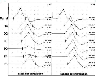
After the S-NCS, the nerves that were displaced by elbow flexion were tracked using ultrasonography and marked as ragged dots when elbow flexion was at 135°. The closest point from the tip of the medial epicondyle was marked as point P’. Marks were then placed along the course of the Figure 1 Angle of the elbow when the ulnar
nerve was dislocated
The ulnar nerve (*) moved onto the tip of the medial epicon-dyle when the elbow was flexed and finally dislocated at 125 degrees of flexion.
Figure 2 Stimulation points for the short-segment ulnar nerve conduction study
tracked ulnar nerve at 2-cm intervals from 6 cm proximal to 4 cm distal to point P’ (ragged dots in figure 2). The S-NCSs were performed again by stimulating the ragged dots.
RESULTS Ultrasonography.
Ulnar nerve
disloca-tion (Type D) occurred in 13 elbows (5.6%) of
13 subjects (8 men, 5 women). Ulnar nerve
sub-luxation (Type S) was found in 58 elbows (24.8%)
of 49 subjects (22 men, 27 women). The mean
elbow angle associated with ulnar nerve
disloca-tion was 99.8° (range 75 to 135). The ulnar nerve
was dislocated at an elbow angle of less than 90°
in 3 elbows; 2 elbows were at 87°, and 1 elbow
was at 75°.
Short-segment nerve conduction study.
All 13 cases
of ulnar nerve dislocation at the elbow exhibited
delayed latency (12 subjects; mean 0.62 msec,
range 0.6 to 0.8 msec) or a conduction block
around the medial epicondyle during the S-NCS
with black dot stimulation. Of the 13 subjects
with conduction block, the amplitude drop points
were P in 6 subjects and P2 in 3 subjects; the mean
amplitude drop rate in these 9 subjects was 68.9%
(range 58.8 to 92.2%). The other 4 subjects
showed complete conduction block (no evoked
compound muscle action potential) at point P2.
One of the 4 subjects with complete conduction
block showed an amplitude drop of 34% at point
P. However, no abnormal findings were detected
in the elbows of the subjects with ulnar nerve
sub-luxation during elbow flexion.
The abnormal findings in the 13 elbows with
ulnar nerve dislocation were transformed to
nor-mal waveforms and latencies using ragged dot
stimulation (figure 3).
DISCUSSION
The prevalence of ulnar nerve
dis-placement in the present study was similar to the
results of our previous study that showed
disloca-tion in 3.8% and subluxadisloca-tion in 20.5% of
sub-jects.
1The results of these two studies showed a
high percentage of ulnar nerve displacement in
comparison with a previous report,
9although this
is probably due to the increased precision of
in-vestigation with high-resolution
ultrasonogra-phy. On the other hand, the percentage of ulnar
nerve displacement in the present study was low
in comparison with that from another report that
found ulnar nerve dislocation in 39 elbows (20%)
and ulnar nerve subluxation in 53 elbows (27%)
Figure 3 Example of short-segment ulnar nerve conduction study
out of 200 healthy elbows.
7Considering that the
report included older subjects (age 20 to 69 years)
than our study (age 20 to 50 years), the higher
percentage reported in that report was likely
re-lated to the loosening of soft tissue in the ulnar
groove due to aging, which would result in a
higher chance of ulnar nerve displacement.
UNE is the second most common upper
ex-tremity compression neuropathy
10; however, the
diagnosis of UNE is not straightforward. Most
physicians suspect UNE when a patient
com-plains of occasional ulnar distribution numbness
and paresthesias; however, these symptoms could
be caused by a variety of conditions, such as
lower cervical radiculopathy, lower trunk
in-volvement of the brachial plexus, Guyon canal
palsy, and even myofascial pain syndrome. The
diagnostic yield of electrodiagnosis of UNE is
low, and the interpretation of the data is often
quite difficult. In our previous study, we
sug-gested the possibility of false negatives in the
de-tection of UNE if a patient has ulnar nerve
dislocation during elbow flexion.
1If a patient is
seen who is suspected of having UNE, but
none-theless shows normal findings in routine NCS,
S-NCS could be used as one of the next steps to
increase sensitivity of the diagnosis. Indeed, the
results of the present study showed that the
S-NCS performed with elbow flexion in patients
with ulnar nerve dislocation at the elbow could
lead to a significant diagnostic error. All elbows
showed conduction block or significant latency
delays in the ulnar nerve at the across-elbow
seg-ment, which is important, because the
identifica-tion of these signs are important criteria for the
diagnosis of UNE.
6Therefore, significant caution
is needed when using S-NCS. Although elbow
flexion at 90 or 135° is recommended during NCS
to straighten the ulnar nerve in the ulnar groove,
the technical error induced by ulnar nerve
dis-location during elbow flexion should not be
over-looked. In the present study, most elbow angles
when the ulnar nerves were dislocated were more
than 90°. Only three elbows were at an angle less
than 90°, and two of the three elbows were very
close to 90°, both at an angle of 87°. This finding
indicates that there is less chance of ulnar nerve
dislocation during NCS at the across-elbow
seg-ment when the degree of elbow flexion is less
than 90°.
To our knowledge, although there have been
no studies that have reported that dislocation of
the ulnar nerve predisposes a patient to ulnar
neu-ropathy, recurring ulnar nerve dislocation at the
elbow may cause bothersome symptoms in some
subjects via repeated irritation or enhanced
expo-sure to injury. Therefore, detection of ulnar nerve
dislocation using ultrasonography would be
help-ful in evaluating patients with normal conduction
velocities in routine nerve conduction study at the
across-elbow segment, despite clinical symptoms
of highly suspected UNE. If ultrasonography is
not available in a clinic, an alternative study
could include S-NCS, which should be performed
with two elbow positions, with one at less than 90
degrees and the other at full flexion. The
appear-ance of conduction delay or block with full
flex-ion would support either dislocatflex-ion or transient
compressive neurapraxia, either of which might
be important in a symptomatic patient after other
causes of symptoms have been excluded. Future
studies are needed to investigate the reliability of
this potential approach and the importance of
dislocation as a predisposing factor for UNE.
S-NCS for assessment of the ulnar nerve at the
across-elbow segment requires special caution
due to the possibility of technical error caused by
elbow position. Many laboratories use a variety
of elbow angles in order to minimize
measure-ment errors due to an ulnar nerve redundancy;
however, such practices may overlook another
source of artifact, especially since we observed
that ulnar nerve dislocation induced by elbow
overflexion could lead to significant technical
er-ror. The results of our study suggest that S-NCS
at the elbow should be performed with the elbow
flexed at less than 90 degrees. Future studies are
needed to investigate the ideal range of the elbow
angle during S-NCS in order to avoid technical
error caused by the position of the elbow.
REFERENCES
1. Kim BJ, Date ES, Lee SH, Yoon JS, Hur SY, Kim SJ. Distance measure error induced by displacement of the ulnar nerve when the elbow is flexed. Arch Phys Med Rehabil 2005;86:809–812.
2. Checkles NS, Russakov AD, Piero DL. Ulnar nerve conduction velocity– effect of elbow position on mea-surement. Arch Phys Med Rehabil 1971;52:362–365. 3. Nelson RM. Effects of elbow position on motor
con-duction velocity of the ulnar nerve. Phys Ther 1980;60: 780–783.
4. Kimura J. Assessment of individual nerves. In: Kimura J, ed. Electrodiagnosis in Diseases of Nerve and Mus-cle: Principles and Practice, 3rd ed. New York: Oxford University Press; 2001:146.
6. Kanakamedala RV, Simons DG, Porter RW, Zucker RS. Ulnar nerve entrapment at the elbow localized by short segment stimulation. Arch Phys Med Rehabil 1988;69:959–963.
7. Okamoto M, Abe M, Shirai H, Ueda N. Morphology and dynamics of the ulnar nerve in the cubital tunnel. Observation by ultrasonography. J Hand Surg 2000;25: 85–89.
8. Azrieli Y, Weimer L, Lovelace R, Gooch C. The utility of segmental nerve conduction studies in ulnar mononeuropathy at the elbow. Muscle Nerve 2003; 27:46–50.
9. Childress HM. Recurrent ulnar-nerve dislocation at the elbow. J Bone Joint Surg Am 1956;38-A:978–984. 10. Campbell WW. Ulnar neuropathy at the elbow.