letters
to
the editor
Depression, intellectual impairment,
and Parkinson disease
To
t h e Editor: Dr. Mayeux and his colleagues make an interesting contribution to the study of intellectual and emotional disturbances in patients with subcortical disease.I However, I would like to comment on several issues, with particular reference to the specificity of their findings to Parkinson disease. For example, 67% of a sample of consecutively admitted neurological patients’ showed evidence of cognitive deficit, emotional disturbance, or both, using instruments similar to those employed by Mayeux et al. This figure is considerably higher than for the general medical populations and may account, in part, for the fact t h a t “when equally disabled patients with other medical diseases were used for comparison, the prevalence of depression in PD was always significantly higher” (page 6471.’ As the authors pointed out, attention and memory are frequently im- paired in depressed patients; this is a common observation even when the cognitive disturbance is not severe enough to warrant a diagnosis of “dementia syndrome with depression.” In addition, we have frequently encountered disturbances in calculations, constructional ability, and memory in patients with early Alzheimer disease, when language, praxis, and gnostic functions were still intact. In practice, the “characteristic” symptoms of “subcortical dementia” seem to describe many patients with mild cognitive impairment of diverse etiology (e.g., ather- osclerotic or demyelinating disease). Adequate descrip- tion of this impairment requires more than the corre- lation of single items from the mental status examination. The most striking aspect of the intellectual and emo- tional disturbances in PD appears to be their surprisingly strong correlations with akinesia and rigidity.l Mayeux et a1 suggest that the degeneration of dopaminergic neurons in the substantia nigra and locus ceruleus cannot account for this finding; yet, they do not consider the possible contribution of the mesolimbic and mesocortical dopamine systems, both of which may be affected in PD,56 and which might account for the observed relation between the movement and psychologic disorders. Their alternative hypothesis of neuronal loss in the tuberal and posterolateral hypothalamus merits further con- sideration, although one might then expect to observe a relationship between cognitive/emotional status and autonomic vegetative symptoms in these patients. Fur- ther exploration of these factors would surely enhance the specificity and value of their findings.Keith D . Cicerone
Psychiatry Seruice, Department of Neurology Memorial Sloan-Kettering Cancer Center Ne w York, N Y
References
1. Mayeux R, Stern Y, Rosen J, Leventhal J . Depression, in- tellectual impairment, and Parkinson disease. Neurology
2. DePaulo J R , Folstein MF. Psychiatric disturbances in neu- rological patients: detection, recognition, and hospital course. Ann Neurol 1978;4:225-8.
(Ny) 1981;31:645-50.
3. Schwab J J , Bialow M, Brown J M , Holzer CE. Diagnosing depression i n medical i n - p a t i e n t s . Ann I n t e r n Med
4. Hoehn MN, Crowley TJ, Rutledge 0 C . Dopamine correlates of neurological and psychological status in untreated par- kinsonism. J Neurol Neurosurg Psychiatry 1976;39:941-51. 5. Price KS, Farley IJ, Hornykiewicz 0. Neurochemistry of Parkinson’s disease: relation between striatal and limbic dopamine. Adv Biochem Psychopharmacol1978;19:293-300.
6. Javoy-Agid F, Agid Y. Is the mesocortical dopaminergic system involved in Parkinson disease? Neurology ( N y ) 1967;67:695-707
1980;30:1326-30.
Reply from the Author: We thank Dr. Cicerone for his comments. He does raise several important points t h a t should be addressed. The paper by DePaulo and Folstein’ was actually quite different from ours. They used a brief neuropsychologic evaluation and a broadly based general questionnaire regarding the health status of the patient. There was no reference to the incidence of depression. Cognitive impairment was present in only one-third of the patients.
The primary point of our investigation was the higher than usual prevalence of depression among parkinsonian patients. In addition, our neuropsychologic measures indicated a direct correlation between this affective dis- order and attention or memory. The depression rating scale was chosen primarily because of its ability to reg- ister the signs and symptoms of a n affective disorder. We cannot agree with Dr. Cicerone’s impression that most neurologic patients have psychiatric complaints, particularly depression.
With regard to his comments about impaired calcu- lation, constructions, and memory in patients with early Alzheimer disease, we wholeheartedly agree, but the distinct characteristic of the “subcortical dementia” seems to be the combination of a n affective disturbance with intellectual impairment.
Lastly, Dr. Cicerone is correct in suggesting that the mesolimbic and mesocortical dopamine systems may be involved in the striking relationship between intellectual ability and akinesia or rigidity in Parkinson disease. Certainly, the work of Javoy-Agid and Agid’ suggests t h a t the mesocortical dopamine system may play a role in the etiology of the emotional and intellectual dis- turbances in Parkinson disease. We did not include this in our discussion of depression because no clear corre- lation has been established between dopamine metab- olism and depression.
Richard Mayeux, M.D.
Columbia University College of Physicians and Surgeons New York. N Y
References
1. DePaulo J R , Folstein MF. Psychiatric disturbances in neu- rological patients: detection, recognition, and hospital course. Ann Neurol 1978;4:225-8.
2. Javoy-Agid F, Agid Y. Is the mesocortical dopaminergic system involved in Parkinson disease? Neurology (Ny )
1980;30:1326-30.
Distal basilar artery occlusion
syndrome
To the Editor: In his recent outline of clinical syndromes associated with distal basilar artery occlusion, Caplan' divided the syndromes into two broad types-rostra1 brainstem infarcts associated with oculomotor and pu- pillary signs, and temporal and occipital lobe infarcts, associated with visual field defects and abnormalities of higher cortical function. He stated t h a t most distal basilar occlusions a r e embolic in nature.
We recently saw two patients with occlusion of the tip of the basilar artery who had clinical syndromes that did not fit either of these two categories, and both were attributed to in situ thrombosis.
One, a 75-year-old man, had a pseudobulbar syndrome, with increasing dysarthria, dysphagia, emotional lability, and unsteady gait but no visual symptoms. On exam- ination, he wept easily and was dysarthric but there was no other cranial nerve abnormality. Ocular move- ments were normal, including convergence, and the pupils were 4 mm, reacting briskly to light. Visual fields were full. There was truncal ataxia and mild left hemi- paresis. CT was normal, but arteriography showed oc- clusion of the distal third of t h e basilar artery and ath- erosclerotic changes in the proximal portion of the artery.
Figure 1. Right transbrachial arteriogram showing termination of the basilar artery at the level of the superior cerebellar arteries. The basilar is narrowed by atherosclerosis in its midportion. T h e posterior cerebral artery is large and arises from the internal carotid. It also is extensively narrowed.
Both posterior cerebral arteries were large and arose from the internal carotid artery. The right one showed atherosclerotic narrowing in the proximal portion (figure 1). The right vertebral artery was relatively clean of atherosclerotic disease, and the left vertebral was hy- poplastic. The patient was treated with intravenous heparin and gradually improved, although he was left with significant truncal ataxia.
The second patient, a 29-year-old woman, had been taking birth control pills for 11 years and also had mig- raine. For several hours, she suffered progressively more severe dysarthria and right-sided numbness and weak- ness. On admission, she was mute and agitated. Visual fields were difficult to assess, but she responded to threat from all directions. Ocular movements were full and the pupils were 3 m m and reacted briskly to light. A severe right hemiparesis involved the face and arm, with t h e leg less severely involved. CT was normal but arteriography revealed 99% stenosis of the tip of the basilar artery (figure 2). The proximal portions of the
Figure 2. Vertebral arteriogram showing severe stenosis of the distal portion of the basilar artery by thrombus.
basilar artery and both vertebral arteries were normal. The patient was given intravenous heparin and gradually improved in the next month. She was left with a mild right hemiparesis and dysarthria, but no visual field cut was ever noted.
The first patient seems to fit into Caplan's rostral brainstem infarct group, since both posterior cerebrals filled from the internal carotid circulation and there were no symptoms or signs referable to the cerebral hemispheres. Nevertheless, no oculomotor or pupillary abnormalities were noted. The second patient had char- acteristics of both the rostral brainstem infarct and temporal and occipital lobe infarct groups. Mutism was
characteristic of high brainstem infarcts, but agitated behavior was attributed by Caplan to cortical infarcts in the distribution of the posterior cerebral artery.
Occlusions of the distal basilar artery cause unusual clinical syndromes which may be difficult to localize. When oculomotor and pupillary abnormalities are pres- ent, or when there are visual field deficits, the diagnosis can be readily made. However, in our two cases, these signs were absent and the localization was suggested in the first case by the pseudobulbar affect and in the second case by the combination of agitation and mutism.
J a m e s C . Grotta, M.D.
The University of Texas Health Science Center at Houston Houston, TX
References
1. Caplan LR. “Top of the basilar” syndrome. Neurology (Ny)
Reply f r o m the Author: Two general axioms might apply to posterior circulation occlusive disease: (1) The rostrocaudal localization of a brainstem lesion rests on the tegmental findings. Lesions restricted to the basal stem which interrupt corticofugal pyramidal pathways a t the level of the cerebral peduncle, basis pontis, or medullary pyramid are difficult, if not impossible, to separate. (2) To understand the pathophysiology of pos- terior circulation ischemia, the locus of the parenchy- matous lesion, the location of the vascular occlusion, and other associated functional problems (spasm, clotting function, collateral circulatory pathways) must all be identified.
In Grotta’s case 1, the clinical findings were limited to dysfunction of pyramidal and cerebellar pathways without tegmental abnormalities, and no laboratory procedure further clarified the rostrocaudal localization. (CT was normal; AER not done). To my eye, the illus- trated arteriogram shows dye within the distal basilar artery but poor opacification more proximally, as if there were a more diffuse proximal or midbasilar stenosis. Though rostra1 brainstem disease could cause dysfunction limited to the basal portion of the midbrain, in this case, peduncular localization of the findings is speculative and, in my estimate, probably wrong. An abnormal AER might corroborate a pontine locus. In case 2, the middle and distal basilar artery were poorly opacified, and the nature of the clinical disorder, migraine, suggested the possibility of more diffuse basilar artery spasm. Only repeat angiography, weeks later, would settle the issue of the nature and location of the vascular lesion. The exact parenchymatous localization is uncertain.
In theory, I agree with Grotta that disease of the basilar apex could occur without tegmental abnormal- ities. I do not agree t h a t his cases are clear-cut examples of that phenomenon.
1980;30:72-9.
Peripheral neuropathy in epileptics
To the Editor: The study of peripheral neuropathy in epileptic patients reported by Swift et all indicated quite clearly that the current population of phenytoin (PH) users do not have an exclusive relation to the devel- opment of neuropathy. Our 1966 paper’ reported on patients treated with PH since it was introduced in 1938. More than 80% of those with neuropathy took daily doses in excess of 300 mg (400 to 700 mg), stim- ulating our cautionary warning about continued high dosage. Additionally, we commented that 95% had re- ceived PH for more than 10 years, 85% for more than 15 years and 65% for more than 20 years.
Thus, when we consider the total body dose (daily dose and length of administration) we suspect that i t was vastly in excess of t h a t taken by the patients of Dr. Swift (although he did not provide this data). In fact, we would be interested to see this data, giving us satisfaction t h a t our paper published 15 years ago did, in fact, engender caution in the daily dosage level of PH.
In relation to high dose, in 1968 we (Lovelace RE and Gonzalez M-unpublished observations) administered the equivalent of a human dose of 700 to 800 mg daily to 100 rats, which were sacrificed over an eight-month period. They were systemically toxic but when conduction velocities were performed terminally on the sciatic-tibia1 nerves in vitro in the Lorente-de-No chamber there was no difference in conduction velocities or refractory periods from the controls. This would support Dr. Swift’s’ view that the acute slowing of nerve conduction velocity in animals is probably membrane related and, therefore, could be reversed when the excised nerve is bathed in mammalian Ringer solution.‘ There may also be species and strain susceptibilities in the effect of PH, a s LeQuesne et a15 used the guinea pig.
Robert E . Lovelace, M D
The Neurological Institute New York. N Y
References
1. Swift TR, Gross JA, Ward C, Crout BO. Peripheral neuropathy in epileptic patients. Neurology (Ny) 1981;31:826-31. 2. Lovelace RE, Horwitz SJ. Peripheral neuropathy in long
term diphenylhydantoin therapy. Arch Neurol 1968;18:69- 77.
3. Marcus DJ, Swift TR, McDonald TF. Acute effects of phenytoin on peripheral nerve function in the rat. Muscle Nerve
4. Lovelace RE. Experimental neuropathy in rats made diabetic with alloxan. Bull Am Assoc Electromyog Electrodiag 1967;14:13.
5. LeQuesne PM, Goldberg V, Vajda F. Acute conduction velocity changes in guinea pigs after administration of diphenylhy- dantoin. J Neurol Neurosurg Psychiatry 1976;39:995-1000. 1981;4:48-50.
Louis R . Caplan, M.D.
~
$
~
~
~
~
~
~
~
~
~
~
~
~
~
$
Harvard Medical School
Boston, MA
Reply f r o m the Author: We appreciate Dr. Lovelace’s comments. We did look a t a n index of total dose of anticonvulsants multiplying the length of administration by the daily dose and found no relationship to the oc-
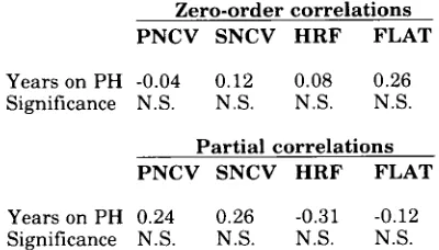
currence of neuropathy. However, a s Dr. Lovelace cor- rectly surmised, almost all of our patients were receiving the same daily dose (300 mg) of phenytoin (PHI, so t ha t the major determinant of total body dose becomes t h e number of years administered. In our original submission of the manuscript to Neurology we included tables which had to be omitted because of space limitations. As can be seen from one of these tables (below), there was no significant effect of PH duration on any of the electro- physiologic tests.
Table. Zero-order and partial correlations of EP measures with PH treatment duration in 40 patients receiving only PH
Zero-order correlations
PNCV SNCV HRF FLAT
Years on PH -0.04 0.12 0.08 0.26 Significance N.S. N.S. N.S. N.S.
Partial correlations
PNCV SNCV HRF FLAT
Years on PH 0.24 0.26 -0.31 -0.12 Significance N.S. N.S. N.S. N.S.
Partial correlations control for the effects of age, fibular length, and the other independent variables ( P H treat- ment duration when computing P H blood level corre- lations and vice versa).
Thomas R . Swift, M.D.
Medical College of Georgia Augusta, GA
Continuous ambulatory simultaneous
EEG/ECG recording
To the Editor: The author’s recommendation regarding the use of continuous ambulatory EEGiECG recording advances th e state of th e art for t h e clinician attempting to differentiate between seizures and syncope.’ However, not all physicians will have access to such a device, and a less complex method may sort out this diagnostic di- lemma. Here we refer to t h e use of the “pale face syn- drome of loss of consciousness”.2
In obtaining a thorough history, including interviews with witnesses, we routinely ask about t he color of t he patient’s face at the time of loss of consciousness. To date, we have studied 20 patients with loss of con- sciousness characterized by the appearance of a pale, ashen, or white face. Once consciousness was lost nine patients had brief seizures, either focal or grand mal.
This “pale face syndrome of loss of consciousness” is a peripheral manifestation of decreased blood flow to t h e head and face. The seizures are due to acutely in- effective blood flow t o the head and brain with sudden
cerebral ischemia. The diminished blood flow to th e head, face, and brain occurs as a result of hypotension or cardiac disease.>
John F . Aita
Midwest Clinic Omaha, N E
References
1. Lai C-W, Ziegler DK. Syncope problem solved by continuous ambulatory simultaneous EEGiECG recording. Neurology
2. Aita JF. Pale face syndrome of loss ofconsciousness. Nebraska (Ny) 1981;31:1152-4.
Medical Journal (in press).
Reply from the Authors: We agree with Dr. Aita t h at the so-called “pale face syndrome of loss of consciousness” is a reliable indicator of syncope due to inadequate blood flow to th e head and brain, and we have found in our recent study of “convulsive syncope” i n blood donors th a t a pale face did occur in most of the cases during th e attack.’ However, vasomotor changes a re also com- monly reported by witnesses of temporal lobe seizures, in which pallor is twice as common as flushing.zJ Fur- thermore, electrical stimulation of limbic structures of the temporal lobe elicits autonomic changes, including cardiovascular responses, such as increase or decrease of blood pressure, tachycardia or bradycardia, and va- somotor changes.4 Therefore, we would express reser- vations in attributing facial pallor during syncope to a cardiovascular event because a temporal lobe seizure cannot be ruled out by this observation alone. Continuous and simultaneous EEG-ECG monitoring may be nec- essary for diagnosis.
Chi-Wan Lai, M.D. Dewey K . Ziegler, M.D.
Department of Neurology University of Kansas Medical Center
Kansas City, K S
References
1. Lin JT, Ziegler DK, Lai C-W, Bayer W. Convulsive syncope in blood donors. Ann Neurol (in Press)
2. Van Buren J M . Some autonomic concomitants of ictal au- tomatism. Brain 1958;81:505-28.
3. Mulder DW, Daly D, Bailey AA. Visceral epilepsy. Arch Intern Med 1954;93:481-93.
4. Gloor P. Physiology of t h e limbic system. (Chap 3 ) In: Penry
J K , Daly DD, eds. Adv Neurol, vol. 11. New York: Raven Press, 1975:27-55.
Correction
“A prospective study of lacunar infarction using com- puterized tomography” by Geoffrey A. Donnan, Brian M. Tress, and Peter F. Bladin, Jan u a ry 1982, p. 54. In figure 7, the pathologic specimen on the right should be turned so th a t the top edge is on th e bottom.
DOI 10.1212/WNL.32.4.458-b
1982;32;458-458-b
Neurology
A prospective study of lacunar infarction using computerized tomography
This information is current as of April 1, 1982
Services
Updated Information &
http://n.neurology.org/content/32/4/458.3.citation.full including high resolution figures, can be found at:
Permissions & Licensing
ssions
http://www.neurology.org/about/about_the_journal#permi (figures,tables) or in its entirety can be found online at: Information about reproducing this article in parts
Reprints
http://n.neurology.org/subscribers/advertise
Information about ordering reprints can be found online:
1526-632X.
American Academy of Neurology. All rights reserved. Print ISSN: 0028-3878. Online ISSN: continuously since 1951, it is now a weekly with 48 issues per year. Copyright © 1982 by the
® is the official journal of the American Academy of Neurology. Published