pISSN 2320-1770 | eISSN 2320-1789
Original Research Article
A prospective study to evaluate the efficacy of 11-13
+6weeks anatomy
scan in detecting fetal structural anomalies compared to
traditional 18-22 weeks scan
Elavarasi Elamaran
1, Saley Daniel
1*, Regi George A. N.
2INTRODUCTION
Congenital anomalies are still one of the leading causes of still birth and neonatal mortality and can be defined as structural or functional anomalies, including metabolic disorders, which are present at the time of birth. Congenital anomalies affect an estimated 1 in 33 infants and result in approximately 3.2 million birth defect related disabilities every year.1
The prevalence of congenital anomalies is grossly under reported in India. According to recent survey, 63 / 1000 live births have serious birth defects in India. The most common severe congenital anomalies are heart defects, neural tube defects and down syndrome.2
The traditional Targeted Imaging For Fetal Anomaly (TIFFA) scan done at 18-22 weeks leads to delay in diagnosis, referral and management.3,4
ABSTRACT
Background: Congenital anomalies are one of the leading causes of infant mortality. Traditional TIFFA scan done at 18-22 weeks leads to delay in diagnosis, referral and management. With high resolution ultrasound and TVS probe, normal and abnormal fetal anatomy could be visualized in early gestation with good accuracy.Objective of present study was to evaluate the efficacy of 11-13+6 weeks anatomy scan in detecting fetal structural anomalies compared to
traditional 18 -22weeks scan and in visualizing the complete normal fetal anatomy
Methods: An Observational study of 300 antenatal patients at Jubilee Mission Medical College for 1 year (Jan-Dec2014) was done. The scan was performed at 11-13+6 weeks by TAS first, if a full fetal anatomy survey not
achieved, TVS added. A mid-trimester fetal anatomy scan was then performed in patients who had not dropped out, miscarried or undergone pregnancy termination at 18-22weeks.
Results: The incidence of anomalies in our study was 3.67% -11 cases; 9 detected at 11-13+6 weeks, 2 were newly
detected at 18-22 weeks. At 11-13+6 weeks anatomy scan, the detection rate of anomalies was 81.8% and complete
fetal anatomy survey was achieved in 92%. Heart and kidneys were not properly visualized in 4% and 12.7%, at 11‐
13+6 weeks compared with 0.7% and 0% at 18‐22 weeks.
Conclusions: The 11-13+6 weeks anatomy scan is an important diagnostic tool which is underutilized and should be
offered to all women as a routine standard of antenatal care. However as fetal anomalies can present at varying gestational age, standard 18-22 weeks anatomy scan cannot be abandoned.
Keywords: Targeted imaging for fetal anomaly, Transabdominal scan, Transvaginal Scan, Ultrasonography
1Department of Obstetrics and Gynecology, 2Department of Radiology, Jubilee Mission Medical College, Thrissur,
Kerala, India
Received: 22 March 2017
Revised: 01 May 2017
Accepted: 02 May 2017
*Correspondence:
Dr.Saley Daniel,
E-mail: [email protected]
Copyright: © the author(s), publisher and licensee Medip Academy. This is an open-access article distributed under the terms of the Creative Commons Attribution Non-Commercial License, which permits unrestricted non-commercial use, distribution, and reproduction in any medium, provided the original work is properly cited.
Since most of the major fetal structures complete its development by 12 weeks, proper visualization of the anatomy is possible at the time of routine NT scan at 11-13+6 weeks.5
Visualizing the complete fetal anatomy during 11-13+6
weeks scan needs high resolution ultrasound machine, well trained and experienced Sonographer.
Early detection of anomalies helps to decide about the option of further management by Chorionic villous sampling and if abnormal, medical termination of pregnancy, or delivery in a setting with specialized surgical and medical care possible.6
METHODS
300 low-risk women with viable singleton pregnancies who attended Obstetrics and Gynaecology department of Jubilee Mission Medical College Thrissur from December 2013 to November 2014 were enrolled in the study, after getting informed consent and approval from Institutional Ethics Committee. Estimated gestational age was calculated based on last menstrual date or previous ultrasound report if periods were irregular or not sure of her last menstrual period. After getting basic information regarding history followed by clinical examination (including height and weight) were subjected to Trans abdominal ultrasound between11-13+6 weeks and if fetal
anatomy survey was not possible to be completed by TAS alone, a TVS was performed after counseling and getting consent from the women. All women enrolled in the study were again subjected to traditional 18-22 weeks anatomy scan using the TAS probe only.7 A single
[image:2.595.312.543.233.682.2]experienced certified radiologist did all the anatomy scans for the study using Voluson 730 pro ultrasound machine with 3.5-5 MHz Trans abdominal transducer and 5-9 MHz Trans vaginal transducer.
Figure 1: Fetus in neutral position in nuchal translucency measurement.
Scanning procedure
11-13+6 weeks scan
• Fetal viability was examined and CRL, BPD, FL
• Nuchal translucency measurement was done as per fetal medicine foundation guidelines.
18-22 weeks follow-up scan
• This was performed by the same examiner and using
[image:2.595.54.282.522.650.2]the same equipment as for the first-trimester scan, following the protocols proposed by the Clinical Standards Committee of the ISUOG.
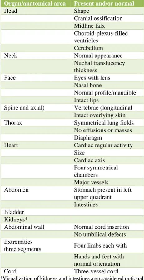
Table 1: Evaluation of fetal anatomy according to the checklist based on ISUOG Practice Guidelines: performance of first-trimester fetal ultrasound
Organ/anatomical area Present and/or normal
Head Shape
Cranial ossification Midline falx
Choroid-plexus-filled ventricles
Cerebellum Neck Normal appearance
Nuchal translucency thickness
Face Eyes with lens Nasal bone
Normal profile/mandible Intact lips
Spine and axial) Vertebrae (longitudinal Intact overlying skin Thorax Symmetrical lung fields
No effusions or masses Diaphragm
Heart Cardiac regular activity Size
Cardiac axis Four symmetrical chambers Major vessels
Abdomen Stomach present in left upper quadrant Intestines Bladder
Kidneys*
Abdominal wall Normal cord insertion No umbilical defects Extremities
three segments Four limbs each with Hands and feet with normal orientation Cord Three-vessel cord
*Visualization of kidneys and intestines are considered optional for completion of scan
Statistical analysis
significance of study parameters between three or more groups of patients.Chi-square/ Fisher Exact test has been used to find the significance of study parameters on categorical scale between two or more groups.8,9
P value ≤0.05 has been considered as statistically significant. The Statistical software namely SAS 9.2, SPSS 15.0, Stata 10.1, MedCalc 9.0.1, Systat 12.0 and R environment ver.2.11.1 were used for the analysis of the data and microsoft word and excel have been used to generate graphs, tables etc.
RESULTS
In the 300 pregnant women studied, most of them belonged to 20-30 years and 13 women 4.3% belonged to advanced maternal age (35 and above), 4 women, 1.3% were obese.
171 women were primi gravida and 129 were multi gravida and all had singleton pregnancies. The mean gestational age for 11-13+6 weeks’ scan was 12 weeks 5days and for 18-22 weeks scant is 20 weeks 1 day.
Table 2: All variables.
Variables Minimum Maximum Mean±SD
Age 17 43 25.57±4.35
BMI 16.24 35.12 21.94±2.92 GA 11w0d 13w6d 12.70±0.71
CRL 45 81 63.84±9.34
[image:3.595.325.532.72.255.2]NT 1 10.9 1.49±0.82
Table 3: NT findings of patients studied.
NT(mm) No. of patients %
<1.5 217 72.3
1.5-2.5 70 23.3
2.5-3.5 9 3.0
>3.5 4 1.3
[image:3.595.313.545.344.513.2]Total 300 100.0
Table 4: Mode of scan used in the study.
Mode of scan
11-14 weeks 18-22weeks No. of
patients %
No. of
patients %
TAS 300 100 292 100
Addl TVS 75 25 0 0
In 25% cases, additional TVS was required in early scan. All the cases in followup scan were done only by TAS.
So, among 11 anomalies detected in our study, early scan detected 9 and follow up scan detected 2 anomalies, which was missed by the early scan. Thus, the detection rate of anomalies by early scan in our study was 81.8%.
Figure 2: Anomalies in present study.
High incidence of anomaly was found in advanced age group women.
Table 5: Early scan-outcome.
Early scan No. of
patients Outcome
Acrania 1 0.3
Anencephaly 1 0.3
Diffuse skin edema 2 0.7
Omphalocele 1 0.3
Single outlet 1 0.3
Thanatophoric dwarfism 1 0.3 Abn NT, absent nasal bone,
single ventricle in heart, absent radius
1 0.3
Renal pelviectasis 1 0.3
Normal scan 291 97.0
Total 300 100.0
Table 6: Follow up scan-outcome.
Follow scan No. of patients (n=292) %
Renal anomaly 1 0.3
Arnold chiari 1 0.3
Renal pelviectasis 1 0.3
Normalscan 289 96.3
Total 292 97.3
Table 7: Advanced age and anomaly.
Age in years Anomalous scan Total scan %
<35 years 7 287 2.4
>35yrs 4 13 13.3
P<0.001**, Significant, Chi-Square test
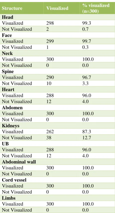
[image:3.595.54.281.477.552.2]Table 8: Individual structures visualization in 11-13+6 weeks scan.
Structure Visualized % visualized (n=300) Head
Visualized 298 99.3 Not Visualized 2 0.7
Face
Visualized 299 99.7 Not Visualized 1 0.3
Neck
Visualized 300 100.0 Not Visualized 0 0.0
Spine
Visualized 290 96.7 Not Visualized 10 3.3
Heart
Visualized 288 96.0 Not Visualized 12 4.0
Abdomen
Visualized 300 100.0 Not Visualized 0 0.0
Kidneys
Visualized 262 87.3 Not Visualized 38 12.7
UB
Visualized 288 96.0 Not Visualized 12 4.0
Abdominal wall
Visualized 300 100.0 Not Visualized 0 0.0
Cord vessel
Visualized 300 100.0 Not Visualized 0 0.0
Limbs
Visualized 300 100.0 Not Visualized 0 0.0
There is a moderately strong correlation suggesting follow up scan was better in achieving complete scan compared to early scan.
Spine, heart, kidneys and urinary bladder were difficult to visualize by early scan and were better visualized with follow up scan
DISCUSSION
In our study, here on, 11-13+6 weeks scan will be referred
as early scan and 18-22 weeks as followup scan. The mean gestational age for early scan was 12 weeks 5 days and for late scan it is 20 weeks 1 day.10,11
In this study, we chose the time period of 111-13+6 weeks
because it is the optimum gestational age to examine fetal anatomy and measure nuchal translucency in the first trimester and also because visualization of fetal anatomy was found to improve with increasing gestational age; from 6% at the 10th gestational week to 75% at the 11th
week, 96% at the 12th week, and 98% at the 13th and 14th
weeks.12,13
We decided, not to exclude chromosomally abnormal cases. Our study was aimed to assess in a single scanning session, the structural anomalies suitable for evaluation at this gestational age, following the checklist mentioned.14
A total of 300 pregnant women completed the study, 292 had both early and follow up anatomy scans while 8 cases with confirmed lethal anomalies by early scan chose to terminate their pregnancies and the anomalies were confirmed after termination.15
The incidence of anomaly in our study was 3.67%. We found anomalies were more in advanced age group women in our study (p<0.001).
Table 9: Difference in ability to visualize individual structures in 11-13+6 weeks scan by mode of
scan and significance.
Structures visualized TAS alone TAS+TVS P value
No. of patients % No. of patients %
Head 248 82.7 298 99.3 <0.001**
Face 271 90.3 299 99.7 <0.001**
Neck 288 96 300 100 <0.001**
Spine 233 77.7 290 96.7 <0.001**
Thorax 292 97.3 300 100 0.004**
Heart 217 72.3 288 96 <0.001**
Abdomen 278 92.7 300 100 <0.001**
Kidneys 188 62.7 262 87.3 <0.001**
Urinary bladder 228 76 288 96 <0.001**
Abdominal wall 281 93.7 300 100 <0.001**
Cord vessels 288 96 300 100 <0.001**
[image:4.595.50.546.582.756.2]Table 10: Mode of scan and ability to achieve complete scan and its significance.
Mode of scan
Complete scan
Incomplete scan
% complete
TAS 225 75 75.0
Addl TVS 276 24 92.0
[image:5.595.49.287.327.398.2]P<0.001**, Significant Chi-Square test
Table 11: GA and ability to achieve complete scan and its significance.
GA Complete scan
Incomplete scan
Total scan
% complete
11-11+6 26 19 45 57.8
12- 12+6 122 2 124 98.4
13-13+6 128 3 131 97.7 P<0.001**, Significant, Chi-Square test
Table 12: Comparing early and follow up scan in achieving complete scan.
USG
Ability to achieve complete scan
% Significance
Early scan 276/300 92.0
0.013* Follow up
scan 290/292 99.32
In present study, the early scan detected 9 anomalies out of 300 cases (3%).
The detected anomalies were 1 case of acrania, 1 case of anencephaly, 2 cases of diffuse skin edema, 1case of omphalocele, 1 case of single cardiac outlet, 1 case of than atophoric dwarfism, 1 case of multiple anomalies with single cardiac ventricle, absent radius and non immunehydrops.
Of the 5 cases who underwent karyotyping, 4 fetuses were found to have abnormal karyotype. Eight patients had termination, upon their request, after the early scan due to the presence of lethal fetal anomalies.16
The detected anomalies in the follow up scan were 3 cases (1%) out of 292 cases.
The 3 cases included the 1 remaining case from the early scan and 2 new cases detected only by the follow up scan (one was a case of Arnold chiari malformation with cranial defect and meningomyelocele and a case of renal anomaly with anhydramnios.17,18
So, among 11 anomalies detected in our study early scan detected 9 and follow up scan detected 2 anomalies, which was missed by early scan. Thus, the detection rate of anomalies by early scan in our study was 81.8%.19,20
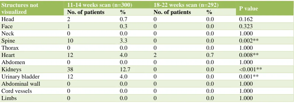
Table 13: Failure to properly visualize different fetal organs by the 11‐14 weeks scan compared to the 18‐22 weeks scan.
Structures not visualized
11-14 weeks scan (n=300) 18-22 weeks scan (n=292)
P value
No. of patients % No. of patients %
Head 2 0.7 0 0.0 0.162
Face 1 0.3 0 0.0 0.323
Neck 0 0.0 0 0.0 1.000
Spine 10 3.3 0 0.0 0.002**
Thorax 0 0.0 0 0.0 1.000
Heart 12 4.0 2 0.7 0.008**
Abdomen 0 0.0 0 0.0 1.000
Kidneys 38 12.7 0 0.0 <0.001**
Urinary bladder 12 4.0 0 0.0 0.001**
Abdominal wall 0 0.0 0 0.0 1.000
Cord vessels 0 0.0 0 0.0 1.000
Limbs 0 0.0 0 0.0 1.000
In present study, early scan was not superior to follow up scan but early anomaly scan helped in detection of many major anomalies when fetuses was less than 50 grams enabling earlier termination. Results of present study are in concurrence with earlier reports which have shown a detection rate of anomalies from 68 to 86.54 % with reference to follow up scan.20
[image:5.595.55.544.464.636.2]Almost, all the previous reported studies available had the same conclusion.22,23
Out of total 300 patients who underwent early anatomy scan, NT was normal (NT <2.28-95 percentile) in 285 patients and raised in 15 patients (NT more than 2.28). The mean NT of fetuses studied was 1.49±0.82. We found abnormal NT in 8 out of 9 cases with anomalies (sensitivity of 88.9).
When we compare early and follow up scan regarding complete visualization of structures, we found follow up scan was better in achieving the complete visualization (p=0.013). Follow up scan was better, especially in visualizing spine, heart, kidney and urinary bladder compared to early can (p <0.05). The heart and kidneys were not properly visualized in 4% and 12.7% of cases, respectively, at the 11‐13+6weeks scan compared with
0.7% and 0% at the 18‐22 weeks scan.24 In early scan,
complete fetal anatomic survey was achieved in 76% of women who had TAS alone and in 92% of women who had additional TVS (p <0.001) and the need for additional TVS was 25% in present study.
We found our ability to achieve complete fetal anatomic survey was improved, when TAS was complemented with TVS, in particular, head, spine, heart, kidney, bladder and limbs were better visualized with TVS (p<0.001). The ability to achieve complete scan increases, as GA age increases, from 58% at 11-11+6
weeks to 98% at 12-14 weeks (p <0.001). Also, we found the need for additional TVS for completion of scan, decreases with increasing GA from 46.2% at 11-11+6
weeks to 18.8% at 12 to 12+6 weeks to 12.5% at 13-13+6
weeks (p<0.001).
However, the study can demonstrate only the added benefit of a TVS assessment in these cases rather than examine the relative sensitivities and specificities of TAS and TVS.25 Various authors have reported, complete fetal
anatomy survey was achieved between 82%-98% depending on the protocol and gestational age at the time of scan.26
The strength of the study includes the completeness of data, usage of good ultrasound machine for better visualization, the checklist for visualization of structures was based on ISUOG protocol for uniformity and standardization, and all the scans for the study were done by a single experienced radiologist with the same machine.27 The limitation of our study was the small
sample size.
CONCLUSION
The benefits of the 11-14 weeks early anatomy scan are unquestionable. It is an important tool in early diagnosis of certain major anomalies, there by suggesting the prognosis of such pregnancies at an early stage. It also
pregnancy. It also helps by detecting cases with increased NT to pick cases at risk for genetic syndromes.
When TVS is used along with TAS, it greatly enhances the ability to achieve complete fetal anatomic survey. Complete visualization of structures increases and need for additional TVS decreases when scan was done between 12-14 weeks compared to 11-12 weeks.
By establishing early anatomy scan as a part of routine antenatal care, helps in detecting major anomalies earlier and offers early counseling, karyotyping and termination of pregnancy if needed. Nevertheless, this early scan cannot rule out certain significant structural anomalies that appear later in pregnancy. Thus, the place of mid trimester fetal anatomy scan in practice remains crucial to complement the late first trimester scan.
Funding: No funding sources Conflict of interest: None declared
Ethical approval: The study was approved by the Institutional Ethics Committee
REFERENCES
1. Sarkar S, Patra C, Dasgupta MK, Nayek K, Karmakar PR. Prevalence of congenital anomalies in neonates and associated risk factors in a tertiary care hospital in eastern India. J Clin Neonatol. 2013;2(3):131-4.
2. Sharma R. Birth defects in India: Hidden truth, need for urgent attention. Indian J Hum Genet. 2013;19:125-9.
3. ACOG. Ultrasonography in pregnancy - Practice Bulletin. Obstet Gynecol. 2009;113:451-61.
4. Cargill Y, Morin L, Bly S, Butt K, Denis N, Gagnon R, et al. Content of a complete routine second trimester obstetrical ultrasound examination and report. J Obstet Gynaecol Can. 2009;31:272-5, 276-80.
5. Windrim R. Fetal alert network: A need for a population based antenatal network. Am J Obstet Gynecol. 2006;195(6):S219.
6. Ebrashy A, EL Kateb A, Momtaz M, EL Sheikhah A, Aboulghar MM, Ibrahim M et al. 13-14-week fetal anatomy scan: a 5-year prospective study. Ultrasound Obstet Gynecol. 2010;35:292-6.
7. Saltvedt S, Almström H, Kublickas M, Valentin L, Grunewald C. Detection of malformations in chromosomally normal fetuses by routine ultrasound at 12 or 18 weeks of gestation: A randomised controlled trial in 39 572 pregnancies. BJOG An Int J Obstet Gynaecol. 2006;113:664-74.
8. Souka AP, Krampl E, Bakalis S, Heath V, Nicolaides KH. Outcome of pregnancy in chromosomally normal fetuses with increased nuchal translucency in the first trimester. Ultrasound Obstet Gynecol. 2001;18:9-17.
guidelines: Performance of first-trimester fetal ultrasound scan. Ultrasound Obstet Gynecol. 2013;41:102-13.
10. Timor-Tritsch IE, Bashiri A, Monteagudo A, Arslan AA. Qualified and trained sonographers in the US can perform early fetal anatomy scans between 11
and 14 weeks. Am J Obstet Gynecol.
2004;191(4):1247-52.
11. Abramowicz JS, Kossoff G, Marsal K, Ter Haar G. Safety Statement, 2000 (reconfirmed 2003). International Society of Ultrasound in Obstetrics and Gynecology (ISUOG). Ultrasound Obstet Gynecol. 2003;21(1):100.
12. Salvesen K, Lees C, Abramowicz J, Brezinka C, Ter Haar G, Maršál K. ISUOG statement on the safe use of Doppler in the 11 to 13+6 week fetal ultrasound
examination. Ultrasound Obstet Gynecol. 2011;37(6):628.
13. Souka AP, Pilalis A, Kavalakis Y, Kosmas Y, Antsaklis P, Antsaklis A. Assessment of fetal anatomy at the 11-14-week ultrasound examination. Ultrasound Obstet Gynecol. 2004;24:730-4.
14. Braithwaite JM, Economides DL. Acceptability by patients of transvaginal sonography in the elective assessment of the first-trimester fetus. Ultrasound Obstet Gynecol. 1997;9:91-3.
15. Hendler I, Blackwell SC, Bujold E, Treadwell MC, Mittal P, Sokol RJ, et al. Suboptimal second-trimester ultrasonographic visualization of the fetal heart in obese women: should we repeat the examination? J Ultrasound Med. 2005;24:1205-9. 16. Hendler I, Blackwell SC, Treadwell MC, Bujold E,
Sokol RJ, Sorokin Y. Does advanced ultrasound equipment improve the adequacy of ultrasound visualization of fetal cardiac structures in the obese gravid woman? Am J Obstet Gynecol. 2004;1616-20.
17. Souka AP, Pilalis A, Kavalakis I, Antsaklis P, Papantoniou N, Mesogitis S et al. Screening for major structural abnormalities at the 11 to 14 week ultrasound scan. Am J Obstet Gynecol. 2006;194:393-6.
18. Yagel S, Achiron R, Ron M, Revel A, Anteby E. Transvaginal ultrasonography at early pregnancy
cannot be used alone for targeted organ ultrasonographic examination in a high-risk population. Am J Obstet Gynecol. 1995;172:971-5. 19. Taipale P, Ämmälä M, Salonen R, Hiilesmaa V.
Learning curve in ultrasonographic screening for selected fetal structural anomalies in early pregnancy. Obstet Gynecol. 2003;101:273-8. 20. Devine PC, Simpson LL. Nuchal translucency and its
relationship to congenital heart disease. Semin Perinatol. 2000;24:343-51.
21. Timor-Tritsch IE, Fuchs KM, Monteagudo A, D’alton ME. Performing a fetal anatomy scan at the time of first-trimester screening. Obstetrics Gynecol. 2009:402-7.
22. Fisher J. First-trimester screening: Dealing with the fall-out. Prenat Diagn. 2011;31:46-9.
23. Salomon LJ, Pizzi C, Gasparrini A, Bernard J-P, Ville Y. Prediction of the date of delivery based on first trimester ultrasound measurements: an independent method from estimated date of conception. J Matern Fetal Neonatal Med. 2010;23(1):1-9.
24. Fong KW, Toi A, Salem S, Hornberger LK, Chitayat D, Keating SJ, et al. Detection of Fetal Structural Abnormalities with US during Early Pregnancy. Radiographics. 2004:157-74.
25. Sepulveda W, Wong AE, Martinez-Ten P, Perez-Pedregosa J. Retronasal triangle: A sonographic landmark for the screening of cleft palate in the first trimester. Ultrasound Obstet Gynecol. 2010;35:7-13. 26. MacOnes GA, Stamilio DM, Odibo A, Cahill A.
Ultrasound screening for fetal spina bifida:
27. Rosati P, Guariglia L, Capelli G. A new mathematical formula for predicting long bone length in early pregnancy. Ultrasound Obstet Gynecol. 2002;19(2):184-9.
Cite this article as: Elamaran E, Daniel S, George RAN.A prospective study to evaluate the efficacy of 11-13+6 weeks anatomy scan in detecting fetal