pISSN 2320-1770 | eISSN 2320-1789
Research Article
Efficacy of fetal echocardiography in prenatal diagnosis
of congenital heart diseases
Mahmoud F. Midan
1, Refaat I. Alsheemy
1, Samia M. Eid
1, Marwa E. Abdel Salam
2*
INTRODUCTION
Congenital heart defects (CHDs) occur in nearly 1% of live births. Being six times more common than chromosomal abnormalities and four times more common than neural tube defects. The incidence of CHD with intrauterine diagnosis ranges from 2.4% to 54%. Some countries have high incidence of CHD because they have instituted an organized policy to perform heart screening by ultrasound systematically.1-4
Prenatal diagnosis of heart defects can lead to changes in medical management that may improve clinical outcomes. For example, decisions to deliver at tertiary care centers with ready access to paediatric medical and surgical specialties are associated with decreased
neonatal morbidity and mortality. Prenatal diagnosis can be particularly important in the case of critical CHDs (those that require surgery or catheterization within the first year of life) that may cause hypoxia and lead to severe organ damage or death in the absence of timely intervention.5,6
Although several risk factors for CHDs have been identified, such as family history, exposure to teratogenic medications, lack of prenatal vitamin and folic acid use, parenteral CHDs and pregestational diabetes, the causes of the majority of CHDs remain unexplained. However the routine use of fetal screening echocardiography in all obstetric population is still controversial.7-9
1
Department of Obstetrics and Gynaecology,Alazhar, New Damietta, Damietta, Egypt
2Department of Obstetrics and Gynaecology, Mansoura International Hospital, Mansoura, Dakahlia, Egypt
Received: 08 March 2016
Accepted: 04 April 2016
*Correspondence:
Dr.Marwa E. Abdel Salam,
E-mail: sweet_marwa24@yahoo.com
Copyright: © the author(s), publisher and licensee Medip Academy. This is an open-access article distributed under the terms of the Creative Commons Attribution Non-Commercial License, which permits unrestricted non-commercial use, distribution, and reproduction in any medium, provided the original work is properly cited.
ABSTRACT
Background: Congenital heart diseases are the commonest fetal congenital defects and until nowadays most of them
are bypassed without prenatal diagnosis to be still considered as unexplained stillbirths or perinatal deaths. In this study, we tried to prove the importance of routine fetal cardiac screening in the ANC visits and also confirming its high accuracy.
Methods: This study was prospective longitudinal one, including doing ISUOG extended fetal cardiac screening for
one hundred foetuses scheduled at certain gestational age visits, whom their half were at risks for CHDs and the other were not, with comparing the results to antenatal and postnatal detailed fetal echocardiography.
Results: The best gestational age for the fetal cardiac screening was at 18-22 weeks gestation. The accuracy of the screening to the antenatal echocardiogram was 96%-100% and to the postnatal one was 96%-98%.
Conclusions: CHDs are still the commonest congenital fetal defects and the antenatal fetal cardiac screening by extended basic views has high accuracy. Making this screening a routine in ANC visits will be of great help in improving the fetal outcome.
Keywords: CHDs, ISUOG, ANC, echocardiogram
Various gestational ages and various methods of antenatal ultrasound assessment of fetal heart are currently available. The four-chamber view is the most basic assessment. This allows a general examination of the heart and the atrioventricular junctions.10 Also there is “basic” and “extended basic” fetal echocardiography which allows adequate evaluation of the outflow tracts. The overall sensitivity of fetal echocardiography ranges from 60% to 100%.11-13
Against this background, a prospective observational study was conducted among two groups of fetuses having of antenatal fetal cardiac screening at multiple gestational ages for each one, comparing each result with antenatal and postnatal detailed echocardiography done by cardiologists to allow calculating its accuracy and value.
METHODS
This was a prospective observational study done through the period from 2013 to 2015 in the department of obstetrics and gynecology of Alazhar University in New Damietta, Egypt. The study population was two groups of fetuses, group A was fetuses with risk factor for having CHDs, and group B was fetuses without any CHDs’ risk factors.
The first visit of all cases was at (18 weeks-22 weeks) gestational age, where all of the following was done: detailed history taking, examination, all basic and risk factor specific investigations, obstetric US scanning ending with cardiac screening. Then cardiologists do fetal echocardiography. The second visit for cardiac screening was at 28 weeks then lastly at 32 weeks. Then postnatal echocardiography for each case was done.
The cardiac screening protocol was adapted from the international society of ultrasound in obstetrics and gynecology (ISOUG) guidelines.14 Fixed experienced examiner has done the screening for all cases using the equipped convex transducer (3-8 MHz) of voluson E8 machine (general electric, medical system, Austria). While the antenatal and postnatal echocardiography was done by fixed cardiologist using echocardiography machine. Then appropriate management plan i.e., mode, time and place of delivery were thoroughly discussed and chosen.
The data obtained was recorded in an investigative report form and computed using SPSS versions 17 under the platform of Microsoft Windows 7. We performed Chi Pearson for categorized variable and Standard Deviation for quantitative variable. We used the significant level of P ‹0.05.
RESULTS
We performed heart examination of 100 fetuses, their half was having CHDs risk factors (group A) and the other half was without (group B). Median age for group
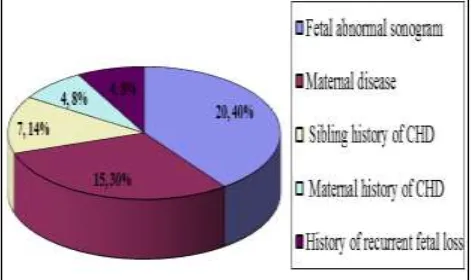
[image:2.595.312.548.143.283.2]A was 25 (±4.9) and group B was 27.3 (± 6.5). Median parity for both groups was approximately 2. Indications for fetal echocardiography in group A were depicted in (Figure 1) with the most common of them was detailed in (Table 1).
Figure 1: The reported risk factors in high risk groups.
Table 1: The fetal abnormal sonogram.
No %
Fetal hydrocephalus (CNS) 4 20% Fetal meningomyelocele (CNS) 2 10% Fetal arrhythmia
(abnormal cardiac examination) 2 10% Abnormal cardiac shadow
(abnormal cardiac examination) 3 15% Non-immune hydrops 3 15% Fetal bilateral renal agenesis 1 5% Fetal omphalocele 1 5%
Fetal ascites 1 5%
Oligohydramnios 1 5%
Polyhydraminos 1 5%
IUGR 1 5%
The cardiac scans were done at three sets of gestational age (18-22 weeks), (28 weeks), and (32 weeks). All views of the heart were obtained in the first one while the other two were variable in obtaining the all views. (Table 3, 4) represents the number of cases in which we have seen the all cardiac views at these two gestational ages. While at (18-22 weeks), all views were seen, may be in one or two sets, but finally all cardiac views were seen.
Table 2: Number of cases completed scans at 28 weeks gestational age.
High risk N=50
Low risk N=50
Chi-square test
X2 p All views seen 35 40
Table 3: Number of cases completed scans at 32 weeks gestational age.
High risk N=50
Low risk N=50
Chi-square test
X2 p
All views seen 10 25
9.9 0.002* Not all views
seen 40 25
We depended on the (18-22 weeks) scans results in comparing it with the cardiologists’ antenatal and postnatal echocardiography to allow calculating the accuracy of the routine cardiac screening done by the obstetrician to the detailed fetal echocardiography. (Table 5, 6) show the results of comparisons done in the two groups.
Table 4: Sensitivity, specificity, PPV and NPV and accuracy of 1st antenatal cardiac scan in relation to
antenatal echocardiography.
High risk Low risk
Sensitivity 80.0 100.0 Specificity 98.0 100.0
PPV 80.0 100.0
NPV 98.0 100.0
Accuracy 96.0 100.0
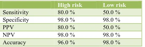
Table 5: Sensitivity, specificity, PPV and NPV and accuracy of 1st antenatal cardiac scan in relation to
postnatal echocardiography.
High risk Low risk
Sensitivity 80.0 % 50.0 % Specificity 98.0 % 98.0 %
PPV 80.0 % 50.0 %
NPV 98.0 % 98.0 %
Accuracy 96.0 % 98.0 %
DISCUSSION
Congenital heart disease (CHD) accounts for approximately 20% of neonatal deaths and 50% of infant deaths and is seen four to five times more frequently in stillbirths and for that fetal echocardiography was introduced in 1980s, and since then; antenatal detection of CHDs remains one of the most challenging issues of prenatal diagnosis many studies have focused on its effectiveness of detecting fetal CHDs, and provided convincing evidence about its reliability and high scan quality.15
We aimed to do Obstetrician study for antenatal fetal cardiac examination as most of the studies that have been done in the field of antenatal detection of fetal congenital heart defects are about the echocardiography which is done by the pediatric cardiologist for the high risk fetuses
as the systemic review and meta- analysis of Yifie et al.16 which studied 82 studies of cardiologists for antenatal fetal echocardiogram, while few number of studies were done for the obstetrician antenatal fetal cardiac screening like the study of Luciane et al.17 in Brazil. Also almost all of these studies were done on risky fetuses only.
Regarding the risk factors of the high risk group whether we discovered them or referred with as an indication for fetal echocardiography; we found the most frequent risk factor was abnormal fetal sonogram (40 %) then maternal disease (30%) then sibling history of CHDs (14%), then maternal history of CHDs (8%) and history of recurrent fetal loss (8%). This is in disagreement with study of Luciane et al which stated that the most common risk factor was maternal metabolic disease (30%), increased nuchal translucency by Clur et al, family history by Emam. While Ozkutlu et al had the same indications arrangements like our study.17-20
In this study, we scheduled the visits starting from 18-22 weeks of gestation, 28 weeks and 32 weeks. Best results of obtaining all of the screening views were in the first visits. Although earlier visits have been tried in different studies, but all are fetal echocardiography for the risky fetuses only, D’Amelio et al studied it at 11-14 weeks, Dolkart and Reimers studied it at 10-15 weeks of gestation and this is for helping in earlier termination of pregnancy as up to 75 % of parents chose that in U.K. where termination of pregnancy is allowed up to 23 weeks but all have concluded that it can never be a practical routine way of antenatal cardiac screening, as it always needs another confirmative study at 18-22 weeks because it was found that minor CHDs were not all correctly early detected.21-23
We calculated the specificity, sensitivity and accuracy of the 18-22 weeks views with antenatal and postnatal echocardiography (which is the most accurate), and to compare these results with other studies’ results was very difficult as most studies are about the detailed echocardiography which is done only for the risk patients, and mostly they were done by cardiologists, but we found nearly similar study of Randall et al who reported wide range from 35% up to 86%,while in this study, the accuracy was in the high risk group 96% antenatally and 96% postnatally, and in the low risk group was 100% antenatally and 98% postnatally differing according to scanning regimen, operators’ skills and equipment. 24
the second trimester among unselected and low risk population as it is not cost effective. 24,25
CONCLUSIONS
Congenital heart diseases are still the commonest congenital anomalies and most of them occur without any risk factor. And because their antenatal diagnosis extremely improves the outcome of their treatment and fetal survival. And because detailed antenatal fetal echocardiography is costly to be routine screening test together with the high accuracy of the obstetrician fetal cardiac sonographic examination; so we recommend it in antenatal care visits.
ACKNOWLEDGEMENTS
First of all, thanks to Allah. Then my deepest appreciation and outmost respect to my professors of obstetrics and gynaecology: Prof. Mahmoud Farouk Midan, prof. Refaat Ibrahim Alsheemy and Prof. Samia Mohamed Eid for their creative stimulation and intellectual teaching.
Funding: No funding sources Conflict of interest: None declared
Ethical approval: The study was approved by the Institutional Ethics Committee
REFERENCES
1. Khoshnood B, Lelong N, Houyel L. Prevalence,
timing of diagnosis and mortality of newborns with congenital heart defects: a population based study. 2012;98(22):1667-73.
2. Yu Z, Xi Y, Ding W, Han S, Cao L, Zhu C, et al.
Congenital heart disease in a Chinese hospital: pre
and postnatal detection, incidence, clinical
characteristics and outcomes. Pediatrics
International. 2011;53(6)1059-65.
3. Ozbarlas N, Erdem S, K¨uc O, ¨ukosmano˘.
Prevalence and distribution of structural heart diseases in high and low risk pregnancies. Anadolu Kardiyol Derg. 2011;11:125-30.
4. Galindo, Herraiz I, Escribano D, Lora D, Melchor
JC, de la Cruz J. Prenatal detection of congenital heart defects: a survey on clinical practice in Spain. Fetal Diagnosis and Therapy. 2011;29(4):287-95.
5. Nelle M, Raio L, Pavlovic M. Prenatal diagnosis and
treatment planning of congenital heart
defects-possibilities and limits. World J Pediatr.
2009;5(1):18-22.
6. Mahle WT, Newburger JW, Matherne GP. Role of
pulse oximetry in examining newborns for congenital heart disease: a scientific statement from the AHA and AAP. Pediatrics. 2009;124(2):823-36.
7. Jenkins KJ, Correa A, Feinstein JA. Noninherited
risk factors and congenital cardiovascular defects: current knowledge: a scientific statement from the American heart association council on cardiovascular
disease in the young: endorsed by the American
academy of pediatrics. Circulation.
2007;115(23):2995-3014.
8. Srinivasan S. Fetal echocardiography. Ind J Pediatr.
2000;67:S20-S5.
9. Comstock CH. What to expect from routine
midtrimester screening for congenital heart disease. Semin Perinatol. 2000;24:331-42.
10. Friedman AH, Kleinman CS, Copel JA. Diagnosis of
cardiac defects: where we’ve been and where we’re going. Prenat Diagn. 2002;22:280-4.
11. Kleinman CS, Hobbins JC, Jaffee CC, Lynch DC,
Talner NS. Echocardiographic studies of the human fetus: prenatal diagnosis of congenital heart disease
and cardiac dysrhythmias. Pediatrics.
1980;65:1059-67.
12. Hsieh CC, Kuo DM, Chiu TH, Hsieh TT. Prenatal
diagnosis of major congenital cardiovascular malformations. Gynecol Obstet Invest. 1996;42:84-7.
13. Cooper MJ, Enderlein Ma, Dyson DC, Roge CL,
Tarnoff H. Fetal echocardiography: retrospective review of clinical experience and anevaluation of indications. Obstet Gynecol. 1995;86:577-82.
14. International society of ultrasound in obstetrics and
gynecology, cardiac screening examination of the fetus: guidelines for performing the basic and extended basic cardiac scan, Ultrasound in Obstetrics and Gynecology. 2006;27:107-13.
15. Volpe P, Tuo G, De Robertis V, Campobasso G,
Marasini M. Fetal interrupted aortic arch: 2D-4D
echocardiography, associations and outcome.
Ultrasound Obstet Gynecol. 2010;35:302-9.
16. Yifie Li, Hua Y, Fang J, Wang C, Qiao L.
Performance of different scan protocols of fetal echocardiography in the diagnosis of fetal congenital heart disease: a systematic review and meta-analysis. PLoS ONE. 2013;8(6):e65484.
17. Luciane A, Edward A, Liliam C, Fernanda S.
Prenatal detection of congenital heart diseases: one-year survey performing a screening protocol in a single reference center in Brazil; cardiology research and practice volume. 2014;175635:5 pages.
18. Clur SAB, Van Brussel PM, Mathijssen IB, Pajkrt E,
Ottenkamp J, Bilardo CM. Audit of 10 years of
referrals forfetal echocardiography. Prenatal
Diagnosis. 2011;31(12):1134-40.
19. Emam SM. High prevalence of complex congenital
cardiac anomalies detected by fetal
echocardiography in a cohort of Saudi women referred for prenatal assessment. Journal of the
Egyptian Society of Parasitology.
2012;42(2):281-90.
20. Ozkutlu S, Akca T, Kafal G, Beksac S. The results of
fetal echocardiography in a tertiary center and comparison of lowand high-risk pregnancies for fetal congenital heart defects. Anadolu Kardiyol Derg. 2010;10:263-9.
21. D’Amelio R, Giorlandino C, Masala L. Fetal
echocardiography using transvaginal and
pregnancy: a comparative study. Prenat Diagn. 1991;11:69-75.
22. Dolkart LA, Reimers FT. Transvaginal fetal
echocardiography in early pregnancy: normative data. Am J Obstet Gynecol. 1992;165:688-91.
23. Sharland GK, Allan LD. Screening for congenital
heart disease prenatally. Results of a 2 1/2-year study in the South East Thames Region. Br J Obstet Gynaecol. 1992;99:220-5.
24. Randall P, Breal S, Hahn S, Khan k, Parsons JM.
Accuracy of fetal echocardiography in the routine detection of congenital heart disease among unselected and low risk populations: a systematic review. BJOG. 2005;122(1):24-30.
25. Berkley EM, Goens MB, Karr S, Rappaport V.
Utility of fetal echocardiography in postnatal management of infants with prenatally diagnosed
congenital heart disease. Prenat Diagn.
2009;29:654-8.
Cite this article as: Midan MF, Alsheemy RI, Eid SM, Salam MEA. Efficacy of fetal