Original Research Article
Diagnosis and management of benign ovarian tumors at the Ignace
Deen Maternity Hospital of Conakry University
Hospital in Guinea
Abdourahamane Diallo
1,2*, Ibrahima Koussy Bah
1,2, Aboubacar Sidiki Magassouba
2,
Mamadou Cellou Diallo
1,2, Telly Sy
1,2INTRODUCTION
Benign ovarian tumors are a common reason for consultation and intervention in gynecology.1,2
Their prevalence is estimated between 14 and 18% in postmenopausal women and around 7% in asymptomatic women during periods of genital activity. Their incidence during pregnancy is between 0.2 and 5%.3 In pre-pubes
and adolescent girls, the prevalence of ovarian cysts greater than 2 cm ranges from 1 to 12%.4 Their onset may
depend on numerous medical treatments, whether they are contraceptives (appearance or prevention of their appearance), endocrinological (stimulator of ovulation, hormonal therapy), or independent of gynecological pathologies (immunosuppressors).3
These cysts may be the origin of symptoms due to their natural evolution or the occurrence of complications.5
The most common complications are torsion, rupture, mechanical compression of neigh-boring organs, intra-cystic hemorrhage.2,6 The risk of recurrence after
ABSTRACT
Background: Benign ovarian tumors are a common reason for consultation and intervention in gynecology. The objective of this was to describe the clinical, ultrasonographic, histological and therapeutic aspects of benign ovarian tumors in the department.
Methods: This is a retrospective and descriptive study of three years and six months from January 1, 2016, to June 30, 2019, which focused on the records of women-operated during this period of benign ovarian tumors.
Results: The incidence of benign ovarian tumors was 12.58%. The circumstances of discovery were dominated by disorders of the menstrual cycle (35.05%) followed by infertility (20.78%), the sensation of a pelvic mass (19.48%), and pelvic pain. (15.58%). The ultrasound report was in favor of a serous cyst in 74% of cases, a mucoid cyst in 14% of cases, a dermoid cyst in 9% and an endometriotic cyst in 3%. Histology revealed a serous cystadenoma in 70.13% of the cases, a mucinous cystadenoma in 16.88% of the cases, a mature poly tissue teratoma in 9.09% of the cases and an endometrial cyst in 3.90 % of the cases. Cystectomy was the most performed surgical procedure (71%).
Conclusions: Benign ovarian tumors are common in our practice. The most common histological forms were serous and mucinous cystadenomas. Conservative treatment has been practiced in the majority of cases.
Keywords: Benign ovarian tumors, Conakry, Diagnosis, Ignace deen, Management
1Department of Gynecology-Obstetrics of the Ignace Deen National Hospital of Conakry University Hospital, Guinea 2Faculty of Health Sciences and Techniques of Gamal Abdel Nasser University of Conakry, Guinea
Received: 13 December 2019
Accepted: 06 January 2020
*Correspondence:
Dr.Abdourahamane Diallo, E-mail: adiallo69gn@gmail.com
Copyright: © the author(s), publisher and licensee Medip Academy. This is an open-access article distributed under the terms of the Creative Commons Attribution Non-Commercial License, which permits unrestricted non-commercial use, distribution, and reproduction in any medium, provided the original work is properly cited.
treatment is between 1 and 19%.5 Ovarian cysts may have
a consequence of female fertility. Still, therapeutic strategies are more infertile than the histological nature of the cyst in young women.7 Benign ovarian tumors may be
associated with an increased long-term risk of mucinous ovarian cancer.8 In contrast, some authors report that
malignant transformation of an ovarian cyst is very rare and that women with ovarian cysts of benign appearance have no increased risk of cancer.5 Although pelvic
ultrasound is the first-line examination in the diagnosis of ovarian tumors, it does not differentiate benign malignancy with 100% reliability. Only histological examination leads to the conclusion of the benignity of an ovarian tumor.9,10
In terms of surgical management, ovarian cystectomy, ovariectomy or adnexectomy are the standard surgical techniques for benign tumors.11
What about the diagnosis and management of benign ovarian tumors in the department?
In the absence of a previous study to answer this question, it was proposed to carry out this study whose objective is to describe the clinical, ultrasonographic, histological, and therapeutic aspects of benign tumors of the ovary in the department.
METHODS
This is a retrospective study and descriptive of three years and six months from 1st January 2016 to 30th June
2019. The study concerned the records of women operated on during this period of benign ovarian tumor.
Inclusion criteria
• All the files of women operated on for benign ovarian tumors containing the report of the histological examination of the surgical specimen were included in the study.
Exclusion criteria
• All non-usable files have been excluded.
We carried out an exhaustive recruitment of all the files meeting the inclusion criteria.
The data was collected by examining the various files selected and reported on the previously established survey form.
The evaluation focused on the socio-demographic characteristics of women (age, parity, marital status, occupation, educational level), the circumstances of discovery, the report of ultrasound and histological examinations, and management.
Statistical analysis
The data were analyzed with the R-software (version 3.4.2). The descriptive analysis consisted of calculating proportions for the qualitative variables, means, standard deviations, and extremes for the quantitative variables.
RESULTS
Frequency of benign ovarian tumors in the department: during this period, 77 files were identified that met the selection criteria among the 612 gynecological interventions performed in the department, i.e., a frequency of 12.58%.
Sociodemographic characteristics
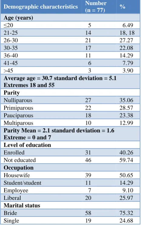
[image:2.595.312.545.386.758.2]The mean age of the patients was 30.7±5.1 years, the extremes of 18 and 55 years, and the range of 21 to 40 years was the most affected (82%). The average parity was 2.1±1.6, with extremes of 0 to 7 years. The majority were out of school (60%), married (75%) and housewives (51%) (Table 1).
Table 1: Distribution of patients according to their socio-characteristics sociodemographic.
Demographic characteristics Number (n = 77) % Age (years)
≤20 5 6.49
21-25 14 18, 18
26-30 21 27.27
30-35 17 22.08
36-40 11 14.29
41-45 6 7.79
>45 3 3.90
Average age = 30.7 standard deviation = 5.1 Extremes 18 and 55
Parity
Nulliparous 27 35.06 Primiparous 22 28.57 Pauciparous 18 23.38 Multiparous 10 12.99
Parity Mean = 2.1 standard deviation = 1.6 Extreme = 0 and 7
Level of education
Enrolled 31 40.26
Not educated 46 59.74
Occupation
Housewife 39 50.65
Student/student 11 14.29
Employee 7 9.10
Liberal 20 25.97
Marital status
Bride 58 75.32
Circumstances of discovery
[image:3.595.53.283.175.278.2]Menstrual disorders ranked first with 35.05%, followed by infertility (20.78%), the sensation of pelvic mass (19.48%), and pelvic pain (15.58%) (Table 2).
Table 2: Distribution of patients according to the circumstances of discovery.
Circumstances of discovery Findings %
Pelvic pain 12 15.58 Pelvic mass 15 19.48 Menstrual cycle disorder 27 35.06 Infertility 16 20.78 Micturition disorders 2 02.60 Fortuitous 5 06.49
Total 77 100.00
Among the 16 patients received for infertility, 6 (37.50%) returned to give birth in the department within two years of the procedure. The tumor was bilateral in 3 patients (3.90%), and 5 patients (6.50%) were received for recurrence of the tumor after the first intervention.
Ultrasound report
[image:3.595.49.284.483.561.2]The ultrasound report was in favour of a serous cyst in 74% of cases, a mucoid cyst in 14% of cases, 9% of the dermoid cyst, and 3% of the endometriotic cyst. The size of the tumor varied from 10 to 19 cm in diameter (Table 3).
Table 3: Distribution of patients according to the ultrasound report.
Report Number %
Serous cyst 57 74.02
Dermoid cyst 7 9.10
Mucoid cyst 11 14.29 Endometriotic cyst 2 2.60
Total 77 100.00
The size of the tumor varied from 10 to 19 cm on ultrasound.
Table 4: Distribution of patients according to histological result.
Result Number %
Serous cystadenoma 54 70.13 Mucinous cystadenoma 13 16.88 Cyst endometrial 3 3.90 Mature poly tissue teratoma 7 9.09
Total 77 100.00
Results of histological examination
The examination revealed a serous cystadenoma of 70.13% of cases, a mucinous cystadenoma in 16.88% of
cases, a mature poly tissue teratoma in 9% of cases, and an endometrial cyst in 3% of cases (Table 4).
A surgical procedure performed
Cystectomy was the most performed surgical procedure (71%) followed by ovariectomy (16%), adnexectomy (10%), and hysterectomy with adnexectomy (3%).
Complications of the cyst
There were five cases of torsion and two cases of compression of the lower urinary tract.
Postoperative complications
There were three cases of anemia (moderate and mild) and two cases of parietal suppuration.
Mean length of stay 3.2±1.4 days with extremes of 2 and 7 days.
DISCUSSION
Benign ovarian tumors are the second leading cause of gynecological intervention in the department after uterine fibromyoma. About one in 10 operated patients is operated on for a benign ovarian tumor. The high prevalence of benign ovarian tumors in the interval from 21 to 40 years reported during this study was also found in the study by Fouedjio et al, at the Yaoundé Central Hospital.2 Younger girls may be affected by this
condition, as in this case of a 14-years-old pre-pubertal girl who underwent a benign mucinous cystadenoma of 9 kg.12 According to Mimoun et al, the prevalence of
ovarian cysts in prepubertal girls is 5.4%.3
The socio-demographic profile of the victims is different from that reported in Yaoundé according to which single people (73%, students (42%) and women in school (100%) are the most frequent.2
These tumors were mainly discovered during a consultation for disorders of the menstrual cycle (amenorrhea, dysmenorrhea, anysomenrrhea), for infertility (primary and secondary) or for pelvic pain. On the other hand, in the study of Fouedjio, the pains constituted the first circumstance of discovery.2 In 6% of
cases, the tumor was accidentally discovered during a routine examination in asymptomatic patients. This observation is similar to that of Laculle-Massin et al.9 At
the end of the clinical examination, torsion of the cyst was suspected in 5 patients, which was confirmed during surgery, thus constituting the first complication as reported by some authors.13 Bottomley et al, found that
cyst torsion accounted for 2.7% of all gynecological emergencies.6 In contrast, Fouedjio et al, reported a
All patients had undergone a preoperative ultrasound. The reports had reported no malignancy criteria for all cases. This testifies to the excellent performance of ultrasound in the diagnosis of ovarian tumors even if it does not replace histology as reported by some authors.9,10 The ultrasound/histology concordance is
around 70 to 80%.10 The findings were most often in
favor of a serous or mucoid cyst. Cases of bilateral tumors were rare, as well as recurrences following a first intervention.
Since the service had no laparoscopy, laparotomy was the only surgical method used, contrary to current recommendations that consider laparoscopy as the standard technique for the surgical management of benign ovarian tumors.14,15 In order to maintain the
fertility of the patients, the cystectomy was performed whenever possible for women during periods of genital activity. This is consistent with the result of Fouedjio who reported 79% of cystectomy.2 Bourdel et al. suggest,
before the menopause, that a conservative treatment is privileged outside an oncological antecedent.14 But, even
a cystectomy can influence the subsequent fertility of the patients by decreasing the ovarian reserve or by creating adhesions that can hinder the uptake of the oocyte by the uterine tubes.4,7,14,16 Some authors propose, for the
conservation of fertility, the freezing of ovarian tissues.13
Dadou et al, demonstrated the feasibility and proper adherence of patients to egg freezing.17
In this series, 4 patients out of 10 who were received for an ovarian tumor on an infertility site returned to give birth in the department within two years. According to some authors, failing to preserve fertility, the conservation of endocrine function is desirable in the premenopausal patients to not increase the risk of cardiovascular diseases in these women.14 For huge
tumors with no obvious cleavage area and after checking the contralateral ovary, ovariectomy was performed. The adnexectomy was reserved for necrotic appendix twists and cases of recurrence with solid adhesions preventing the separation of the tumor from the rest of the appendix. Both cases of hysterectomy involved two women over 50 years of age with ovarian cysts, uterine myomas, and hydrosalpinx. The follow-up was complicated by 3 cases of moderate to mild anemia that did not require blood transfusion and two cases of parietal suppuration involving two obese women, one of whom was diabetic.
No histological examination was done in extemporaneous. All examinations were performed on the surgical specimens deposited in the anatomy pathology laboratory after the procedure. Histological examination revealed more frequently serous cystadenoma or mucinous cystadenoma. This finding is different from that of Dadoun et al, who report 50% of patients who had a dermoid cyst, 25% a mucinous cyst, and 25% of other patients an endometrioma.17 Akhras et
al, reported the case of a 72-year-old woman who underwent a mucinous cystadenoma weighing 27 kg.18
The patients' stay in the ward after the procedure was relatively brief, with an average stay of 3.2 days. Steady patients with reverted transit were released on the second or third postoperative day.
CONCLUSION
Benign ovarian tumors are common in our practice. Women aged 21 to 40, out of school, married, and homemakers were the most affected. It was noted that there was good agreement between the ultrasound report and the histological result. Serous and mucinous cystadenomas were common. Laparotomy was the only surgical method used. Conservative treatment has been practiced in the majority of cases. Operative follow-ups were usually straightforward with a brief stay at the hospital after the procedure. The implementation of laparoscopic surgery and extemporaneous histological examination may improve the management of patients suffering from ovarian tumors in the department.
Funding: No funding sources Conflict of interest: None declared
Ethical approval: The study was approved by the Institutional Ethics Committee
REFERENCES
1. Dimassi K, Bettaieb H, Derbel M, Triki A, Gara MF. Ovarian cysts: sonographic score of malignancy. The Pan African Med J. 2014;18:215.
2. Fouedjio JH, Fouelifack FY, Nnang NA, Mbu RE. Epidemiological, clinical and therapeutic aspects of presumed benign ovarian tumors at Yaounde Central Hospital. Pan African Med J. 2016;25(207):1-6. 3. Mimoun C, Fritel X, Falconer A, Deffieux X,
Dumont A, Huchon C. Epidemiology of suspected benign ovarian tumors. J Gynecol Obstet Biol Reprod. 2013;42:722-9.
4. Lansac J, Marret H. Ovarian tumors. In: Gynecology for the practitioner. 9th ed. Paris: Elsevier Masson;
2018:205-19.
5. Deffieux X, Thubert T, Hu Cchon, Demoulin G, Rivain AL, Faivre E, et al. Complications of suspected benign ovarian tumors. J Gynecol Obstet Biol Reprod. 2013;42:816-32.
6. Bottomley C, Bourne T. Diagnosis and management of ovarian cyst accidents. Best Pract Res Clin Obstet Gynaecol. 2009;23:711-24.
7. Aubard Y, Poirot C. Ovarian tumors presumed benign and fertility. J Gynecol Obstet Biol Reprod. 2013;42:794-801.
8. Guleria S, Jensen A, Toender A, Kjaer SK. Risk of epithelial ovarian cancer among women with benign ovarian tumors: a follow-up study. Cancer Causes Control. 2019:1-7.
10. Marret H, Cayrol M. Ultrasound, and doppler in the diagnosis of presumed benign ovarian tumors. J Gynecol Obstet Biol Reprod. 2013;42:730-43. 11. Borghese B, Marzouk P, Santulli P, de Ziegler D.
Surgical treatments of presumed benign ovarian tumors. J Obstet Reprod Biol. 2013;42:786-93. 12. Biçer S, Erkul Z, Demiryilmaz I, Peker N. A 9-kg
ovarian mucinous cystadenoma in a 14-year-old premenarchal girl. Am J Case Rep. 2014;15:326-9. 13. Sevestre H, Ikoli JF, AL Thakfi W. Pathological
anatomy and cytology of supposedly benign ovarian tumors. J Gynecol Obstet Biol Reprod. 2013;47:715-21.
14. Bourdel N, Canis M. Therapeutic strategies of suspected benign ovarian tumors. J Gynecol Obstet Biol Reprod. 2013;42:802-15.
15. Mbaye M, Cissé ML, Kane Gueye SM, Faye Diemè ME, Diouf AA, Guèye M, et al. First results of gynecological coelioscopy at the University Hospital Of Dakar (CHU): prospective series of 128 cases. J Obstet Gynaecol Can. 2012;34(10):939-46.
16. Legendre G, Catala L, Morinière C, Lacoeuille C, Boussion F, Sentilhes L, et al. Relationship between
ovarian cysts and infertility: what surgery and when? Fertil Steril. 2014;101(3):608-14.
17. Dadoun Y, Azais H, Keller L, d'Orazio E, Collinet P, Decanter C. Systematic proposal of fertility preservation by freezing of eggs in case of benign ovarian tumor. Obstet Gynecol Fertil Senol. 2017;57:1-7.
18. Akhras LN, Akhras LN, Faroog S, AlSebay L. A 27-kg giant ovarian mucinous cystadenoma in a 72-year-old postmenopausal patient: a case report. Am J Case Rep. 2019;20:1601-6.
Cite this article as: Diallo A, Bah IK, Magassouba AS, Diallo MC, Telly S.Diagnosis and management of benign ovarian tumors at the Ignace Deen