Original Research Article
Evaluation of endometrial pathology in women with postmenopausal
bleeding by saline infusion sonography and hysteroscopy
Shikha Bharti*, Rupali Dewan
INTRODUCTION
Postmenopausal bleeding (PMB) is a common problem representing 5% of all gynaecological outpatient
attendance.1 Common causes of postmenopausal bleeding
are atrophic endometritis or vaginitis (30%), exogenous
estrogens (30%), endometrial cancer (15%),
endometrial/cervical polyps (10%), and endometrial
hyperplasia (5%).2 Up to 80% of women with
postmenopausal bleeding and endometrial thickness more than 5 mm have endometrial pathology and most
pathological lesions have focal growth pattern.3
Dilatation and curettage (D and C) and other blind endometrial sampling techniques fail to detect most of the
focally growing lesion therefore further diagnostic work up for focal pathology is required.
Imaging plays vital role in the assessment of patients with PMB after excluding other clinically obvious causes related to the vagina, vulva or uterine cervix.
Transvaginal ultrasound (TVS), saline infusion
sonohysterography (SIS) and hysteroscopy (HS) are the imaging modalities used in the assessment and follow up of the patient with PMB.
Transvaginal ultrasonography (TVS) plays an important role as an initial modality. TVS is a simple, innocuous, relative non-invasive method that can be applied in
ABSTRACT
Background: Postmenopausal bleeding (PMB) is a common problem representing 5% of all gynaecological outpatient attendance. Objective of this study was to determine diagnostic performance of saline infusion sonography and hysteroscopy for evaluation of endometrial lesions in postmenopausal bleeding.
Methods: Being a prospective cross-sectional study, the present study was conducted on 46 postmenopausal women with bleeding, admitted to department of obstetrics and gynecology VMMC and Safdarjang Hospital, New Delhi, India. After TVS, all patients with ET >4 mm underwent SIS and then scheduled for hysteroscopy when there was no active bleeding. Sensitivity, specificity, positive predictive value and negative predictive value were calculated to compare the diagnostic accuracy of SIS and hysteroscopy.
Results: Most commonly found endometrial lesions were polyp (39.13%) and endometrial hyperplasia (28.26%) among our study population consisting of 46 postmenopausal women (mean age 56.72±6.6 years). Overall sensitivity rates were 86.84% for SIS and 97.37% for hysteroscopy, while the overall specificity rates were 50% for both SIS and hysteroscopy. Hysteroscopy had PPV and NPV of 90.24% and 80% respectively whereas PPV and NPV were 89.19% and 44.44% for SIS.
Conclusions: As an easy to perform, safe and well tolerated procedure yielding high diagnostic accuracy, SIS seems to be comparable to hysteroscopy for endometrial evaluation.
Keywords: Endometrial pathology, Hysteroscopy, Postmenopausal bleeding
Department of Obstetrics and Gynecology, VMMC and Safdarjung Hospital, New Delhi, India
Received: 28 February 2020
Revised: 03 April 2020
Accepted: 10 April 2020
*Correspondence:
Dr.Shikha Bharti,
E-mail: shikhaa.bharti@gmail.com
Copyright: © the author(s), publisher and licensee Medip Academy. This is an open-access article distributed under the terms of the Creative Commons Attribution Non-Commercial License, which permits unrestricted non-commercial use, distribution, and reproduction in any medium, provided the original work is properly cited.
almost all the women with postmenopausal bleeding. TVS is a highly sensitive but less specific test and carries a false negative rate of 8% for detection of endometrial carcinoma.4 Saline infusion sonohysterography (SIS) is
more specific, less invasive, well-tolerated and cost-effective imaging modalities which requires no anaesthesia, less time consuming with minimal morbidity. SISH is a diagnostic procedure that enhance endometrial imaging by using saline as a contrast media and also utilizes the uterine distension property of saline,
thereby showing structural abnormalities of
endometrium.5 Few studies have suggested that SIS can
replace hysteroscopy as a method of diagnosing focal lesions in the uterine cavity with high accuracy.6 Taking
into consideration these high diagnostic accuracies, in addition to wide availability and reproducibility, TVS and SISH when used initially in patients with PMB may obviates the need for sophisticated or invasive procedures.
“Gold standard” procedure is hysteroscopy which allows direct visualisation and biopsy of diffuse or focal abnormalities of the endometrium. It is a well- tolerated, accurate, and sensitive outpatient procedure with a high predictive value in the investigation of postmenopausal bleeding has been well documented.7 Need of anaesthesia
and risk of perforation are the shortcomings of hysteroscopy.
Limited literature is available regarding use of SIS as an alternative standard test for evaluation of endometrial pathology in women with postmenopausal bleeding. Studies concerning utility of SIS as a novel screening technique for evaluation of endometrial pathology could assist clinician to decide on the procedure or combination of procedures that would best benefit patients.8 Therefore,
the present study was designed to compare the diagnostic accuracy of SIS and HS in the detection of uterine cavity abnormalities associated with abnormal uterine bleeding among postmenopausal women.
METHODS
This prospective cross-sectional study was carried out in the department of obstetrics and gynecology VMMC and Safdarjang hospital. Postmenopausal patients presenting with abnormal uterine bleeding were subjected to detail clinical history and examination. Patients on hormone replacement therapy, with coagulopathies, with cervical
and vaginal malignancy, ovarian cancers, current pelvic inflammatory diseases were not included in the study. Patients with endometrial thickness >4 mm on TVS taken for SIS followed by hysteroscopy. For SIS, under all aseptic precaution a sterile speculum was introduced in the posterior vaginal wall and anterior lip of the cervix held with vulsellum.
Cervix cleansed with povidone- iodine solution. A Foley’s catheter of 8-12 Fr size was introduced inside uterine cavity and balloon inflated with 4 ml sterile saline solution. Balloon was slightly retraced to close the internal cervical os and transvaginal probe introduced again. Continuous scanning was made in two orthogonal planes while injecting the normal saline. Amount 10-20 ml.
Endometrial thickness was measured by adding anterior and posterior endometrial thickness excluding anechoic fluid part. Findings of SIS were expressed as normal endometrium, focal thickening of endometrium and
homogenously or heterogeneously thickened
endometrium.
Hysteroscopy was performed with a 4 mm Storz rigid hysteroscope and the uterine cavity was distended with a low flow, high pressure (max 100 mmHg) normal saline infusion system. The images were viewed on a high-resolution color monitor. Hysteroscope was guided through the endocervical canal into the uterine cavity under visual control.
Hysteroscopic appearance of endometrium was
categorized as: normal endometrium/suggestive atrophy, heterogenous diffuse thickness, focal abnormality, homogenous diffuse thickness. Endometrial biopsy was performed at the end of hysteroscopy by a directed biopsy.
Histologic diagnosis was given by an investigator who was blinded to ultrasonographic and hysteroscopic finding.
RESULTS
Among 46 postmenopausal study subjects, mean age was 56.72 years with a mean duration of menopause 4.54 years and mean BMI 27.98.
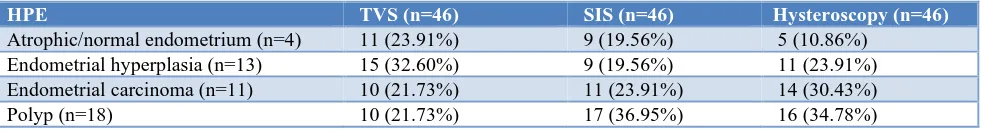
Table 1: TVS, SIS and hysteroscopy with histopathology.
HPE TVS (n=46) SIS (n=46) Hysteroscopy (n=46)
Atrophic/normal endometrium (n=4) 11 (23.91%) 9 (19.56%) 5 (10.86%)
Table 1 shows hysteroscopy to be the technique with most accurate to identify almost all endometrial
pathologies with respect to endometrial biopsy
considered as gold standard.
TVS and SIS both revealed false negative results as they over diagnosed normal endometrium as abnormal but SIS was more accurate than TVS in diagnosing individual endometrial abnormalities like endometrial polyp, endometrial hyperplasia and endometrial carcinoma.
TVS missed out many cases of focal endometrial lesions (endometrial polyp) while hysteroscopy revealed false positive results of endometrial carcinoma.
Table 2 shows that SIS was comparable to hysteroscopy in diagnosing polyps and endometrial carcinoma.
It diagnosed 17 polyps and 11 heterogeneously thickened endometrium while hysteroscopy diagnosed 16 polyps and 14 heterogeneously thickened endometrium.
Table 3 shows that SIS was more accurate in diagnosing polyp as it diagnosed 15/18 (32.6%) cases correctly. 9 cases of endometrial carcinoma were diagnosed correctly, 1 mis-diagnosed as homogenous thickened endometrium and 1 case as focal thickening of endometrium when compared with histopathological findings.
Table 2: Comparison of SIS and hysteroscopy findings in endometrial evaluation.
Hysteroscopy findings
SIS
Atrophic (9) Homogenous thick endometrium (9)
Heterogenous thick endometrium (11)
Focal thickening of endometrium (17)
Atrophic (5) 5 (10.86%) 0 (0%) 0 (0%) 0 (0%)
Homogenous thick
endometrium (11) 2 (4.34%) 7 (15.21%) 1 (2.17%) 1 (2.17%)
Heterogenous thick
endometrium (14) 2 (4.34%) 2 (4.34%) 10 (21.73%) 0 (0%)
Benign focal abnormality (16) 0 (0%) 0 (0%) 0 (0%) 16 (34.78%)
Table 3: Comparison of endometrium evaluation on SIS and HPE.
Endometrial HPE
SIS findings
Normal findings Abnormal findings
Atrophic/normal endometrium (n=9)
Homogenous thick endometrium (n=9)
Heterogenous thick endometrium (n=11)
Focal thickening (n=17)
Atrophy/ normal
endometrium (n=4) 4 (8.69%) 0 (0%) 0 (0%) 0 (0%)
Endometrial carcinoma
(n=11) 0 (0%) 1 (2.17%) 9 (19.56%) 1 (2.17%)
Hyperplasia (n=13) 3 (6.52%) 7 (15.21%) 2 (4.34%) 1 (2.17%)
Polyp (n=18) 2 (4.34%) 1 (2.17%) 0 (0%) 15 (32.60%)
Table 4: Diagnostic accuracy parameters of TVS, SIS, hysteroscopy for evaluation of endometrial pathology with respect to histopathological diagnosis.
TVS SIS Hysteroscopy
Polyp Sensitivity 55.56% 83.33% 88.89%
Specificity 100.00% 93.33% 100%
Endometrial hyperplasia Sensitivity 53.89% 53.85%% 53.85%
Specificity 80.49% 94.29% 89.19%
Endometrial carcinoma Sensitivity 45.45% 81.82% 72.70%
Specificity 87.50% 94.54% 85.37%
Normal endometrium Sensitivity 100.00% 100% 100%
DISCUSSION
Table 4 shows diagnostic accuracy of each imaging method for evaluation of endometrial pathology. Concerning normal endometrium sensitivity was 100% for each method while specificity was 85.71% for TVS, 89.36% for SIS and 97.62% for hysteroscopy. For polyp hysteroscopy was most accurate (88.89% sensitive and 100% specific) followed by SIS (83.33% sensitive and 93.33% specific). For endometrial hyperplasia all imaging modalities were equally sensitive but SIS was more specific (94.29%) followed by hysteroscopy (89.19%). For endometrial carcinoma SIS was found to be most accurate (81.82% sensitive and 94.54% specific).
SIS had been found superior to conventional transvaginal ultrasound examination with regard to endometrial evaluation in postmenopausal bleeding women in this study and various other studies as well. Reason behind is increased sonographic contrast of endometrial cavity after instillation of sterile fluid in uterine cavity. This enables delineation of endometrial abnormalities that could have been missed on transvaginal sonography.
Hysteroscopy was superior to both conventional ultrasound examination and SIS with regard to evaluating endometrial pathology. Hysteroscopy helps in direct visualization of whole uterine cavity and when combined with guided biopsy allows accurate identification of endometrial pathology.
For endometrial evaluation in postmenopausal women with bleeding saline infusion sonography was superior to
transvaginal sonography and almost similar to
hysteroscopy in diagnostic accuracy. It is easy to perform, well tolerated, less invasive and does not require anesthesia also.
Considering this all it can be used as an alternative procedure in place of hysteroscopy, histopathology remaining the gold standard investigation in post-menopausal bleeding women. Present study completely agrees with study by Bingol et al, Chawla et al, Epstein et al and Karsidag et al.9-12 Karlsson et al, found SIS
superior than TVS and hysteroscopy both for diagnosing endometrial abnormalities.3
CONCLUSION
SIS can evaluate endometrial abnormalities more definitely. It may be regarded as primary method in the
detection of endometrial abnormalities among
postmenopausal bleeding women. Diagnostic accuracy of saline infusion sonography is comparable to hysteroscopy
for diagnosing endometrial polyp and normal
endometrium. Considering the excellent correlation between SIS and hysteroscopy, it can be used as an
Funding: No funding sources Conflict of interest: None declared
Ethical approval: The study was approved by the Institutional Ethics Committee
REFERENCES
1. Danero S, Ricci MG, La Rosa R, Massafra C,
Franchi F, Pitino C, et al. Critical review of dilatation and curettage in the diagnosis of malignant pathology of the endometrium. Eur J Gynaecol Oncol. 1986;7:162-5.
2. O’Connell LP, Fries MH, Zeringue E, Brehm W.
Triage of abnormal postmenopausal bleeding: a comparison of endometrial biopsy and transvaginal sonohysterography versus fractional curettage with hysteroscopy. Am J Obstet Gynecol. 1998;178:956-61.
3. Karlsson B, Gransberg S, Wikland M, Ylostalo P,
Torvid K, Marsal K, et al. Transvaginal
ultrasonography of the endometrium in women with postmenopausal bleeding- a nordic multicenter study. Obstet Gynecol Survey. 1996;51(2):100-1.
4. Smith-Bindman R, Kerlikowske K, Feldstein VA,
Subak L, Scheidler J, Segal M, et al. Endovaginal ultrasound to exclude endometrial cancer and other
endometrial abnormalities. JAMA.
19984;280(17):1510-7.
5. De Kroon CD, De Bock GH, Dieben SW, Jansen
FW. Saline contrast hysterosonography in abnormal uterine bleeding: a systematic review and meta-analysis. BJOG. 2003;110:938-47.
6. Weber G, Mere E, Bahlmanw E, Riisch B.
Evaluation of different transvaginal sonographic
diagnostic parameters in women with
postmenopausal bleeding. Ultrasound Gynecol
Obstet. 1998;12(4):265-70.
7. Dubinsky T, Stroehlein K, Abu-Ghazzeh Y, Parvey
H, Maklad N. Prediction of benign and malignant endometrial disease: hysterosonographic pathologic correlation. Radiol. 1999;210(2):393-7.
8. Widrich T, Bradley L, Mitchinson A, Collins R.
Comparison of saline infusion sonography with office hysteroscopy for the evaluation of the
endometrium. AMC J Gynecol Obstet.
1996;174(4):1327-34.
9. Bingol B, Gunenc M, Gedikbasi A, Guner H,
Tasdemir S, Tiras B. Comparison of diagnostic accuracy of saline infusion sonohysterography, transvaginal sonography and hysteroscopy in postmenopausal bleeding. Arc of Gynecol Obstet. 2010;284(1):111-7.
10. Chawla I, Vohra P, Tripathi S, Singh P. To evaluate
investigation of women with postmenopausal bleeding and endometrium >5 mm. Ultrasound Obstet Gynecol. 2001;18(2):157-62.
12. Karageyim Karsidag AY, Buyukbayrak EE, Kars B,
Unal O, Turan MC. Transvaginal sonography, sonohysterography and hysteroscopy for evaluation of focal intrauterine lesions in women with recurrent postmenopausal bleeding after dilatation and curettage. Arch Gynecol Obstet. 2010;281:637-43.
Cite this article as: Bharti S, Dewan R.Evaluation of endometrial pathology in women with postmenopausal bleeding by saline infusion sonography and