DOI: 10.1534/genetics.108.099986
A Screen for Modifiers of Notch Signaling Uncovers Amun, a Protein
With a Critical Role in Sensory Organ Development
Nevine A. Shalaby, Annette L. Parks, Eric J. Morreale, Marisa C. Osswalt,
1Kristen M. Pfau, Eric L. Pierce
2and Marc A. T. Muskavitch
3Biology Department, Boston College, Chestnut Hill, Massachusetts 02467
Manuscript received January 25, 2009 Accepted for publication May 11, 2009
ABSTRACT
Notch signaling is an evolutionarily conserved pathway essential for many cell fate specification events during metazoan development. We conducted a large-scale transposon-based screen in the developing Drosophila eye to identify genes involved in Notch signaling. We screened 10,447 transposon lines from the Exelixis collection for modifiers of cell fate alterations caused by overexpression of the Notch ligand Delta and identified 170 distinct modifier lines that may affect up to 274 genes. These include genes known to function in Notch signaling, as well as a large group of characterized and uncharacterized genes that have not been implicated in Notch pathway function. We further analyze a gene that we have named
Amun and show that it encodes a protein that localizes to the nucleus and contains a putative DNA glycosylase domain. Genetic and molecular analyses ofAmunshow that altered levels ofAmun function interfere with cell fate specification during eye and sensory organ development. Overexpression of Amun decreases expression of the proneural transcription factor Achaete, and sensory organ loss caused by Amun overexpression can be rescued by coexpression of Achaete. Taken together, our data suggest that Amun acts as a transcriptional regulator that can affect cell fate specification by controlling Achaete levels.
T
HE Notch pathway is a highly conserved signaling cascade that controls cell fate specification by inducing or inhibiting the adoption of cell fates in many contexts. Disruption of pathway function gener-ally results in disruption of cell fate specification during development (Artavanis-Tsakonaset al.1999; Portin2002; Schweisguth 2004; Bray 2006; Nichols et al.
2007b). Despite a wealth of literature addressing many aspects of Delta–Notch signaling, numerous findings indicate that additional molecular components and mechanisms that affect the pathway remain to be dis-covered.
Drosophila Notch is a type I transmembrane receptor protein with two known ligands, Delta and Serrate, which are also type I transmembrane proteins (Fiuza
and Arias 2007; D’Souza et al. 2008). Upon ligand
binding, the Notch extracellular domain (NotchECD) enters the Delta-expressing cell via trans-endocytosis (Parks et al. 2000; Itoh et al. 2003; Nichols et al.
2007a). The transmembrane-bound Notch intracellular domain then undergoes two proteolytic cleavages. The first is thought to be mediated by an ADAM metalloprotease, Kuzbanian (Panand Rubin1997; Sotilloset al.1997;
Lieber et al. 2002), while the second is mediated by
the g-secretase complex, which includes Presenilin (Fortini2002; Selkoeand Kopan2003). These
cleav-ages release the Notch intracellular domain into the cytoplasm, which subsequently translocates into the nucleus where it forms a transcriptional co-activation complex that includes Suppressor of Hairless and Mastermind (Mam). These complexes activate expres-sion of Notch target genes [e.g.,Enhancer of split-Complex, orE(spl)-C, genes] in a variety of contexts (Kopan2002;
Schweisguth2004; Bray2006). Regulation of Notch
signaling occurs on several different levels (Schweisguth
2004; Bray2006). For example, many proteins of the
endocytic (e.g., Auxilin, Dynamin, Epsin, Numb, and Rab11) and ubiquitylation (e.g., Neuralized, Mind-bomb1, and Deltex) machinery affect ligand and receptor localization as well as activation of ligand-dependent signaling and downregulation of the receptor (reviewed in Chitnis2006; LeBorgne2006; Nichols et al.2007b; Brou2009). Post-translational modification
of proteins can also play a role in Notch regulation. For example, Notch itself can be modified by O-fucosyl transferase (a glycosyltransferase and chaperone), Fringe (a glycosyltransferase), and proteases such as Furin (Nicholset al.2007b; Stanley2007; Irvine2008). Supporting information is available online athttp://www.genetics.org/
cgi/content/full/genetics.108.099986/DC1.
1Present address:Infinity Pharmaceuticals, 780 Memorial Dr., Cambridge, MA 02139.
2Present address: Department of Medicine, Hematology Division, Brigham and Women’s Hospital, Harvard Medical School, Boston, MA 02115.
3Corresponding author: Biology Department, Boston College, 140 Commonwealth Ave., Chestnut Hill, MA 02467.
E-mail: muskavit@bc.edu
Drosophila wing, eye, and bristle development are excellent contexts in which to study mechanisms of Notch-mediated development and uncover additional components in the pathway. Notch can act in either an inductive or an inhibitory manner to promote the proper adoption of various cell fates during develop-ment of these tissues. In the developing eye, Notch signaling first promotes formation of a single R8 photoreceptor cell within a group of equivalent pro-neural cells and then prevents neighboring cells from adopting the R8 fate, thereby restricting the number of R8 photoreceptors to one per ommatidium (Bakerand
Yu1997; Baker2000; Leeet al.2000). Notch signaling is
subsequently required for differential specification of the R3/R4 photoreceptor fates (DelAlamoand M lod-zik2006) and for induction of the R7 photoreceptor,
cone cell, and primary pigment cell fates (Cooperand
Bray2000; Tomlinsonand Struhl2001; Tsudaet al.
2002; Carthew2007; Nagarajand Banerjee2007).
In the bristle organ, development is initiated with the expression of the basic helix–loop–helix (bHLH) tran-scription factors, Achaete and Scute, within groups of developmentally equivalent proneural cells. Within these proneural groups, sensory organ precursors (SOPs) are singled out, and Delta–Notch signaling by SOPs re-presses Achaete expression in neighboring cells, pre-venting them from adopting the SOP fate. SOPs then divide into two cells, pIIa and pIIb. The daughters of pIIa form the shaft and socket of the mature bristle organ, while the granddaughters of pIIb (daughters of pIIIb) form the neuron and sheath cells. Notch signal-ing occurs between pIIa and pIIb and each successive pair of daughter cells to prevent adoption of inappro-priate fates. For example, Notch signaling prevents the adoption of the pIIb fate in the presumptive pIIa cell and inhibits adoption of the neuronal fate in the presumptive sheath cell (Parks and Muskavitch
1993; Gho et al. 1996, 1999; Guo et al. 1996; Wang et al.1997; Reddyand Rodrigues1999; LeBorgneand
Schweisguth2003). Mis-regulation of Notch signaling
during bristle organ development can lead to a variety of phenotypic defects, depending on the developmental stage during which disruption occurs. For example, reduction of Notch signaling during SOP specification results in the specification of multiple SOPs, leading to the development of multiple bristle organs, whereas reduction of Notch signaling during differentiation of the neuron and sheath cells leads to the specification of two neurons and no sheath cells (Hartenstein and
Posakony1990; Parksand Muskavitch1993).
To identify additional functions that modulate Notch signaling during development, we designed a large-scale genetic screen using the Exelixis transposon collection housed in the Artavanis-Tsakonas laboratory at the Harvard Medical School (Artavanis-Tsakonas
2004). Our screen is based on the ability of genes to modify cell fate changes that result from Delta
over-expression in the retina during development. We confirm the identification of 170 individual transposon insertions that potentially affect a total of 274 genes. We further characterize a phenotypic suppressor of Delta overexpression encoded byCG2446, a gene that we have namedAmun(for an ancient Egyptian god also referred to as ‘‘the hidden one’’). The Amun protein contains a DNA glycosylase domain, and Amun loss-of-function phenotypes include bristle and eye defects. Overexpres-sion of Amun inhibits the formation of microchaeta sense organs by downregulating Achaete protein within microchaeta proneural equivalence groups. Our data therefore suggest that Amun is a nuclear factor that can regulate Achaete levels to control cell fate specification during sensory organ development.
MATERIALS AND METHODS
Fly stocks and culture: The Exelixis transposon collection (Thibaultet al.2004) and all stocks from our laboratory were
maintained using standard procedures. All crosses were performed at 25°, unless otherwise noted.
Drosophila strains used:The following strains were used for the screen: the Exelixis collection housed in the laboratory of Spyros Artavanis-Tsakonas (Artavanis-Tsakonas 2004);
GMR-Gal4(Hayet al.1994; Freeman1996);34B-Gal4(Ingham
and Fietz1995);C96-Gal4(Gustafsonand Boulianne1996),
a gift from Barry Yedvobnick, Emory University (Atlanta); UAS-DeltaWT(on chromosome 2; Jacobsenet al. 1998); and
UAS-DeltaDICD(also known as DeltaD; Huppertet al.1997). The
following strains were used for the study ofCG2446(Amun):
eyeless(ey)-Gal4(Boseet al. 2006) (Bloomington Drosophila
Stock Center); decapentaplegic (dpp)-Gal4/TM6B(Staehling
-Hamptonet al.1994);pannier(pnr)-Gal4/TM3(Heitzleret al.
1996), a gift from Gines Morata, Centro de Biologı´a Molecular Severo Ochoa (Madrid); patched (ptc)-Gal4 (Speicher et al.
1994) (Bloomington Drosophila Stock Center);scabrous(sca) -Gal4/CyO(Mlodziket al.1990), a gift from Andrea Brand,
University of Cambridge (Cambridge, UK); UAS-myr-mRFP/ TM6B(Bloomington Drosophila Stock Center);stripeMD710(sr)
-Gal4/TM6B(Callejaet al.2002; Usuiet al.2004), a gift from
Pat Simpson, University of Cambridge (Cambridge, UK); UAS-AmunRNAiandUAS-Dicer2(Dietzlet al.2007), obtained from
the Vienna Drosophila RNAi Center; and P[lArB]A101.IF3 (neurA101-LacZ)/TM3(Bellenet al.1989), a gift of Hugo Bellen,
Baylor College of Medicine (Houston).
Screen:The screen was performed by assaying the effects of each of 10,447 transposon insertions from the Exelixis stock collection (supporting information, Table S1) (Artavanis
-Tsakonas 2004) on the eye phenotype of GMR-Gal4
UAS-DeltaWT/1(GMR.DeltaWT/1) flies. The collection is composed of four different transposon types: threepiggyBac-based trans-posons (PB, RB, and WH) and one P-element-based trans-poson (XP). Two of the four transposons contain at least one upstream activating sequence (UAS) cassette (Brand and
Perrimon 1993). The WH transposon contains a single
carrying autosomal or viable X-linked insertions to
GMR.DeltaWT-bearing virgin females and scoring the F1
progeny for changes in the rough-eye phenotype. Modifying transposons were categorized as enhancers or suppressors of weak, moderate, or strong intensity. Of 798 primary screen modifiers, 284 were retested withGMR.DeltaWT/1to con-firm modification. A negative secondary test was performed by crossing confirmed modifiers to flies carrying theGMR-Gal4
transgene alone to eliminate modifiers that affect eye de-velopment in the same manner as seen in the primary screen. Positive secondary analyses were performed to prioritize the candidate modifiers using phenotypes that result from expression of a dominant-negative Delta variant created by truncation of the Delta intracellular domain (DeltaDICD; Huppertet al.1997) in the developing wing vein [34B-Gal4
UAS-DeltaDICD/1 (34B.DeltaDICD)] or wing margin [ UAS-DeltaDICD/1; C96-Gal4/1 (C96.DeltaDICD)]. We assessed enhancement and suppression of both of these phenotypes.
Annotation of hits: All high-priority modifiers were anno-tated by aligning the relevant transposon-flanking sequence (Thibault et al. 2004) against the Drosophila melanogaster
genome (FB2007_03 Dmel Release 5.4) using the FlyBase BLAST website (http://flybase.bio.indiana.edu/blast). A 10-kb genome browser snapshot was taken, and transposon-specific criteria were used to assess which genes were potentially affected by the transposon insertion. A gene was considered potentially disrupted if the transposon was inserted within the transcription unit or within 2 kb of the 59-end or 1 kb of the 39-end of the transcription unit. In addition, for UAS-containing transposons (XPandWH), a gene was considered a potential target for UAS-directed expression if it was within 5 kb of the transposon insertion site (and ‘‘downstream’’ of the UAS), unless there was a potential RNA polymerase II transcription stop site between the UAS and the gene in question. Genes identified as possible modifiers were placed into functional categories using previously published data when available and/or FlyBase (FB2007_03 Dmel Release 5.4) Gene Ontology terms.
Constructs and transgenic stocks:A full-length cDNA clone of CG2446 (Amun) in the pOT2 vector (GH02702) was acquired from the Berkeley Drosophila Genome Project. A
SalI–NotI fragment ofAmunwas created by using the forward primer (59-GTCGACATGTCCAACGGCAAGGCG-39) and re-verse primer (59-GTGCGGCCGCGATTCGCTGCGCAG-39) (IDTDNA, http://www.idtdna.com), and the PCR fragment was purified and ligated into blunt-end TOPO (Invitrogen), restricted with SalI and NotI, and ligated into the Gateway vector pENTR1A (Invitrogen). Lambda recombinase (Invi-trogen) was used to insertAmuninto the following Gateway destination vectors (obtained from the Drosophila Genome Resource Center, Indiana University, Bloomington, IN): pTW (containing a UAS promoter) and pTWR [containing a UAS promoter and a monomeric Red Fluorescent Protein (mRFP) C-terminal tag].w1118transgenics containing these constructs
were generated by Genetics Services (Cambridge, MA).
Molecular confirmation of the P{XP}d03329 insertion site:
Genomic DNA was prepared using a standard procedure (Parks et al. 2004). Inverse PCR was performed using a
protocol adapted from the Bloomington Drosophila Stock Center (http://flystocks.bio.indiana.edu/pdfs/Exel_links/ 5__fly_iPCR_XP_pub.pdf). Briefly, genomic DNA was di-gested withSau3AI and ligated to create circular DNA, and primer pairs 31A-31B and 51A-51B were used to amplify sequences flanking the transposon. PCR products were then sequenced, and BLAST was used to match the sequences obtained against theD. melanogastergenome. Two-sided PCR was also performed using a standard procedure (Parkset al.
2004). For amplification of the 59-end of the P{XP}d03329
transposon insertion, 52B (forward) and d03329 reverse flank 59-AGTCGCACACACAGAGACGTAGTT-39 (reverse) primers were used. For amplification of the 39-end of the transposon, d03329 forward flank 59-ATGGGAATGACGAACGACGACGAA-39 (forward) and XP-3SEQ (reverse) primers were used.
Immunohistochemistry: Primary antibodies used (all mouse monoclonals) were the following: anti-Cut at 1:5 [Developmental Studies Hybridoma Bank (DSHB); Iowa University, Iowa City, IA]; anti-Achaete at 1:5 (DSHB and a gift from Teresa Orenic, University of Illinois, Chicago); and 22C10 at 1:100 (a gift from Seymour Benzer, California Institute of Technology, Pasadena, CA). Secondary antibodies used were Alexa488-conjugated goat anti-mouse at 1:500 (Molecular Probes, Eugene, OR) and horseradish-peroxi-dase-conjugated goat anti-mouse at 1:1000 ( Jackson Immu-noresearch, West Grove, PA). Peroxidase activity was visualized using 3, 39-diaminobenzidine. b-Galactosidase activity was detected using Fe(CN)/X-gal staining solution (Hartenstein
and Posakony1990). Imaginal discs were stained as in Parks
et al. (1997), except TPBS (0.3% Triton X-100, 0.02 M Na2HPO4, 0.14 M NaCl, pH 7.6) was used as the buffer for
Achaete staining. Vybrant DyeCycle Green stain (Molecular Probes) was used to assess AmunTRFP (Amun C-terminally-tagged with monomeric Red Fluorescent Protein) subcellular localization. For cone cell analysis,15 retinas were dissected, and the number of cone cells in 20 ommatidia per retina was counted, providing an average of 300 ommatidia for each genotype tested. Student’st-test was used (two-tail distribution and two sample unequal variants; Microsoft Excel 2004) to compare the number of cone cells per ommatidium between genotypes. The SP5 Leica confocal microscope and Adobe Photoshop 7.0 were used to process images.
Gain-of-function clones: For ectopic clonal expression,
hs-Flp; Act5C.y1.Gal4/CyO virgins were crossed to UAS-Amun::RFP/TM6Bmales. F1first to second instar larvae were
incubated at 37° for 1 hr to induce clones. Six hours after puparium formation (APF) nota were dissected and stained as in Parkset al.(1997) except TPBS was used as the buffer.
Phenotypic assessment of transgenic adults: Adult wings and nota were submerged in mineral oil, and pictures were taken on a Zeiss Axioskop and Zeiss Stemi SV11 using the Zeiss AxioCam camera and Zeiss AxioCam Plug-In software, Version 1.0. Adult eye pictures were taken using the Leica MZ16 In-Focus system. All images were assembled using Adobe Photo-shop 7.0.
Protein alignments:Alignments were created in VectorNTI (Suite 7.1, for Mac OS X) using the AlignX program with the following protein sequences:D. melanogasterAmun (CG2446), NP_727552.1;Drosophila simulansGD15978, XP_002106704.1;
Danio rerio Zgc:112496, AAH91543.1; Xenopus tropicalisLOC 100145131, NP_001120112.1;Equus caballusLOC100066977, XP_001497177.1; Bos taurusLOC516108, XP_594248.3; and
Monodelphis domesticaLOC100020910, XP_001373236.1.Homo sapiens N-methylpurine-DNA glycosylase (MPG), NP_00 1041636.1;H. sapiensMutYH, NP_001041636.1;Mycobacterium tuberculosis ultraviolet N-glycosylase/AP lyase (Pdg), NP_ 338328.1; Bacillus subtilis DNA-3-methyladenine glycosylase (AlkA), YP_176647.1; D. melanogaster 8-oxoguanine DNA glycosylase (OGG1), NP_572499.2; H. sapiens 8-oxoguanine DNA glycosylase (OGG1), NP_002533.1; H. sapiens Nth1, NP_002519.1; andD. melanogasterNth1, NP_610078.2.
RESULTS
array and regulates the specification of most, if not all, cell types within each ommatidium (see Introduction). To identify additional components of the Delta–Notch signaling pathway, we designed a genetic modifier screen based on overexpression of Delta in the developing retina under control of the GMR promoter, which drives gene expression posterior to the morphogenetic furrow (Hayet al.1997). TheGMR-Gal4driver initiates
expression in rows 4–6 of the developing eye disc (data not shown) when the photoreceptors R2, R3, R4, R5, and R8 are already present and photoreceptors R1 and R6 are joining the ommatidial precluster (Wolffand
Ready 1993). GMR-driven wild-type Delta expression
(GMR-Gal4 UAS-DeltaWT/1 or GMR.DlWT/1) leads to an adult eye that is glossy and reduced in size with irregular ommatidial spacing (Figure 1B). This pheno-type is suitable for the identification of both suppressor and enhancer mutations and provides a suitable sensitized background for a genetic modifier screen.
To understand how mis-regulation of Notch signaling by Delta overexpression leads to the observed adult eye phenotype, we examined the fates of two cell types induced by Notch signaling (see Introduction) after GMR-Gal4expression is initiated: the R7 photoreceptor and the non-neuronal cone cells. We used the lacZ reporter XA12 to detect R7 photoreceptors (Van
Vactor et al. 1991). In control third larval instar eye
discs, we found a single XA12-positive cell per omma-tidium, whereas in GMR.DlWT/1 eyes, we often detected multiple XA12-positive cells per ommatidium (seeFigure S1). This indicates that overexpression of Delta results in the specification of excess R7 cells, suggesting an increase in Notch inductive signaling in this context. Wild-type ommatidia possess four cone cells, which strongly express Cut protein at 24 hr APF. In
contrast to the increase in R7 cells, anti-Cut immuno-labeling reveals a decrease in the number of cone cells per ommatidium inGMR.DlWT/1retinas, as well as an overall disorganization of the ommatidial array (Figure 1B9). The average number of cone cells per ommatid-ium (n¼140) is 3.34 in theGMR.DlWT/1eye, which is significantly less than the invariant number of four cone cells per ommatidium in wild-type eyes (Figure 1A9; see also Figure 3D). The decrease in cone cell numbers indicates a decrease in Notch inductive signaling in this context. Dominant-negative phenotypes resulting from overexpression of wild-type Delta have been observed previously in the notum (T. R. Parody, T. Zhong and
M. A. T. Muskavitch, unpublished results) and in the
wing pouch (DeCelisand Bray1997; Micchelliet al.
1997; Li and Baker 2004). The presence of Notch
signaling gain-of-function and loss-of-function phenotypes in the GMR.DlWT/1 eye may be due to differential expression of GMR-Gal4 in different cell types or may reflect situations in which different cell types respond to ectopic Delta expression by Notch activation in some instances and by Notch inhibition in others. Taken together, our analyses indicate that overexpression of Delta posterior to the morphogenetic furrow causes specific cell fate changes for at least two cell types. These cell fate changes are consistent with either an increase in Notch signaling (R7 photoreceptor specification) or a decrease in Notch signaling (cone cell specification), depending on the developmental context assessed. The GMR.DlWT/1 genotype therefore provides an ideal genetic background for screening for modifiers of the effects of increased or decreased Notch signaling on the specification of distinct, well-characterized retinal cells.
A screen for suppressors and enhancers of Delta-dependent cell fate changes: We performed a genetic
Figure 1.—Suppressors and enhancers of
DeltaWT overexpression in the Drosophila eye. (A–D) Adult eyes. (A9–D9) Twenty-four-hour APF retinas stained with anti-Cut antibody to de-tect cone cells (green). (A and A9) A wild-type eye possesses an organized array of ommatidia (A); each ommatidium has four cone cells (A9). (B and B9) AGMR.DeltaWT/1eye is small, glossy, and rough, with disorganized ommatidia (B) and an average of 3.34 cone cells/ommatidium (B9). (C and C9) AP{XP}d04859/GMR.DeltaWT
eye. P{XP}d04859 mediates overexpression of
Vha68-2. Adults have a larger, less glossy eye (C) with an average of 3.5 cone cells/ommatidium (C9); P,0.005 compared withGMR.DlWT/1. (D and D9) AP{XP}d10593/GMR.DeltaWTeye.
P{XP}d10593 mediates overexpression of Hr38. Adults have a smaller eye with loss of pigmenta-tion and more disorganized ommatidia (D) with an average of 2.89 cone cells/ommatidium (D9);
modifier screen by assaying the effect of each of 10,447 transposon insertions on theGMR.DlWT/1 eye phe-notype (seeTable S1 andTable S3) from the Exelixis stock collection (Thibault et al. 2004). The
trans-posons in this collection could act either through UAS-mediated overexpression of neighboring genes or by transposon-mediated reduction of gene function due to insertional gene disruption or antisense transcript synthesis (see materials and methods). Modifiers
classified as ‘‘suppressors’’ yielded larger eyes with a more hexagonal appearance to the ommatidial array compared to GMR.DlWT/1 (Figure 1C). Modifiers classified as ‘‘enhancers’’ yielded a smaller, flatter eye with a ‘‘shinier’’ or ‘‘smoother’’ surface and/or loss of pigmentation compared to GMR.DlWT/1 (Figure 1D). We hypothesized that several classes of genes would be isolated as modifiers, including genes that directly regulate the Notch pathway (e.g., genes involved in processing, trafficking, and expression of Notch path-way members), genes that act in signaling pathpath-ways that interact with the Notch pathway (e.g., Ras/EGFR signal-ing), and genes that function in eye development independently of Notch signaling.
A total of 798 transposons modified theGMR.DlWT/1
phenotype in our primary screen (Table S1). Among these primary hits, 66% of the modifiers were UAS-containingXPtransposons (seematerials and methods),
although XPs make up only 21% of the transposons screened (Table 1). The prevalence of XPs recovered suggests that the GMR.DlWT/1 phenotype is more easily modified by expression of neighboring genes via one of the UAS elements present in theXPtransposon than by transposons that lack UAS elements and are more likely to disrupt genes by creating hypomorphic and/or null insertion alleles. Of the 798 modifying transposons, we chose to further analyze 284 trans-posons, most of which were classified as strong or mod-erate modifiers. Among these 284 primary hits, 260 transposons passed retesting against GMR.DlWT/1
(91% confirmation rate), and these were subsequently crossed to GMR-Gal4 to eliminate Delta-independent
modifiers. This resulted in 170 ‘‘confirmed’’ modifiers, including 92 suppressors, 62 enhancers, 9 enhancers/ suppressors (which exhibit aspects of both enhance-ment and suppression), and 7 modifiers that were lethal in combination withGMR.DlWT/1(Table 1,Table S2, andTable S3). If we assume a similar confirmation rate for all 798 primary hits, we would predict our final hit rate as 6.9%. Our hit rate is comparable to the hit rate of 3.94% obtained by Kankelet al. (2007) in a screen of
15,500 lines from the same collection for phenotypic modification of reduced Notch signaling during wing-margin development using aC96-Gal4 UAS-MamDN(a dominant-negative Mam variant) genetic background.
All 170 confirmed modifiers were crossed into two additional genetic backgrounds to assess their ability to modify Delta-dependent phenotypes affecting the wing vein and the wing margin. Expression of DeltaDICD (Huppertet al.1997) under the control of the34B-Gal4
driver (Ingham and Fietz 1995) (34B.DeltaDICD)
causes the development of thickened wing veins (Figure 2B). DeltaDICD expression under control of the C96-Gal4 driver (Gustafsonand Boulianne1996) results
in notches in the wing margin (Figure 2E). Both phenotypes reflect reduced Notch signaling. Among the 170 confirmed modifiers, 20 enhanced and 11 suppressed the 34B.DeltaDICD wing vein phenotype (see Figure 2, C and D, for examples;Table S2), while 33 enhanced and 25 suppressed theC96.DeltaDICD wing-notching phenotype (see Figure 2, F and G, for examples; Table S2). Taken together, our secondary screen data indicate that theGMR.DlWT-based primary screen enriched for phenotypic modifiers of Notch-signaling-associated developmental defects.
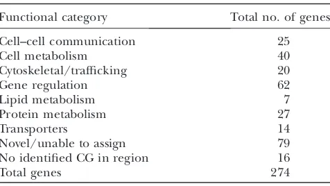
Identification of loci that are dominant modifiers of Delta overexpression: To understand the functional relevance of the recovered modifiers, we sorted genes potentially affected by these transposons into seven functional categories (cell–cell communication, cell metabolism, cytoskeletal/trafficking, gene regulation, lipid metabolism, protein metabolism, and transport) on the basis of previous published studies or on gene
TABLE 1
Screen statistics of 170 confirmed modifying transposons
Transposon type
Total in collection
Total screened
Passed retest and
negative 2°testa Enhancers Suppressors Lethal
Enhancers/ Suppressors
PB 3,548 2,421 7 2 4 1 0
RB 3,288 2,228 3 0 3 0 0
WH 5,637 3,632 8 0 7 0 1
XP 3,715 2,166 152 60 78 6 8
Total 16,188 10,447 170 62 92 7 9
A total of 10,447 transposons were screened, and 284 of the 798 primary hits were retested againstGMR.DeltaWT/1. Of these, 260 passed retesting and were subsequently crossed toGMR-Gal4(negative 2°test). A final set of 170 lines passed the negative secondary test. Enhancers/suppressors yield phenotypic characteristics associated with enhancement and suppression.
a
ontology terms associated with each gene in FlyBase. Our criteria for identifying affected genes varied by transposon type and are described inmaterials and methods. On the basis of these criteria, 152 modifying
transposons had potential effects on 274 genes (195 genes with known or putative function and 79 genes of unknown function), 16 transposons resided in regions with no annotated genes, and two transposons have no sequence data available. We note that a few of the transposons have not been definitively placed at a unique site within the genome, and although most of the transposon insertion sites are accurate (Kankelet al.
2007), molecular characterization of all transposons would be required to positively confirm their assigned insertional positions. The distribution among func-tional categories of gene(s) potentially affected by these modifying transposons is summarized in Table 2.
The 30 transposon insertions associated with cell–cell communication proteins include genes encoding known Notch pathway members (e.g.,numb,kuzbanian; Bray2006), as well as genes that have been recovered
previously from Notch-based screens, such as patched, Ras85D, andpuckered(Rottgenet al.1998; Mulleret al.
2005; Mahoney et al. 2006). We also identified genes
encoding a number of cell–cell communication pro-teins not previously implicated in Notch signaling, such as the phosphataseGilgamesh, the two immunoglobulin superfamily membersFasciclin 2andImpL2, and the two hormone-receptor-like genesHr38 and Hr39. Overex-pression of Hr38 by P{XP}d10593 enhances the GMR.DlWT/1 adult phenotype (Figure 1D), as well as the cone cell phenotype (Figure 1D9) (average of 2.89 cone cells/ommatidium;P,0.005). Overexpression of Hr38 also enhances the wing notching phenotype of C96.DeltaDICD(Figure 2F) and is lethal in combina-tion with34B.DeltaDICD.These data suggest that
over-expression ofHr38reduces net Notch signaling in all of these contexts. Hr38 has been shown to mediate an ecdysteroid signaling pathway that is distinct from that involving the classical ecdysone receptor (EcR) (Baker et al.2003). Ecdysteroids are hormones found only in arthropods that induce signals required for postembry-onic development (Kozlovaand Thummel2000). The
recovery of Hr38 as a Notch antagonist from our screen is not surprising considering that Notch signaling and EcR-mediated ecdysone signaling have recently been shown to act antagonistically in oogenesis during the switch from endoreplication (whole-genome amplifica-tion without cell division) to amplificaamplifica-tion (amplifica-tion of specific genes only, without cell division) (Sun et al. 2008). These results, considered in light of the
Figure 2.—Secondary tests for confirmed
modifiers of the GMR.DeltaWT/1 phenotype. (A) A wild-type adult wing. (B) Adult34B-Gal4 UAS-DeltaDICD/1 (34B.DeltaDICD) wings dis-play thickened wing veins, consistent with reduced Notch signaling. (C) Adult
34B.DeltaDICD/1;P{XP}d11183/1 wings ex-hibit enhancement of the34B.DeltaDICD wing-vein-thickening phenotype. P{XP}d11183
disrupts karst. (D) Adult P{XP}d03329/1; 34B.DeltaDICD/1 wings exhibit suppression of the 34B.DeltaDICD wing vein-thickening phenotype. P{XP}d03329 mediates overexpres-sion of Amun and CG1837. (E) Adult UAS-DeltaDICD/1;C96-Gal4/1(C96.DeltaDICD) wings display notches along the wing margin, typical of reduced Notch signaling. (F) Adult
P{XP}d10593/UAS-DeltaDICD;C96-Gal4/1 wings exhibit enhancement of the C96.DeltaDICD
wing notching phenotype. P{XP}d10593 medi-ates overexpression of Hr38. (G) Adult UAS-DeltaDICD/1;C96-Gal4/P{XP}d07162wings exhibit suppression of theC96.DeltaDICDwing notching phenotype.P{XP}d07162
disruptsCysteine string protein.
TABLE 2
Functional classification of 274 candidate genes potentially disrupted by 170 GMR.DeltaWT-modifying transposons
Functional category Total no. of genes
Cell–cell communication 25
Cell metabolism 40
Cytoskeletal/trafficking 20
Gene regulation 62
Lipid metabolism 7
Protein metabolism 27
Transporters 14
Novel/unable to assign 79
No identified CG in region 16
Total genes 274
emergence of Hr38 and Hr39 from our screen, suggest that multiple hormones and hormone receptors may work along with Notch signaling to specify cell fates in a variety of developmental contexts.
We recovered 22 transposons affecting genes that are likely to play roles in cytoskeletal regulation and/or intracellular trafficking. Ligand and receptor endocy-tosis, NotchECD trans-endocytosis, and intracellular trafficking are core regulatory elements in the activation and regulation of Notch signaling (see Introduction). Genes identified in this class include those encoding myosin-related proteins (jaguar/Myosin VI,myosin heavy chain, and myosin binding subunit) and actin-binding proteins (fimbrin, formin3, and diaphanous), peanut (a septin),short stop(a cytoskeletal protein),Cysteine string protein (a putative chaperone), and karst (b Heavy-spectrin). We also recoveredVha68-2, a subunit of the v-ATPase proton pump complex. v-ATPases are known to function in vesicle trafficking, membrane fusion, and acidification of organelles (Dow 1999). Interestingly, P{XP}d04859, which overexpressesVha68-2, suppresses theGMR.DlWT/1adult rough-eye phenotype (Figure 1C), the cone cell phenotype (average of 3.5/ommatid-ium cone cells; P , 0.005) (Figure 1C9), and the C96.DeltaDICDwing-margin-notching phenotype (data not shown), suggesting that overexpression ofVha68-2 may increase Notch pathway activity in more than one context. Vha68-2 could play important roles during Notch signaling; for example, it may be required to maintain the pH necessary for dissociation of internal-ized Delta/NotchECD complexes and/or for proper intracellular trafficking of Notch and Delta proteins.
Our largest class of modifiers (excluding those affecting unknown genes) fall into the gene expression and transcriptional regulation (‘‘gene regulation’’) category. This group contains 67 independent trans-poson insertions affecting genes including pipsqueak, longitudinals lacking (lola), lilliputian, split ends (spen), tramtrack,TATA binding protein, andSuppressor of Triplole-thal. Many of these genes are known to have roles during eye development (Neufeld et al.1998; Wittweret al.
2001; Voasand Rebay2004). Some, likelolaandspen,
have been implicated previously in Notch signaling and are thought to antagonize the Notch pathway (Ferres
-Marcoet al.2006; Doroquezet al.2007). Others, such
aslilliputian andtramtrack, are known to have interac-tions with other pathways, such as the Ras and ecdyste-roid pathways, that are known to influence Notch signaling (see above) (Sundaram 2005; Hasson and
Paroush 2006). We also identified genes encoding
many largely uncharacterized proteins including CG9650, a zinc-finger-containing putative transcription factor that has been implicated in axon guidance in the embryo (McGovernet al.2003). The modifying
trans-poson,P{XP}d03295, overexpresses the entire protein and enhances theGMR.DlWT/1 eye, resulting in an eye devoid of pigment containing necrotic regions,
indicative of cell death (data not shown). In contrast, analysis of cone cell development reveals that P{XP}d03295 significantly increases the number of cone cells per ommatidium in theGMR.DlWT/1 back-ground (average of 3.87 cone cells/ommatidium; P ,
0.005) (data not shown). This increase of cone cell number suggests that overexpression of CG9650 in-creases net Notch signaling during cone cell specification. The resulting necrotic eye, however, implies thatCG9650 has additional roles during Drosophila eye development and/or in cell viability.
In summary, our screen has identified numerous known components or mediators of Notch signaling, as well as genes not previously associated with the path-way, many of which can be linked to specific signaling functions on the basis of their known roles in processes like intracellular trafficking or transcriptional regula-tion. We anticipate that this modifier collection will provide a rich resource for further investigations of the molecular mechanisms of Notch signaling.
P{XP}d03329 suppresses theGMR.DlWT/1 pheno-type via overexpression of CG2446: We chose to char-acterize one suppressor, P{XP}d03329, in more detail because of the previous implication of an adjacent gene in Notch signaling.P{XP}d03329is located between two open reading frames,CG1837andCG2446, and could potentially mediate overexpression of either gene. WhileCG1837is an uncharacterized gene,CG2446has been associated previously with Notch signaling in the adult eye and bristle organ (Abdelilah-Seyfried et al.
2001; Mulleret al.2005).EP(X)1503, a UAS-containing
transposon insertion (Rorth 1996) upstream of the CG2446 open reading frame, has been identified as a modifier in several Drosophila screens (Abdelilah
-Seyfriedet al.2001; Bourbonet al.2002; Brodyet al.
2002; Mulleret al.2005; Zhuet al.2005). In one screen, EP(X)1503 expressed under control of the sca-Gal4 driver modified Notch and Hairless (a Notch pathway inhibitor; Schweisguthand Lecourtois1998)
loss-of-function phenotypes affecting adult bristles (A bdeli-lah-Seyfriedet al.2001). Overexpression ofCG2446in
UAS-Amun suppresses the GMR.DeltaWT/1 eye phenotype in a manner similar to P{XP}d03329: To verify the genomic insertion position ofP{XP}d03329, we used inverse PCR and two-sided PCR to map the insertion to the X chromosome at coordinate 11,610,026 (FB2006_01 Dmel Release 5.1), which is 200 bp displaced from the insertion site previously annotated by Exelixis (Thibault et al. 2004). To confirm that
overexpression ofAmuncauses the suppression of the GMR.DlWT/1 eye phenotype, we generated a trans-gene placing a full-length Amun open reading frame under the control of a UAS regulatory cassette and generated transgenic lines. Co-expression of UAS-Del-taWTandUAS-AmununderGMR-Gal4control results in suppression of theGMR.DlWT/1 adult rough-eye phe-notype to an extent similar to that seen withP{XP}d03329 (data not shown). We analyzed cone cells ofGMR.DlWT/1; UAS-AmunWT/1 24-hr APF retinas and found signif-icant suppression of cone cell loss (an average of 3.73 cone cells/ommatidium;P , 0.005) (Figure 3, C and D). We also observed mild suppression when P{XP}d03329was crossed toGMR.DlWT(Figure 3, B and D). Since loss of cone cells results from decreased Notch signaling, these results suggest that Amun func-tion potentiates Notch signaling required for cone cell induction in the developing Drosophila eye. This in-terpretation is further corroborated by the observation that overexpression of Amun mediated by EP(X)1503 suppresses inhibition of Notch signaling that results from Hairless overexpression in the eye (Mulleret al.
2005).
Amun is a nuclear protein and has a conserved DNA glycosylase domain: Amunis located cytogenetically at 10D6-10D7 and encodes a 550-amino-acid (aa) protein
with a predicted molecular weight of 58.4 kDa. The entire protein sequence is highly conserved among drosophilid species (seeFigure S2). The amino-termi-nal region of the protein also exhibits significant conservation among a set of putative orthologs from vertebrate and invertebrate species. In contrast, the carboxyl-terminal region of the protein exhibits no sequence similarity in animals beyond the drosophilids (seeFigure S2), and it contains a predicted coiled-coil domain between aa 448 and aa 481 (Figure S2, region shaded in gray). Coiled-coil domains are thought to mediate protein–protein interactions and are found in proteins with diverse biological functions such as vesicle trafficking, cell signaling, and transcriptional regulation (Yu 2002). The amino-terminal region of Amun
in-cludes a putative DNA glycosylase domain (aa 116–151) that is highly conserved across phyla (Figure 4A). DNA glycosylases initiate an evolutionarily conserved base excision repair pathway by excising mismatched or altered bases that result from processes including oxidation, deamination, alkylation, and methylation (Dizdaroglu 2005). In addition, DNA glycosylases
have been shown to act as transcriptional regulators (Choiet al.2002; Cortazaret al.2007).
Protein sequences of DNA glycosylases vary signifi-cantly in size and possess little to no sequence conser-vation within their amino and carboxyl termini. However, many share a DNA glycosylase domain con-sisting of a leucine–proline–glycine–valine/isoleucine– glycine ‘‘hairpin loop’’ sequence flanked by two helices (a-helix–hairpin loop–a-helix, or HhH, domain) fol-lowed by a glycine/proline-rich region and a conserved, catalytically active aspartic acid, which donates an electron during DNA base excision (Figure 4A) (Krokan Figure 3.—Overexpression of Amun
sup-presses the GMR.DeltaWT/1 cone cell pheno-type. (A–C) Twenty-four-hour APF retinas stained with anti-Cut antibody (green) to detect cone cells. (A) AGMR.DeltaWT/1retina exhib-its a disorganized array of ommatidia with an average of 3.34 cone cells/ommatidium. (B) A
et al. 1997; Scharer and Jiricny 2001). Since the
conserved sequence of Amun possesses a DNA glyco-sylase domain, we asked whether the protein localizes to the nucleus, where a DNA glycosylase/transcriptional regulator would be predicted to function. We engi-neered an RFP-tagged version of Amun and expressed it transgenically in Drosophila tissues, including salivary glands (Figure 4B); the notum (see Figure 8B); wing, eye, and leg imaginal discs; and S2 cells (data not shown). We find that overexpressed AmunTRFP local-izes to the nucleus in all tissues examined (we note that an antibody against the endogenous protein will be required to confirm that endogenous Amun is also nuclear), consistent with possible functions as a DNA glycosylase and/or transcriptional regulator.
Amun overexpression and reduction of Amun expression by RNA interference cause bristle defects:
To understand the role of Amun during development, we examined the effects of loss of Amun function, as well as Amun overexpression, in different tissues during development. To examine the loss-of-function pheno-types ofAmun, we usedUAS-AmunRNAitransgenic flies obtained from the Vienna Drosophila RNAi Center (Dietzlet al.2007). To show that this RNA interference
(RNAi) strain can effectively reduce Amun protein expression, we co-expressed AmunTRFP andAmunRNAi in the notum under the control ofpnr-Gal4(see below) and examined discs for the presence of RFP (seeFigure S4for a diagram of thepnr-Gal4expression domain). In the absence ofAmunRNAi, robust RFP accumulation is seen within thepnrexpression domain (Figure 5A). In contrast, when AmunRNAi is co-expressed, a severe reduction in RFP is observed within thepnrexpression domain (Figure 5B). Therefore,AmunRNAieffectively
blocks Amun overexpression. Importantly, phenotypes induced by AmunRNAi can be rescued by overexpres-sion of AmunTRFP (see below), suggesting that Amun-RNAialso has effects on endogenous Amun levels.
Ubiquitous overexpression of Amun or AmunRNAi using theAct5C-Gal4driver results in lethality between the first and second larval instar, suggesting thatAmunis an essential gene necessary for aspects of embryonic and/or early larval development (data not shown). This is consistent with previous reports that Amun is an essential gene expressed during embryonic and larval development (Bourbonet al.2002; Brodyet al.2002).
WhenAmunRNAiis expressed under the control of ey-Gal4 (a transgene that drives expression in the early
Figure 4.—Amun contains a putative DNA
glycosylase domain and localizes to the nu-cleus. (A) Comparison of the Amun HhH DNA glycosylase domain to other known DNA glycosylases. The sequences used were the fol-lowing: MPG, H. sapiens; MutY, H. sapiens; Pdg, M. tuberculosis; AlkA, B. subtilis; OGG1,
D. melanogaster; Ogg1,H. sapiens; Nth1,D. mela-nogaster; Nth1, H. sapiens. There is little se-quence similarity among these proteins; however, they share a conserved DNA-binding motif that consists of twoa-helices (purple cyl-inders denote the approximate locations of these helices) connected by a hairpin loop with the consensus sequence LPG(V/I)G followed by a glycine/proline-rich region (green high-light) and a catalytically active aspartic acid res-idue (D, red highlight). The conserved H/N residue (blue highlight), following the catalytic D residue, differentiates between monofunc-tional (N) and bifuncmonofunc-tional (H) glycosylases. Blue-highlighted L, P, and V residues are part of the consensus HhH domain. Red-highlighted white G residues are com-pletely conserved in these DNA glycosylases. (B and B9) A dpp-Gal4/UAS-AmunTRFP third larval instar salivary gland. AmunTRFP (red) localizes to nuclei (B), as indicated by the DNA dye Vybrant Green (B9).
Figure5.—AmunRNAieffectively reduces AmunTRFP
pro-tein expression. Third larval instar wing/notal imaginal discs. (A) A pnr-Gal4 UAS-AmunTRFP/1 disc (pnr.AmunTRFP). Overexpression of AmunTRFP can be detected via RFP expression (red) in thepnr expression domain. (B) A
antennal-eye imaginal disc; Boseet al.2006), we observe
a reduction in eye size (seeFigure S3) consistent with a role for Amun in eye development (see above). Re-duction of Amun levels under control of the sca-Gal4 or ptc-Gal4 driver results in multiple bristle defects including missing, supernumerary, and misplaced macrochaetae, as well as probable shaft-to-socket trans-formations (Figure 6, E and F, respectively). Reduction of Amun under control of sr-Gal4 (a transgene that drives expression in a subset of microchaeta rows in the medial and lateral notum; Callejaet al.2002) and pnr-Gal4(aP-element insertion in thepnrgene that drives expression in the 10 medial microchaeta stripes; H eit-zler et al. 1996) results in disorganized and smaller
microchaetae (Figure 6, G and H, respectively). Impor-tantly, as shown in Figure 6I, co-overexpression of AmunTRFP with AmunRNAi rescues the loss-of-func-tion phenotypes shown in Figure 6H, suggesting that the AmunRNAiphenotype results from specific reduc-tion of endogenous Amun funcreduc-tion.
Overexpression of AmunTRFP under sca-Gal4 con-trol results in a combination of missing, extra, and misplaced macrochaetae (data not shown), whereas overexpression underptc-Gal4control results in missing macrochaetae (Figure 6B). This range of bristle pheno-types is consistent with previous results obtained by overexpressingAmuninEP(X)1503flies undersca-Gal4 control (see above) (Abdelilah-Seyfriedet al.2001).
The most severe phenotype that we observed is the complete loss of microchaetae when AmunTRFP is expressed usingsr-Gal4(Figure 6C) orpnr-Gal4(Figure 6D). These data indicate that both reduced and in-creased Amun expression levels lead to cell fate speci-fication and/or morphogenetic defects in the Drosophila eye and/or notum.
Figure6.—Amunloss-of-function, gain-of-function, and
res-cue experiments demonstrate a function for Amun during sen-sory organ development. (A) A wild-type adult notum. There are 10 organized rows of notal microchaetae including and be-tween the two rows containing the dorsocentral macrochaetae (aDC and pDC). (B–D)AmunTRFP overexpression pheno-types (assessed following growth at 27°). (E–H) Loss-of-function phenotypes that result from UAS-AmunRNAi
expression (27° unless otherwise noted). (B) A ptc-Gal4/1; UAS-AmunTRFP/1 notum results in loss of aSC (arrow) and pSC macrochaetae. (C) Asr-Gal4/UAS-AmunTRFPnotum ex-hibits severe loss of microchaetae in stripes 2 and 3 (highlighted with square brackets). (D) Apnr-Gal4 UAS-AmunTRFP/1notum exhibits severe loss of microchaetae across the central notum from stripe 1 to stripe 4 (highlighted with square brackets). (E) AUAS-AmunRNAi/1;UAS-Dicer2/ sca-Gal4notum exhibits
Loss of microchaetae is due to loss of bristle sensory organ precursor cells: Loss of bristles following Amun overexpression could result from loss of SOPs or could reflect the loss of socket and shaft cells that would result from the adoption of the pIIb cell fate by the pIIa cell (resulting in multiple neurons and/or sheath cells in each organ). To determine whether Amun plays a role in the latter decision, we used MAb22C10 to stain neuron and shaft cells in the developing notum. In control nota at 31 hr APF, we detected a regular array of microchaeta neurons (Figure 7A). In contrast, there were few or no neurons or shaft cells discernible within thepnrexpression domain following overexpression of Amun (Figure 7B), suggesting that Amun acts upstream of pIIa/pIIb specification during the development of the bristle organ. We then asked whether Amun plays a role in SOP specification. To test this possibility, we assayed SOP specification in the presence and absence
of pnr-driven AmunTRFP (pnr.AmunTRFP) using
neurA101, a
lacZinsertion in theneuralizedgene (Bellen et al.1989), to mark SOP cells. Analysis of 15-hr APF nota stained forb-galactosidase activity reveals a regular array of microchaeta SOPs in control nota (Figure 7C) and the absence of SOPs within thepnrexpression domain inpnr.AmunTRFPnota (Figure 7D). This suggests that
overexpression of Amun in the notum interferes with either the specification of SOPs or the formation of the proneural clusters within which SOPs are specified.
Ectopic Amun expression downregulates the pro-neural transcription factor Achaete: To determine whether failure of SOP specification in regions of elevated Amun expression is due to the absence of SOP proneural groups, we used Achaete immunolabel-ing to detect microchaeta proneural equivalence groups in the developing notum. Achaete is a bHLH transcription factor required for SOP specification within proneural equivalence groups (see Introduc-tion). We compared the pattern of Achaete expression in sr.AmunTRFP nota with that in control nota using
sr.mRFP (a myristylated monomeric RFP; Andersen et al. 2005). Achaete-positive cells are detected in regions of control nota expressing mRFP at 9 hr APF (Figure 8A). In contrast, Achaete protein levels are severely reduced in the sr expression domain (micro-chaeta rows 2 and 3) following overexpression of AmunTRFP (Figure 8B). Similar results are obtained with expression of AmunTRFP under control of pnr-Gal4 (data not shown). These results suggest that the loss of microchaetae shown in Figure 6, C and D, is due to the absence of proneural groups and that over-expression of Amun downregulates levels of Achaete expression.
We investigated whether the downregulation of Achaete by Amun overexpression is cell autonomous or nonautonomous by overexpressing AmunTRFP ran-domly throughout the disc in gain-of-function clones (Glittenberget al.2006) under control of the Act5C-Gal4driver. Adults developing from larvae with gain-of-function clones exhibit numerous small patches of notal microchaeta loss (data not shown). Immunohistochemical analysis of clones in pupae reveals reductions in Achaete levels in cells that express AmunTRFP within micro-chaeta proneural groups. At clone borders, strong Achaete staining is frequently detected in wild-type cells directly adjacent to AmunTRFP-positive cells, which generally lack Achaete expression (Figure 8C). These observations indicate that overexpression of Amun exerts cell-autonomous effects on Achaete protein levels. This effect could reflect direct action of Amun on Achaete levels or an indirect action of Amun via other factors that regulate Achaete levels in the notum (seediscussion).
We then asked whether or not loss of Achaete is responsible for the bristle-loss phenotype observed when Amun is overexpressed. Indeed, we find that
Figure 7.—Amun-induced loss of notal microchaetae is
due to the loss of bristle organs and sensory organ precursors in the developing notum. (A and B) Nota from 31-hr APF pu-pae (27°) stained with MAb22C10 to detect neurons and shaft cells. (A) A wild-type notum has organized rows of microchae-ta neurons and shaft cells. (B) Apnr-Gal4 UAS-AmunTRFP/1 notum lacks staining for neurons within thepnrexpression domain (outlined by dashes), indicating that the absence of external shafts is not due to the transformation of shaft/ socket cells into neurons. (C and D) Nota from 15-hr APF pu-pae (27°) stained forb-galactosidase activity. TheneurA101lacZ
insertion in theneuralizedgene is used to mark SOPs. (C) A
neurA101notum exhibits wild-type rows of microchaete SOPs.
overexpression of Achaete and AmunTRFP within the pnr domain results in a significant rescue of the pnr.AmunTRFP microchaeta-loss phenotype (Figure
6J). This result demonstrates that the primary cause of microchaeta loss induced by Amun overexpression is the loss of Achaete expression.
In contrast to Amun overexpression, reduction of Amun function usingsr-Gal4.AmunRNAicauses smaller and disorganized microchaetae, but does not cause any apparent changes in microchaeta number, as might be expected if loss of Amun function affected Achaete levels. Indeed, we could not detect any overt changes in notal Achaete immunolabeling following knockdown of Amun using sr-Gal4 (data not shown). There are several possible explanations for this observation. First, it is possible that Amun does affect Achaete expression levels, but the change in level is not easily recognized with immunolabeling. Second, it is possible that ex-pression of endogenous Achaete levels can be main-tained by very low levels of Amun function and that knockdown mediated byAmunRNAiis not sufficient to cause a discernible effect on Achaete expression.
These observations suggest that while Amun can clearly affect Achaete expression and SOP specification, Amun probably also plays roles in other aspects of bristle development. Because Achaete is not known to be involved in bristle development beyond SOP forma-tion (Bertrandet al.2002), we suggest that Amun can
act through other factors to control sensory organ development following SOP specification.
DISCUSSION
Drosophila continues to play a leading role in the discovery of genes and mechanisms implicated in de-velopmental processes mediated by, or associated with, the Notch signaling pathway. We present the results of a transposon screen for the effects of loss-of-function and gain-of-function mutations in a genetic background sensitized for Delta-mediated cell fate changes. In addition, we characterize Amun, a nuclear protein iden-tified as a suppressor in our screen. We show that Amun suppresses a dominant-negative effect of Delta over-expression on cone cell induction in the eye, suggesting
Figure8.—Amun overexpression
downreg-ulates Achaete levels in a cell-autonomous manner. (A and B) Nota from 9-hr APF pupae (27°) stained with anti-Achaete antibody (green). (A, A9, and A$) A sr-Gal/UAS-myr-mRFP
notum. RFP (red) reflects expression of mRFP within thesrdomain. Achaete-positive cells (arrows) mark the microchaeta proneu-ral groups during this developmental stage. (B, B9, and B$) A sr-Gal4/UAS-AmunTRFP notum shows a severe reduction or absence of Achaete expression (arrowheads) within thesrexpression domain where AmunTRFP is overexpressed. (C, C9, and C$) A 6- to
7-hr APF notum stained with anti-Achaete antibody (green in C and C$). AmunTRFP
that Amun can positively regulate Notch signaling in this context. Alternatively, Amun may function in a parallel or intersecting pathway to affect cone cell development. We also provide evidence that Amun can function early during the cellular patterning under-lying mechanosensory bristle development by down-regulating the expression of the proneural transcription factor Achaete (see below). The identification and initial characterization of Amun function reflect the potential of the ensemble of 170 transposon insertions identified in our screen for discovery of additional factors that affect Notch signaling mediated development.
A screen for Notch-mediated development: The Exelixis collection covers 50% of Drosophila genes (Thibault et al.2004) and contains many new alleles
for genes that may prove to be involved in the Delta– Notch signaling pathway (e.g., Kankelet al.2007 and this
work) or other developmental pathways. The collection has also been screened by Kankel et al. (2007) in a
search for modifiers of aNotchloss-of-function signaling phenotype in the wing margin using C96-driven MamDN. Among the 170 modifiers that we identified, 29 lines were also recovered by Kankelet al.(2007) (see
Table S2) and 141 lines were recovered only in our screen. Among the putative genes recovered in both screens are several known Notch pathway members and genes that have been previously recovered from Notch-based screens (e.g., numb, wingless, puckered, and Ras85D). In addition, several genes that had not been implicated previously in Notch signaling were identified in both screens, supporting roles for their encoded products during Notch-mediated development. These genes include peanut (a septin), Oatp30B (an ion channel), Indy(a transporter), andHr38 (a hormone receptor). Of potentially equal interest are the 11 transposon lines that modified phenotypes in both of the secondary tests in this work (Table S2). Genes potentially disrupted by these transposons includekarst (bHeavy-spectrin),bifocal(a cytoskeletal regulator), dia-phanous(an actin-binding protein), and caudal (a tran-scriptional regulator). Further characterization of these genes, as well as other genes recovered in our screen, will help provide a deeper understanding of the mechanisms that govern the Notch signaling pathway.
The function of Amun during Drosophila sensory organ development: A number of our results suggest that Amun is required for cell fate determination during Notch-mediated bristle organ development. Reduction of Amun function and Amun protein overexpression in the developing notum, using several Gal4 drivers in-cluding pnr, ptc, sca, and sr, generate defects during microchaeta and macrochaeta development. Substan-tial loss of microchaetae is observed in the nota of adults that express Amun under pnr-Gal4 or sr-Gal4 control during development. Immunohistochemical analysis of developing nota and the Achaete expression rescue experiments demonstrates that this loss of
microchae-tae is due to loss of the bHLH transcription factor Achaete. The expression patterns of the proneural proteins Achaete and Scute are best characterized for the dorsocentral macrochaetae, for whichcis-regulatory elements control the expression of these genes in specific patterns to establish proneural clusters (re-viewed in Modolelland Campuzano1998 and Calleja et al. 2002). These enhancer elements are thought to be activated directly by members of several signaling pathways, including Decapentaplegic and Wingless, as well as by other factors including Pannier (Pnr), Daughterless (Da), Chip, and members of the Iroquois complex (Araucan and Caupolican) (reviewed in Bertrandet al. 2002). The expression ofachaete/scute
is antagonized by several factors, including U-shaped and dCtBP, both of which bind Pnr to form a transcrip-tional corepressor complex (Cubaddaet al.1997; Sato
and Saigo2000; Biryukovaand Heitzler2008; Stern et al. 2009); Extramacrochaetae (Emc), which forms a heterodimer with Da to inactivate it (Elliset al.1990; vanDorenet al.1991); and the E(spl)-C proteins, which
are downstream targets of Notch signaling (deCelis et al.1996). In microchaeta proneural groups, Achaete is also known to be repressed by Hairy (Ohsako et al.
1994; van Doren et al. 1994), as well as by Notch
signaling (Parkset al.1997). We demonstrate here that
the effect of Amun overexpression on Achaete levels is cell autonomous, suggesting that the action of Amun on achaete expression could be direct. However, while it is tempting to speculate that Amun regulates Achaete levels by directly binding tocis-regulatory elements that affect achaete expression, we cannot rule out the possibilities that Amun functions by repressing an activator of achaete (e.g., Da or Chip), by activating a repressor of achaete (e.g., Emc, Hairy, or the Notch pathway), or by destabilizingachaetemRNA or protein.
Reductions in Amun function by RNA interference result in small and disorganized microchaetae. In contrast to the Amun overexpression phenotype, the small microchaeta phenotype is not easily attributable to changes in Achaete expression, given that Achaete has no known roles in bristle development subsequent to SOP specification. It has been shown that bristle shaft size can be correlated with several processes. First, both the shaft and socket cells undergo endoreplication to form polyploid nuclei that are required to form the elongated shaft structure. The degree of endoreplica-tion has been correlated with shaft size (Edgar and
Orr-Weaver 2001; Weng et al. 2003). Second, shaft
length can be affected by mutations in genes that affect actin bundle formation necessary for proper elongation of the shaft (Tilneyet al.2000). Third, there is a period
of rapid protein synthesis during sensory bristle develop-ment that enables the shaft and socket cells to generate the high levels of protein required for the development of the socket and shaft structures (Lambertsson1998;
include small bristles [which exports mRNA from the nucleus into the cytoplasm (Korey et al. 2001)] and
the Minute loci [genes encoding ribosomal proteins (Lambertsson 1998; Saeboe-Larssen et al. 1998;
Marygoldet al.2007)], which can affect bristle shaft
length. Preliminary data suggest that Amun is unlikely to affect endoreplication. We measured nuclei of micro-chaetae that develop in regions of the notum expressing sr-drivenAmunRNAiand found no consistent effects on nuclear size as compared to the nuclei of cells of micro-chaetae in regions devoid ofAmunRNAi(data not shown). We therefore favor the notion that Amun may be required for transcriptional regulation of specific genes involved in growth and elongation of the shaft or for the elevated levels of mRNA and protein synthesis required for shaft development.
Amun as a DNA glycosylase and transcriptional regulator: Our finding that Amun can affect Achaete expression levels, together with our identification of Amun as a nuclear protein with a putative DNA glyco-sylase domain, are consistent with the hypothesis that Amun functions as a transcriptional regulator. While DNA glycosylases are best known for repair of damaged and mismatched bases, recent work indicates that they also play roles in transcriptional regulation. The mam-malian DNA glycosylase thymine DNA glycosylase (TDG) acts as a transcriptional co-activator, when bound to CREB-binding protein (CBP) and p300 (Tiniet al.2002),
to enhance CBP-activated transcription in cell culture (Cortazaret al.2007). It also acts as a transcriptional
corepressor when bound to thyroid transcription factor-1 (TTF1) to repress TTF1-activated transcription in cell culture (Cortazaret al.2007; Kovtunand McMurray
2007). The Arabidopsis DNA glycosylase DEMETER is required to activate expression of the maternalMEDEA allele, an imprinted maternal gene essential for viability (Choiet al.2002). In light of these studies, the nuclear
localization of Amun is suggestive of a function for Amun as a transcriptional regulator.
In summary, we demonstrate that Amun is a nuclear protein essential for organismal viability and proper cell fate specification during metamorphosis of Drosophila tissues, including the eye and mechanosensory organs. We suggest that Amun affects at least two distinct processes during bristle organ development because of the distinct loss-of-function and gain-of-function bristle phenotypes associated withAmun. One pathway is critical for regulation of Achaete protein levels, and the other pathway affects sensory organ bristle shaft size. Because the sequence of Amun contains a putative DNA glycosylase domain, we reason that Amun may act as a transcriptional regulator, as previously demonstrated for other DNA glycosylases (e.g., TDG and DEMETER). Further characterization of Amun is necessary to iden-tify distinct transcriptional targets and pathways on which it may act and to decipher its potential function as a DNA glycosylase during Drosophila development.
We thank S. Artavanis-Tsakonas for allowing us to screen the Exelixis collection and M. Kankel, D. Dimlich, and G. Doughty for help collecting males. We thank K. Klueg for generating theGMR-Gal4 UAS-DeltaWT stock. We thank T. Orenic, P. Simpson, H. Bellen, B. Yedvobnick, A. Brand, G. Morata, the Developmental Studies Hybrid-oma Bank, the Bloomington Drosophila Stock Center, the Drosophila Genome Resource Center, and the Vienna Drosophila RNAi Center for stocks and reagents. We are grateful to P. R. Hiesinger for discussion and critical reading of the manuscript and technical assistance. We also thank J. Rosenberg for technical assistance. This work was funded by National Institutes of Health grant GM33291 and the DeLuca Professorship awarded to M.A.T.M.
LITERATURE CITED
Abdelilah-Seyfried, S., Y. M. Chan, C. Zeng, N. J. Justice, S. Younger -Shepherdet al., 2001 A gain-of-function screen for genes that affect the development of the Drosophila adult external sensory organ. Genetics157:455–456.
Andersen, R., Y. Li, M. Resseguie and J. E. Brenman, 2005 Calcium/calmodulin-dependent protein kinase II alters structural plasticity and cytoskeletal dynamics in Drosophila. J. Neurosci.25:8878–8888.
Artavanis-Tsakonas, S., 2004 Accessing the Exelixis collection. Nat. Genet.36:207.
Artavanis-Tsakonas, S., M. D. Randand R. J. Lake, 1999 Notch signalling: cell fate control and signal integration in develop-ment. Science284:770–775.
Baker, K. D., L. M. Shewchuk, T. Kozlova, M. Makishima, A. Hassellet al., 2003 The Drosophila orphan nuclear receptor DHR38 mediates an atypical ecdysteroid signaling pathway. Cell
113:731–742.
Baker, N. E., 2000 Notch signaling in the nervous system: pieces still missing from the puzzle. BioEssays22:264–273.
Baker, N. E., and S. Y. Yu, 1997 Proneural function of neurogenic genes in the developing Drosophila eye. Curr. Biol.7:122–132. Bellen, H. J., C. J. O’Kane, C. Wilson, U. Grossniklaus, R. K. Pearsonet al., 1989 P-element mediated enhancer detection: a versatile method to study development in Drosophila.Genes Dev.3:1288–1300.
Bertrand, N., D. S. Castro and F. Guillemot, 2002 Proneural genes and the specification of neural cell types. Nat. Rev. Neuro-sci.3:517–530.
Biryukova, I., and P. Heitzler, 2008 Drosophila C-terminal bind-ing protein dCtBP is required for sensory organ prepattern and sharpens proneural transcriptional activity of the GATA factor Pnr. Dev. Biol.323:64–75.
Bose, A., B. Kahali, S. Zhang, J. M. Lin, R. Allada et al., 2006 Drosophila CK2 regulates lateral-inhibition during eye and bristle development. Mech. Dev.123:649–664.
Bourbon, H. M., G. Gonzy-Treboul, F. Peronnet, M. F. Alin, C. Ardourelet al., 2002 A P-insertion screen identifying novel X-linked essential genes in Drosophila. Mech. Dev.110:71–83. Brand, A. H., and N. Perrimon, 1993 Targeted gene expression as a
means of altering cell fates and generating dominant pheno-types. Development118:401–415.
Bray, S. J., 2006 Notch signalling: a simple pathway becomes com-plex. Nat. Rev. Mol. Cell Biol.7:678–689.
Brody, T., C. Stivers, J. Nagle and W. F. Odenwald, 2002 Identification of novel Drosophila neural precursor genes using a differential embryonic head cDNA screen. Mech. Dev.
113:41–59.
Brou, C., 2009 Intracellular trafficking of Notch receptors and ligands. Exp. Cell Res.315:1549–1555.
Calleja, M., O. Renaud, K. Usui, D. Pistillo, G. Morataet al., 2002 How to pattern an epithelium: lessons from achaete-scute regulation on the notum of Drosophila. Gene292:1–12. Carthew, R. W., 2007 Pattern formation in the Drosophila eye.
Curr. Opin. Genet. Dev.17:309–313.