DOI: 10.1534/genetics.108.089243
Rtf1-Mediated Eukaryotic Site-Specific Replication Termination
T. Eydmann,* E. Sommariva,* T. Inagawa,* S. Mian,
†A. J. S. Klar
‡and J. Z. Dalgaard*
,1*Marie Curie Research Institute, The Chart, Oxted RH8 0TL, United Kingdom,†Lawrence Berkeley National Laboratory, Life Sciences Division, Berkeley, California 94720-8265 and‡National Cancer Institute, Gene Regulation and Chromosome Biology Laboratory, Frederick
Cancer Research and Development Center, Frederick, Maryland 21702-1201 Manuscript received March 18, 2008
Accepted for publication June 30, 2008
ABSTRACT
The molecular mechanisms mediating eukaryotic replication termination and pausing remain largely unknown. Here we present the molecular characterization of Rtf1 that mediates site-specific replication termination at the polarSchizosaccharomyces pombebarrierRTS1. We show that Rtf1 possesses two chimeric myb/SANT domains: one is able to interact with the repeated motifs encoded by theRTS1element as well as the elements enhancer region, while the other shows only a weak DNA binding activity. In addition we show that the C-terminal tail of Rtf1 mediates self-interaction, and deletion of this tail has a dominant phenotype. Finally, we identify a point mutation in Rtf1 domain I that converts theRTS1element into a replication barrier of the opposite polarity. Together our data establish that multiple protein DNA and protein–protein interactions between Rtf1 molecules and both the repeated motifs and the enhancer region ofRTS1are required for site-specific termination at theRTS1element.
D
NA replication is a highly complex process whereby genetic information and epigenetic chromatin states are duplicated, sister chromatid cohesion is estab-lished, and DNA damage repair is performed. Although there is a general understanding of the factors and mechanisms by which eukaryotic DNA replication is initiated, very little is known about the molecular pro-cesses underlying replication pausing and termination. Most replication termination occurs randomly when converging replication forks meet in termination zones between active origins (Santamariaet al.2000). How-ever, at special genetic elements, site-specific replication termination or pausing is deliberately induced. One class of such elements is the barriers present in the polymerase I-transcribed rDNA arrays from yeasts to metazoans (reviewed by Hyrien 2000; Codlin and Dalgaard2003). At these replication barriers, a family of transcription termination factors mediate site-specific termination of replication forks moving in one direction while allowing replication forks moving in the other direction to pass unhindered; the factors include TTF1 (mouse and human; Gerberet al.1997; Lopez-Estrano et al. 1998), Reb1 (Schizosaccharomyces pombe; Sanchez -Gorostiagaet al.2004), as well as the unrelated protein Fob1 (Saccharomyces cerevisiae; Brewer and Fangman 1988; Linskens and Huberman1988; Kobayashiand Horiuchi1996). While the biological function(s) of the Reb1/TTF1 barriers has not been establishedexperi-mentally, the Fob1 barrier has a dual function: It acts (i) to prevent collision between replication and polymerase I transcription machinery, which otherwise leads to genetic instability, by ensuring that the two types of forks move in the same direction within the polymerase I transcriptional unit (Takeuchiet al.2003) and (ii) to induce recombi-nation and establishment of cohesion between sister chromatids to prevent unequal crossovers and genetic instability (Kobayashiand Horiuchi1996; Huanget al. 2006).
Interestingly, the S. pombe RTS1 element located in the mating-type region is closely related to the rDNA barriers:
i. RTS1is polar, acting on replication forks moving in thecenII-distal direction, and its biological function is to optimize the replication-coupled recombina-tion event that underlies mating-type switching (Figure 1A; Dalgaardand Klar2001)
ii. Replication forks stalled atRTS1 induce recombi-nation (Ahnet al.2005; Lambertet al.2005). iii. The cis-acting sequences are related (Figure 1B).
First,RTS1region B contains four repeated60-bp motifs each possessing polar barrier activity (Codlin and Dalgaard2003). Similar rDNA motifs, which in the metazoan system are called SALboxes, are required for barrier activity (Gerber et al. 1997; Lopez-Estrano et al. 1999; Sanchez-Gorostiaga et al.2004). For theS. pombeReb1 and the metazoan TTF1 factors, these rDNA barrier motifs have been shown to act as binding sitesin vitro(Melekhovets et al. 1997; Zhaoet al. 1997; Lopez-Estrano et al. 1998; Sanchez-Gorostiagaet al.2004).
Sequence data from this article have been deposited with the EMBL/ GenBank Data Libraries under accession no. DS:[57973].
1Corresponding author: Marie Curie Research Institute, The Chart,
Oxted RH8 OTL, Surrey, UK. E-mail: [email protected]
iv. In addition, an60-bp enhancer called region A, characterized by a purine-rich upper and a pyrimidine-rich lower strand has been defined forRTS1. Region A does not possess any independent barrier activity, but mediates in vivo a fourfold enhancement of region B activity by promoting a functional interac-tion between the motifs (Codlin and Dalgaard 2003). Similarly, for the metazoan rDNA elements, in vitroexperiments have established the presence of a GC-rich sequence flanking one of theSALboxes, which is required for contrahelicase activity. Like the RTS1 region A, this GC-rich sequence is character-ized by an asymmetrical distribution of purines and pyrimidines on the two DNA strands (Putterand Grummt2002).
v. Both theS. pombe rDNA barrier andRTS1 require Swi1 and Swi3 factors for activity, while the S. cerevisiaeFob1 rDNA barrier depends on the homo-logs, Tof1 and Csm3 (Mohantyet al.2006).
Finally, it should be noted that recently a Reb1-independent, but putatively Sap1-dependent barrier where replication pausing is observed was defined within theS. pomberDNA barrier (Kringsand Bastia 2005; Mejia-Ramirez et al. 2005; Krings and Bastia 2006). Interestingly, Sap1 has also been shown to bind in the mating-type region (Arcangioliand Klar 1991), but thesmt-0 deletion that removes thecis-acting Sap1 binding sites does not affect the replication barriers in the mating-type region (Dalgaardand Klar2000).
Here we characterize thetrans-acting factor Rtf1 that is required forRTS1 function. Rtf1 is a paralog of the S. pombe Reb1 protein required for rDNA replication barrier activity as well as polymerase I transcription termination, and thus it is a new member of the Rtf1/ Ttf1/Reb1 protein family. We address the molecular mechanism by which Rtf1 mediates site-specific replica-tion terminareplica-tion atRTS1.
MATERIALS AND METHODS
UV mutagenesis: Logarithmically growing cells (strain JZ183) were plated on either sporulation (PMA1) or rich (YEA) media-containing plates and directly irradiated with UV (24mJ; 55% survival) using a Stratalinker (Stratagene).
PMA1 plates were incubated at 30°for 5 days and then stained with iodine vapor for identification of mutants.
YEA plates were incubated for 4 days at 30°, replicated to PMA1, followed by 2 days of incubation at 30°, and then stained with iodine vapor.
Iodine staining was performed as described by Moreno
et al. (1991).
The genetic screen used for identification of the dominant rtf1mutant was done in a similar fashion, except thatrtf11
-plasmid pBZ136 had been introduced in the strain JZ183.
Strain construction and isolation:Strains were constructed using methods described by Moreno et al. (1991). The
genotypes of the strains are described in the supplemental data.
2D-gel analysis of replication intermediates: Strains were grown either in YEA or AALeu (plasmid-containing strains) media. DNA from logarithmically growing cells was isolated as described by Huberman et al. (1987). Replication
inter-mediates were enriched using BND cellulose (Sigma; Kiger
and Sinsheimer1969), digested with restriction enzymes and
analyzed on two-dimensional agarose gels (Brewer and
Fangman1987). A probe specific to the 0.8-kbRTS1fragment
(Dalgaardand Klar2001) was used for the Southern
anal-ysis. Signals were quantified using a phosphorimager and Quantity One software (Biorad). For each gel the intensity of ascending part of the Y arc was used for normalizing the pause-and termination-signal intensities. The quantification method is described in full in Codlinand Dalgaard(2003).
Protein expression, purification, and gel-shift assays: Do-mains II (aa 244–466) and I1II (aa 94–466) are expressed using the Studier expression systems (Studierand Moffatt
1986). Domain I (aa 94–256) was expressed using the pMAL expression system (New England Biolabs). Partial purification was done using an amylose column or a Ni21column (domains I1II) followed by an amylose column (DiGuanet al.1988;
Petty1996). Gel shifts were obtained as described by Sambrook
and Russell(2001). For each figure, all lanes displayed in a
given panel were run on the same gel. For a more complete description refer to the supplemental text.
Two-hybrid analysis: Rtf1 segments were cloned into S. cerevisiaetwo-hybrid vectors, pGADT7 and pGBKT7 (MATCH-MAKER Gal4 two-hybrid system3, BD Biosciences Clontech). The analysis was performed as described (Bartelet al.1993)
usingS. cerevisiaestrain AH109.
RESULTS
complementation groups, named replication t er-mination factors (rtf), were isolated in this screen (Dalgaard and Klar 2000). The majority of the mutations, 28 of 30, belong to thertf1complementation group described here. The sporulation levels observed for the identifiedrtf1mutants varied from 31 to 61%, compared to 4.8% observed in the parental strain ( JZ183) and 65% in the wild-type h90 control strain ( JZ1). Importantly, haploid meiosis is not observed in these strains, establishing that derepression of the silenced donor loci,mat2Pandmat3M, does not occur (data not shown). Furthermore, Southern analysis of themat1region of these strains detected increased levels ofmat1DSB, as expected from a partial restoration of themat1imprint (Figure 1D). Subsequently, subcloning
and complementation studies identifiedrtf1as the open reading frame SPAC22F8.07C defined in the S. pombe genome project (supplemental data). A complete rtf1 null mutation was constructed by replacing the rtf1 open reading frame with the ura41gene (strain SC7). Analysis of the chromosomal as well as the plasmid-borne RTS1 shows that Drtf1 abolishesRTS1 function (Figure 1, E and F).
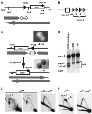
Definition of functional Rtf1 domains: The large number of isolatedrtf1alleles allowed us to define the functional domains of the Rtf1 protein. The alleles include 10 single amino acid (aa) substitutions, six frameshifts (one in an intron splice junction), and four nonsense mutations (Figure 2, A and B; supplemental Figure S1). All the mutants isolated in the initial screen Figure1.—Isolation ofrtf1mutants. (A) Line
drawing displaying the wild-typemat1region on chromosome II. The positions of the imprint (solid circle), theRTS1element (triangle) and theMPS1(horizontal bracket) are given. Shaded arrows indicate the directions by which the repli-cation forks are moving within themat1region, as well as the polarity of theRTS1replication bar-rier. (B) Graphic outline of the RTS1 subele-ments. Region A (box) and the four repeated region B motifs (triangles; rep1, -2, -3, and -4) are shown. (C) Graphic outline of the genetic screen used for isolation of the rtf1 mutants. (Top) The rearranged mating-type region of the JZ183 strain; the site-specific terminator RTS1has been deleted at thecen-proximal side ofmat1and inserted at thecen-distal side in the inverted orientation. (Top inset) Colonies of strain JZ183 stain yellow with iodine vapor. (Bot-tom) Mutagenesis (vertical arrow) ofreplication termination factor (rtf) genes abolishes RTS1 function and leads to a partial restoration of the wild-type direction of replication at mat1 (shaded arrows). Thus, imprinting (solid circle) and mating-type switching are partly reestab-lished. (Bottom inset) Colonies of rtf1 strains stain black with iodine vapor (strain JZ184, Fig-ure 2 legend). (D)rtf1 mutations partly restore mat1 imprinting. Southern analysis of HindIII-digested chromosomal DNA (Dalgaard and
Klar1999). A probe specific to themat1P
Hin-dIII fragment was utilized. Signals that corre-spond tomat1,mat2P, andmat3Mfragments are indicated. The mat2P and mat3M are detected due to partial homology.mat1 imprinted DNA is fragile during purification, where hydrolysis at the imprint leads to the formation of a dou-ble-stranded break (DSB). The generated frag-ments are indicated within the panel. The difference in the molecular sizes of the DSB fragfrag-ments from wild-type (lane 1) and mutant strain (lane 3) is due to the transposition ofRTS1. (E) Rtf1 is required forRTS1function. 2D-gel analysis of replication intermediates at the wild-typeRTS1locus in wild-type ( JZ1) andDrtf1(SC11) strains. The genomic position of theNsiI restriction fragment analyzed is indicated in A. Stall (S) and termination (T) signals observed for wild-type replication intermediates are indicated. Note that theRTS1element is replicated in both directions; however, we have earlier shown that while the marjority of replication forks move in the permissive direction, the small fraction of replication forks in the nonpermissive direction is stalled and terminated (Codlinand Dalgaard2003). (F) 2D-gel analysis of plasmid-borneRTS1from wild-type (SC1) andDrtf1
(SC46) strains. Earlier published experiments have established that theRTS1element at this position in the plasmid is replicated in both orientations (Codlinand Dalgaard2003). Thus, stalling and termination signals are observed originating from forks
Figure2.—Bioinformatics analysis of the Rtf1 amino acid sequence. (A) Graphic outline of the position of the two c-myb-like domains
(blue boxes) and their structural motifs (white ellipses) as well as identified mutations. In the gel-shift experiment presented below domain II encompasses the C-terminal tail. The positions of missense and frameshifts/nonsense mutations are given at the top and bottom, respectively. The position of the dominant mutation (strain ES8, R346*) is highlighted (red arrow). The strain names and iden-tified mutations are JZ184,W405G; JZ185,S340F; JZ221,Q300*; JZ223,R293K; JZ226,L129F; JZ229,S154L; JZ231,T131*frame-1; JZ232, L162Y; JZ237,Q175*; JZ238,P136L; JZ240,Q145*; JZ241,L318*; JZ242,K200*frame-1; JZ243,G183E; JZ245,T420*frame11; JZ249, M343R; JZ250,Q147*splice junction; JZ251,P252S; JZ252,T420*frame-1; JZ253,L129*frame-1; and JZ254,P252S.(B) Alignment of the two conservedc-myb-like domains of the Reb1/Rtf1/TTF1 protein family to the human c-myb protein sequence. Domains I and II are highlighted in light and dark blue, respectively. Proteins aligned areS. pombeReb1 (Q9P6H9), Eta2 (BAC54905),S. cerevisiae Reb1 (CAA84992), Reb1L (NP_010309),Homo sapiensDMTF1 (AAH07447), andH. sapiensTTF1 (AAI04640). Residues that display .40% conservation are highlighted. Thea-helices shown above the alignment are those predicted for the Rtf1 domain using the PhD program package. The last sequence shown is that ofM. musculusc-myb (Mmc-myb_1H89). The positions of thea-helices in the three-dimensional structure of mouse c-myb (Weinsteinet al.1986; Ogataet al.1993; Tahirovet al.2002) are displayed below
were recessive (data not shown). The distribution of point mutations suggested that in addition to the known myb motif, an additional functional domain might be present, thus, we employed bioinformatics for its identification. The Rtf1 sequence (CAF31329; SpRtf1) and related sequences were used to search a nonredundant protein sequence database through the World Wide Web interface to the PSI-BLAST program (default parameter settings). An400-aa Rtf1 segment showed statistically significant similarity to proteins from a variety of species (E-value > 0.05) and was retained for further analysis. Previously, an 200-aa conserved segment (here domain II) encompassing the two myb/SANT motifs was identified in Mus musculus TTF1 (MmTTF1),S. cerevisiaeReb1, andM. musculus c-myb (Everset al. 1995, which refers to the two myb/ SANT motifs as domains I and II). The myb motif is an
50-aa sequence which folds into a domain consisting of three helices characterized by tryptophan (Trp) residues essential for DNA binding. In the case of this protein family, mutation of Trp668 to Lys (W668K) in MmTTF1 was found to abolish binding of the dsDNA recognition sequence (Everset al.1995). In addition, a subclass of the myb motifs called the SANT motif has been shown to interact with histone tails (Boyer et al. 2004). Interestingly, the two domain II c-myb motifs of Rtf1 are identified on the sequence level to belong to this subclass. A more careful examination of the PSI-BLAST output revealed that the conserved domain II, present in the second half of the protein, displayed similarity to a putatively related domain in the first half, i.e., the400-aa Rtf1/Reb1 conserved segment can be divided into two structurally related regions both pre-dicted to interact with DNA via myb-like folds (Figure 2, A and B; domains I and II). A careful computational analysis, using a hidden Markov model, the Conserved Domain Database, and the PhD structural predictions establishes that this family of proteins possesses two chimeric putative DNA-binding domains, both display-ing an overall similarity to metazoan c-myb. These two domains potentially contain in total five structural myb motifs, two of which might also be SANT motifs (sup-plemental text and Figure 2, A and B).
Rtf1 domain I can bind toRTS1regions A and B:To characterize the DNA-binding specificities of the two domains, fusion proteins between a 63 His-tagged maltose binding protein (MBP) and Rtf1 segments encompassing domain I, domain II, and the chimeric domains (domains I1II) were purified (supplemental Figure S2A). Using the domain I, gel-shift assays were performed with a labeled dsDNA oligonucleotide cor-responding to motif 4 from region B (Codlin and Dalgaard 2003; Figure 3A). The analysis detected several sharply defined mobility shifts characteristic of protein binding, and potentially of more than one molecule. It should be noted that Western analysis of shifted material verifies that the shift is due to binding of
domain I (supplemental Figure S2B), and that binding can be outcompeted with excess cold-specific compet-itor (supplemental Figure S2C). Furthermore, gel shifts with dsDNA oligonucleotides resembling three shorter segments of motif 4 establish that domain I binds to the middle third of the motif (Figure 3B; left). A linker scanning mutagenesis of motif 4 has earlier defined two linker substitutions that abolish motif 4 barrier activity in vivo(Codlinand Dalgaard2003). We used the five dsDNA oligonucleotides synthesized for that study to further identify sequences within motif 4 required for Rtf1 domain I binding. Interestingly, none of the substitutions completely abolished binding (data not shown; Codlin and Dalgaard 2003). However, gel-shift assays using the rep4-mut3 substitution, which in vivo abolishes barrier activity, leads to a marked reduction in the amount of shifted material (Figure 3B; right). Together these experiments establish that the main domain I binding site is located in the middle third of motif 4.
Interestingly, in this part of motif 4, purines and pyrimidines are distributed asymmetrically between the two strands. As mentioned in the introduction theRTS1 element possesses an enhancer region characterized by an asymmetric distribution of pyrimidines and purines. We decided to investigate if domain I also displays an affinity for region A dsDNA (Figure 3A; right). Again, gel-shift assays detected DNA binding. The binding could somewhat be outcompeted with poly I:C but not poly G:C, thus displaying some specificity. Western analysis of shifted material verifies that the shift is due to binding of domain I (supplemental Figure S2B), and that binding can be outcompeted with excess cold-specific competitor (supplemental Figure S2C). How-ever, the domain I displays a lower affinity for region A (Kd¼3467 nm) than for motif 4 dsDNA (Kd¼549 nm;
Figure 4). Importantly, assays with the segment contain-ing the chimeric domains detected similar bindcontain-ing specificities as observed for domain I only; shifts of a slightly reduced intensity are observed for all four region B motifs as well as for the enhancer region A (Figure 3C, domains I1II).
found that when the rep4-mut4 mutation is introduced, the shift is abolished, showing that the detected in-teraction is sequence specific (data not shown; Figure 3C). This substitution, which also abolishes motif 4
barrier functionin vivo(Codlinand Dalgaard2003), affects the sequence which shows similarity to the bind-ing sequence defined forS. pombeReb1 (Melekhovets et al.1997). Thus, the observations are consistent with Figure3.—DNA-binding specificities of the two Rtf1 c-myb-like domains. A key above the panels defines the experimental
Rtf1 domain II interacting with the motif’s Reb1-like recognition sequence (Codlinand Dalgaard2003).
Importantly, we have previously established that a single motif can act as a weak replication barrier, and
that in the absence of region A, the introduction of additional motifs has an additive effect on the overall barrier activity (Codlin and Dalgaard 2003). The datasets are therefore consistent with Rtf1 molecules binding each of the four repeats present in region B in vivo. We also establish that domain I, but not domain II, can interact specifically but with a lesser affinity with region A dsDNA. This potentially allows at least five Rtf1 molecules to act atRTS1(seediscussion).
Domain I is involved in establishment of the polarity of theRTS1barrier activity:To gain further insight into the mechanism of Rtf1-mediated replication termina-tion atRTS1, we decided to investigate thein vivoactivity of mutantrtf1alleles, containing aa substitutions. The analyzed domain II point mutations either strongly reduced or abolished barrier activity (mutations rtf1-S340F, rtf1-R293K, and rtf1-M343R; data not shown). However, while abolishment of the wild-type barrier activity is observed in the six mutant domain I alleles, a novel barrier signal could be observed in some; the signal is the strongest in the rtf1-S154L genetic back-ground (Figure 5A), is detectable in the rtf1-L162Y strain (supplemental Figure S2D), barely detectable in the rtf1-P136L strain, and is absent for rtf1-L129Fand rtf1-G183E (data not shown). When theSacI–PstI frag-ment is analyzed, the wild-type signal is located close to the apex on the ascending part of the Y arc (Figure 5A, inset), however, the novel signal is located on the de-scending part (Figure 5A, middle). This novel barrier signal is strongest when only the cis-acting region B is present; for unknown reasons the presence of region A causes a reduction of the signal intensity (compare Figure 5A and 5B). There are two possible explanations for the appearance of this novel barrier signal: either the forks replicate through the RTS1 sequence and pause at ade novosite outside the element or theRTS1 barrier activity has inverted its polarity now pausing replication forks moving in the opposite direction (we conclude that only replication pausing occurs as we do not observe any termination signal). To discriminate between the two possibilities, we first excluded that replication forks were stalling at a different position within the plasmid DNA. An analysis of an empty plas-mid detected no barrier signal (supplemental Figure S2E), thus, theRTS1 cis-acting sequence is still required for Rtf1-S154L-mediated pausing. We also verified that
Figure4.—Characterization of domain I binding to
dou-ble-stranded region A and repeat 4. (A) Gel shift of ds repeat 4 DNA while titrating domain I. (B) Gel shift of ds region A DNA while titrating domain I. (C) Hill plot of the data points obtained above. The binding to repeat 4 and region A DNA fit a simple model with a Hill coefficient of 1.45 and 1.16, respec-tively, characteristic of low or no synergistic binding. The dis-sociation constant (Kd) for domain I binding to repeat 4 DNA
is determined as 549 nm. Binding to region A is slightly weaker
the novel Rtf1-S154L barrier is dependent onswi11 and swi31
activities (supplemental Figure S2F), and that the novel signal could be observed when the element was cloned in both orientations within the plasmid (Figure 5, A, B, and D). Again in the presence of region A, the barrier intensity of the signal is lower and only clearly visible when located close to the middle of the fragment (Figure 5D; also a relative difference in intensity is observed for the wild-type barrier in the two orienta-tions, supplemental Figure S2G; left). Finally, we
ex-cluded that the novel barrier is due to ‘‘collisions’’ with polymerase II transcription initiated at the flanking nmt1 promoter, similar to the collisions recently ob-served between transcription forks initiated by poly-merase III and replication forks (Krings and Bastia 2006). Changes between repressed ‘‘low-level’’ and in-duced ‘‘high-level’’nmt1-promoter mediated polymerase II transcription has no effect on the wild-type RTS1 activity (supplemental Figure S2G). However, while we observed no effect of polymerase II transcription on Figure 5.—(A) The domain I
Rtf1-S154L barrier activity, when the transcription forks move in the same direction as the paused replication forks (Figure 5C), a reduction of the barrier activity is observed when the transcription occurs in the opposite
direction (Figure 5E). A possible explanation is that transcription displaces Rtf1-S154L molecules bound to the DNA. We then investigated the second possibility that the polarity of theRTS1barrier has changed in the Rtf1-S154L genetic background. We utilized the method where the polarity of a replication barrier can be established by analyzing overlapping restriction fragments of replica-tion intermediates such that the posireplica-tion of the barrier is moved from one end of the DNA fragment to the other. This analysis was done for plasmids containing RTS1-derived elements in both orientations, and it veri-fied that the polarity of the Rtf1-S154L barrier is in-verted (Figure 5, A and D). To investigate the possibility that the change in polarity was due to the S154L mutation affecting domain I DNA binding, we purified the mutant domain and analyzed its binding to motif 4 and region A dsDNA. We observed gel-shift signals using the S154L-domain I at lower concentrations than ob-served with the wild-type domain I (Kd¼264 nmand 343
nm for motif 4 and region A, respectively; Figure 6), establishing that the mutant domain is binding with a greater affinity than the wild-type domain. However, at the lower protein concentrations we also observed a smaller Hill coefficient in both cases: 1.0 and 0.71 vs. 1.41 and 1.14 for motif 4 and region A, respectively (Figures 4 and 6). At higher protein concentrations there is no linear fit but a stronger negative coopera-tivity. Thus, the mutation affects the domain’s ability to form multimeric complexes with both region A and motif 4.
The Rtf1 C-terminal region is required for function and can mediate dimerization/polymerization:Finally, a genetic screen for dominant mutants was conducted. A multicopy plasmid carrying the rtf1 gene was trans-formed into the JZ183 strain, and the obtained strain was mutagenized. One mutant with increased iodine staining was isolated. Analysis of RTS1 replication intermediates verified that there is a complete loss of replication barrier activity in this mutant (supplemental data; supplemental Figure S2J). By crossing the isolated mutant strain with theDrtf1strain (SC8), and observing that no crossovers occurred in 27 tetrads analyzed, it was established that the mutation is closely linked to Rtf1 (data not shown). Sequence analysis of the rtf1 gene detected a mutation introducing a nonsense codon at position 346, leading to a 120-aa truncation of the Rtf1 protein. Transformation of the strain with an rtf11
Figure6.—Characterization of domain I-S154L binding to
double-stranded motif 4 and region A DNA. (A) Gel shift of ds motif 4 and region A DNA while titrating domain I-S154L. (B) Hill plot of the data points obtained above, only data points for the five lowest concentrations were used for the linear fit. The Hill coefficient of 1.0 and 0.71 were obtained for motif 4 and region A, respectively. The dissociation constant Kdfor
plasmid (pBZ136) verified that the isolated strain carried a partiallyrtf11
-dominant mutation (Figure 7A; strain ES8). One possible model for the partially dominant effect of this truncation is that it inhibits a functionally important dimerization or oligomeriza-tion of the Rtf1 molecules. To test this hypothesis, we employed a two-hybrid analysis. A self-interaction could be detected with the 127-aa C-terminal region of Rtf1 that includes one of the myb-sant domains (Figure 7B). However, this interaction was masked by the presence of DNA-binding domains, probably because the fusion proteins could bind at other positions in theS. cerevisiae genome with greater affinity than at the reporter genes used for the assay. Thus, our genetic analysis shows that the Rtf1 C-terminal region is required forRTS1 func-tion, and the two-hybrid results establish that this is through a role in Rtf1 dimerization or polymerization.
DISCUSSION
The analysis presented here allows us to propose a model for Rtf1-mediated impediment of replication fork
progression atRTS1(Figure 8A). In summary, the pre-sented data suggest that at least five Rtf1 molecules can bind to the double-stranded RTS1 element through interactions involving both of the protein’s myb domains but mainly promoted by domain I (Figures 2, 3, 4, and 8A, top). Importantly, Rtf1 is able to interact both with the repeated region B motifs and the enhancer region A. The Rtf1 binding to the cis-acting sequences might be stabilized through protein–protein interactions be-tween Rtf1 molecules involving the Rtf1 C-terminal domain (Figure 6). One possibility is that Rtf1 DNA binding at multiple sites within theRTS1 in combina-tion with interaccombina-tions between Rtf1 molecules acts as a topological constraint for DNA unwinding by the repli-cative helicase (Figure 7A). Such a constraint could be augmented by DNA looping, a property which already has been observed for c-myb; the c-myb and c/EBP transcription factor complex together mediate DNA looping required for transcriptional activation (Tahirov et al.2002). In addition, binding of multiple Rtf1 mole-cules within region B combined with the interaction between Rtf1 molecules could act to recruit Rtf1 to the Figure7.—The Rtf1 C-terminal tail is
lower affinity site within the region A dsDNA. Indeed, the dominant phenotype of the Rtf1 allele lacking the C-terminal region (Figure 7) combined with the obser-vation that region A has no intrinsic barrier activity but mediates a cooperative enhancement of the region B activity (Codlinand Dalgaard2003) strongly support a role of C-terminal domain’s self-interaction in re-cruitment of Rtf1 to the enhancer region A. One possibility we are investigating is that the protein interacts with single-stranded DNA formed at region A when the DNA is unwound by the replicative helicase (T. Eydmann and J. Z. Dalgaard, unpublished observation).
We identify a domain I mutation that changes the polarity ofRTS1 (Figure 5). When the Rtf1 domain I mutations that cause this inversion of the barrier’s polarity are superimposed on the known structure of c-myb in complex with its dsDNA-binding site, it is evident that the mutation is not located on the DNA-binding surface (Figure 8B). When the initial Kd is
estimated for this domain, we find that it is lower than that of the wild-type domain, suggesting a stronger DNA affinity; however, at the higher protein concentrations we observe a decreased affinity and a Hill coefficient,1. Thus while the mutation does not significantly affect the initial complex formation, the mutant protein does display a decreased ability to form a multimeric com-plex. The characteristics of this rtf1 allele add some support to the model that unknown protein–protein interaction(s) involving domain I and replication pro-tein(s) are affected by the mutation. Among the replication proteins, the replicative helicase [mini-chro-mosome maintenance proteins (MCMs), reviewed by Takahashiet al. 2005], as well as Rtf2 (Codlin and Dalgaard 2003), Swi1, and Swi3 factors are likely candidates. Swi1 and Swi3 travel with the replication fork (Katouet al.2003; Noguchiet al.2004) and act at MPS1to coordinate pausing of leading-strand replica-tion in response to a lagging-strand signal (Figure 1A; Vengrovaand Dalgaard2004). The identification of a swi1-rtf mutation, which only affects termination of replication atRTS1but not at other replication barriers establishes that suchRTS1-specific interactions involv-ing replication fork proteins do occur (Codlin and
Dalgaard 2003; Krings and Bastia 2004). This parallels the situation in Escherichia coli, where the transacting factor Tus is thought to mediate replication termination through direct interactions with the repli-cative helicase DnaB (Muluguet al.2001). Importantly, the observation that in the Rtf1-S154L genetic back-ground there is a loss of replication termination activity affecting the forks moving in one direction, but a gain of replication pausing activity acting on forks moving in the other, shows that the proposed Rtf1 domain I interactions are of importance when the element is replicated in both directions: Wild-type Rtf1 domain I interactions are required for efficient replication termi-nation of the forks moving in one direction, but also must act to prevent pausing of the forks moving in the opposite.
Finally it should be noted that the identification of two DNA binding domains within the Rtf1 protein could have implications for understanding the molec-ular mechanisms underlying a wide range of activities attributed to the Reb1/TTF1/Rtf1 protein family; poly-merase II transcription activation (Carmenand Holland 1994; Graham and Chambers 1994; Packham et al. 1996; Wangand Warner1998), polymerase I transcrip-tion activatranscrip-tion/repression (Wanget al. 1990), and ter-mination (Lang and Reeder 1993; Lang et al. 1994; Masonet al.1997; Melekhovetset al.1997; Zhaoet al. 1997), as well as chromatin insulator function (Fourel et al.2001). Interactions with double-stranded DNA as well as dynamic changes in these interactions could play an important role for all these molecular processes.
We thank our colleagues at the Marie Curie Research Institute for helpful suggestions and interactions. A special thanks to Rob Cross, Natalie Mansfield, S. Jack Carlisle, Doug Drummond, Sonya Vengrova, and Michael Bonaduce for technical assistance. This work was supported by the Intramural Research Program of the National Cancer Institute of the National Institutes of Health (A.J.S.K.), the Marie Curie Cancer Care (J.Z.D.) and the Association of International Cancer Research ( J.Z.D.).
LITERATURE CITED
Ahn, J. S., F. Osmanand M. C. Whitby, 2005 Replication fork
block-age byRTS1at an ectopic site promotes recombination in fission yeast. EMBO J.24:2011–2023.
Figure8.—(A) Model of Rtf1’s
medi-ated termination of replication atRTS1. (B) Model of c-myb in complex with its target DNA. c-myb residues (Ogata
Arcangioli, B., and A. J. Klar, 1991 A novel switch-activating site
(SAS1) and its cognate binding factor (SAP1) required for effi-cient mat1 switching in Schizosaccharomyces pombe. EMBO J.
10:3025–3032.
Arcangioli, B., 1998 A site- and strand-specific DNA break confers
asymmetric switching potential in fission yeast. EMBO J. 17:
4503–4510.
Bartel, P. L., C.-T. Chien, R. Sternglanxand S. Fields, 1993 Using
the two-hybrid system to detect protein-protein interactions, pp. 153–179 inCellular Interaction in Development: A Practical Approach, edited by D. A. Hartley. Oxford University Press, Oxford.
Boyer, L. A., R. R. Latekand C. L. Peterson, 2004 The SANT
do-main: A unique histone-tail-binding module? Nat. Rev. Mol. Cell Biol.5:158–163.
Brewer, B. J., and W. L. Fangman, 1987 The localization of
replica-tion origins onARSplasmids inS. cerevisiae.Cell51:463–471. Brewer, B. J., and W. L. Fangman, 1988 A replication fork barrier at
the 39end of yeast ribosomal RNA genes. Cell55:637–643. Carmen, A. A., and M. J. Holland, 1994 The upstream repression
sequence from the yeast enolase gene ENO1 is a complex regu-latory element that binds multiple trans-acting factors including REB1. J. Biol. Chem.269:9790–9797.
Codlin, S., and J. Z. Dalgaard, 2003 Complex mechanism of
site-specific DNA replication termination in fission yeast. EMBO J.
22:3431–3440.
Dalgaard, J. Z., and A. J. Klar, 1999 Orientation of DNA
replica-tion establishes mating-type switching pattern inS. pombe.Nature
400:181–184.
Dalgaard, J. Z., and A. J. Klar, 2000 swi1 and swi3 perform
im-printing, pausing, and termination of DNA replication in S. pombe.Cell102:745–751.
Dalgaard, J. Z., and A. J. Klar, 2001 A DNA replication-arrest site
RTS1regulates imprinting by determining the direction of rep-lication atmat1inS. pombe.Genes Dev.15:2060–2068.
diGuan, C., P. Li, P. D. Riggsand H. Inouye, 1988 Vectors that
fa-cilitate the expression and purification of foreign peptides in
Escherichia coli by fusion to maltose-binding protein. Gene67:
21–30.
Evers, R., A. Smid, U. Rudloff, F. Lottspeich and I. Grummt,
1995 Different domains of the murine RNA polymerase I-specific termination factor mTTF-I serve distinct functions in transcription termination. EMBO J.14:1248–1256.
Fourel, G., C. Boscheron, E. Revardel, E. Lebrun, Y. F. Huet al.,
2001 An activation-independent role of transcription factors in insulator function. EMBO Rep.2:124–132.
Gerber, J. K., E. Gogel, C. Berger, M. Wallisch, F. Mulleret al.,
1997 Termination of mammalian rDNA replication: polar ar-rest of replication fork movement by transcription termination factor TTF-I. Cell90:559–567.
Graham, I. R., and A. Chambers, 1994 A Reb1p-binding site is required
for efficient activation of the yeastRAP1gene, but multiple binding sites for Rap1p are not essential. Mol. Microbiol.12:931–940. Huang, J., I. L. Brito, J. Villen, S. P. Gygi, A. Amon et al.,
2006 Inhibition of homologous recombination by a cohesin-associated clamp complex recruited to the rDNA recombination enhancer. Genes Dev.20:2887–2901.
Huberman, J. A., L. D. Spotila, K. A. Nawotka, S. M.el-Assouliand
L. R. Davis, 1987 Thein vivoreplication origin of the yeast 2
microns plasmid. Cell51:473–481.
Hyrien, O., 2000 Mechanisms and consequences of replication fork
arrest. Biochimie82:5–17.
Katou, Y., Y. Kanoh, M. Bando, H. Noguchi, H. Tanakaet al.,
2003 S-phase checkpoint proteins Tof1 and Mrc1 form a stable replication-pausing complex. Nature424:1078–1083.
Kiger, Jr., J. A., and R. L. Sinsheimer, 1969 Vegetative lambda
DNA. IV. Fractionation of replicating lambda DNA on benzoy-lated-naphthoylated DEAE cellulose. J. Mol. Biol.40:467–490. Kobayashi, T., and T. Horiuchi, 1996 A yeast gene product, Fob1
protein, required for both replication fork blocking and recom-binational hotspot activities. Genes Cells1:465–474.
Krings, G., and D. Bastia, 2004 swi1- andswi3-dependent and
inde-pendent replication fork arrest at the ribosomal DNA of Schizosac-charomyces pombe.Proc. Natl. Acad. Sci. USA101:14085–14090.
Krings, G., and D. Bastia, 2005 Sap1p binds to Ter1 at the
ribo-somal DNA of Schizosaccharomyces pombe and causes polar rep-lication fork arrest. J. Biol. Chem.280:39135–39142.
Krings, G., and D. Bastia, 2006 Molecular architecture of a
eukary-otic DNA replication terminus-terminator protein complex. Mol. Cell. Biol.26:8061–8074.
Lambert, S., A. Watson, D. M. Sheedy, B. Martinand A. M. Carr,
2005 Gross chromosomal rearrangements and elevated recom-bination at an inducible site-specific replication fork barrier. Cell
121:689–702.
Lang, W. H., B. E. Morrow, Q. Ju, J. R. Warnerand R. H. Reeder,
1994 A model for transcription termination by RNA polymer-ase I. Cell79:527–534.
Lang, W. H., and R. H. Reeder, 1993 TheREB1site is an essential
component of a terminator for RNA polymerase I in Saccharomy-ces cerevisiae.Mol. Cell. Biol.13:649–658.
Linskens, M. H., and J. A. Huberman, 1988 Organization of
repli-cation of ribosomal DNA in Saccharomyces cerevisiae. Mol. Cell. Biol.8:4927–4935.
Lopez-Estrano, C., J. B. Schvartzman, D. B. Krimerand P. Hernandez,
1998 Co-localization of polar replication fork barriers and rRNA transcription terminators in mouse rDNA. J. Mol. Biol.
277:249–256.
Lopez-Estrano, C., J. B. Schvartzman, D. B. Krimerand P. Hernandez,
1999 Characterization of the pea rDNA replication fork barrier: putativecis-acting and trans-acting factors. Plant. Mol. Biol.40:
99–110.
Mason, S. W., M. Wallischand I. Grummt, 1997 RNA polymerase I
transcription termination: similar mechanisms are employed by yeast and mammals. J. Mol. Biol.268:229–234.
Mejia-Ramirez, E., A. Sanchez-Gorostiaga, D. B. Krimer, J. B.
Schvartzman and P. Hernandez, 2005 The mating type
switch-activating protein Sap1 Is required for replication fork ar-rest at the rRNA genes of fission yeast. Mol. Cell. Biol.25:8755– 8761.
Melekhovets, Y. F., P. S. Shwedand R. N. Nazar, 1997 In vivo
analyses of RNA polymerase I termination inSchizosaccharomyces pombe.Nucleic Acids Res.25:5103–5109.
Mohanty, B. K., N. K. Bairwaand D. Bastia, 2006 The
Tof1p-Csm3p protein complex counteracts the Rrm3p helicase to con-trol replication termination ofSaccharomyces cerevisiae.Proc. Natl. Acad. Sci. USA103:897–902.
Moreno, S., A. Klarand P. Nurse, 1991 Molecular genetic analysis
of fission yeastSchizosaccharomyces pombe.Methods Enzymol.194:
795–823.
Mulugu, S., A. Potnis, J. Shamsuzzaman, K. Taylor, K. Alexander
et al., 2001 Mechanism of termination of DNA replication of
Escherichia coliinvolves helicase-contrahelicase interaction. Proc. Natl. Acad. Sci. USA98:9569–9574.
Noguchi, E., C. Noguchi, W. H. McDonald, J. R. YatesIII and P.
Russell2004 Swi1 and Swi3 are components of a replication
fork protection complex in fission yeast. Mol. Cell. Biol. 24: 8342–8355.
Ogata, K., H. Kanai, T. Inoue, A. Sekikawa, M. Sasaki et al.,
1993 Solution structures of Myb DNA-binding domain and its complex with DNA. Nucleic Acids Symp. Ser.29:201–202. Packham, E. A., I. R. Grahamand A. Chambers, 1996 The
multifunc-tional transcription factors Abf1p, Rap1p and Reb1p are required for full transcriptional activation of the chromosomalPGKgene inSaccharomyces cerevisiae.Mol. Gen. Genet.250:348–356. Petty, K. J., 1996 Metal-chelate affinity chromatography, unit
10.11B inCurrent Protocols in Molecular Biology, edited by F. M. Ausubel. John Wiley & Sons, Malden, MA.
Putter, V., and F. Grummt, 2002 Transcription termination factor
TTF-I exhibits contrahelicase activity during DNA replication. EMBO Rep.3:147–152.
Sambrook, J., and D. W. Russell, 2001 Molecular Cloning: A Laboratory
Manual.Cold Spring Harbor Laboratory Press, Cold Spring Harbor, NY. Sanchez-Gorostiaga, A., C. Lopez-Estrano, D. B. Krimer, J. B.
Schvartzmanand P. Hernandez, 2004 Transcription
Santamaria, D., E. Viguera, M. L. Martinez-Robles, O. Hyrien, P.
Hernandezet al., 2000 Bi-directional replication and random
termination. Nucleic Acids Res.28:2099–2107.
Studier, F. W., and B. A. Moffatt, 1986 Use of bacteriophage T7
RNA polymerase to direct selective high-level expression of cloned genes. J. Mol. Biol.189:113–130.
Tahirov, T. H., K. Sato, E. Ichikawa-Iwata, M. Sasaki, T. Inoue
-Bungoet al., 2002 Mechanism of c-Myb-C/EBP beta
coopera-tion from separated sites on a promoter. Cell108:57–70. Takahashi, T. S., D. B. Wigleyand J. C. Walter, 2005 Pumps,
para-doxes and ploughshares: mechanism of the MCM2–7 DNA heli-case. Trends Biochem. Sci.30:437–444.
Takeuchi, Y., T. Horiuchiand T. Kobayashi, 2003
Transcription-dependent recombination and the role of fork collision in yeast rDNA. Genes Dev.17:1497–1506.
Vengrova, S., and J. Z. Dalgaard, 2004 RNase-sensitive DNA
mod-ification(s) initiates S. pombe mating-type switching. Genes Dev.
18:794–804.
Wang, H., P. R. Nicholsonand D. J. Stillman, 1990 Identification
of aSaccharomyces cerevisiaeDNA-binding protein involved in tran-scriptional regulation. Mol. Cell. Biol.10:1743–1753.
Wang, K. L., and J. R. Warner, 1998 Positive and negative
autore-gulation of REB1transcription inSaccharomyces cerevisiae.Mol. Cell. Biol.18:4368–4376.
Weinstein, Y., J. N. Ihle, S. Lavuand E. P. Reddy, 1986 Truncation
of the c-myb gene by a retroviral integration in an interleukin 3-dependent myeloid leukemia cell line. Proc. Natl. Acad. Sci. USA
83:5010–5014.
Zhao, A., A. Guo, Z. Liuand L. Pape, 1997 Molecular cloning and
analysis ofSchizosaccharomyces pombeReb1p: sequence-specific rec-ognition of two sites in the far upstream rDNA intergenic spacer. Nucleic Acids Res.25:904–910.