DOI: 10.1534/genetics.106.061705
Note
An Efficient Genetic Screen in Drosophila to Identify Nuclear-Encoded
Genes With Mitochondrial Function
T. S. Vivian Liao,*
,1Gerald B. Call,
†,1Preeta Guptan,
†Albert Cespedes,
†Jamie Marshall,
†Kevin Yackle,
†Edward Owusu-Ansah,
†Sudip Mandal,
†Q. Angela Fang,
†Gelsey L. Goodstein,
†William Kim
†and Utpal Banerjee*
,†,‡,2*Molecular Biology Institute,†Department of Molecular, Cell and Developmental Biology and‡Department of Biological Chemistry, University of California, Los Angeles, California 90095
Manuscript received June 8, 2006 Accepted for publication July 6, 2006
ABSTRACT
We conducted a screen for glossy-eye flies that fail to incorporate BrdU in the third larval instar eye disc but exhibit normal neuronal differentiation and isolated 23 complementation groups of mutants. These same phenotypes were previously seen in mutants forcytochrome c oxidase subunit Va. We have molecularly characterized six complementation groups and, surprisingly, each encodes a mitochondrial protein. Therefore, we believe our screen to be an efficient method for identifying genes with mitochondrial function.
M
ITOCHONDRIAL function is essential for a number of important cellular processes, such as the generation of ATP (reviewed in Ackerman andTzagoloff 2005), apoptosis (reviewed in Newmeyer
and Ferguson-Miller 2003), and the regulation of
aging (reviewed in Ohta 2003). Recently, work from
our laboratory has shown that the metabolic status of a cell, controlled by mitochondrial function, also regu-lates the G1–S checkpoint in mitosis (Mandal et al.
2005). This is evident intenured(tend) mutants, which contain a null mutation in the gene encoding cyto-chrome c oxidase subunit Va of complex IV of the mitochondrial electron transport chain. This mutation causes a reduction in ATP generated in mutant cells to
40% of wild-type levels, triggering the activation of AMPK and p53, which leads to the eventual down-regulation of cyclin E. A G1–S mitotic checkpoint is
then enforced, preventing cells from reentering the cell cycle (Mandalet al. 2005).
Adult tend mutants have glossy eyes lacking lens material, normally secreted by cone and pigment cells of the eye. In earlier studies, the lens-depleted glossy-eye phenotype was described inlozengemutants, which are defective in cone and pigment cell specification
(Floreset al. 1998). However, in the case oftend, such
defects are caused by the lack of a sufficient number of precursor cells from which cone and pigment cells arise (Mandalet al. 2005).
The events that give rise to the tend adult eye phe-notype occur in larval eye development. The larval Drosophila eye imaginal disc undergoes two distinct phases of proliferation (Wolff and Ready 1991).
During the early larval stages prior to differentiation, all cells divide frequently and asynchronously, appar-ently without regulation. In the third larval instar, the morphogenetic furrow sweeps across the eye disc to pattern the cells (Readyet al. 1976). Within the
mor-phogenetic furrow, cells arrest in G1and, following the
passage of the furrow, either differentiate into neuronal photoreceptors or reenter the cell cycle and undergo a terminal round of division termed the second mitotic
wave (SMW). This generates the precursors for cone and pigment cell lineages (Wolff and Ready 1991).
Misregulation of the SMW will lead to a loss of these accessory cell types. In the third larval instar, cells mutant fortendanterior to the furrow slow down in cell cycle progression, while those posterior to the furrow fail to cross the G1–S checkpoint due to impaired ATP
production (Mandal et al. 2005). Therefore, in
addi-tion to undergoing fewer rounds of mitosis prior to the passage of the furrow, tend mutants lack the SMW and, as a result, have a dramatically reduced number of accessory cells. Importantly, intendmutant cells, cell
1These authors contributed equally to this work.
2Corresponding author: Department of Molecular, Cell and Develop-mental Biology, 2204 Life Science, 621 Charles E. Young Dr. South, Los Angeles, CA 90095. E-mail: [email protected]
divisions in second and early third instar eye discs are not affected. As a result,tendmutants give rise to large clones in the adult eye. This is different from the phenotype resulting from mutant clones in the basic cell cycle machinery, which would not be able to give rise to any mutant clones. Furthermore, early patterning and differentiation of neurons are also not affected in
tend, as mutant clones express pan-neuronal, as well as cell-type-specific, markers and extend axons normally to the optic lobe (Mandalet al. 2005).
On the basis of the nature of thetendphenotype, we hypothesized that a simple screen that identifies glossy-eye flies, followed by a secondary screen for a subset of the mutants in which cells exhibit proliferation defects but have relatively normal patterning in the third larval instar, will potentially identify nuclear genes that en-code mitochondrial proteins. Our hypothesis is sup-ported by our earlier finding that, liketend, flies mutant in the mitochondrial components pdsw, mRpL4, and
mRpL17exhibit a glossy-eye phenotype and fail to in-corporate BrdU in the larval eye disc but stain normally with the ELAV antibody (Mandal et al. 2005). In this
article, we show that an unbiased screen for glossy-eye mutants that exhibit these larval phenotypes is remark-ably effective in identifying genes with mitochondrial function.
We conducted aneyeless-flp/FRT-based mitotic recom-bination screen (Newsomeet al. 2000) on chromosome
arm 3R (Figure 1).y w ey-flp; FRT 82Bmales were mu-tagenized with 25 mm ethyl methanesulfonate (EMS)
and crossed toy w ey-flp; FRT82B P[w1
] cl-R3/TM6B Tb, y1
females to generate adult eye clones. ey-flp drives mitotic recombination between the FRT-containing
chromosomes under the control of theeyelessenhancer, specifically in the embryonic anlagen of the developing eye (Newsomeet al. 2000). The resulting clones of cells
will be homozygous for the newly induced mutation and are white due to the lack of thew1 gene essential for
pigmentation. A total of 75,000 adult flies were screened for the mosaic glossy-eye phenotype. Ninety mutants were isolated. Complementation analysis was then per-formed by crossing each of the 90 mutants with one another and looking for lethality in the offspring. This yielded a total of 46 complementation groups. All mutants were balanced over aTM6B Tb, y1
chromosome and maintained as stocks.
Next, third larval instar eye imaginal discs from the identified glossy-eye mutants were assayed for BrdU incorporation and stained with the neuron-specific ELAV antibody (Yaoet al. 1993). Larval eye-disc clones
were generated by crossing the mutant stocks to y w ey-flp; FRT82B Ubi-GFP RpS3/TM6B Tb, y1
flies. Clones homozygous for the mutation are negatively marked by their lack of GFP. The 46 complementation groups isolated from the glossy-eye screen fall into four catego-ries on the basis of this characterization. Ten mutant groups exhibit both wild-type neuronal patterning and BrdU incorporation, indicating that the glossy-eye phe-notype is due to mutations in genes that function after the third instar and are required for cone and pigment cell specification. Three mutant groups display abnor-mal ELAV staining patterns while cells incorporate BrdU normally. Since the formation of cone cells re-quires a combination of signals from differentiated neuronal photoreceptors (Floreset al. 2000), this
phe-notype likely occurs because of defects in the patterning
Figure1.—Crossing scheme
for genetic screen. A total of 75,000 chromosomes were mu-tagenized and screened for the adult glossy-eye phenotype. A total of 90 glossy-eye mutants were isolated, and larval eye-disc clones were induced to study their BrdU incorporation and ELAV expression. Twenty-three failed to incorporate BrdU but expressed ELAV normally and were studied further.m* repre-sents the EMS-induced muta-tion. Thecl-R3mutation is cell lethal and is used to eliminate cells that are homozygous for the chromosome that does not containm*. Similarly, for larval clones, cells homozygous for the RpS3 (Minute) mutation are eliminated. Cells heterozy-gous for the cl-R3 and RpS3
of specific photoreceptors. Ten mutant groups exhibit defective ELAV staining patterns in early clusters as well as defects in BrdU incorporation, which can be attrib-uted to mutations in genes required for specification of multiple cell types in the eye. Finally, 23 mutant groups fail to incorporate BrdU properly but show essentially normal patterning of neuronal clusters emerging from
the furrow. They were further analyzed as part of this study.
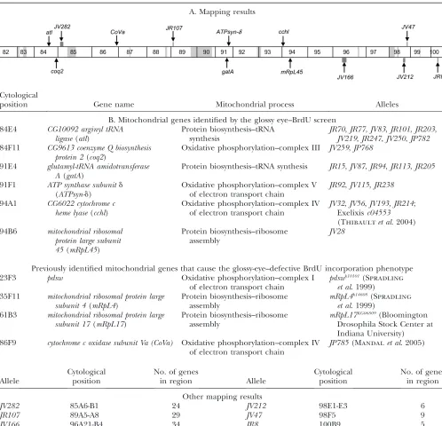
We have mapped all mutants from this last category to either available deficiencies on chromosome 3R from the Bloomington and Exelexis collections (Thibault et al. 2004) or attributed their map positions to gaps within the deficiency kit (Table 1A). We have cloned
TABLE 1
Mapping results and mutations in nuclear-encoded genes with mitochondrial function that exhibit a G1–S block in the
third larval instar with normal neuronal differentiation
A. Mapping results
Cytological
position Gene name Mitochondrial process Alleles
B. Mitochondrial genes identified by the glossy eye–BrdU screen 84E4 CG10092 arginyl tRNA
ligase(atl)
Protein biosynthesis–tRNA synthesis
JR70,JR77,JV83,JR101,JR203,
JV219,JR247,JV250,JP782
84F11 CG9613 coenzyme Q biosynthesis protein 2(coq2)
Oxidative phosphorylation–complex III JV259,JP768
91E4 glutamyl-tRNA amidotransferase A(gatA)
Protein biosynthesis–tRNA synthesis JR15,JV87,JR94,JR113,JR205
91F1 ATP synthase subunitd
(ATPsyn-d)
Oxidative phosphorylation–complex V of electron transport chain
JR92,JV115,JR238
94A1 CG6022 cytochrome c heme lyase(cchl)
Oxidative phosphorylation–complex IV of electron transport chain
JV32,JV56,JV193, JR214; Exelixisc04553
(Thibaultet al. 2004)
94B6 mitochondrial ribosomal protein large subunit 45(mRpL45)
Protein biosynthesis–ribosome assembly
JV28
Previously identified mitochondrial genes that cause the glossy-eye–defective BrdU incorporation phenotype 23F3 pdsw Oxidative phosphorylation–complex I
of electron transport chain
pdswk10101(Spradling
et al. 1999) 35F11 mitochondrial ribosomal protein large
subunit 4(mRpL4)
Protein biosynthesis–ribosome assembly
mRpL4k14608(Spradling
et al. 1999) 61B3 mitochondrial ribosomal protein large
subunit 17(mRpL17)
Protein biosynthesis–ribosome assembly
mRpL17KG06809(Bloomington
Drosophila Stock Center at Indiana University) 86F9 cytochrome c oxidase subunit Va (CoVa) Oxidative phosphorylation–complex IV
of electron transport chain
JP785(Mandalet al. 2005)
Allele
Cytological position
No. of genes
in region Allele
Cytological position
No. of genes in region
Other mapping results
JV282 85A6-B1 24 JV212 98E1-E3 6
JR107 89A5-A8 29 JV47 98F5 9
JV166 96A21-B4 34 JR8 100B9 5
six mutant complementation groups, consisting of 24 alleles, and obtained sequence information on the mo-lecular lesions of 15 of the alleles (Figure 2). As the guiding hypothesis of this screen was to identify chondrial mutations, we sequenced potentially mito-chondrial genes within candidate regions as determined through BLAST analysis (Altschul et al. 1990) and
MitoProt scores, which predict potential mitochondrial localization on the basis of the presence of a mitochon-drial localization sequence (Clarosand Vincens1996).
Using this method, we identified four mutants in
CG6022, a homolog ofcytochrome c heme lyase(cchl); two mutant alleles of CG9613, a homolog of coenzyme Q biosynthesis protein 2(coq2); nine mutants inCG10092, a homolog of arginyl tRNA ligase (atl); three mutant alleles ofATP synthase-dsubunit(ATPsyn-d); five mutants inglutamyl-tRNA amidotransferase A(gatA); and one mu-tant allele ofmitochondrial ribosomal protein large subunit 45(mRpL45). Combined with previously described mu-tants inpdsw,mRpL4,mRpL17, andtend, all mutations exhibiting the glossy-eye phenotype with defective BrdU incorporation and wild-type neuronal patterning cloned thus far could be rationalized as having roles in the mitochondrion (see below and Figure 3 and Table 1B). Note that this screen identified the first mutant alleles forCoVa,coq2,atl,ATPsyn-d, gatA, andmRpL45. Single alleles of cchl, pdsw, mRpL4, and mRpL17 had been identified as lethal mutations in either the Exelixis col-lection or the Berkeley Drosophila Genome Gene Disruption Project, but were not previously genetically characterized (Spradling et al. 1999; Thibaultet al.
2004) (Table 1B).
CCHL: Within the electron transport chain, CCHL participates in the function of complex IV by attaching a heme group to apocytochromec, thereby converting it to a functional holocytochrome c and enabling its import through the inner membrane of the mitochon-drion (reviewed in Kranzet al. 1998). InSaccharomyces cerevisiae, strains lacking or mutant in CCHL have increased levels of apocytochromec in the cytoplasm (Dumontet al. 1991). From the screen, we have isolated
four alleles ofcchl(Figure 2) on the basis of their failure to complement Exelixisc04553(Thibaultet al. 2004),
which harbors a lethal insertion incchl. The first allele,
JR214, harbors a T.C transition resulting in the amino acid substitution M25T.JV56 contains a C . T transi-tion, resulting in a premature stop codon at Q65. The remaining two alleles, JV193 and JV32, have G . A transitions at the first and last base pair of intron 2, respectively, and thus may result in the insertion of 19 amino acids if splicing cannot occur normally. All of the mutations occur within the heme lyase domain of the protein. Drosophila CCHL bears 53% identity to the human homolog and 42% identity to the S. cerevisiae
homolog. According to MitoProt predictions, CCHL has a 28% likelihood of localizing to the mitochon-drion; this relatively low likelihood may be due to the
fact that mitochondrial heme lyases lack an N-terminal targeting signal and instead are directed to the mito-chondrion by an internal localization sequence in the third quarter of the protein (Diekertet al. 1999). This is
further supported by the fact that MitoProt also predicts a very low (4.8%) chance of mitochondrial localization for theS. cerevisiaeCCHL.
ATPsyn-d: Studies in Escherichia coli show that F1F0
-ATP synthase, complex V of the electron transport chain, generates ATP through cooperation between a ‘‘rotor’’ and a ‘‘stator’’ (reviewed in Boyer1997; Weber et al. 2004). The stator consists of twob-subunits and one
d-subunit (reviewed in Dunnet al. 2000), and loss of the d-binding site on thea-subunit of F1results in impaired
growth and ATP synthesis in vivo(Weber et al. 2004).
Three alleles ofATPsyn-dwere isolated from the screen (Figure 2).JV115contains a G.A transition that results in a premature stop codon at W54, whileJR92has a C.
T transition that prematurely terminates the protein at Q83. A molecular lesion for the third allele,JR238, has not been found; presumably, it is located within a reg-ulatory region of the gene. The mutations all lie within the ATP synthase-d domain of the protein. MitoProt analysis suggests that Drosophila ATPsyn-d has a 92% likelihood of localizing to the mitochondria.
COQ2: coq2 encodes the enzyme parahydroxyben-zoate (PHB):polyprenyltransferase (Ashbyet al. 1992).
As part of a multi-step process involved in ubiquinone biosynthesis, PHB:polyprenyltransferase catalyzes the transfer of a polyprenyl group from polyprenyl diphos-phate to parahydroxybenzoate to form 3-polyprenyl-4-hydroxybenzoate (Winrow and Rudney 1969). Once
synthesized, ubiquinone functions as an electron trans-porter between complexes I, II, and III of the electron transport chain (Green1966). Two alleles ofcoq2were
identified from the screen (Figure 2). The first allele,
JP768, contains a 94-bp deletion mutation starting at A100 that causes a frameshift in the remaining se-quence. All amino acids downstream of the deletion are altered, and a premature stop codon is introduced after the 232nd amino acid. JV259has a T.A trans-version that results in a premature stop codon at L189. Both mutations fall within the UbiA prenyltransferase domain of COQ2. Drosophila COQ2 has 45% identity to the S. cerevisiae homolog and a 97% likelihood of being imported into the mitochondrion according to MitoProt analysis.
GatA: Aminoacylation studies have shown that there is no detectable glutaminyl–tRNA synthase activity in cyanobacteria, Gram-positive bacteria, and chloroplasts and mitochondria of plants and animals (Wilcoxand
Nirenberg 1968; Schon et al. 1988). Instead, these
organisms and organelles synthesize glutamine by mis-charging a tRNAGln with glutamate and then using
glutamyl–tRNA amidotransferases to convert the gluta-mate to glutamine (Schonet al. 1988). According to
low probability (56%) of localizing to the mitochon-drion. To determine the exact subcellular location of GatA, we constructed a GatA–GFP fusion protein, transfected Drosophila S2 cells with the construct, and co-immunostained the cells with a GFP antibody and MitoTracker Orange CM-H2TMRos, a dye that localizes to the mitochondrion in response to its membrane potential. This colocalization assay shows that GatA is indeed a mitochondrial protein (Figure 4, A–C). Thus, both functional and localization studies and the nature of other mutants isolated by our screen establish that GatA has a mitochondrial function. Five alleles ofgatA
were isolated from our screen; we have located molec-ular lesions in four alleles (Figure 2).JR94has a 168-bp deletion in exon 5.JR15harbors a G.T transversion, resulting in the amino acid substitution Q54H.JR113
has a C . G transversion that causes the substitution H278D. Finally, JV87 has three point mutations, G.T, T.G, and G.A, which result in the following amino acid changes: Q53H, A58S, and V74I. All of the mutations lie within the putative amidase domain of the protein.
ATL:It has been determined thatS. cerevisiaehas two genes encoding arginyl tRNA ligase (ATL), with one functioning in the cytoplasm and the other,MSR1, in the mitochondrion (Tzagoloffand Shtanko 1995).
The Drosophila genome has three genes encoding proteins with potential arginyl tRNA ligase activity on the basis of Gene Ontology annotation in FlyBase (http://www.flybase.org): atl, Aats-arg, and CG8097
(Drysdale and Crosby2005). BLAST analysis shows
that ATL is 36% identical to Msr1p, while AATS-arg and CG8097 are 28 and 24% identical, respectively. Accord-ing to MitoProt analysis, ATL is only 8.4% likely to localize to the mitochondrion. This low number did not support a mitochondrial function for ATL. To pinpoint its subcellular location, we constructed an ATL–GFP fusion protein and demonstrated that it colocalizes with MitoTracker Orange CM-H2TMRos to the mitochon-drion by immunostaining S2 cells transfected with the fusion protein (Figure 4, D–F). Thus, ATL’s subcellular localization, similarity to Msr1p, and its suite of phe-notypic similarities to other known mitochondrial mu-tants, indicate that this protein functions in the mitochondrion. Our screen has identified nine alleles ofatl, and we have located molecular lesions in two of these nine alleles (Figure 2).JR70has a G.A transition resulting in the amino acid change G182R.JV219 con-tains a 12-bp in-frame deletion starting at V266. Both mutations occur within the catalytic arginyl tRNA core domain.
Figure2.—Map positions and molecular lesions of identified mutations. Deficiency stock names and their approximate
Figure 3.—Representative adult and larval
phenotypes of mutants from mapped comple-mentation groups. (A–N) Eyes of adult flies ex-amined by scanning electron microscopy (A–G) and bright-field microscopy (H–N). An eye con-taining mock clones is mosaic in color (H), but the entire eye is faceted as in wild-type tissue (A). In eyes mutant for the indicated mitochon-drial genes, cells heterozygous for the mutation are faceted (B–G) and red (I–N) as in mock clones, while homozygous clones are glossy and white. In all images, the representative allele is in-dicated in parentheses and posterior is to the left. (O–U) 30-min BrdU incorporation (red) in third instar larval eye discs with mock (O) and mutant (P–U) clones. Armadillo (blue) marks the mor-phogenetic furrow. In an eye disc with mock clones (O), both green and nongreen tissue are wild type. BrdU is randomly incorporated ante-rior to the furrow (‘‘a’’); posteante-rior to the furrow, a single band of BrdU incorporation (arrowhead) marks the SMW. In mutant eye discs, BrdU incor-poration is lost in homozygous mutant tissue, marked by the lack of GFP, both anterior and pos-terior to the furrow. Incorporation remains nor-mal in clones heterozygous for the mutation (green). For comments on the apparent local nonautonomy of this phenotype (arrow), see Mandal et al. (2005). (V–B9) ELAV antibody
mRpL45: mRpL45 is a protein of 599 amino acids with a molecular weight of 26 kDa (reviewed in Graack
and Wittmann-Liebold 1998). At least 50
mitochon-drial ribosomal proteins exist within the S. cerevisiae
mitochondrial ribosome, among which at least 39 are classified as large subunit proteins. In humans, these ribosomal subunits help synthesize 13 mitochondrial proteins essential for oxidative phosphorylation, and mutations in the genes encoding them are associated with many disorders (reviewed in Sylvesteret al. 2004).
According to MitoProt, mRpL45 has an 88% chance of localizing to the mitochondrion. One mutant allele of mRpL45 was identified from the screen (Figure 2).JV28
harbors a 9-bp deletion and a 7-bp insertion in exon 2 that results in a frameshift starting at V230 and a premature stop codon at the 279th amino acid. The mutation occurs within the TIM44 domain, which is involved in transporting the protein across the inner membrane of the mitochondrion (reviewed in Pfanner
and Geissler2001).
The generation of ATP in eukaryotic cells is largely dependent on oxidative phosphorylation in mitochon-dria (Figure 5). ATP originates from the oxidation of NADH and FADH2 via the electron transport chain, which consists of a series of four electron transporting complexes (I–IV) and the terminal complex V, which generates ATP. Complexes I and II transfer electrons, from NADH and FADH2, respectively, to ubiquinone (CoQ) via a series of iron–sulfur clusters. Ubiquinone, which is a mobile carrier, transfers these electrons to complex III. Cytochrome c, another mobile carrier protein, then delivers the electrons from complex III to complex IV where it reduces dioxygen to water. Concurrent with electron transport, complexes I, II, and IV pump protons from the matrix into the inter-membrane space, forming an electrochemical proton gradient across the inner membrane, which is used to generate ATP by ATP synthase or complex V. Five
mu-tants discussed in this study—tend,pdsw,coq2,cchl, and
ATP syn-d—function either within or between com-plexes of the electron transport chain. These mutants likely activate the G1–S checkpoint as described fortend
(Mandal et al. 2005). The remaining mutants—atl, gatA,mRpL4,mRpL17, andmRpL45—function in diverse roles within the mitochondrial protein biosynthesis path-ways. The mitochondrial genome encodes 13 proteins that are essential for oxidative phosphorylation, and defects within the protein translation system are likely to cause downstream effects in energy production and subsequently affect the G1–S transition of the cell.
The screen described here is the most efficient in any higher eukaryote for identifying nuclear-encoded genes that function in the mitochondrion. All six mutants mapped thus far are likely to have roles in mitochon-drial processes. This is further supported by the fact that previously describedP-element mutants inpdsw,mRpL4, and mRpL17also exhibit the same phenotypes as mu-tants isolated from our screen (Mandal et al. 2005).
This is an independent confirmation that this screen is uniquely capable of selecting for disruptions in cell cycle regulation in the developing eye disc, caused by the activation of a metabolic checkpoint initiated by the mitochondrion. However, not all of the mutants isolated in this screen are expected to be disruptions in mitochondrial proteins. In Mandal et al. (2005), we
described a pathway linking mitochondrial processes to cell cycle regulation in the nucleus. Two key members of this pathway, AMPK and p53, are known cytosolic proteins. This pathway is not yet fully understood, and we expect that our screen will also provide missing cytosolic links, in addition to mitochondrial ones de-scribed here. Our mapping results show that the screen is very likely to have identified some nonmitochondrial genes. For example, JV212 maps to 98E1-98E3 (Table 1, A and B), a region encompassing six genes, none of which bears homology to other mitochondrial genes or
Figure 4.—GatA and ATL localize to
contains a mitochondrial localization sequence. This is again true for JR8 (Table 1, A and B), which maps to 100B9, another region containing five genes that are not likely to be mitochondrial.
An additional advantage to our screen is that it may be a more accurate predictor of mitochondrial function than the currently available in silico databases. For example, MitoProt predicts relative low probabilities of mitochondrial localization for CCHL (28%), Pdsw (10%), GatA (56%), and ATL (8.4%). We have demon-strated through immunostaining in this study that both GatA and ATL localize to the mitochondrion. Pdsw, a NADH dehydrogenase, and CCHL have been shown in previous functional studies to play roles in complexes I and IV, respectively, of the electron transport chain. Although MitoProt will be able to reliably predict mitochondrial localization for the proteins that fit its algorithm, our screen provides the more relevantin vivo
information.
This study identified several new mutations in genes that function in either oxidative phosphorylation or mitochondrial protein biosynthesis. Consistent with our earlier observation using tend (Mandal et al. 2005),
these new mutants exhibit normal early cell divisions, a G1–S block at the third larval instar, and proper
neuronal differentiation. The new mutants help to further establish that attenuated mitochondrial func-tion, caused by a loss in one of its many individual
components, will result in an enforcement of the G1–S
checkpoint of the cell cycle. The fact that each of the characterized mutants with this suite of phenotypes encodes a protein with a mitochondrial function sug-gests that our method can serve as a general screen for identifying nuclear-encoded genes with mitochondrial function. This study represents yet another example of the power of Drosophila genetics and mutant screens in the discovery and refinement of important cellular processes.
We thank Julia Thompson, Kha Nguyen, and Ryan Skophammer for their help with the screen and the Bloomington Stock Center and the Exelixis Stock Center for fly stocks. This study was supported by National Institutes of Health grant R01-EY08152 to U.B. U.B. is a Howard Hughes Medical Institute (HHMI) professor and G.C. is an HHMI instructor. We acknowledge HHMI for supporting research efforts by our undergraduate students, six of whom (A.C., J.M., K.Y., Q.A.F., G.L.G., and W.K.) are included here as authors.
LITERATURE CITED
Ackerman, S. H., and A. Tzagoloff, 2005 Function, structure, and
biogenesis of mitochondrial ATP synthase. Prog. Nucleic Acid Res. Mol. Biol.80:95–133.
Altschul, S. F., W. Gish, W. Miller, E. W. Myersand D. J. Lipman,
1990 Basic local alignment search tool. J. Mol. Biol.215:403– 410.
Ashby, M. N., S. Y. Kutsunai, S. Ackerman, A. Tzagoloffand P. A.
Edwards, 1992 COQ2 is a candidate for the structural gene
encoding para-hydroxybenzoate:polyprenyltransferase. J. Biol. Chem.267:4128–4136.
Figure5.—Schematic of complexes involved in oxidative phosphorylation in the mitochondrion. Proteins highlighted in pink
Boyer, P. D., 1997 The ATP synthase: a splendid molecular
ma-chine. Annu. Rev. Biochem.66:717–749.
Claros, M. G., and P. Vincens, 1996 Computational method to
predict mitochondrially imported proteins and their targeting sequences. Eur. J. Biochem.241:779–786.
Diekert, K., G. Kispal, B. Guiardand R. Lill, 1999 An internal
targeting signal directing proteins into the mitochondrial inter-membrane space. Proc. Natl. Acad. Sci. USA96:11752–11757. Drysdale, R. A., and M. A. Crosby, 2005 FlyBase: genes and gene
models. Nucleic Acids Res.33:D390–D395.
Dumont, M. E., T. S. Cardillo, M. K. Hayesand F. Sherman,
1991 Role of cytochrome c heme lyase in mitochondrial import and accumulation of cytochrome c in Saccharomyces cerevisiae. Mol. Cell. Biol.11:5487–5496.
Dunn, S. D., D. T. McLachlinand M. Revington, 2000 The second
stalk of Escherichia coli ATP synthase. Biochim. Biophys. Acta
1458:356–363.
Flores, G. V., A. Daga, H. R. Kalhorand U. Banerjee, 1998
Loz-enge is expressed in pluripotent precursor cells and patterns multiple cell types in the Drosophila eye through the control of cell-specific transcription factors. Development125:3681–3687. Flores, G. V., H. Duan, H. Yan, R. Nagaraj, W. Fuet al., 2000
Com-binatorial signaling in the specification of unique cell fates. Cell
103:75–85.
Graack, H. R., and B. Wittmann-Liebold, 1998 Mitochondrial
ribo-somal proteins (MRPs) of yeast. Biochem. J.329(Pt 3): 433–448. Green, D. E., 1966 Comprehensive Biochemistry, pp. 309–326. Elsevier,
Amsterdam.
Kranz, R., R. Lill, B. Goldman, G. Bonnardand S. Merchant,
1998 Molecular mechanisms of cytochrome c biogenesis: three distinct systems. Mol. Microbiol.29:383–396.
Mandal, S., P. Guptan, E. Owusu-Ansah and U. Banerjee,
2005 Mitochondrial regulation of cell cycle progression during development as revealed by the tenured mutation in Drosophila. Dev. Cell9:843–854.
Newmeyer, D. D., and S. Ferguson-Miller, 2003 Mitochondria:
re-leasing power for life and unleashing the machineries of death. Cell112:481–490.
Newsome, T. B., B. Asling and B. J. Dickson, 2000 Analysis of
Drosophila photoreceptor axon guidance in eye-specific mosaics. Development127:851–860.
Ohta, S., 2003 A multi-functional organelle mitochondrion is
in-volved in cell death, proliferation and disease. Curr. Med. Chem.
10:2485–2494.
Pfanner, N., and A. Geissler, 2001 Versatility of the mitochondrial
protein import machinery. Nat. Rev. Mol. Cell Biol.2:339–349. Ready, D. F., T. E. Hansonand S. Benzer, 1976 Development of the
Drosophila retina, a neurocrystalline lattice. Dev. Biol.53:217– 240.
Schon, A., C. G. Kannangara, S. Goughand D. Soll, 1988 Protein
biosynthesis in organelles requires misaminoacylation of tRNA. Nature331:187–190.
Spradling, A. C., D. Stern, A. Beaton, E. J. Rhem, T. Lavertyet al.,
1999 The Berkeley Drosophila Genome Project gene disrup-tion project: singleP-element insertions mutating 25% of vital Drosophila genes. Genetics153:135–177.
Sylvester, J. E., N. Fischel-Ghodsian, E. B. Mougeyand T. W.
O’Brien, 2004 Mitochondrial ribosomal proteins: candidate
genes for mitochondrial disease. Genet. Med.6:73–80. Thibault, S. T., M. A. Singer, W. Y. Miyazaki, B. Milash, N. A.
Dompeet al., 2004 A complementary transposon tool kit for
Drosophila melanogaster using P and piggyBac. Nat. Genet.
36:283–287.
Tzagoloff, A., and A. Shtanko, 1995 Mitochondrial and
cyto-plasmic isoleucyl-, glutamyl- and arginyl-tRNA synthetases of yeast are encoded by separate genes. Eur. J. Biochem. 230:
582–586.
Weber, J., A. Muharemagic, S. Wilke-Mounts and A. E. Senior,
2004 Analysis of sequence determinants of F1Fo-ATP synthase in the N-terminal region of alpha subunit for binding of delta subunit. J. Biol. Chem.279:25673–25679.
Wilcox, M., and M. Nirenberg, 1968 Transfer RNA as a cofactor
coupling amino acid synthesis with that of protein. Proc. Natl. Acad. Sci. USA61:229–236.
Winrow, M. J., and H. Rudney, 1969 The incorporation of
p-hydroxybenzoic acid and isopentenyl pyrophphate into ubiqui-none precursors by cell-free preparations of rat tissues. Biochem. Biophys. Res. Commun.37:833–840.
Wolff, T., and D. F. Ready, 1991 The beginning of pattern
forma-tion in theDrosophilacompound eye: the morphogenetic furrow and the second mitotic wave. Development113:841–850. Yao, K. M., M. L. Samson, R. Reevesand K. White, 1993 Gene elav
of Drosophila melanogaster: a prototype for neuronal-specific RNA binding protein gene family that is conserved in flies and humans. J. Neurobiol.24:723–739.